Article

Bioprospecting Fluorescent Pseudomonas from the Brazilian Amazon for the Biocontrol of Signal Grass Foliar Blight

by
Samara Nunes Campos Vicentini
1,
Giselle de Carvalho
2,
Loane Dantas Krug
1,
Tiago Calves Nunes
3,
Abimael Gomes da Silva
1,
Silvino Intra Moreira
4,
Lucas Matheus de Deus Paes Gonçalves
1,
Tatiane Carla Silva
1 and
Paulo Cezar Ceresini
1,*
1
Department of Crop Protection, Agricultural Engineering and Soil (Departamento de Fitossanidade Engenharia Rural e Solos), UNESP—São Paulo State University, Ilha Solteira 15385-000, Brazil
2
Biomass Innovation Institute, SENAI, Três Lagoas 79640-250, Brazil
3
Federal Institute of Mato Grosso, IFMT, Juína 78320-000, Brazil
4
Department of Plant Pathology, UFLA Federal University of Lavras, Lavras 37200-000, Brazil
*
Author to whom correspondence should be addressed.
Agronomy 2022, 12(6), 1395; https://doi.org/10.3390/agronomy12061395
Submission received: 1 May 2022 / Revised: 23 May 2022 / Accepted: 28 May 2022 / Published: 10 June 2022
(This article belongs to the Special Issue Microbial Control of Crop Diseases: Limitations and Optimizations)

Abstract

:
Biological control is considered the only viable integrated disease management practice for controlling the widely distributed and destructive foliar blight and sudden death disease caused by the fungus Rhizoctonia solani AG-1 IA on signal grass (Urochloa brizantha) pastures. Since major signal grass varieties are highly susceptible and fungicide sprays are not labeled for grass pasture cropping systems, biological control is sought as an alternative for managing this fungal disease. In this study, 24 fluorescent Pseudomonas isolates obtained from naturally suppressive soils from the Amazon biome were bio-prospected for their role as biocontrol agents against R. solani AG-1 IA. Based on in vitro antagonism, three isolates (Amana, Poti, and Yara) were selected for further in vivo assays. Multilocus phylogenetic analysis indicated that Amana and Yara were grouped into the Pseudomonas putida group while Poti was grouped into the Pseudomonas asplenii group, and could well constitute a new Pseudomonas species. For in vivo biocontrol assays, the biocontrol agents were applied either via seed-treatment or via foliar spray. All three isolates produced siderophores and solubilized phosphate, while Amana and Poti showed protease and chitinase in vitro activity. Foliar application of P. putida Amana from Amazonian suppressive soils resulted in a significant reduction of the foliar blight disease severity on signal grass. We discuss further steps for the development and labeling of Pseudomonas-based biofungicides for managing the foliar blight disease on signal grass pastures in Brazil.

1. Introduction

The foliar blight and sudden death caused by Rhizoctonia solani anastomosis group AG-1 IA on signal grass (Urochloa brizantha) pastures have been very destructive diseases in the South American Amazon biome since its early description in 1990s [1]. The R. solani AG-1 species complex causes extremely important diseases on other Poaceae hosts in South American countries, such as banded leaf and sheath blight diseases on corn and sheath blight of rice [2].
The foliar blight on signal grass emerged for the first time in Urochloa pastures and expanded towards areas previously cropped with upland rice in the Colombian Llanos in the 1990′s following the demands for expansion of extensive livestock farming [3]. In Brazil, the disease has been reported in grass pastures from the widely cultivated and highly susceptible U. brizantha cv. Marandu and U. decumbems, in Acre, Maranhão, northern Mato Grosso, Rondônia, southern Pará and Tocantins, all in the Amazon [4,5,6]. Its emergence followed the large expansion of signal grass cultivation towards areas previously cropped with susceptible hosts to R. solani AG-1, such as cowpea, rice, and soybeans [7].
The disease can be very destructive under the high temperatures and humidity prevalent in the Amazon, quickly evolving from initial symptoms of leaf blight and collar rot to the complete death of signal clamps [1,6]. Diversification of forage species and cultivars in outbreak pastures is of limited efficacy because resistant varieties are lacking [6,8]. Large-scale pasture restoration with a non-host crop is not an effective option either due to the pathogen’s broad host range and long-term survival in the soils [7]. Because signal grass forages represent 80 to 90% of the 200 million hectares of cultivated pastures in Brazil, the large-scale deployment of modern site-specific systemic curative fungicides for managing the disease is not sustainable for the environment nor safe for the livestock health and is restricted only to seed production fields [6,8]. Besides, fungicide resistance could rapidly emerge in the pathogen population as a result of the enormous selection pressure resulting from the large-scale deployment of the fungicide active [9].
Therefore, the lack of economically viable, ecologically sustainable, and minimally effective strategies for disease management makes biological control one of the best alternatives for controlling the foliar blight and sudden death on signal grass. Biological control could contribute to restoring the natural balance between the populations of R. solani AG-1 IA and soil antagonists, resulting in disease suppression even in the presence of the pathogen´s inoculum [10].
Our study focuses on the potential biocontrol role of fluorescent bacteria from the genus Pseudomonas previously obtained from undisturbed and possibly naturally suppressive soils within areas of native rainforest from the Amazon. These fluorescent Pseudomonas with biocontrol abilities would be well adapted to that agroecosystem when delivered as biofungicides against the signal grass foliar blight disease on adjacent pasture areas.
Fluorescent Pseudomonas comprises Gram-negative aerobic bacteria with versatile metabolism and high capacity for adaptation to different agroecosystems [11,12]. Members of Pseudomonas fluorescens and P. putida groups are amongst the most common fluorescent Pseudomonas species described as antagonistic to plant pathogens [13,14]. Antagonistic Pseudomonas species are aggressive niche competitors, showing high multiplication and colonization capacity, either in the rhizosphere, in the phylloplane, or inside plant tissues [11].
In addition, fluorescent Pseudomonas bacteria with biocontrol capabilities produce fungal pathogen—inhibiting enzymes and a wide variety of bioactive metabolites such as antibiotics and siderophores that are low molecular weight, high-affinity iron-chelating compounds [15]. Pseudomonas antibiotics include DAPG, phenazine, pyrrolnitrin, pyoluteorin, and volatile hydrogen cyanide [15,16,17,18]. Siderophores include pyoverdine, pyochelin, and ferripyoverdine [19] and are also associated with the bacteria fluorescence [20]. Another biocontrol mechanism exerted by fluorescent Pseudomonas against fungal plant pathogens is the ability to induce systemic resistance by activating the plant defense systems [12,21]. Several studies have already described the antagonistic potential of fluorescent Pseudomonas in the biocontrol of Rhizoctonia-like diseases such as potato black scurf and root rot caused by R. solani AG-3 PT, and AG-4 HGI [22]; the maize foliar blight and banded leaf diseases [23], and the rice sheath blight [24], both caused by R. solani AG-1 IA; the damping-off disease on Chinese cabbage caused by R. solani AG 2-1 [17]; the root rot disease on snap beans caused by R. solani AG 4-HGI [16], and the wheat root rot caused by R. solani AG-8 and R. oryzae [25].
So far, there have been no studies focused on the biological control of the foliar blight disease caused by R. solani AG-1 IA on signal grass pastures in Brazil. Therefore, the aim of our study was to determine whether Pseudomonas fluorescent bacteria obtained from soils in the Amazon Biome of Mato Grosso and Rondônia States have the potential for controlling the foliar blight and sudden death diseases caused by R. solani AG-1 IA on signal grass.

2. Materials and Methods

2.1. Sampling and Isolation of Pseudomonas Species from Amazonian Soils

Soil samples were collected in 2016 at a 10 cm depth, under undisturbed and potentially suppressive soils from Paranaíta County, in the Teles Pires river basin, Northern Mato Grosso (25 samples) and in Colorado do Oeste County, southern Rondônia (12 samples), both in the Amazon Biome. Isolation was carried out using soil dilution at 10−6 on Petri dishes containing S2 medium [26], followed by incubation at 23 °C for 48 h. The plates were examined under UV light at 310 nm for fluorescent colonies with greenish-colored halos, which were transferred to S2 medium.

2.2. Potential of Fluorescent Pseudomonas from the Amazon Soils as Biocontrol Agents

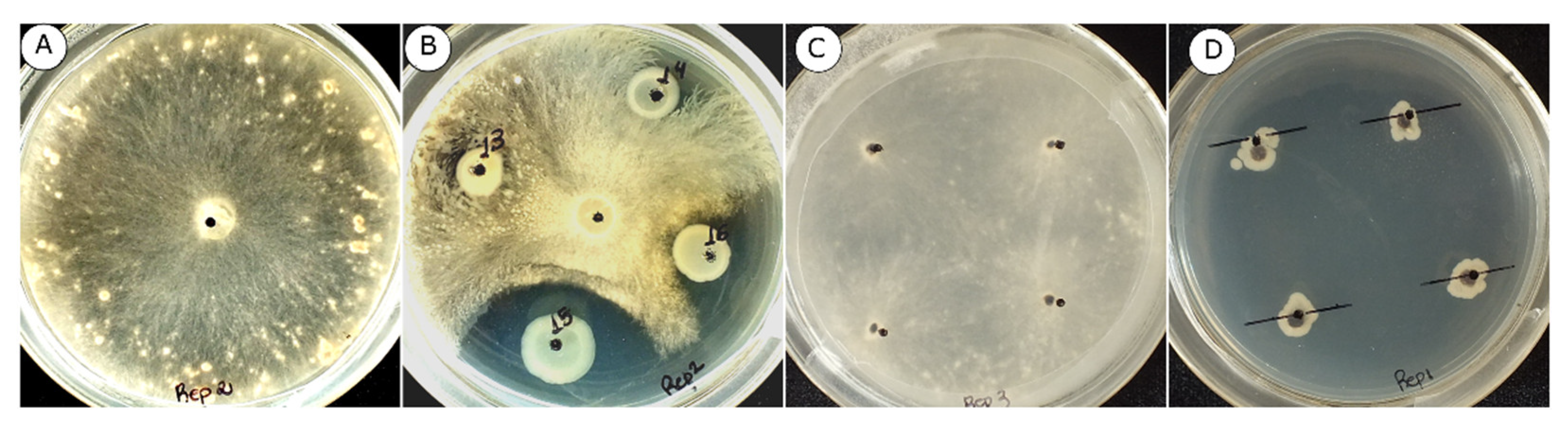
2.2.1. In Vitro Mycelial Growth Inhibition of R. solani AG-1 IA

The antagonism of fluorescent Pseudomonas on the mycelial growth of R. solani AG-1 IA isolates MTUB01C, MTUB04E, and MTUB05A obtained from Brazil, Mato Grosso state, was determined by transferring a 7 mm diameter disk from a 7 day old culture grown on potato dextrose agar (PDA) medium at 28 °C to the center of Petri dishes with King’s B agar medium (KB). Shortly after, 10 µL aliquots of bacterial suspensions previously cultured for 14 h in Luria-Bertani medium (LB) were incubated in a rotatory shaker at 190 rpm for 14 h at 28 °C, with the concentration adjusted to 6.2 × 10⁸ ufc.mL−1 (OD620 = 0.8), were deposited close the edge of the plate, in four equidistant points. The experimental design was completely randomized with five replicates for each treatment (N = 24 Pseudomonas isolates and a negative check). After 72 h incubation at 28 °C, the antagonism of the Pseudomonas isolates against R. solani AG-1 IA was documented by photographing the growth plates using a digital camera coupled to a 20 cm high monopod. The percentage of the mycelial growth inhibition of the pathogen was determined with the aid of the image analysis program Assess, APS (ASSESS: Image Analysis Software for Plant Disease Quantification; Department of Plant Science, University of Manitoba, Winnipeg, Manitoba, Canada) [27].

2.2.2. In Vitro Inhibition of R. Solani AG-1 IA Sclerotial Germination

Sclerotia of the three R. solani AG1-IA isolates (MTUB01C, MTUB04E, and MTUB05A) cultured in PDA medium for seven days at 28 °C and 12 h photoperiod were transferred to Erlenmeyer flasks containing 20 mL of liquid LB medium with 200 µL of the bacterial suspension obtained in the previous step and incubated at 28 °C in a rotatory shaker at 190 rpm for 24 h. After this period, the flasks were transferred to Petri dishes with PDA medium and incubated at 28 °C for three days. The experimental design was completely randomized; each experimental plot was composed of a plate containing four sclerotia, with four replicates. The inhibitory action of Pseudomonas on the sclerotia germination was determined by observing if there was mycelial growth in relation to the control treatment for each bacterial strain, which was composed only of sclerotia in LB medium without the inoculation of Pseudomonas, kept under same conditions as the other treatments.

2.2.3. In Vivo Biocontrol of Rhizoctonia Foliar Blight by Fluorescent Pseudomonas Delivered as Seed Treatment

Based on the previous step of in vitro analysis, for this study, three isolates of fluorescent Pseudomonas were used, which were selected as potential biocontrol agents. The bacterial isolates were grown in tubes containing 20 mL of liquid LB culture medium, incubated at 28 °C in a rotatory shaker at 190 rpm for 14 h. The bacterial suspension was centrifuged at 5000 rpm for 10 min and then suspended in fresh LB medium. The process was carried out three times, but after the last centrifugation sterile water was added to the suspension and the concentration was adjusted to 6.2 × 10⁸ ufc. mL−1 (OD620 = 0.8). The three different bacterial suspensions were applied separately to seeds of U. brizantha cv. Marandu, one hour before sowing.
Sowing was carried out in pots with a capacity of 500 mL containing commercial substrate and vermiculite in the proportion of de 3:1. The application of the chemical fertilizer with NPK 10-10-10 was carried out on the plants approximately 20 days after germination.
In the experiment, eight treatments were performed: one treatment for each Pseudomonas tested (Amana, Poti, and Yara) with R. solani inoculation; one treatment for each Pseudomonas tested without R. solani inoculation; a control treatment with R. solani inoculation only; and a control treatment without inoculation. The inoculum of R. solani was produced using sterilized rice grains transferred to clean Petri dishes containing colony discs with seven days of R. solani growth. The rice grains were incubated on the plates for five days at 28 °C and 12 h photoperiod until the fungal mycelium colonization. The inoculation was performed 30 days after the emergence of the plants. The inoculum consisted of a grain of colonized rice, which was placed on the leaves close to the base of the plants, with the inoculation of one leaf per plant. After inoculation, the plants were kept in a humid chamber in a greenhouse, with a daytime temperature of 30 °C (±3 °C) and a night temperature of 25 °C (±3 °C). The experiment was designed in randomized blocks, with four replicates consisting of five plants. The experiment was repeated once. The evaluation of disease symptoms was performed seven days after inoculation, photographing the set of plants from each experimental plot, determining an infected leaf area with the aid of image analysis software Assess, APS (ASSESS: Image Analysis Software for Plant Disease Quantification, Department of Plant Science, University of Manitoba, Winnipeg, Manitoba, Canada) [27].

2.2.4. In Vivo Biocontrol of Rhizoctonia Foliar Blight by Fluorescent Pseudomonas Delivered by Foliar Spray

The same bacteria isolates used in seed treatment were used in this study. Bacterial suspensions were obtained as previously described and were sprayed on the leaves and soil around the plant (10 mL of suspension per replicate). In the experiment, eight treatments were performed: one treatment for each Pseudomonas tested (Amana, Poti, and Yara) with R. solani inoculation; one treatment for each Pseudomonas tested without R. solani inoculation; a control treatment with R. solani inoculation only; and a control treatment without inoculation. The inoculum of the pathogen was produced as previously described. Inoculation of the pathogen was carried out 72 h after inoculation of the bacterial suspensions. After inoculation, the plants were kept in a humid chamber, in a greenhouse with a daytime temperature of 30 °C (±3 °C) and a night temperature of 25 °C (±3 °C). The experiment was designed in random blocks, with five replicates consisting of five plants. The experiment was repeated once. The evaluation of disease symptoms was performed seven days after inoculation, photographing the set of plants from each experimental plot, determining an infected leaf area with the aid of image analysis software Assess, APS (ASSESS: Image Analysis Software for Plant Disease Quantification, Department of Plant Science, University of Manitoba, Winnipeg, Manitoba, Canada) [27].

2.3. Phylogenetics Identification of Fluorescent Pseudomonas

2.3.1. DNA Extraction

Pure bacterial colonies N = 24 were transferred to glass test tubes containing LB medium and incubated at 28 °C in a rotatory shaker at 190 rpm for 12 h. The resulting liquid cultures were subsequently centrifuged for five minutes at 10,000 rpm and the pellets harvested for DNA extraction using “GenEluteTM Bacterial Genomic DNA Kits” (Sigma Aldrich, St. Louis, MO, USA) according to the manufacturer’s instructions. DNA yield was quantified using a NanoDrop® 2000c spectrophotometer (Thermo Fisher Scientific, Waltham, MA, USA) and diluted to a final concentration of 25 ng µL−1.

2.3.2. PCR Reactions and Sequencing of the 16S rDNA Region and rpoB Genes

PCR reactions were conducted in a final volume of 30μL containing ultra-pure distilled water, 25 ng of total DNA, 0.1 μM of each primer, 0.2 mM of each dNTP, 2 mM of MgCl 2, 2.5 μL of 10× buffer and 1U of Taq DNA Polymerase (Sigma-Aldrich, USA). The amplifications were performed in a ProFlex thermal cycler (Applied Biosystems, Bedford, MA, USA) using the following cycling conditions: initial denaturation at 95 °C for 5 min, followed by 35 cycles at 95 °C for 30 s, annealing temperature ranging from 54 °C to 63 °C (according to the primer used) for 1 min and 72 °C for 1 min; and final extension at 72 °C for 5 min (Table 1). The PCR products were sequenced on an ABI 3730xl DNA Analyzer (Applied Biosystems) at the Biological Resources and Genomics Center—CREBIO at UNESP Jaboticabal Campus. The sequences were analyzed using the Geneious R 9.0.5 software (Biomatters, New Zealand) and compared with the 16S rDNA region and rpoB genes sequences only from type species of Pseudomonas available at the GenBank/NCBI databases. These sequences were used to test hypotheses regarding the phylogenetic identification of the fluorescent Pseudomonas isolates obtained in our study.

2.3.3. Phylogenetics Identification

The sequences from 16S rDNA region and rpoB genes (N = 24 from each region) were used for phylogenetic analyses. Using the neighbor-joining method and the HKY genetic distance model implemented in the Geneious 9.0.5 software, we reconstructed the phylogenetic tree. Bootstrap supporting values for the tree branching were obtained from 1,000 replications of the iterations. Sequences from the P. aeruginosa lineage obtained from GenBank/NCBI were used as an outgroup for the tree and sequences from the Pseudomonas fluorescence lineage, including the major groups P. fluorescens (subgroups P. chlororaphis, P. corrugata, P. fluorescens, P. fragi, P. gessardii, P. jessenii, P. mandelii and P. protegens), P. putida, P. asplenii, P. lutea, and P. syringae were used as comparison species [14,31,32,33,34,35]. A total of 77 sequences of both 16S rDNA region and rpoB genes from these type species of Pseudomonas were included to reconstruct this phylogeny (Supplementary Materials S1).

2.4. Potential Biological Control Mechanisms

2.4.1. Protease, Chitinase, and Cellulase Activity Measurements

The detection of in vitro protease activity was determined by the degradation of casein in skimmed milk agar culture medium (casein peptone 5 g.L−1 ; yeast extract 2.5 g.L−1; skim milk powder 1 g.L−1; dextrose 1 g.L−1; agar 10.5 g.L−1; final pH 7.0 at 25 °C). Bacterial suspensions of the isolates Amana, Poti, and Yara, obtained as previously described, were deposited in three equidistant points on Petri dishes. An experimental unit consisted of a single plate of skimmed milk agar culture medium containing three bacterial suspension aliquots of 10 µL each. The experimental design was completely randomized with three replicates and the experiment was repeated once. Experiments for detection of cellulase and chitinase activities were designed as described to protease activity. Cellulase activity was determined by checking for clear halos around colonies grown on carboxymethyl cellulose agar (CMCA) plates [36]. Chitinase activity was determined by checking the colonies growth patterns for clear halos on chitin agar (CA) medium [37].

2.4.2. Detection of Siderophore Production

Siderophore production by isolates of fluorescent Pseudomonas was determined using the chrome azurol (CAS), as described formerly [38]. Three aliquots of 10 µL suspension of Amana, Poti, and Yara isolates were used for this experiment, obtained as previously described. The aliquots were deposited on CAS medium and incubated for 48 h at 30 °C. Afterwards the colonies were screened for yellow-orange zones indicating siderophores production. An experimental unit consisted of a single CAS medium plate containing three aliquots with 10 µL of suspension from each bacterial isolate. The experimental design was completely randomized with three replicates and the experiment was repeated once.

2.4.3. Detection of Phosphate Solubilization

To evaluate P solubilization ability of isolates Amana, Poti, and Yara it was used GL solid medium (glucose 10 g.L−1; yeast extract 2 g.L−1; agar 15 g.L−1) supplemented with phosphate source (CaHPO4 0.89 g.L−1). Three aliquots with 10 µL of suspension of Pseudomonas were used for this experiment, obtained as previously described. After 24 h of inoculation, clear halos were observed around colonies grown on GL medium with phosphate source [39]. As the control. GL medium was used without a phosphate source. An experimental unit consisted of a single plate GL solid medium containing three aliquots with 10 µL of suspension, one aliquot per each of the the three isolates (Amana, Poti, and Yara). The experimental design was completely randomized with three replicates, and the experiment was repeated once.

3. Results

3.1. Mycelial Growth and Sclerotia Germination of R. Solani AG-1 IA

The first step to assess the potential of fluorescent Pseudomonas (from undisturbed and possibly naturally suppressive soils within areas of native rainforest from the Amazon) as biocontrol agents against the signal grass foliar blight disease was to select strains capable of inhibiting mycelial growth and sclerotia germination of the pathogen R. solani AG-1 IA. There was no difference between replicates of these experiments. The effect of the interaction experiment*isolates was also non-significant, indicating reproducibility of the observations, regardless of the replicates (Table 2, Figure 1a,b). Combining the two replicates, the joint analysis of the data indicated significant differences in mycelial growth inhibition among Pseudomonas isolates (p ≤ 0.01). The mean inhibition of the pathogen´s mycelial growth by Pseudomonas isolates ranged from 5.9 to 41.8%. The Pseudomonas isolates Yara, Poti and Amana inhibited R. solani AG-1 IA mycelial growth the most (Supplementary Materials S2). There were also significant differences among Pseudomonas isolates considering the inbition of sclerotia germination there (p ≤ 0.05) (Table 2, Figure 1c,d), which ranged from 10.8% to 100.0%. Out of the 24 Pseudomonas isolates tested, seven showed 100% inhibition of sclerotia germination. Amana, Poti, and Yara isolates inhibited 65.0, 79.2, and 100.0% of the sclerotia germination, respectively (Supplementary Materials S2).
These three isolates of Pseudomonas were then chosen for the subsequent in vivo experiments.

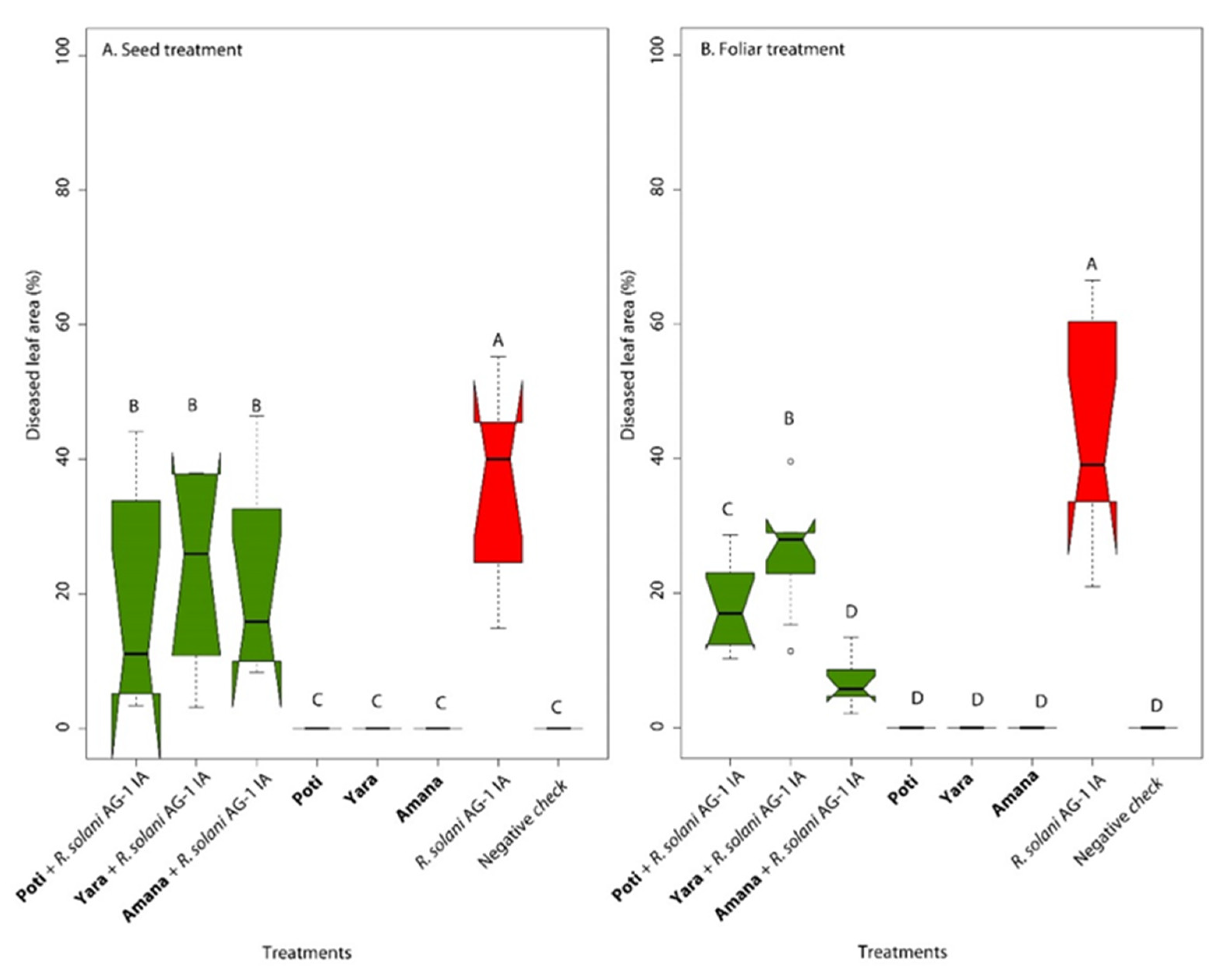
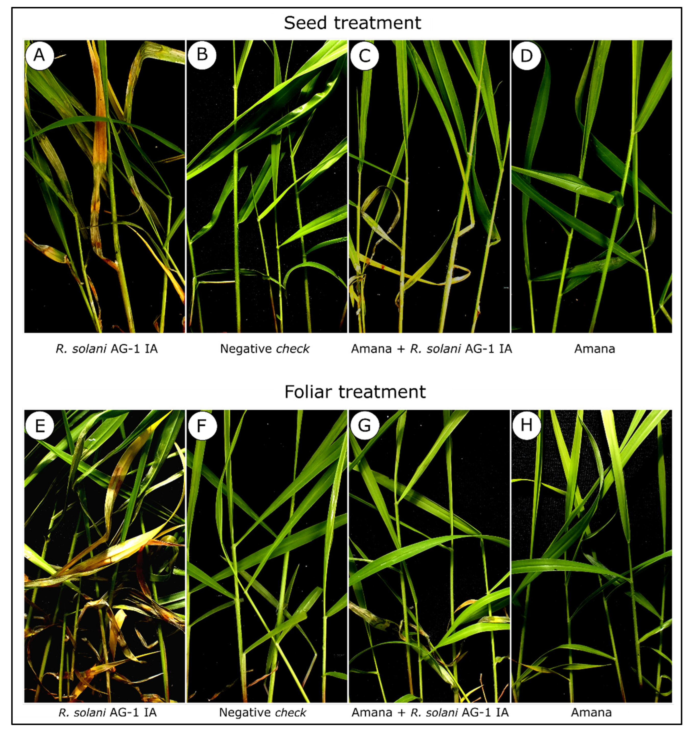
3.2. Potential of Pseudomonas spp. for in vivo Biocontrol of the Leaf Blight Disease on Signal Grass

3.2.1. Seed Treatment with Fluorescent Pseudomonas

There was no difference between replicates of the experiment of seed treatment. The effect of the interaction experiment*isolates was also non-significant, indicating reproducibility of the observations, regardless of the replicates. Combined analyses of the two experiments indicated the significance of the treatment effect at p ≤ 0.01. All three isolates of Pseudomonas used as biocontrol agents significantly reduced the severity of leaf blight on signal grass similarly (no significant difference among bacterial isolates by the Scott Knott test at p ≤ 0.05 (Table 3, Figure 2 and Figure 3).

3.2.2. Foliar Application of Fluorescent Pseudomonas

Significant differences were also detected between treatments at p ≤ 0.01, considering the foliar application of the biocontrol agents, resulting in decreased disease severity (Table 3, Figure 2 and Figure 3). While on the non-treated positive control, the severity of leaf blight reached a maximum of 43.1% on average—a significant reduction in disease severity to as low as 6.9% was detected on the treatment with Amana, while the treatments with Poti and Yara resulted in 17.7 and 25.6% of diseased leaf area, respectively.

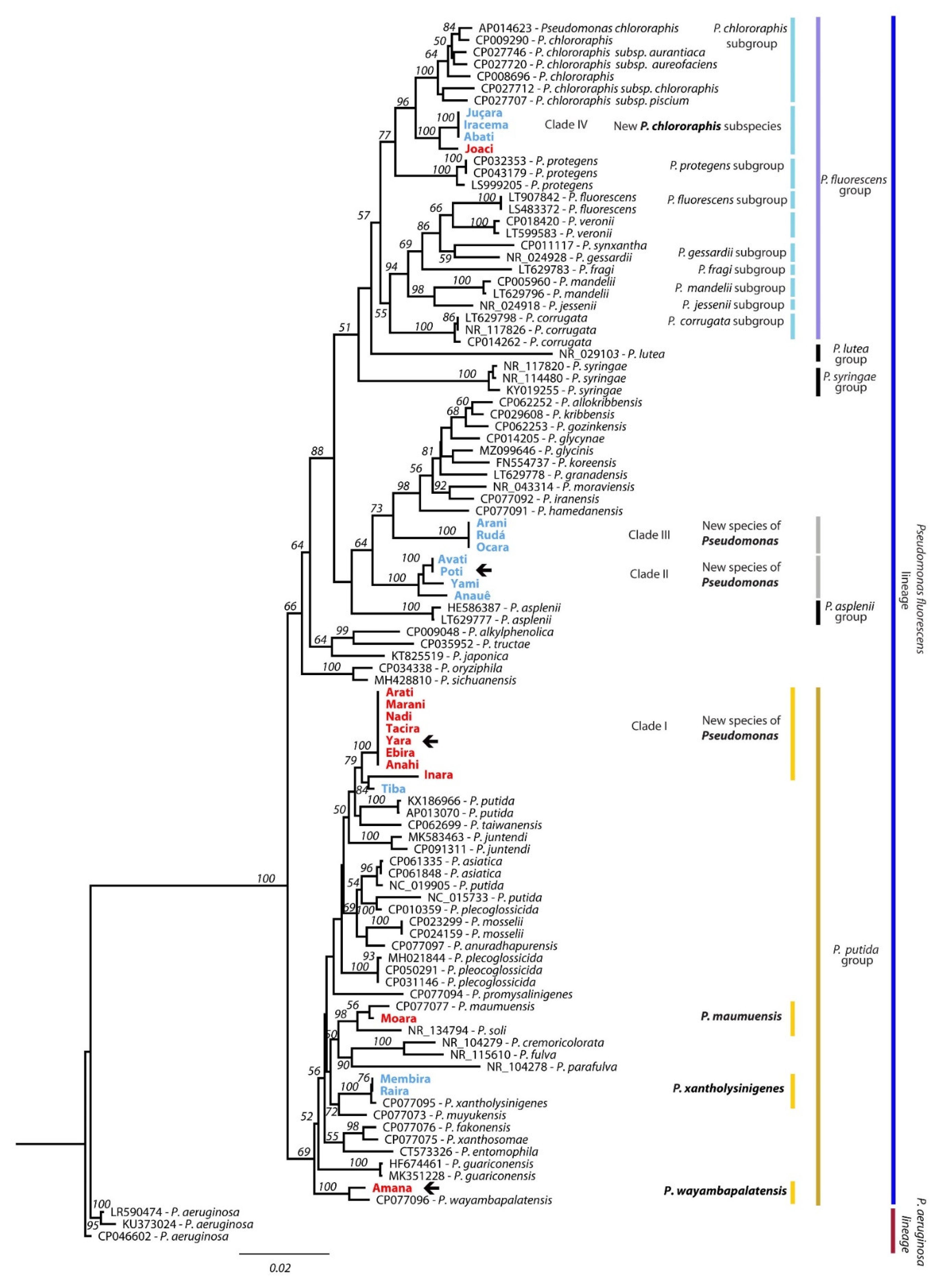
3.3. Molecular Identification of Fluorescent Pseudomonas from Amazonian Soils

The phylogenetic tree constructed indicated support for the main phylogenetic groups and subgroups within the Pseudomonas fluorescens lineage (Figure 4), accordingly to former phylogenetic reconstructions [31,32,33,34]. Fourteen isolates from our study, including Amana and Yara, which were selected as potential biocontrol agents, fell within the broad Pseudomonas putida group clade. While the isolate Amana grouped with P. wayambapalatensis, however, there was bootstrap support for a clade (I) grouping the isolate Yara with Arati, Ebira, Anahi, Tacira, Nadi, Marani, Inara, and Tiba isolates, possibly comprising a new species not yet described within this broad P. putida group. Additionally within the P. putida group, the isolate Moara grouped with P. maumuensis and P. soli, while the isolates Membira and Raira grouped with P. xantholysigenes.
Another two clades with bootstrap support, which comprised the isolates Poti, Avati, Yami, and Anauê (clade II), and Ocara, Arani, and Rudá (clade III) showed close similarity with the Pseudomonas asplenii group included in this analysis. However, they could well consist of two independent species not yet described within the P. asplenii group.
A fourth clade (clade IV) with bootstrap support contained the isolates Abati, Iracema, Juçara, and Joaci, and could consist of a new subspecies comprising the P. chlororaphis subgroup within the P. fluorescens group.

3.4. In Vitro Detection of Protease, Cellulase, and Chitinase Activities of Fluorescent Pseudomonas Isolates

While only Amana and Poti showed protease activity, as for chitinase, all three Pseudomonas isolates (Amana, Poti, and Yara) showed enzymatic activity. In constrast, these three isolates did not show any cellulase activity (Table 4, Figure 5).

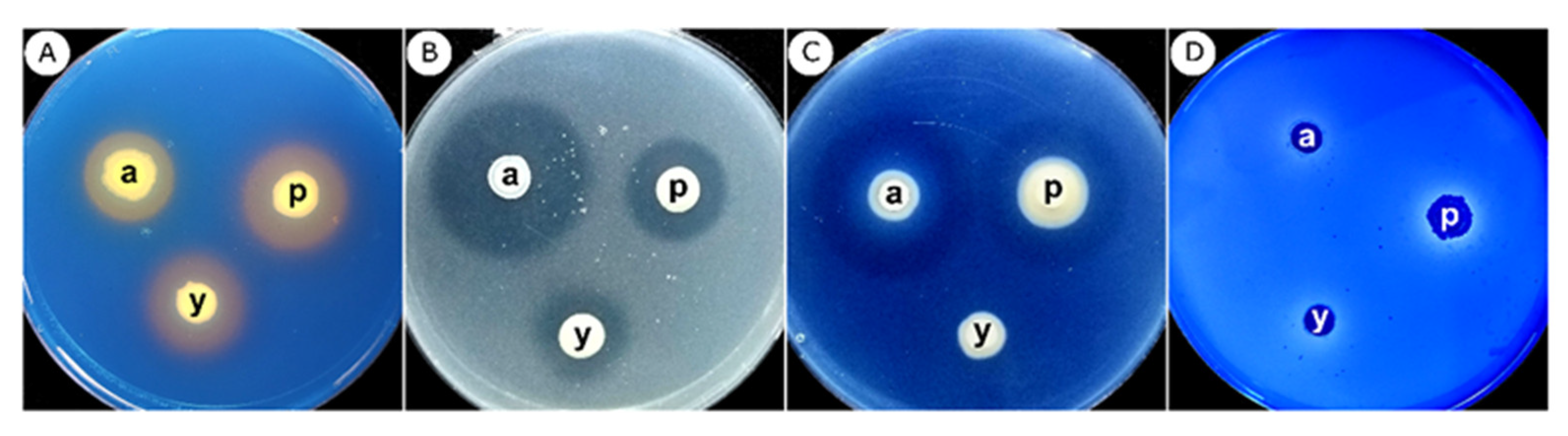
3.5. Qualitative Detection of Siderophore Production and Phosphate Solubilization

Amana, Poti, and Yara produced siderophores in culture medium with a low amount of iron. The three isolates also solubilized phosphate in vitro (Table 4, Figure 5).

4. Discussion

In this study, 24 isolates of fluorescent Pseudomonas obtained from naturally suppressive soils from the Amazon biome were bio-prospected for their role as biocontrol agents of the foliar blight disease caused by R. solani AG-1 IA on signal grass. From the initial set of 24 isolates screened, three (Amana, Poti, and Yara) were singled out, considering their ability for inhibiting fungal mycelial growth and sclerotia germination.
The phylogenetic analysis based on the joint analysis of the 16S [40] and rpoB [30,41,42] genes indicated a number of strains from yet undescribed fluorescent Pseudomonas species or subspecies, including the isolate Poti (independent but closely related to P. asplenii), which showed potential as a biocontrol agent. In fact, the microbial diversity from undisturbed soils of the Amazon biome is probably rich in unknown bacterial species, as we detected [43]. In addition, the two other Pseudomonas isolates with biocontrol potential, Amana (closely related to P. wayambapalatensis) and Yara (an independent clade closely related to the species P. putida) fell into the Pseudomonas putida species complex.
Seed or foliar application of P. putida Amana, Poti, and Yara resulted in significant disease control, causing a pronounced reduction in the severity of the foliar blight and sudden death, and thus, were further analyzed for their mechanisms of biocontrol. All three isolates produced siderophores, while Amana and Poti showed protease and chitinase in vitro activity. None of the isolates had cellulase activity.
The ability of species of Pseudomonas to produce extracellular compounds, such as siderophores, improves rhizosphere colonization by the bacteria, depriving potential plant pathogens of iron, thus inhibiting their growth, in addition to possibly ensuring competitive advantages assisting in the growth and development of plants [44]. In fact, siderophore-producing fluorescent Pseudomonas inhibited the mycelial growth of Pyricularia oryzae, the causal agent of rice blast, and of R. solani AG-1 IA, which is also associated with the rice sheath blight disease [45], besides foliar blight on signal grass.
Our results are also in line with a previous study that reported a positive correlation between the inhibition of mycelial growth of Fusarium oxysporum by fluorescent Pseudomonas with chitinase and protease activity and a negative correlation between the inhibition of the pathogen’s mycelial growth and the bacterial production of cellulase [46]. No cellulase activity was also reported for the antagonism of fluorescent Pseudomonas against Pythium aphanidermatum [47].
In addition to the potential as a biocontrol agent, it is suitable that selected bacterial or fungal species present other favorable characteristics for plant growth and development, such as the ability to solubilize phosphate. Phosphate-solubilizing bacteria favor the uptake of phosphorus by plants through the mineralization process, converting organic phosphorus to inorganic phosphorus, a form more available to plants and an alternative to the application of chemical fertilizers in agriculture [48,49]. In our study, the fluorescent Pseudomonas Amana, Poti, and Yara showed phosphate solubilization capacity.
Both the seed treatment and the foliar application of the biocontrol agents resulted in a considerable reduction of disease severity. The application of fluorescent Pseudomonas via seed treatment followed by foliar spraying significantly reduced by 75% the severity of rice sheath blight caused by R. solani AG-1 IA [50]. However, in our study, the seed treatment delayed signal grass germination and seedling emergence. This fact might be explained by the intrinsic high dormancy associated with seeds of most tropical forage grasses under alternating light and temperature regimes, and extreme variations in soil moisture, but especially when in contact with soil microorganisms and organic acids [51].
Therefore, foliar spray of fluorescent Pseudomonas seemed the best way of applying the biocontrol agents for managing leaf blight on signal grass. The main factor that can explain the effectiveness of foliar sprays is the rapid multiplication and high survival capacity of bacteria in the phylloplane [52,53]. In addition, bacterial cells and their extracellular compounds can be absorbed by stomatal pores and can be transported to different regions of plants, contributing to their strengthening and combating plant pathogens that may come into contact [54,55].
Although in our screening, we have not tested the ability of the fluorescent Pseudomonas to induce systemic resistance (ISR), this mechanism has already been demonstrated in several studies in different plant species [47]. ISR is based on a series of mechanisms that can promote the activation of plant defense mechanisms in response to the attack of plant pathogens. Nandakumar, Babu, Viswanathan, Raguchander, and Samiyappan [50] reported that a single application of fluorescent Pseudomonas resulted in ISR in rice plants when inoculated with R. solani AG-1 IA. In addition, the combination of four application methods (seed, root, soil, and foliar) increased the ISR, as well as the production of chitinase by the bacterial biocontrol agent.
Despite several reports of the biocontrol efficacy of fluorescent Pseudomonas, there are no biofungicides with P. putida as an active ingredient labeled in Brazil [56]. One of the greatest challenges for labeling fluorescent Pseudomonas-based biofungicides is the lack of studies that provide a reliable and stable industrial formulation, since the bacterial species do not produce resistant spores that usually insure the long-term stability and viability of formulation. However, a successful industrial-scale formulation of a P. putida-based biofungicide (formulated with the bacterial strain B 2017, in particular) for the management of F. oxysporum, R. solani and Sclerotinia sclerotiorum diseases has already been reported in Spain [57]. The development of a stable formulation of the Pseudomonas-based biocontrol agents selected in our study for managing the signal grass foliar blight is warranted. Further research on the topic should include field tests of the biofungicides formulations obtained thereafter.
Even though, in some cases, the level of disease control with the application of microorganisms under greenhouse conditions may be at levels below what is necessary to avoid large yield losses in the field, the main purpose of using biological control is not its isolated effect but an integration of methods to improve the efficacy of their use [58].

5. Conclusions

Based on in vitro antagonism, three isolates (Amana, Poti, and Yara) were selected for further in vivo assays.
Multilocus phylogenetic analysis indicated that Amana and Yara grouped into the Pseudomonas putida group, while Poti was grouped into the Pseudomonas asplenii group and could well constitute a new Pseudomonas species.
All three isolates produced siderophores and solubilized phosphate, while Amana and Poti showed protease and chitinase in vitro activity.
Foliar application of P. putida Amana from Amazonian suppressive soils resulted in a significant reduction of the foliar blight disease severity on signal grass.

Supplementary Materials

The following supporting information can be downloaded at: https://www.mdpi.com/article/10.3390/agronomy12061395/s1, Supplementary Materials S1. Alignment of sequence data from 16S rDNA region and rpoB genes from unknown fluorescent Pseudomonas species from Amazon soils used to reconstruct a phylogeny with the major groups of the P. fluorescens lineage. Supplementary Materials S2. Rhizoctonia solani AG-1 IA mycelial growth and sclerotia germination inhibition by fluorescent Pseudomonas isolates.

Author Contributions

Conceptualization, P.C.C.; methodology, P.C.C., G.d.C., S.I.M. and S.N.C.V.; software, S.N.C.V., T.C.N. and A.G.d.S.; validation, S.N.C.V., A.G.d.S., T.C.S. and P.C.C.; formal analysis, S.N.C.V. and A.G.d.S.; investigation, S.N.C.V., L.D.K., L.M.d.D.P.G. and T.C.S., resources, S.N.C.V. and G.d.C.; data curation, P.C.C., S.N.C.V. and A.G.d.S.; writing—original draft preparation, S.N.C.V. and P.C.C.; writing—review and editing, P.C.C.; visualization, P.C.C.; supervision, P.C.C.; project administration, P.C.C.; funding acquisition, P.C.C. All authors have read and agreed to the published version of the manuscript.

Funding

This research was funded by FAPESP (São Paulo Research Foundation, Brazil), Grant/Award Numbers: 11/50150-3, 11/23050-8, 13/11944-0, and 17/03403-0; CNPq (Brazilian National Council for Scientific and Technological Development), Grant/Award Number: Pq-1D 313825/2018-1; CAPES (Coordination for the Improvement of Higher Level Personnel), Grant/Award Numbers: CAPES/Pro-equipment Program 775202/2012, CAPES/AUXPE Program 88881.593505/2020-01—UNESP Ilha Solteira Campus, CAPES PrInt/UNESP, CAPES studentship Program 001).

Institutional Review Board Statement

Not applicable.

Informed Consent Statement

The Brazilian Ministry of Environment/Chico Mendes Institute for Biodiversity Conservation—ICMBio, issued the permit # 40794-4 authorizing the collection of botanical and microbiological material from the Amazon biome.

Data Availability Statement

The 16S and rpoB sequencing data that supports the phylogenetic findings of this study is available as Supplementary material S1 will also be available, upon publication, at the GenBank/NCBI database. The phenotypic data on disease severity presented in this study are available upon request from the corresponding author. The data are not publicly available due to the authors’ option.

Acknowledgments

We received funding for travel support from the company Geo Clean Indústria Comércio de Produtos Químicos, Araraquara, SP, Brazil, which specializes in chemical and biological products for plant nutrition and protection, and the logistic support from its CEO and Agronomist Marcos Cesar Costa for sampling soils in Mato Grosso.

Conflicts of Interest

The authors declare no conflict of interest. The funders had no role in the design of the study; in the collection, analyses, or interpretation of data; in the writing of the manuscript, or in the decision to publish the results.

References

  1. Kelemu, S.; Miles, J.W.; Bonilla, X.P.; Badel, J.L. Sources of resistance in species of Brachiaria to foliar blight disease caused by Rhizoctonia solani. Trop. Grassl. 1995, 29, 257–262. [Google Scholar]
  2. González-Vera, A.D.; Bernardes-de-Assis, J.; Zala, M.; McDonald, B.A.; Correa-Victoria, F.; Graterol-Matute, E.J.; Ceresini, P.C. Divergence between sympatric rice- and maize-infecting populations of Rhizoctonia solani AG-1 IA from Latin America. Phytopathology 2010, 100, 172–182. [Google Scholar] [CrossRef] [PubMed] [Green Version]
  3. Chavarro Mesa, E.; Ceresini, P.C.; Ramos Molina, L.M.; Pereira, D.A.S.; Schurt, D.A.; Vieira, J.R.; Poloni, N.M.; McDonald, B.A. The Urochloa foliar blight and collar rot pathogen Rhizoctonia solani AG-1 IA emerged in South America via a host shift from rice. Phytopathology 2015, 105, 1475–1486. [Google Scholar] [CrossRef] [PubMed] [Green Version]
  4. Duarte, M.d.L.R.; Albuquerque, F.C.; Sanhueza, R.M.V.; Verzignassi, J.R.; Kondo, N. Etiologia da podridão do coleto de Brachiara brizantha em pastagens da Amazônia. Fitopatol. Bras. 2007, 32, 261–265. [Google Scholar] [CrossRef] [Green Version]
  5. Verzignassi, J.R.; Fernandes, C.D. Doenças em Forrageiras; Divulgação Técnica; Embrapa Gado de Corte: Campo Grande, Brazil, 2001; n. 50; 3p. [Google Scholar]
  6. Vieira Júnior, J.R.; Fernandes, C.F.; Marreios, J.A.A.; Sangi, S.C.; Fonseca, A.S.; Silva, D.S.G.; Alves, T.C.; Matos, A.I.; Maia, D. Levantamento da ocorrência da morte-das-pastagens em Rondônia; Comunicado Técnico; Embrapa Rondônia: Rodovia, Brazil, 2014; n. 394; 5p. [Google Scholar]
  7. Chavarro-Mesa, E.; Ceresini, P.; Pereira, D.; Vicentini, S.; Silva, T.; Ramos-Molina, L.; Negrisoli, M.; Schurt, D.; Vieira Júnior, J.R. A broad diversity survey of Rhizoctonia species from the Brazilian Amazon reveals the prevalence of R. solani AG-1 IA on signal grass and the new record of AG-1 IF on cowpea and soybeans. Plant Pathol. 2020, 69, 455–466. [Google Scholar] [CrossRef]
  8. Marchi, C.E.; Fernandes, C.D.; Verzignassi, J.R. Doenças em plantas forrageiras; Documentos; Embrapa Gado de Corte: Campo Grande, Brazil, 2011; n. 187; 28p. [Google Scholar]
  9. Lucas, J.A.; Hawkins, N.J.; Fraaije, B.A. The evolution of fungicide resistance. Adv. Appl. Microbiol. 2015, 90, 29–92. [Google Scholar]
  10. Nega, A. Review on Concepts in Biological Control of Plant Pathogens. J. Biol. Agric. Healthc. 2014, 4, 23. [Google Scholar]
  11. Weller, D.M. Pseudomonas biocontrol agents of soilborne pathogens: Looking back over 30 years. Phytopathology 2007, 97, 250–256. [Google Scholar] [CrossRef] [Green Version]
  12. Zago, V.C.P.; De-Polli, H.; Norma, G. Pseudomonas spp. Fluorescentes—Bactérias Promotoras de Crescimento de Plantas e Biocontroladoras de Fitopatógenos em Sistemas de Produção Agrícola; Documentos; Embrapa Agrobiologia: Rodovia, Brazil, 2000; n. 127; 32p. [Google Scholar]
  13. Ferreira, E.P.B.; Voss, M.; Santos, H.P.; De-Polli, H.; Neves, M.C.P.; Rumjanek, N.G. Diversidade de Pseudomonas fluorescentes em diferentes sistemas de manejo do solo e rotação de culturas. Agrárias-Rev. Bras. Ciências Aggrárias 2009, 4, 140–148. [Google Scholar] [CrossRef] [Green Version]
  14. Höfte, M. The Use of Pseudomonas spp. as Bacterial Biocontrol Agents to Control Plant Disease; Köhl, J., Ravensberg, W., Eds.; Microbial Bioprotectants for Plant Disease Management; Burleigh Dodds Science Publishing: Cambridge, UK, 2022; pp. 1–72. [Google Scholar]
  15. Panpatte, D.G.; Jhala, Y.K.; Shelat, H.N.; Vyas, R.V. Pseudomonas fluorescens: A Promising Biocontrol Agent and PGPR for Sustainable Agriculture. In Microbial Inoculants in Sustainable Agricultural Productivity: Vol. 1: Research Perspectives; Singh, D.P., Singh, H.B., Prabha, R., Eds.; Springer: New Delhi, India, 2016; pp. 257–270. [Google Scholar]
  16. D’aes, J.; Hua, G.K.H.; De Maeyer, K.; Pannecoucque, J.; Forrez, I.; Ongena, M.; Dietrich, L.E.P.; Thomashow, L.S.; Mavrodi, D.V.; Höfte, M. Biological Control of Rhizoctonia Root Rot on Bean by Phenazine- and Cyclic Lipopeptide-Producing Pseudomonas CMR12a. Phytopathology 2011, 101, 996–1004. [Google Scholar] [CrossRef] [Green Version]
  17. Olorunleke, F.E.; Hua, G.K.H.; Kieu, N.P.; Ma, Z.; Höfte, M. Interplay between orfamides, sessilins and phenazines in the control of Rhizoctonia diseases by Pseudomonas sp. CMR12a. Environ. Microbiol. Rep. 2015, 7, 774–781. [Google Scholar] [CrossRef]
  18. Vinay, J.U.; Naik, M.K.; Rangeshwaran, R.; Chennappa, G.; Shaikh, S.S.; Sayyed, R.Z. Detection of antimicrobial traits in fluorescent pseudomonads and molecular characterization of an antibiotic pyoluteorin. 3 Biotech 2016, 6, 227. [Google Scholar] [CrossRef] [PubMed] [Green Version]
  19. Páez, M.; Martínez-Nieto, P.; Bernal-Castillo, J. Siderophore producing Pseudomonas as pathogenic Rhizoctonia solani and Botrytis cinerea antagonists. Univ. Sci. 2005, 10, 65–74. [Google Scholar]
  20. Dimkpa, C. Microbial siderophores: Production, detection and application in agriculture and environment. Endocytobiosis Cell Res. 2016, 27, 7–16. [Google Scholar]
  21. Chen, C.; BÉLanger, R.R.; Benhamou, N.; Paulitz, T.C. Defense enzymes induced in cucumber roots by treatment with plant growth-promoting rhizobacteria (PGPR) and Pythium aphanidermatum. Physiol. Mol. Plant Pathol. 2000, 56, 13–23. [Google Scholar] [CrossRef]
  22. Bautista, G.; Mendoza Rivera, H.; Uribe-Vélez, D. Biocontrol of Rhizoctonia solani in native potato (Solanum phureja) plants using native Pseudomonas fluorescens. Acta Biológica Colomb. 2007, 12, 19–32. [Google Scholar]
  23. Rana, A.; Sahgal, M.; Kumar, P. Biocontrol Prospects of Pseudomonas fluorescens AS15 against Banded Leaf and Sheath Blight Disease of Maize under Field Condition in Conducive Soil. Natl. Acad. Sci. Lett. 2019, 42, 425–428. [Google Scholar] [CrossRef]
  24. Nagendran, S.; Kulanthaivelu, S.; Sundararajan, T. Assessment on antagonistic potential of Bacterial bio agents Pseudomonas fluorescens and Bacillus subtilis against Rhizoctonia solani Kühn. An incitant of Sheath blight of rice. J. Entomol. Zool. Stud. 2019, 7, 128–142. [Google Scholar]
  25. Mavrodi, O.V.; Walter, N.; Elateek, S.; Taylor, C.G.; Okubara, P.A. Suppression of Rhizoctonia and Pythium root rot of wheat by new strains of Pseudomonas. Biol. Control 2012, 62, 93–102. [Google Scholar] [CrossRef]
  26. Gould, W.D.; Hagedorn, C.; Bardinelli, T.R.; Zablotowicz, R.M. New Selective Media for Enumeration and Recovery of Fluorescent Pseudomonads from Various Habitats. Appl. Environ. Microbiol. 1985, 49, 28–32. [Google Scholar] [CrossRef] [Green Version]
  27. Lamari, L. Assess 2.0: Image Analysis Software for Plant Disease Quantification; American Phytopathological Society: St. Paul, MN, USA, 2008. [Google Scholar]
  28. Milling, A.; Smalla, K.; Maidl, F.X.; Schloter, M.; Munch, J.C. Effects of transgenic potatoes with an altered starch composition on the diversity of soil and rhizosphere bacteria and fungi. Plant Soil 2005, 266, 23–39. [Google Scholar] [CrossRef]
  29. Spilker, T.; Coenye, T.; Vandamme, P.; LiPuma, J.J. PCR-based assay for differentiation of Pseudomonas aeruginosa from other Pseudomonas species recovered from cystic fibrosis patients. J. Clin. Microbiol. 2004, 42, 2074–2079. [Google Scholar] [CrossRef] [Green Version]
  30. Tayeb, L.A.; Ageron, E.; Grimont, F.; Grimont, P.A.D. Molecular phylogeny of the genus Pseudomonas based on rpoB sequences and application for identification of isolates. Res. Microbiol. 2005, 156, 763–773. [Google Scholar] [CrossRef]
  31. Garrido-Sanz, D.; Meier-Kolthoff, J.P.; Göker, M.; Martín, M.; Rivilla, R.; Redondo-Nieto, M. Genomic and Genetic Diversity within the Pseudomonas fluorescens Complex. PLoS ONE 2016, 11, e0150183. [Google Scholar] [CrossRef] [Green Version]
  32. Girard, L.; Lood, C.; Höfte, M.; Vandamme, P.; Rokni-Zadeh, H.; van Noort, V.; Lavigne, R.; De Mot, R. The Ever-Expanding Pseudomonas Genus: Description of 43 New Species and Partition of the Pseudomonas putida Group. Microorganisms 2021, 9, 1766. [Google Scholar] [CrossRef] [PubMed]
  33. Hesse, C.; Schulz, F.; Bull, C.T.; Shaffer, B.T.; Yan, Q.; Shapiro, N.; Hassan, K.A.; Varghese, N.; Elbourne, L.D.H.; Paulsen, I.T.; et al. Genome-based evolutionary history of Pseudomonas spp. Environ. Microbiol. 2018, 20, 2142–2159. [Google Scholar] [CrossRef] [PubMed]
  34. Mulet, M.; Lalucat, J.; García-Valdés, E. DNA sequence-based analysis of the Pseudomonas species. Environ. Microbiol. 2010, 12, 1513–1530. [Google Scholar]
  35. Vásquez-Ponce, F.; Higuera-Llantén, S.; Pavlov, M.S.; Marshall, S.H.; Olivares-Pacheco, J. Phylogenetic MLSA and phenotypic analysis identification of three probable novel Pseudomonas species isolated on King George Island, South Shetland, Antarctica. Braz. J. Microbiol. 2018, 49, 695–702. [Google Scholar] [CrossRef]
  36. Ghose, T.K. Measurement of cellulase activities. Pure Appl. Chem. 1987, 59, 257–268. [Google Scholar] [CrossRef]
  37. Ahmadi, K.; Yazdi, M.; Najafi, M.; Shahverdi, A.R.; Faramarzi, M.; Zarrini, G.; Behravan, J. Optimization of Medium and Cultivation Conditions for Chitinase Production by the Newly Isolated: Aeromonas sp. Biotechnology 2008, 7, 266–272. [Google Scholar]
  38. Schwyn, B.; Neilands, J.B. Universal chemical assay for the detection and determination of siderophores. Anal. Biochem. 1987, 160, 47–56. [Google Scholar] [CrossRef]
  39. Souchie, E.; Abboud, A. Solubilização de fosfato por microrganismos rizosféricos de genótipos de Guandu cultivados em diferentes classes de solo. Semin. Ciências Agrárias 2007, 28, 11. [Google Scholar] [CrossRef] [Green Version]
  40. Anzai, Y.; Kim, H.; Park, J.Y.; Wakabayashi, H.; Oyaizu, H. Phylogenetic affiliation of the pseudomonads based on 16S rRNA sequence. Int. J. Syst. Evol. Microbiol. 2000, 50 Pt 4, 1563–1589. [Google Scholar] [CrossRef] [PubMed] [Green Version]
  41. Adékambi, T.; Drancourt, M.; Raoult, D. The rpoB gene as a tool for clinical microbiologists. Trends Microbiol. 2009, 17, 37–45. [Google Scholar] [CrossRef]
  42. Peix, A.; Valverde, A.; Rivas, R.; Igual, J.M.; Ramírez-Bahena, M.H.; Mateos, P.F.; Santa-Regina, I.; Rodríguez-Barrueco, C.; Martínez-Molina, E.; Velázquez, E. Reclassification of Pseudomonas aurantiaca as a synonym of Pseudomonas chlororaphis and proposal of three subspecies, P. chlororaphis subsp. chlororaphis subsp. nov., P. chlororaphis subsp. aureofaciens subsp. nov., comb. nov. and P. chlororaphis subsp. aurantiaca subsp. nov., comb. nov. Int. J. Syst. Evol. Microbiol. 2007, 57, 1286–1290. [Google Scholar]
  43. Cenciani, K.; Lambais, M.R.; Cerri, C.C.; Azevedo, L.C.C.; Feigl, B.J. Bacteria diversity and microbial biomass in forest, pasture and fallow soils in the southwestern amazon basin. Rev. Bras. Ciência Solo 2009, 33, 907–916. [Google Scholar] [CrossRef]
  44. Vellasamy, S.; Nithya, K.; Hariharan, H.; Jayaprakashvel, M.; Balasubramanian, N. Biocontrol Mechanisms of Siderophores against Bacterial Plant Pathogens; Kannan, V.R., Bastas, K.K., Eds.; Sustainable Approaches for Controlling Plant Pathogenic Bacteria; CRC Press: Boca Raton, FL, USA, 2015; pp. 167–186. [Google Scholar]
  45. Reddy, B.P.; Rani, J.; Reddy, M.S.; Kumar, K.V.K. Siderophore isolation and its Biocontrol action against Rice Fungal Pathogens. Biochem. Mol. Biol. 2019, 1, 1–6. [Google Scholar]
  46. Rathore, R.; Vakharia, D.N.; Rathore, D.S. In vitro screening of different Pseudomonas fluorescens isolates to study lytic enzyme production and growth inhibition during antagonism of Fusarium oxysporum f. sp. cumini, wilt causing pathogen of cumin. Egypt. J. Biol. Pest Control 2020, 30, 57. [Google Scholar] [CrossRef]
  47. Bernardes, F.S.; Patrício, F.R.A.; Santos, A.S.; Freitas, S.d.S. Indução de resistência sistêmica por rizobactérias em cultivos hidropônicos. Summa Phytopathol. 2010, 36, 115–121. [Google Scholar] [CrossRef]
  48. Oteino, N.; Lally, R.D.; Kiwanuka, S.; Lloyd, A.; Ryan, D.; Germaine, K.J.; Dowling, D.N. Plant growth promotion induced by phosphate solubilizing endophytic Pseudomonas isolates. Front. Microbiol. 2015, 6, 745. [Google Scholar] [CrossRef] [Green Version]
  49. Prasanna, A.; Deepa, V.; Balakrishn, P.; Deecaraman, M.; Sridhar, R.; Dhandapani, P. Insoluble Phosphate Solubilization by Bacterial Strains Isolated from Rice Rhizosphere Soils from Southern India. Int. J. Soil Sci. 2011, 6, 134–141. [Google Scholar] [CrossRef] [Green Version]
  50. Nandakumar, R.; Babu, S.; Viswanathan, R.; Raguchander, T.; Samiyappan, R. Induction of systemic resistance in rice against sheath blight disease by Pseudomonas fluorescens. Soil Biol. Biochem. 2001, 33, 603–612. [Google Scholar] [CrossRef]
  51. Souza, F.H.D. Produção de Sementes de Gramíneas Forrageiras Tropicais; Embrapa Pecuária Sudeste: São Carlos, Brazil, 2001; p. 43. [Google Scholar]
  52. Akter, S.; Kadir, J.; Juraimi, A.S.; Saud, H.M. In vitro evaluation of Pseudomonas bacterial isolates from rice phylloplane for biocontrol of Rhizoctonia solani and plant growth promoting traits. J. Environ. Biol. 2016, 37, 597–602. [Google Scholar] [PubMed]
  53. Austin, B.; Dickinson, C.H.; Goodfellow, M. Antagonistic interactions of phylloplane bacteria with Drechslera dictyoides (Drechsler) Shoemaker. Can. J. Microbiol. 1977, 23, 710–715. [Google Scholar] [CrossRef] [PubMed]
  54. Kumar, K.; Raju, S.; Reddy, M.; Kloepper, J.; Lawrence, K.; Groth, D.; Miller, M.; Sudini, H.; Du, B. Evaluation of Commercially Available PGPR for Control of Rice Sheath Blight Caused by Rhizoctonia solani. J. Pure Appl. Microbiol. 2009, 3, 485–488. [Google Scholar]
  55. Subashri, R.; Gurusamy, R.; Sakthivel, N.; Raman, B.; Sakthivel, B.; Maheshwari, D. Biological Control of Pathogens and Plant Growth Promotion Potential of Fluorescent Pseudomonads; Maheshwari, D.K., Ed.; Bacteria in Agrobiology: Disease Management; Springer: Berlin/Heidelberg, Germany, 2012; pp. 77–110. [Google Scholar]
  56. MAPA. AGROFIT. Available online: http://extranet.agricultura.gov.br/agrofit_cons/principal_agrofit_cons (accessed on 20 December 2021).
  57. Oliver, C.; Hernández, I.; Caminal, M.; Lara, J.M.; Fernàndez, C. Pseudomonas putida strain B2017 produced as technical grade active ingredient controls fungal and bacterial crop diseases. Biocontrol Sci. Technol. 2019, 29, 1053–1068. [Google Scholar] [CrossRef]
  58. Ghini, R.; Bettiol, W. Proteção de plantas na agricultura sustentável. Cad. De Ciência Tecnol.—CC&T 2000, 17, 61–70. [Google Scholar]
Figure 1. In vitro inibition of Rhizotonia solani AG-1 IA mycelial growth and sclerotia germination by fluorescent bacteria of the Pseudomonas genus. (A) R. solani AG-1 IA growing singly. (B) Confront between R. solani AG-1 IA and Pseudomonas. (C) Sclerotial germination of R. solani AG-1 IA growing singly. (D) Complete inhibition of sclerotia germination by fluorescent Pseudomonas.
Agronomy 12 01395 g001
Figure 2. Disease severity on plants of Urochloa brizantha cv Marandu inoculated with Rhizoctonia solani AG1-IA and treated or not with fluorescent Pseudomonas on seeds (A) or leaves (B).
Agronomy 12 01395 g002
Figure 3. Distinct disease severity levels of foliar blight caused by Rhizoctonia solani AG-1 IA on Urochloa brizantha cv. Marandu. (A) positive check inoculated with the pathogen, (B) negative check without pathogen´s inoculation, (C) seed treatment with fluorescent Pseudomonas Amana followed by inoculation of the pathogen, and (D) seed application of bacterial biocontrol agent solely. In (E) positive check inoculated with the pathogen, (F) negative check without pathogen´s inoculation, (G) foliar treatement of fluorescent Pseudomonas Amana followed by inoculation of the pathogen inoculation and (H) foliar application of the bacterial isolate solely.
Agronomy 12 01395 g003
Figure 4. Phylogenetic tree based on the 16S and rpoB gene sequences from the Pseudomonas fluorescens lineage, including most of the phylogenetic groups and subgroups within this lineage. The tree was constructed with the Geneious 9.0.5 software using the neighbour-joining reconstruction method and the HKY genetic distance model. The data in the branches indicate bootstrap supporting values, from a total of 1000 replications, with only values above 50% presented. The P. aeruginosa sequences were used as an outgroup. Arrows indicate the three potential biocontrol agents selected in our study. Isolates marked in red were obtained from soils in Rondônia, while isolates in blue were obtained from soils from Mato Grosso State.
Agronomy 12 01395 g004
Figure 5. In vitro experiments for determining potential biological control mechanisms associated with the activity of the fluorescent Pseudomonas isolates Amana (a), Poti (p), and Yara (y) from suppressive Amazonian soils. (A) production of siderophores, (B) phosphate solubilization, (C) protease and (D) chitinase activities.
Agronomy 12 01395 g005
Table 1. Primer sequences and annealing temperature used to amplify by PCR reactions the fragments of 16S rDNA and rpoB genes.
GenePrimersSequences (5′–3′)Length (bp)Annealing Temperature (°C)ReferencesFragment Length (bp)
16S rDNAF311-PsCTGGTCTGAGAGGATGATCAGT2263Milling et al., 2004 [28]1150
R1459-PsAATCACTCCGTGGTAACCGT20
16S rDNAPAGSFGACGGGTGAGTAATGCCTA1954Spilker et al., 2004 [29]618
PAGSRCACTGGTGTTCCTTCCTAA
rpoBLAPSTGGCCGAGAACCAGTTCCGCGT2260Tayeb et al., 2005 [30]1229
LAPS27CGGCTTCGTCCAGCTTGTTCAG22
Table 2. ANOVA analyses of Rhizoctonia solani AG-1 IA mycelial growth and sclerotia germination inhibition by fluorescent Pseudomonas isolates from suppressive Amazon soils.
FactorSource of VariationdfMean SquareFp
Inhibition of Mycelial growthExperiments (E)1116.192.1560.142 NS
Treatments (T)232017.8037.4380.000 *
E × T2319.520.3620.997 NS
Error67253.89
Treatments232017.8038.170.000 *
Error69652.85
Inhibition of sclerotia germinationExperiments (E)1226.881.9151.074 NS
Treatments (T)234216.8835.580.000 *
E × T23127.211.0740.378 NS
Error192118.49
Treatments234216.8835.160.000 *
Error216119.92
* Significant by F test at p ≤ 0.001; not significant (NS). Each experiment was repeated once.
Table 3. Analysis of variance of the effects of seed or foliar treatments of fluorescent Pseudomonas isolates for controlling the foliar blight caused by Rhizoctonia solani AG-1 IA on signal grass.
ExperimentsSource of VariationdfMean SquareFp
Seed treatmentExperiments (E)145.640.4840.4900 NS
Treatments (T)71639.8517.4050.0000 *
Blocks3256.102.7180.0556 NS
E × T7167.451.7770.1155 NS
Error4594.21
Treatments71639.8515.5090.000 *
Blocks7149.261.4120.222 NS
Error28105.74
Foliar treatmentExperiments (E)10.00360.000*0.9930 NS
Treatments (T)72562.0455.2820.0000 *
Blocks479.661.7190.1576 NS
E × T710.270.2220.9789 NS
Error6046.34
Treatments72562.0457.640.000 *
Blocks941.240.9230.507 NS
Error6344.44
* Significant by F test at p ≤ 0.05 and not significant (NS). Each experiment was repeated once.
Table 4. Production of siderophores and lytic enzymes from Amana, Poti, and Yara isolates.
Pseudomonas IsolatesProduction of SiderophoresPhosphate SolubilizationEnzymatic Activity
ProteaseChitinaseCellulase
Amana++++-
Poti++++-
Yara++-+-
+: Presence of halos in the culture media indicating the production of siderophores or substrate degradation by enzymatic activity. -: Absence of halos in the culture media.
Publisher’s Note: MDPI stays neutral with regard to jurisdictional claims in published maps and institutional affiliations.

