Article

Host and Tissue Affiliations of Culturable Endophytic Fungi Associated with Xerophytic Plants in the Desert Region of Northwest China

School of Life Sciences, Hebei University, Baoding 071002, China
*
Author to whom correspondence should be addressed.
Agronomy 2022, 12(3), 727; https://doi.org/10.3390/agronomy12030727
Submission received: 25 January 2022 / Revised: 9 March 2022 / Accepted: 10 March 2022 / Published: 17 March 2022

Abstract

:
Endophytic fungi isolated from plants under drought stress have been evidenced to confer hosts adaptive benefits to withstand drought. In this study, endophytic fungi associated with five typical desert shrubs in different tissue from extremely arid desert in Northwest China were investigated based on ITS sequence analysis. A total of 158 endophytic fungal stains were isolated from 1088 tissue segments of 12 samples, and 28 taxa represented by 25 species and 15 genera were identified as Ascomycetes. Alternaria sp. was the dominant genus with generic abundance ranging from 20% to 65%. The colonization rate of root was significantly lower, but the root-endophytic fungi (19 species) conversely presented a higher diversity than stem and leaf (11 and 7 species, respectively). Endophytic fungi had pronounced relative host and tissue preferences, while tissue explained more endophytic fungal variation than plant species. Additionally, soil pH, organic carbon, and phosphatase elicited significant responses from fungal species, which significantly affected the species richness of Fusarium redolens, Alternaria chlamydospore, Didymella glomerata, and Xylariales sp. This research provides a basis for the further understanding of the ecological distribution of endophytic fungi associated with xerophytic plants and their potential application for vegetative restoration and agricultural cultivation in drylands.

1. Introduction

Endophytic fungi are a group of microorganisms that colonize healthy internal plant tissue, such as roots and shoots of living plants, without causing obvious infection symptoms to the hosts [1,2]. These fungi can be vertically propagated along with plant seed inheritance, and the continuum of spores can also infect plants by means of deforming, infiltrating, or decomposing plant epidermal cell walls [3,4]. Endophyte assemblages harbor an extensive host and habitat range, which have been reported in different land plants and terrestrial ecosystems ranging from tropical rainforests to polar regions [5,6,7,8]. Especially in the face of adversity habitats, the symbiosis of endophytes can facilitate the survival of early plants in stressful environments with harsh and poor conditions [9,10]. This intimate association of symbiosis with host plants is insinuated to play critical roles in the biological interrelationships that underlie all ecosystems [11]. Furthermore, the establishment of a particular fungus–plant association can confer thermotolerance, drought resistance, and other important benefits that enhance survival, primary productivity, and plant community structure [12,13].
Endophytic fungi have been found to have broad distribution and high colonization in arid desert environments [11,14]. Particularly, the ecological roles of these assemblages that confer biological protection to hosts under drought and heat tolerance stress have already been recognized [15,16]. Studies have demonstrated that the extended hyphae of symbiotic fungi can promote plant water retention and nutrient transport in arid environments [17,18]. Additionally, endophytic fungi may improve the survival rate of host plants in arid areas by changing the respiration rate and controlling stomatal opening and closing of plants [19,20]. Plants in desert areas are exposed to many environmental stresses, including low water availability, high salinity, high diurnal temperature differences, high irradiance, and nutrient deprivation [21]. The associations of endophytic fungi with host plants are thought to be important in the establishment and survival of desert plants in stressful environments [22,23]. However, the ecological adaptation strategy of plant-fungal symbionts to the arid desert is still unclear.
Recently, root-symbiotic fungi—such as mycorrhizae and dark septate endophytes—have received increasing attention in arid lands [24], which has greatly enriched our understanding of the geographic and host affiliations of fungi that are symbiotic with ecologically important plants in semiarid desert [25,26]. However, the occurrence and distribution pattern of fungal endophytes in different tissues of desert plants across arid depressions are poorly studied. Massimo et al. [27] evaluated the diversity, host affiliations, and distributions of endophytic fungi associated with photosynthetic tissues of desert woody plants in the Sonoran Desert and found that endophytic fungal community composition differed among host species, but not as a function of tissue type. Sun et al. [21] surveyed fungal communities in the stems and leaves of eight plant species from a desert area in China and discovered that isolated species harbored a low diversity but relatively high colonization rate in desert areas. However, most of the investigations have restrictively focused on abundance of aboveground stem and leaf tissue endophytes. In arid ecosystems, different physiological responses of exposed aboveground and belowground root systems to environmental stress were suspected to produce differentiation of endophytic fungi community in tissues. Moreover, studies have demonstrated that endophytic fungi, to a certain extent, exhibit relative specificity across different host and tissue affiliations [28,29]. Therefore, a stimulatory consideration and comparison of aboveground and underground endophytic community appeared to be necessary.
As a consequence of the reliance of fungi on the host as an environment for growth, plant nutrient supply directly affects the diversity of fungal community [30]. However, plant growth and variations can often be influenced by soil nutrient availability [31]. Therefore, the belowground environment and subsequent host changes can affect the diversity of endophytic fungi in plants [32,33,34]. The root-associated fungi—which are ultimately influenced by soil nutrient retention—play an important role in soil microbial communities and are involved in a variety of functions associated with soil ecosystems [35]. Whereas relatively little is known about the factors affecting the occurrence, diversity, and abundance of endophytes for plants that flourish in desert environments. Thus, an understanding of the real diversity of endophytic fungi associated with desert plants and their relationship with adjacent soil factors would be a primary prerequisite before exploring ecological functioning of endophytic fungi [36]. Studying the relationship between underground endophytic fungi and driving soil properties can help to understand the response of plants and endophytes to drought resistance.
The Anxi Extremely Arid Desert of the National Nature Reserve in Gansu Province, Northwest China, is located at the intersection of an Asian temperate desert, extremely arid desert, and a typical desert region, which is the only natural reserve that focuses on the protection of extremely arid desert ecosystems and their associated biodiversity [37]. Plant species such as Reaumuria soongorica (Pall.) Maxim. (Tamaricaceae), Sympegma regelii Bge. (Chenopodiaceae), Ephedra przewalskii Stapf (Ephedraceae), Nitraria sphaerocarpa Maxim. (Zygophyllaceae), and Salsola passerina Bge. (Chenopodiaceae) are typical endemic xerophytic shrubs of the extremely arid desert, and they are characterized as being highly resistant to drought and poor conditions. These desert plants have developed a series of morphological and physiological strategies to adapt to the harsh arid environments, such as highly curled and folded leaves, developed root systems, low transpiration, and strong water retention capacity [38, 39, 40]. Nevertheless, these plants have also coevolved special characteristics and associations with endophytic fungi over the long-term evolution in a desert ecosystem [41]. It is speculated that the presence of these endophytic fungi confer the ability of drought tolerance to the host and make these plants pioneers in the extremely arid environment [42,43]. However, to our knowledge, no reports on the endophytic fungal diversity associated with these prevalent plant species in extremely arid desert are available.
In this study, species diversity and composition of culturable endophytic fungi associated with five xerophytic shrubs in extremely arid desert were investigated respectively in stem, leaf, and root tissue niches. We addressed the following questions: (1) What is the diversity and composition of culturable endophytic fungal communities in desert shrubs in extremely arid desert areas? (2) How are the endophytic fungal communities distributed among different host plants and tissues? (3) What are the main factors affecting the ecological distribution of endophytic fungi? We hypothesized that the composition of the endophytic fungal community was highly correspondent to species and tissues of host plants, and key edaphic drivers would determine the composition of root endophytic fungi. By exploring diversity of endophytes and influence of edaphic factors in extremely arid soils, we expect to provide broader perspectives in endophyte–plant interactions with arid regions.

2. Materials and Methods

2.1. Sampling Site

The Anxi Extremely Arid Desert of the National Nature Reserve in Gansu Province (40° N 96° E), Northwest China, is located at the intersection of an Asian temperate desert, extremely arid desert, and a typical desert region. The average annual temperature is 7.8–10 °C, and the average annual precipitation is no more than 52.0 mm [44], which is far less than the average annual evaporation of 2754.9 mm. The soils are dark chestnut and sandy chestnut [45]. The vegetation is dominated by typical super-xerophytes, including E. przewalskii, R. soongorica, S. regelii, N. sphaerocarpa, and S. passerina.

2.2. Plant Materials and Soil Sampling

Plant shoot materials—including stem and leaf of E. przewalskii, R. soongorica, S. regelii, N. sphaerocarpa, and S. passerine—were collected at three sampling patches within Anxi site in July 2018. At each sampling patch, five healthy plants for each plant species were selected were selected at random at least 100 m apart from another. Subsequently, the adjacent soils containing the fine roots were also collected at a depth of 0–30 cm. All the collected shoot materials were immediately stored in a sterile polythene bags, labeled accordingly, and delivered to the laboratory under refrigerated conditions. The stem and leaf samples were processed within 24 h for the isolation of endophytic fungi. The resultant 75 soil samples adjacent to the fine roots were collected from the sampling site and then sealed in plastic bags and transported to the laboratory in an insulated container. All the soil samples were sieved (<2 mm mesh size) before processing to remove stones, coarse roots, and other litter. Fine roots were picked from each sample and prepared for root endophytic fungi isolation and identification. Part of the sieved soil sample from each replicate was stored at 4 °C for enzyme analyses, and the other subsample was air-dried at room temperature to determine soil variable properties.

2.3. Soil Physico-Chemical Characteristics

Soil pH was determined with a digital pH meter (PHS-3C, INESA, Shanghai, China) in a (1:2.5, soil:water) suspension. Soil organic carbon (SOC) was estimated by the combustion method, with samples being heated in a muffle furnace (TMF–4–10 T, Gemtop, Shanghai, China) at 550 °C for 4 h [46]. Total nitrogen (TN) and total phosphorus (TP) contents were determined using the Kjeldahl method and vanadium molybdate blue colorimetric method, respectively [47]. Briefly, the dried soil sample (0.2 g) was digested in a mixed solution including perchloric acid (12.7 mol/L), sulfuric acid (18 mol/L), and water (10:1:2) by the Mars 6 microwave reaction system (CEM Corporation, Matthews, NC, USA) until a transparent solution was obtained. After cooling, the dissolving solution was diluted with ultrapure water to constant volume and then filtered. Finally, the TN and TP mass fractions of the samples were examined by a continuous flow analyzer (SmartChem 200, Alliance, France).
Soil urease (U) activity was determined using the method of Hoffmann and Teicher [48]. Specifically, a 10 g portion of each soil sample was placed in a volumetric flask, and then 2 mL of toluene was added to the sample which was held for 15 min to inhibit the activity of special microorganisms and eliminate interference. Subsequently, to each sample, we successively added urea (10% aqueous solution) and citric acid-phosphoric acid buffer (0.01 mol/L citric acid solution, 0.2 mol/L disodium hydrogen phosphate solution, pH = 6.7). At this point, samples were cultured at 38 °C for 3 h. After centrifugation (6000× g), the supernatant was diluted, and sodium phenol and sodium hypochlorite were added for enzyme activity reaction. The reaction solution was finally measured at 578 nm, and the urease activity results were expressed as µg NH4+-N released from 1 g soil over a period of 3 h.
Soil acid phosphatise (ACP) and alkaline phosphatise (ALP) were detected following the method of Tarafdar and Marschner [49]. Soil samples of 1 g were placed in a volumetric flask and 0.3 mL toluene was added to inhibit the activity of specific microorganisms and eliminate interference. The mmolp-nitrophenyl phosphate (pNPP) (1 mg/mL aqueous solution) and buffer solution (0.01 mol/L acetic acid buffer for acid phosphatase, pH = 5.2; 0.5 mol/L sodium bicarbonate buffer for alkaline phosphatase, pH = 8.5) were then added and cultured at 30 °C for 1.5 h. At the end of culturing, sodium hydroxide was added to stop the reaction, followed by calcium chloride solution. After filtration, the supernatant was determined by colorimetry at 410 nm, and the unit of phosphatase activity (Eu) was mmolp-nitrophenyl phosphate (pNPP) g−1 soil h−1 that was released by phosphatase.

2.4. Isolation of Endophytic Fungi

Endophytic fungi were isolated from fresh disease-symptom-free samples. The plant materials were washed with deionized water and surface sterilized by dipping them in 70% ethanol for 5 min and then 5% sodium hypochlorite for 2 min under agitation and then washed three times in sterile distilled water. The leaf pieces, young stem segments, and root segments were transferred to potato dextrose agar (PDA) culture medium (Potato powder 3 g, dextrose 20 g, agar 14 g, distilled water 1 L) supplemented with antibiotics (ampicillin and streptomycin sulphate) and kept at 27 °C in the dark [50,51]. Overall, 150 root segments, 60 stem segments, and 60 leaf pieces (R. soongorica and S. passerina) from each plant were cultured. To make sure the isolates are true endophytes, 200 μL of the distilled water left in the final step were coated on PDA midium as a contrast and the absence of any microbial growth was observed as an effective surface sterilization. Mycelium growing from the cut ends of plant segments were transferred to new PDA plates and pure cultured in the dark at 27 °C.
The purified strains were examined periodically for macroscopic morphology and growth pattern measurements. The morphological characteristics such as colony color, size, texture, surface features, and characteristics of matrix and mycelium extension were recorded. The colony diameter was measured every two days for 10 d to test the growth patterns, and the measurement was performed for at least 3 replicates per isolate. For the observation of fungal sporulation structure, the cover glass was inserted into the culture medium at a 45° tilt at the edge of the colony to continue culture. The characteristics of hyphal, sporulation structure—as well as the spore morphology and color—were observed under BS53 microscope (Olympus, Tokyo, Japan). Finally, fungal isolates were initially divided into different ‘morphotypes’ based on morphological features such as colony morphology, pigmentation, growth pattern, spore structures, and hyphal characteristics with the help of standard mycological manuals [52]. One representative strain for each morphotype was selected for further molecular identification using internal transcribed spacer (ITS) region sequences.

2.5. Molecular Identification of Endophytic Fungi

Fresh mycelia (approximately 50 mg) were scraped from the surface of PDA plates, and DNA was extracted using a genomic DNA extraction kit (Solarbio, Beijing, China). Nuclear ribosomal ITS regions, which are considered a universal barcode marker for fungi, were amplified using ITS4 (5′-TCC TCC GCT TAT TGA TAT GC-3′) and ITS5 (5′-GGA AGT AAA AGT CGT AAC AAGG-3′) primers [53]. PCR was performed in 20 μL volumes containing 3.5 μL of fungal genomic DNA, 0.5 μL of each primer, 10 μL of 2× Es Taq Master Mix, and 5.5 μL of ddH2O. PCR cycling was performed in a Life ECOTM system (BIOER, Beijing, China) using the following program: initial denaturation at 94 °C for 5 min; followed by 35 cycles of 94 °C 1 min, 55 °C 1 min, 72 °C 1 min; and a final incubation at 72 °C for 10 min. Finally, the PCR products were purified and sequenced. Clustal X (v.1.81) was used to perform the sequence alignment, and the maximum likelihood tree was drawn with MEGA 6 [54]. DNA sequences were compiled and deposited in GenBank with accession numbers MK809916–MK809968.

2.6. Diversity of the Endophytic Fungal Community

The colonization rate (CR) corresponds to the number of endophytic fungi colonized inside host plants and was calculated as the total number of plant segments infected by one or more fungi divided by the total number of segments incubated [21]. The relative frequency (RF) was calculated as the number of isolates of one species divided by the total number of isolates [55].
Diversity of endophytic fungal community was assessed using indices of Shannon–Weaver index (H) [56] and Simpson index (1-D) [57]. Dominance was calculated with Simpson’s dominance (D) [58] measures. Evenness index (J) was used for the determination of uniformity of the endophytic fungi [59]. The formulae were as follows
H = −∑(Pi)(InPi)
D = ∑(Pi)2
where, the ratio ‘Pi’ is the frequency of colonization of the taxon in the sample.
J = H/In(S)
where, ‘S’ is the total number of fungi isolated.

2.7. Statistical Analysis

Variations among soil factors and colonization rate of endophytic fungi were assessed by one-way analysis of variance (ANOVA), and mean comparisons were performed via the Tukey HSD test (p < 0.05). Venn diagrams and heatmaps based on the abundance of culturable fungal endophytes were processed and formulated using R with packages venndiagram and pheatmap. Variation partitioning was performed to estimate the proportion of variation in abundance of endophytic fungi explained by plant species and tissues, for which ‘vegan’ package in R [60] were used. The relationships between the measured soil parameters and the frequency occurrence of the root endophytic fungi were analyzed using canonical correspondence analysis (CCA), which was conducted using CANOCO 4.5.

3. Results

3.1. Soil Properties

Soil parameters under different plants were determined (Table S1). Results showed that soil pH was weakly alkaline (7.75–8.39) and varied significantly among plant species. Total nitrogen was 0.68–0.88 mg g−1, total phosphorus ranged between 0. 48 and 0.55 mg g1, soil organic carbon was 3.36–23.30 mg g−1. No significant difference in ACP and U were found as a function of host plant. The availability of ALP was significantly greater (p < 0.05) under N. sphaerocarpa and S. regelii than E. przewalskii and S. passerina.

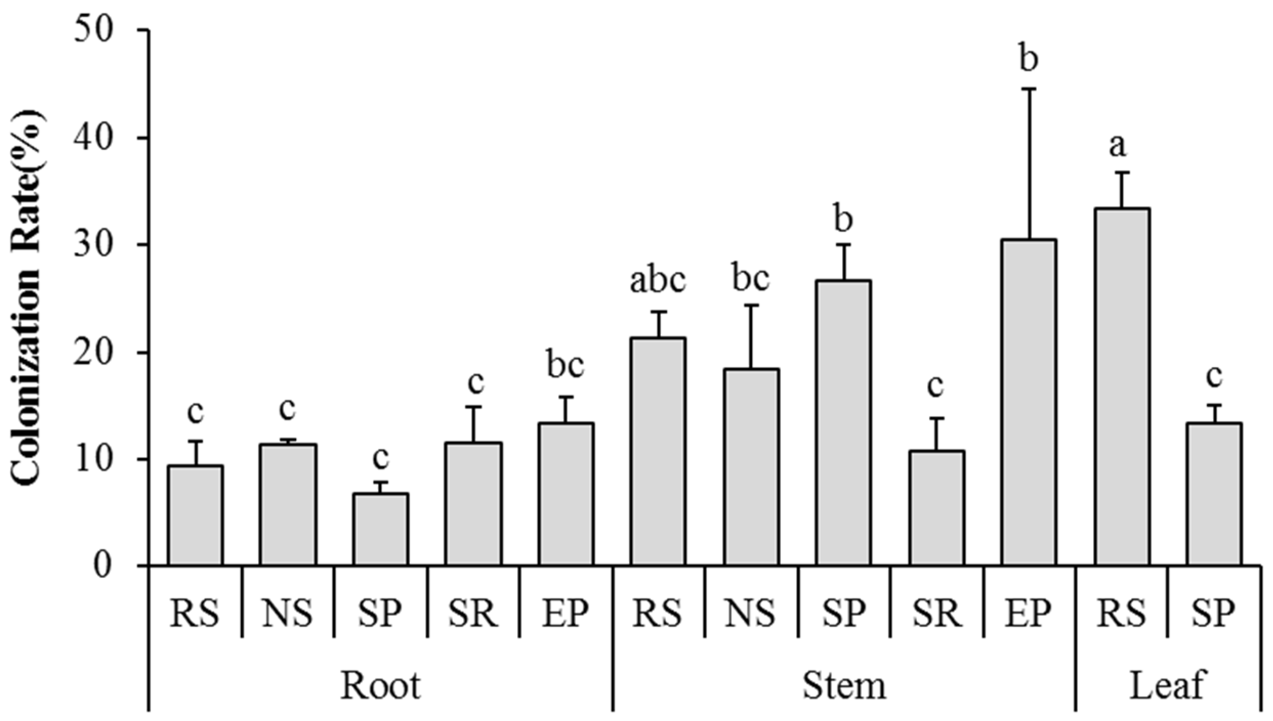
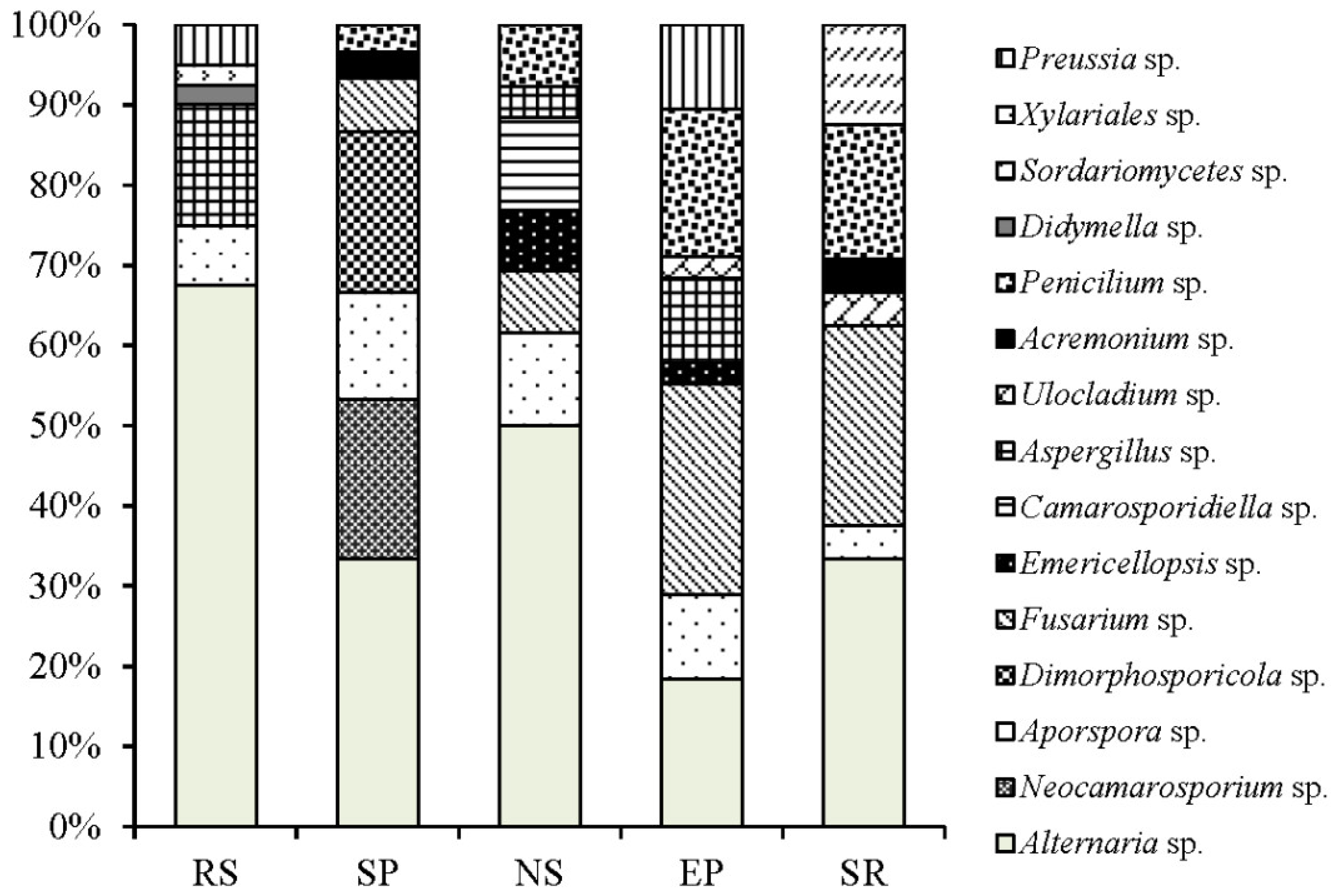
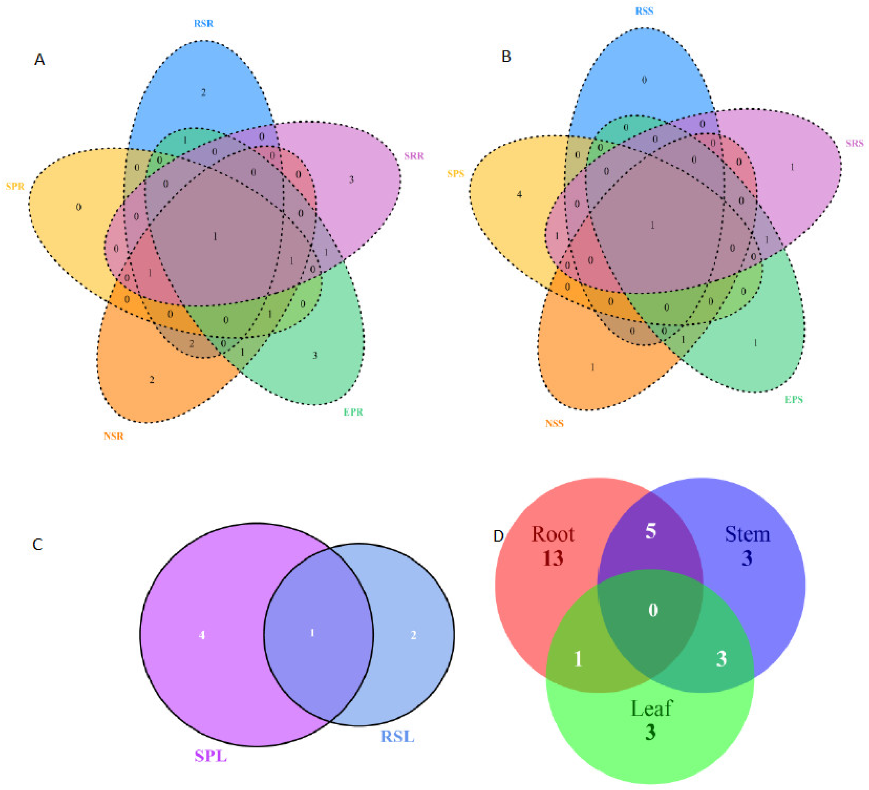
3.2. Community of Endophytic Fungi

A total of 158 endophytic fungi were isolated from 1088 tissue segments of all tested plants growing naturally in extremely arid desert of Anxi (Table 1). The CR of different plant tissues ranged from 7% to 33% (Figure 1). There was no significant difference of root CR among five plants. While the CR of stem in E. przewalskii and S. passerina were significantly higher (p < 0.05) than S. regelii, and the CR of leaves in R. soongorica was significantly higher than that of S. passerina. Overall, the CR of aboveground tissues had a higher value than that of belowground roots (Figure 1).
The endophytic fungal isolates were identified to 28 taxa represented by 25 species, belonging to 15 genera based on ITS sequence analysis (Table 1). The phylogenesis of the maximum likelihood tree based on the ITS4-5.8S-ITS5 rDNA was shown in Figure S1. The composition of endophytic fungal genera from different plants varied (Figure 2). Genus of Alternaria sp. and Aporspora sp. were all presented among five plants, and Alternaria sp. was the dominant genus which accounting for a proportion and generic abundance ranged from 20% to 65% (Figure 2). Dimorphosporicola sp. and Neocamarosporium sp. were only discovered in S. passerina, and Xylariales sp. and Didymella sp. were endemic to the R. soongorica. Additionally, Camarosporidiella sp. and Sordariomycetes sp. were only found in N. sphaerocarpa and S. regelii, respectively.
Endophytic fungal species differ significantly in different host plants. Of 25 species, 11 fungal species were respectively isolated in the rhizosphere of S. passerina, N. sphaerocarpa, and E. przewalskii; 10 species from R. soongorica; and 10 species from S. regelii (Table 1; Figure 3). Among them, the root of five plants shared only one species of Aporspora terricola with a varied RF of 5.56–44.44% (Table 1; Figure 3A). Alternaria alternate was dominantly and commonly isolated from all stem and leaf tissues of plants with a RF range from 22.22% to 100% (Table 1; Figure 3B,C). Except Alternaria alternate, the dominant endophytic fungi of R. soongorica was Aspergillus fumigatiaffinis showing RF of 25%; the dominant endophytic fungi of S. passerina were Aporspora terricola and Dimorphosporicola tragani, showing RF of 44.44% and 30.77%, respectively (Table 1). Aporspora terricola, Alternaria terricola, and Camarosporidiella eufemiaea were dominant in the plant of N. sphaerocarpa, which accounted for 18.75%, 18.75%, and 30% of RF, respectively. Similarly, Fusarium redolens and Penicilium chrysogenum in plant of E. przewalskii with RF of 25% and 33.33%, and Alternaria chlamydospora and Penicilium chrysogenum in plant of S. regelii with RF of 22.22%, respectively.
The fungal communities differed significantly as a function of tissue type (Table 1; Figure 3D). No endophytic fungi coexisted in three tissues of samples. The endophytic fungi of the root tissue occupied the most abundant which owned 13 species. Among the 13 root-fungus, 8 taxa were considered as dark septate endophytes (DSE), which were characterized by melanized structures and dark septate hyphae in culture (Figure S2). Camarosporidiella eufemiaea, Alternaria brassicae, and Neocamarosporium salicornicola were only isolated from the stems of plants, while Neocamarosporium salsolae, Aspergillus flavus, and Acremonium Sclerotigenum were restricted to the leaves of R. soongoric and S. passerina (Table 1; Figure 3D). The endophytic fungal community of roots from five plants was clustered together, while the endophytic fungi of shoot tissues (stems and leaves) was clustered to another group (Figure 4).

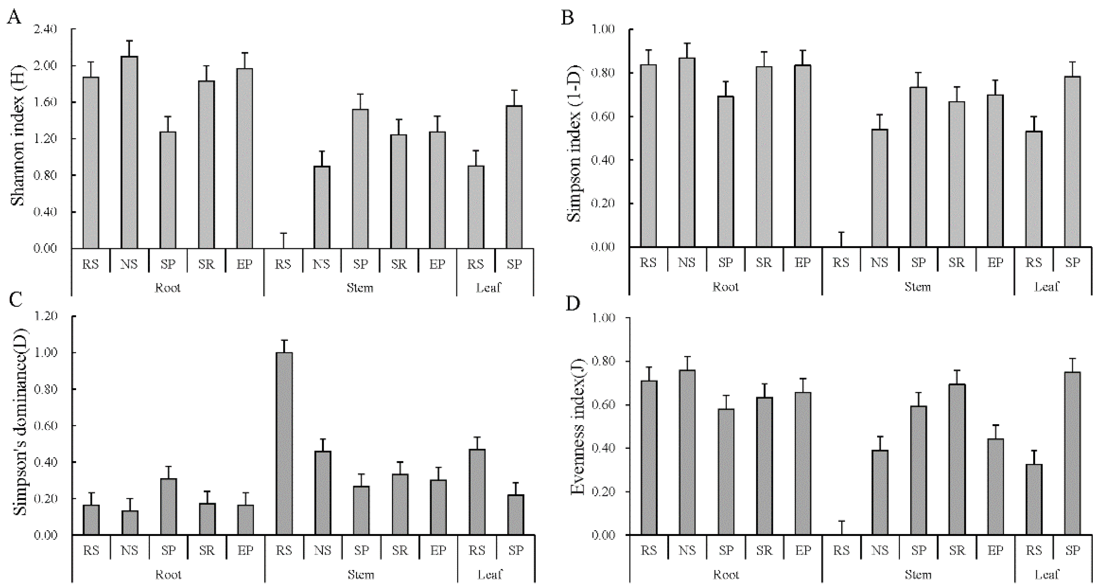
3.3. Diversity Indices of Endophytic Fungi

The root tissue diversity was considerably higher than that of the aboveground stem and leaf tissue except that S. passerina quantified larger leaf diversity than stems followed by roots (Figure 5). The highest values of the Shannon–Weiner and Simpson index were recorded in N. sphaerocarpa roots (2.10 and 0.87), while stems (1.52 and 0.73) and leaves (1.56 and 0.78) of S. passerina recorded the highest value among five plants (Figure 5A,B). The dominance and evenness indices are indicators to characterize the composition of endophytic community. The greater ecological dominance represented highly uneven distribution of species within the community. Evenness and equitability indices were highest in root of N. sphaerocarpa, and evenness of root tissues was considerably higher than that of the aboveground tissue except the stem of S. regelii and the leaf of S. passerina (Figure 5D). The Simpson’s dominance in stem of R. soongorica was the highset, thus representing highly uneven distribution of species in fungal community (Figure 5C).

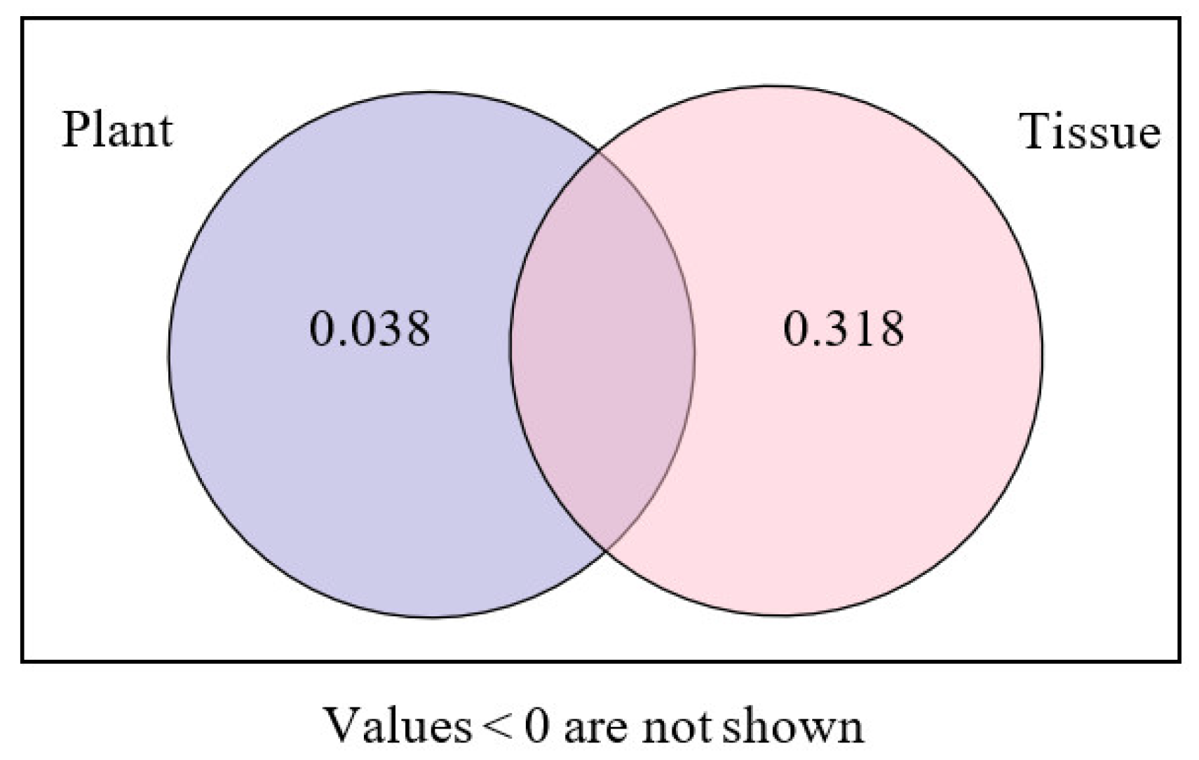
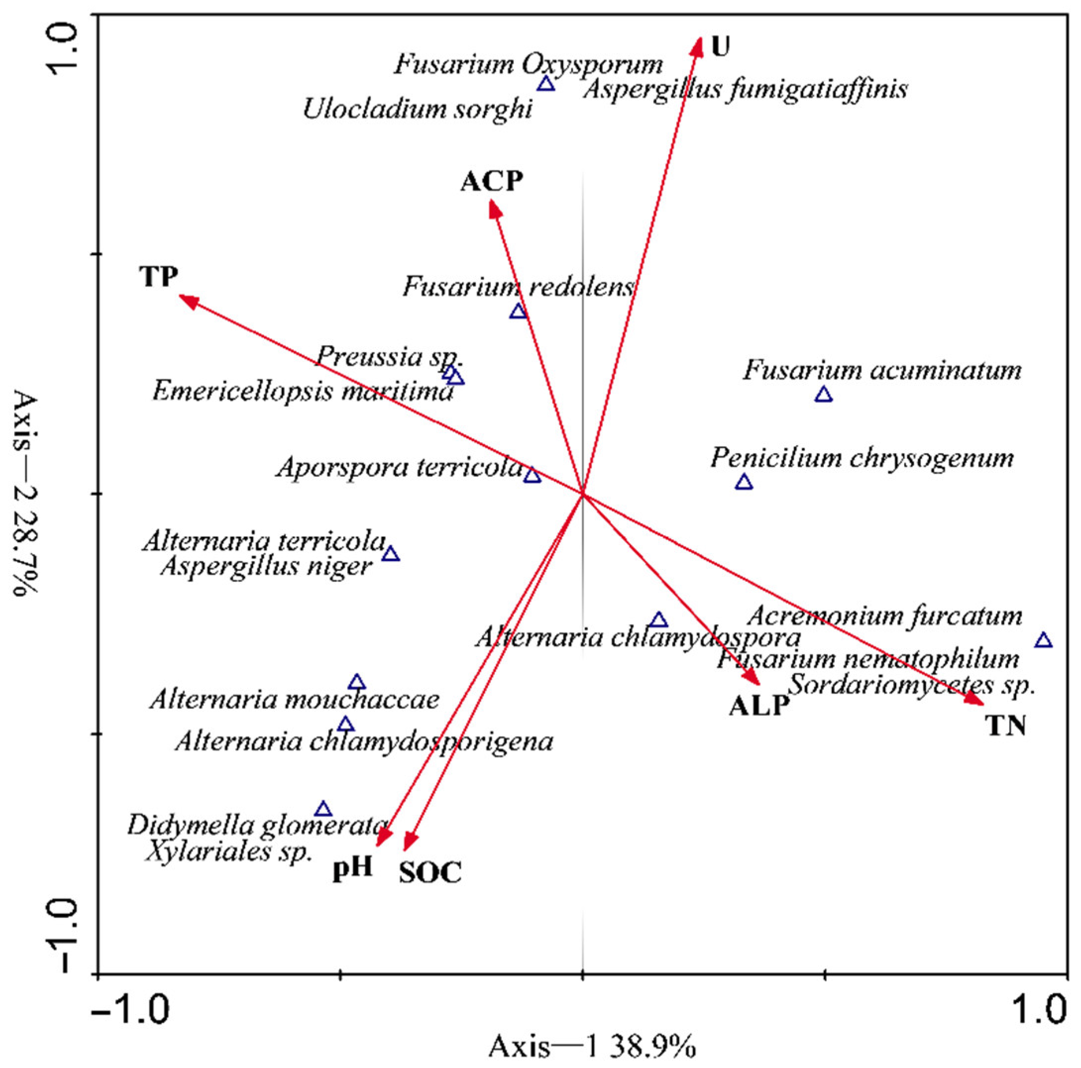
3.4. Variation Partitioning and Canonical Correlation Analysis

Variance partitioning analysis was performed to quantify the contribution of plant species and tissues on abundance of endophytic fungi (Figure 6). The complete set of plant cover and tissues explained 27% of the variation of endophytic fungi. There were no simultaneous effects of two types of dominant habitat factors to explain the variation of fungi communities. The pure variability of plant tissue on endophytic fungi explained 31.8%, which is much more than that of plant species. Therefore, endophytic fungi probably exhibit stronger tissue specificity.
Root fungal communities including 19 taxa (16 on species level and 3 on genus level) were calculated along with soil physicochemical properties using canonical correlation analysis (CCA), of which the first and second axes explained 67.6% of the variation (Figure 7). The frequency of occurrence of Fusarium redolens and Alternaria chlamydospora were closely correlated to ACP and ALP, respectively. Emericellopsis maritime, Preussia sp., and Aporspora terricola were positively affected by TP and negatively affected by TN, while Acremonium furcatum, Fusarium nematophilum, and Sordariomycetes sp. were affected positively by TN and negatively by TP. Moreover, the occurrences of Didymella glomerata and Xylariales sp. were affected by pH and SOC.

4. Discussion

4.1. Endophytic Fungal Community

In the present study, endophytic fungal strains isolated from different tissues of R. soongorica, N. sphaerocarpa, S. passerina, S. regelii, and E. przewalskii were subdivided by 25 species belonging to 15 genera based on morphological and molecular identification. This level of endophytic fungal diversity associated with xerophytic shrubs in extremely arid desert Northwest China was significantly lower than that grasses and sedges in semiarid regions and forests in other temperate zones [14,61,62,63]. Considering the special habitat of arid areas, it seems that the harsh environmental conditions of the extremely arid desert really drive down the growth and diversity of endophytic fungi. Arnold and Lutzoni [64] also detected a low infection rate of endophytic fungal communities in the Sonoran Desert. Meanwhile, compared to previous investigations in similar extreme environment [65,66,67], the diversity of these fungal communities is still relatively high in arid desert, which may be associated with microenvironmental heterogeneity that is characteristic of arid desert habitats [68,69,70].
Interestingly, the colonization rate of root tissue was significantly lower than that of shoots among the five plants, but the root-endophytic fungi conversely presented a higher diversity than stem and leaf. This probably can be related to the way in which endophytic fungi are transmitted in arid environments [27,64,71]. Most root associated symbiosis, belonging to the Class 4 of the nonclavicipitaceous endophytes, is horizontally transferred from plant to plant [2,72]. In desert habitats, vegetation is sparsely distributed with low diversity and density, but the belowground systems of plants are well developed, forming patches of nutrient-rich resources [73]. Thus, the distribution of extended root systems and nutrient status in a desert environment may lead to higher endophytic fungal diversity in the roots than shoot tissues by providing more host and substrate for infection of these fungi [74,75].
Among 28 taxa, all cultured strains belong to Ascomycota and half of the species were Pleosporales, which is consistent with previous reports that Pleosporales have been frequently isolated and found in arid or semi-arid environments [76]. Furthermore, most Ascomycota members are saprophytic and therefore the main decomposers in soil, which are able to decompose large amounts of refractory organic matter, thus playing an important role in nutrient cycling [77]. A. alternata was the most frequently isolated from all plants and was dominated in all stem and leaf tissues, which was consistent with the results of Sun et al. [21] and Massimo et al. [25], and A. alternata was also isolated from other symptomless halophytes as a dominant species [78,79,80]. The taxa of Neocamarosporium sp. have been reported to be isolated from desert plants in the central desert of Iran and can promote the agronomic parameters of tomato and cucumbers under different levels of salinity and drought stress [81]. Aspergillus was identified as the most common genus in the Hyoscyamus muticus L. (Egyptian henbane), which is one of the desert medicinal plants of family Solanaceae [67], while Emericellopsis maritime was reported as a DSE fungi in the roots of desert plant Lycium ruthenicum [82]. Didymella glomerata, Acremonium sp. and Xylariales sp. have been found in stems and roots of halophytes in desert habitats [83,84], and Ulocladium sp. was reported as halophilic fungi of desert soils in Saudi Arabia [85]. Moreover, the taxa of Preussia have been isolated from several species of desert plants, such as Hedysarum scoparium, Lycium ruthenicum, Aloe vera, and even Cactaceae plants [69,82,86,87]. Environments contain most microorganisms required for the assembly of the plant microbiome, and have been considered as the primary origin of the plant microbiome [88]. Therefore, the relatively consistent environmental conditions in desert habitats may explain the certain degree of similarity in plant microflora. The genera of Penicillium and Fusarium have been isolated as common endophytic fungi from a wide range of different host plants [89,90], while Aporospora terricola, Dimorphosporicola tragani, and Camarosporidiella eufemiaea were reported for the first time in extremely arid deserts.

4.2. Host Affiliations and Tissue Specificity

Previous studies have assessed the association of fungal endophytes with plant species and indicating that endophytic fungi may prefer specific hosts [26,28]. In our study, the endophytic species differed among the five host plants, some fungal species exist simultaneously within two or more plants, while several endophytic fungi such as Dimorphosporicola sp. and Camarosporidiella sp. were detected only in one host plant. This result supported the conclusion that endophytic fungi have no host specificity but may exhibit host preference. The five endemic desert plants in this study belong to different family levels. Although desert endophytes may undergo particularly strong selection to act as host-generalists, several investigations have declared that partial endophytes probably harbored host preferences at the plant family level [21,91]. Another reason explaining the host preference of endophytes can be attributed to the different enzymes produced by endophytic fungi [92]. Some endophytic fungi can produce enzymes that dissolve a variety of plant cell walls, allowing them to enter and colonize a wide range of hosts, but the fungus presenting host specificity may only produce a specific enzyme that permits the endophyte to infect only certain host plant cells [93,94].
Except host preference, tissue specificity of endophytes has already been demonstrated [95,96,97]. In this study, heatmaps of the relative abundance of culturable fungi revealed differentiated occurrence patterns between shoots (stems and leaves) and roots tissue niches, and the variation partitioning analysis also conducted a more pure variability of plant tissue (31.8%) on endophytic fungi than plant species (3.8%). These findings suggest that plant tissue niche has more influence on the endophytic fungal community than host affiliations in desert environments. Differences in endophytic assemblages associated with different tissue types might be a reflection of individual tissue preferences for dominant taxa [98], and might indicate their capacity for utilizing or surviving on a specific substrate [99,100]. Moreover, the differentiation of endophytic fungal communities in shoot and root tissues may be related to the environmental variation and heterogeneity between above- and belowground ecosystems in desert [25,77]. The light intensity, humidity, and temperature of the habits in which different tissues are survived and chemical activity of different tissues against fungal infections can absolutely lead to differentiation in endophytic fungal community and tissue specificity [101,102].

4.3. Root Endophytic Assemblages and Influence of Soil Factors

Fungal symbiosis is essential for most plants to absorb nutrients from their root systems, and these associations of symbiosis can facilitate plant growth by increasing the absorption of soil water and mineral elements [82]. In this study, eight root-fungal isolates of 13 taxa, characterized by melanized structures and dark septate hyphae in culture, were identified as dark septate endophytes (DSE) fungi, which suggested that DSE fungi was dominant in this extremely arid desert environment. Previous studies on desert vegetation have demonstrated that DSE fungi can form symbiotic relationships with most desert plants such as Ammopiptanthus mongolicus, Gymnocarpos przewalskii, Hedysarum scoparium, Psammochloa villosa, Hedysarum leave, and Artemisia ordosica [26,69,103,104], accompanying typical dark septate mycelium and melanized microsclerotia in plant root cells. Li et al. [105] have demonstrated that DSE fungi isolated from desert plants showed high tolerance to water deficiency in vitro and enhanced the host root growth under drought conditions through altered root development. In addition, the melanin synthesized by DSE can enhance the survival and competitiveness of endophytic fungi in adverse environments and protect them from ultraviolet light and lysozymes [106]. The accumulation of melanin and prevalence DSE in plants in the desert ecosystem provides a theoretical basis for enhancing the stress resistance of DSE to host plants.
The endophytic composition of a plant clearly depends on host identity as well as on the geographic location of the host [107]. A large number of studies have demonstrated the substantial effects of soil nutrient conditions and pH levels on soil fungal communities [44,108]. In this study, the frequency of occurrence of Fusarium redolens and Alternaria chlamydospora were closely correlated to ACP and ALP, respectively. Soil enzymes are the main exudates of soil microorganisms and plant roots, and the activity of phosphatase can be used to characterize soil phosphorus cycling [69]. Partial endophytic fungi can produce a diverse battery of enzymes; therefore, the association of theses fungi with host plants is required to facilitate the efficiency of nutrient decomposition and acquisition in arid environments [109]. Moreover, the occurrences of Didymella glomerata and Xylariales sp. were affected by pH and SOC. It has been well documented that pH is the most important factor that affects the quantity and diversity of soil fungi, and fungi prefer to live in slightly acidic environments [37,110]. Soil organic matter is the dominant carbon source for microbes [111], and soil carbon sources have been identified as key ecological drivers of microbial community dynamics [112]. Although pH was lightly alkaline in our study, the important role of SOC in an arid ecosystem can have a greater positive effect on microbial growth than other factors [77]. In our studies, Emericellopsis maritime, Preussia sp. Aporspora terricola responded positively to TP and negatively to TN, while Acremonium furcatum, Fusarium nematophilum, and Sordariomycetes sp. were positively affected by TN and negatively by TP. This was probably related to the metabolic process of fungi under different resource utilization. Studies have contributed to our understanding that differences in resource availability could have hampered the growth of some, and stimulated the growth of other fungal species [113]. Depending on whether plants grow in soil with low or high nutrient availability; hence, the growth of fungal communities associated with host plants may vary. Underground microenvironments in arid ecosystems play an important role in the material cycle, energy flow, and multi-element balance of ecosystems. Therefore, the influence of the soil environment should be considered to evaluate the impact of fungal endophytic communities on ecosystem functioning.

5. Conclusions

This study elucidated the ecological process of endophytic fungi associated with xerophyte shrubs across different plant tissue niche levels in the desert region of Northwest China. We found that endophytic fungal species demonstrated pronounced host and tissue preferences, and the community of endophytic fungi was differentiated obviously between above- and belowground tissues. Root endophytic fungi revealed the highest species diversity, and responded strongly to soil nutrient availability and enzymatic activity, such as soil pH, organic carbon, and phosphatase. Plant–fungal interactions and ecological distribution patterns of endophytic fungi associated with host affiliations represented a critical element of desert plants adapting to extremely arid desert environment. Therefore, our findings would be helpful in the exploitation of fungal resources in desert habitats and provide evidence for the application of fungal biotechnology in vegetation restoration and agricultural optimization.

Supplementary Materials

The following supporting information can be downloaded at: https://www.mdpi.com/article/10.3390/agronomy12030727/s1, Figure S1: Maximum Likelihood tree based on rDNA ITS region sequences of 28 endophytic fungal isolates. Numbers (%) on the main branches represent bootstrap support values (for 1000 iterations). The scale bar shows a distance equal to 5% nucleotide diversity; Figure S2: Morphological characteristics of endophytic fungi characterized by melanized structures and dark septate hyphae in culture isolated from the roots of five host plants. (A–H) Colonies of different endophytic fungi, and (a–h) microscopic morphology of endophytic fungi. Scale bars (a–h) = 50 μm; Table S1: Physico-chemical characteristics of soils. Different lowercase letters represent significant differences among different plant species (p < 0.05). TN, total nitrogen; TP, total phosphorus; SOC, soil organic carbon; ACP, acid phosphatase; ALP, alkaline phosphatase; U, urease.

Author Contributions

Conceptualization, Y.Z. and X.H.; Methodology, Y.Z. and X.H.; Formal analysis, Y.Z. and Q.H.; Investigation, Y.Z., Q.H. and K.Z.; Resources, Y.Z. and K.Z.; Writing—original draft preparation, Y.Z.; Writing—review and editing, Y.Z., Q.H. and K.Z.; Supervision, X.H.; Project administration, X.H.; Funding acquisition, X.H. All authors have read and agreed to the published version of the manuscript.

Funding

This research was funded by the National Natural Science Foundation of China, grant number 31770561.

Institutional Review Board Statement

Not applicable.

Informed Consent Statement

Not applicable.

Data Availability Statement

Not applicable.

Acknowledgments

We greatly appreciate the support of Anxi Extremely Arid Desert of the National Nature Reserve for their invaluable assistance on this experiment. We thank the students of Yedong Li and Wenbin Wang for sampling and laboratory work.

Conflicts of Interest

The authors declare no conflict of interest.

References

  1. Petrini, O. Fungal Endophytes of Tree Leaves. In Microbial Ecology of Leaves; Brock/Springer Series in Contemporary Bioscience; Andrews, J.H., Hirano, S.S., Eds.; Springer: New York, NY, USA, 1991; pp. 179–197. [Google Scholar]
  2. Rodriguez, R.J.; White, J.; Arnold, A.E.; Redman, R. Fungal endophytes, diversity and ecological roles. New Phytol. 2009, 182, 314–330. [Google Scholar] [CrossRef]
  3. Balla, K.M.; Troemel, E.R. Caenorhabditis elegansas a model for intracellular pathogen infection. Cell. Microbiol. 2013, 15, 1313–1322. [Google Scholar] [CrossRef] [PubMed] [Green Version]
  4. Yoo, J.-H. Review of Disinfection and Sterilization—Back to the Basics. Infect. Chemother. 2018, 50, 101–109. [Google Scholar] [CrossRef] [PubMed]
  5. Stewart, C.R.A.; Doilom, M.; Taylor, J.E. Analysis of fungal endophytes in Scottish Sitka spruce plantations shows extensive infections, novel host partners and gives insights into origins. For. Pathol. 2019, 49, e12471. [Google Scholar] [CrossRef] [Green Version]
  6. Arnold, A.E.; Miadlikowska, J.; Higgins, K.L.; Sarvate, S.D.; Gugger, P.; Way, A.; Hofstetter, V.; Kauff, F.; Lutzoni, F. A phylogenetic estimation of trophic transition networks for ascomycetous fungi: Are lichens cradles of symbiotrophic fungal diver-sification? Syst. Biol. 2009, 58, 283–297. [Google Scholar] [CrossRef] [PubMed] [Green Version]
  7. Pickles, B.J.; Gorzelak, M.A.; Green, D.S.; Egger, K.N.; Massicotte, H.B. Host and habitat filtering in seedling root-associated fungal communities: Taxonomic and functional diversity are altered in ‘novel’ soils. Mycorrhiza 2015, 25, 517–531. [Google Scholar] [CrossRef] [PubMed]
  8. Zhang, T.; Yao, Y.-F. Endophytic Fungal Communities Associated with Vascular Plants in the High Arctic Zone Are Highly Diverse and Host-Plant Specific. PLoS ONE 2015, 10, e0130051. [Google Scholar] [CrossRef] [PubMed] [Green Version]
  9. Kumar, A.; Verma, J.P. Does plant—Microbe interaction confer stress tolerance in plants: A review? Microbiol. Res. 2018, 207, 41–52. [Google Scholar] [CrossRef]
  10. Gunatilaka, L.A.A. Natural products from plant-associated microorganisms, Distribution, structural diversity, bioactivity and implications of their occurrence. J. Nat. Prod. 2006, 69, 509–526. [Google Scholar] [CrossRef] [Green Version]
  11. Ali, A.H.; Radwan, U.; El-Zayat, S.; El-Sayed, M.A. Desert plant-fungal endophytic association: The beneficial aspects to their hosts. Biol. Forum Int. J. 2018, 10, 138–145. [Google Scholar]
  12. Marasco, R.; Rolli, E.; Ettoumi, B.; Vigani, G.; Mapelli, F.; Borin, S.; Abou-Hadid, A.F.; El-Behairy, U.A.; Sorlini, C.; Cherif, A.; et al. A Drought Resistance-Promoting Microbiome Is Selected by Root System under Desert Farming. PLoS ONE 2012, 7, e48479. [Google Scholar] [CrossRef] [PubMed]
  13. Sieber, T.N.; Grünig, C.R. Fungal Root Endophytes. In Plant Roots—The Hidden Half, 4th ed.; Eshel, A., Beeckman, T., Eds.; CRC Press: Boca Raton, FL, USA, 2013; pp. 38–49. [Google Scholar]
  14. Loro, M.; Valero-Jiménez, C.; Nozawa, S.; Márquez, L. Diversity and composition of fungal endophytes in semiarid Northwest Venezuela. J. Arid Environ. 2012, 85, 46–55. [Google Scholar] [CrossRef]
  15. Hubbard, M.; Germida, J.; Vujanovic, V. Fungal endophytes improve wheat seed germination under heat and drought stress. Botany 2012, 90, 137–149. [Google Scholar] [CrossRef]
  16. Singh, L.P.; Gill, S.S.; Tuteja, N. Unraveling the role of fungal symbionts in plant abiotic stress tolerance. Plant Signal. Behav. 2011, 6, 175–191. [Google Scholar] [CrossRef] [Green Version]
  17. Fellbaum, C.R.; Mensah, J.A.; Cloos, A.J.; Strahan, G.E.; Pfeffer, P.E.; Kiers, T.; Bücking, H. Fungal nutrient allocation in common mycorrhizal networks is regulated by the carbon source strength of individual host plants. New Phytol. 2014, 203, 646–656. [Google Scholar] [CrossRef]
  18. Prieto, I.; Armas, C.; Pugnaire, F.I. Water release through plant roots: New insights into its consequences at the plant and ecosystem level. New Phytol. 2012, 193, 830–841. [Google Scholar] [CrossRef]
  19. Rilling, M.C.; Mummey, D.L. Mycorrhizas and soil structure. New Phytol. 2006, 171, 41–53. [Google Scholar] [CrossRef]
  20. Sikes, B.A.; Cottenie, K.; Klironomos, J.N. Plant and fungal identity deteimines pathogen protection of plant roots by arbuscular mycorrhizas. J. Ecol. 2009, 97, 1274–1280. [Google Scholar] [CrossRef] [Green Version]
  21. Sun, X.; Ding, Q.; Hyde, K.; Guo, L. Community structure and preference of endophytic fungi of three woody plants in a mixed forest. Fungal Ecol. 2012, 5, 624–632. [Google Scholar] [CrossRef]
  22. White, J.F.; Torres, M.S.; Johnson, H.; Irizarry, I.; Tadych, M.A. Functional View of Plant Microbiomes: Endosymbiotic Systems That Enhance Plant Growth and Survival. In Advances in Endophytic Research; Verma, V., Gange, A., Eds.; Springer: New Delhi, India, 2013; pp. 425–439. [Google Scholar]
  23. Rodriguez, R.J.; Woodward, C.; Redman, R.S. Adaptation and Survival of Plants in High Stress Habitats via Fungal Endophyte Conferred Stress Tolerance. In Cellular Origin, Life in Extreme Habitats and Astrobiology; Seckbach, J., Grube, M., Eds.; Springer Science and Business Media LLC: Berlin/Heidelberg, Germany, 2010; Volume 17, pp. 461–476. [Google Scholar]
  24. Fracchia, S.; Krapovickas, L.; Aranda-Rickert, A.; Valentinuzzi, V. Dispersal of arbuscular mycorrhizal fungi and dark septate endophytes by Ctenomys cf. knighti (Rodentia) in the northern Monte Desert of Argentina. J. Arid Environ. 2011, 75, 1016–1023. [Google Scholar] [CrossRef]
  25. Herrera, J.; Khidir, H.H.; Eudy, D.M.; Porras-Alfaro, A.; Natvig, D.O.; Sinsabaugh, R.L. Shifting fungal endophyte communities colonize Bouteloua gracilis: Effect of host tissue and geographical distribution. Mycologia 2010, 102, 1012–1026. [Google Scholar] [PubMed] [Green Version]
  26. Hou, L.; He, X.; Li, X.; Wang, S.; Zhao, L. Species composition and colonization of dark septate endophytes are affected by host plant species and soil depth in the Mu Us sandland, northwest China. Fungal Ecol. 2019, 39, 276–284. [Google Scholar] [CrossRef]
  27. Massimo, N.C.; Devan, M.M.N.; Arendt, K.R.; Wilch, M.H.; Riddle, J.M.; Furr, S.H.; Steen, C.; U’Ren, J.M.; Sandberg, D.C.; Arnold, A.E. Fungal Endophytes in Aboveground Tissues of Desert Plants: Infrequent in Culture, but Highly Diverse and Distinctive Symbionts. Microb. Ecol. 2015, 70, 61–76. [Google Scholar] [CrossRef] [PubMed]
  28. Del Olmo-Ruiz, M.; Arnold, A.E. Interannual variation and host affiliations of endophytic fungi associated with ferns at La Selva, Costa Rica. Mycologia 2014, 106, 8–21. [Google Scholar] [CrossRef]
  29. Sandberg, D.C.; Battista, L.J.; Arnold, A.E. Fungal Endophytes of Aquatic Macrophytes: Diverse Host-Generalists Characterized by Tissue Preferences and Geographic Structure. Microb. Ecol. 2014, 67, 735–747. [Google Scholar] [CrossRef]
  30. Bi, J.; He, D. Research advances in effects of plant on soil microbial diversity. Chin. Agric. Sci. Bull. 2009, 9, 244–250. [Google Scholar]
  31. Sardans, J.; Peñuelas, J. The Role of Plants in the Effects of Global Change on Nutrient Availability and Stoichiometry in the Plant-Soil System. Plant Physiol. 2012, 160, 1741–1761. [Google Scholar] [CrossRef] [Green Version]
  32. Eschen, R.; Hunt, S.; Mykura, C.; Gange, A.C.; Sutton, B.C. The foliar endophytic fungal community composition in Cirsium arvense is affected by mycorrhizal colonization and soil nutrient content. Fungal Biol. 2010, 114, 991–998. [Google Scholar] [CrossRef]
  33. David, A.S.; Seabloom, E.W.; May, G. Plant Host Species and Geographic Distance Affect the Structure of Aboveground Fungal Symbiont Communities, and Environmental Filtering Affects Belowground Communities in a Coastal Dune Ecosystem. Microb. Ecol. 2016, 71, 912–926. [Google Scholar] [CrossRef]
  34. Hartley, S.E.; Gange, A.C. Impacts of Plant Symbiotic Fungi on Insect Herbivores: Mutualism in a Multitrophic Context. Annu. Rev. Entomol. 2009, 54, 323–342. [Google Scholar] [CrossRef]
  35. Liu, D.; Huang, Y.; Sun, H.; An, S. The restoration age of Robinia pseudoacacia plantation impacts soil microbial biomass and microbial community structure in the Loess Plateau. Catena 2018, 165, 192–200. [Google Scholar] [CrossRef]
  36. Strobel, G.A. Methods of discovery and techniques to study endophytic fungi producing fuel-related hydrocarbons. Nat. Prod. Rep. 2014, 31, 259–272. [Google Scholar] [CrossRef] [PubMed] [Green Version]
  37. Wang, M.; Shi, S.; Lin, F.; Jiang, P. Response of the soil fungal community to multi-factor environmental changes in a temperate forest. Appl. Soil Ecol. 2014, 81, 45–56. [Google Scholar] [CrossRef]
  38. Liu, Y.B.; Li, X.R.; Li, M.M.; Liu, D.; Zhang, W.L. Leaf (or assimilation branch) epidermal micromorphology of desert plant in arid and semi-arid areas of China. Chin. J. Plant Ecol. 2016, 40, 1189–1207. [Google Scholar]
  39. Zhang, J.; Wu, B.; Zhu, Y.; Li, Y.; Lu, Q.; Yao, B. Responses of Nitraria tangutorum to water and photosynthetic physiology in rain enrichment scenario. Acta Ecol. Sin. 2013, 33, 172–177. [Google Scholar] [CrossRef]
  40. Li, S.; Su, P.; Zhang, H.; Zhou, Z.; Shi, R.; Gou, W. Hydraulic Conductivity Characteristics of Desert Plant Organs: Coping with Drought Tolerance Strategy. Water 2018, 10, 1036. [Google Scholar] [CrossRef] [Green Version]
  41. Alsharif, W.; Saad, M.M.; Hirt, H. Desert microbes for boosting sustainable sgriculture in extreme environments. Front. Microbiol. 2020, 11, 1666. [Google Scholar] [CrossRef]
  42. Lugo, M.A.; Reinhart, K.; Menoyo, E.; Crespo, E.M.; Urcelay, C. Plant functional traits and phylogenetic relatedness explain variation in associations with root fungal endophytes in an extreme arid environment. Mycorrhiza 2014, 25, 85–95. [Google Scholar] [CrossRef]
  43. Rodrigues, A.; Bacci, M.; Mueller, U.G.; Ortiz, A.; Pagnocca, F.C. Microfungal “weeds” in the leafcutter ant symbiosis. Microb. Ecol. 2008, 56, 604–614. [Google Scholar] [CrossRef]
  44. Wang, J.J.; Qiang, W.; Liu, H.Y.; Ge, J.L.; Zuo, Y.L.; He, X.L. Effects of plants pecies on diversity of arbuscular mycorrhizal fungi in extremely arid desert environment. Mycosystema 2017, 36, 861–869. [Google Scholar]
  45. Kimmins, J.P. Forest Ecology; China Forestry Publishing House: Beijing, China, 2005; Volume 258, pp. 17–43. [Google Scholar]
  46. Heiri, O.; Lotter, A.F.; Lemcke, G. Loss on ignition as a method for estimating organic and carbonate content in sediments: Reproducibility and comparability of results. J. Paleolimnol. 2001, 25, 101–110. [Google Scholar] [CrossRef]
  47. Bao, S.D. Agrochemical Analysis of Soil; Chinese Agricultural Press: Beijing, China, 2000; pp. 44–49. (In Chinese) [Google Scholar]
  48. Hoffmann, G.G.; Teicher, K. A colorimetric technique for determining urease activity in soil. Dung Boden 1961, 95, 55–63. [Google Scholar] [CrossRef]
  49. Tarafdar, J.C.; Marschner, H. Phosphatase activity in the rhizosphere and hyphosphere of VA mycorrhizal wheat supplied with inorganic and organic phosphorus. Soil Biol. Biochem. 1994, 26, 387–395. [Google Scholar] [CrossRef]
  50. Zhan, F.; He, Y.; Li, T.; Yang, Y.-Y.; Toor, G.S.; Zhao, Z. Tolerance and Antioxidant Response of a Dark Septate Endophyte (DSE), Exophiala pisciphila, to Cadmium Stress. Bull. Environ. Contam. Toxicol. 2014, 94, 96–102. [Google Scholar] [CrossRef] [PubMed]
  51. Parmar, S.; Li, Q.; Wu, Y.; Li, X.; Yan, J.; Sharma, V.K.; Wei, Y.; Li, H. Endophytic fungal community of Dysphania ambrosioides from two heavy metal-contaminated sites: Evaluated by culture-dependent and culture-independent approaches. Microb. Biotechnol. 2018, 11, 1170–1183. [Google Scholar] [CrossRef] [PubMed]
  52. Maadon, S.N.; Wakid, S.A.; Zainudin, I.I.; Rusli, L.S.; Mohd zan, M.S.; Hasan, N.A.; Abu Shah, N.A.; Rohani, E.R. Isolation and identification of endophytic fungi from UiTM reserve forest, Negeri Sembilan. Sains Malays. 2018, 47, 3025–3030. [Google Scholar] [CrossRef]
  53. White, T.J.; Bruns, T.; Lee, S.; Taylor, J.W. Amplification and Direct Sequencing of Fungal Ribosomal RNA Genes for Phylogenetics. In PCR Protocols, a Guide to Methods and Applications; Innis, M.A., Gelfand, D.H., Sninsky, J.J., White, T.J., Eds.; Academic Press, Inc.: New York, NY, USA, 1990; pp. 315–322. [Google Scholar]
  54. Tamura, K.; Stecher, G.; Peterson, D.; Filipski, A.; Kumar, S. MEGA6, molecular evolutionary genetics analysis version 6.0. Mol. Biol. Evol. 2013, 30, 2725–2729. [Google Scholar] [CrossRef] [Green Version]
  55. Yuan, Z.L.; Rao, L.B.; Chen, Y.C.; Zhang, C.L.; Wu, Y.G. From pattern to process, species and functional diversity in fungal endophytes of Abies beshanzuensis. Fungal Biol. 2011, 115, 197–213. [Google Scholar] [CrossRef]
  56. Shannon, C.E.; Weaver, W. The Mathematical Theory of Communication; University of Illinois Press: Urbana, IL, USA, 1949. [Google Scholar]
  57. Simpson, E.H. Measurement of species diversity. Nature 1949, 163, 688. [Google Scholar] [CrossRef]
  58. Caruso, T.; Pigino, G.; Bernini, F.; Bargagli, R.; Migliorini, M. The Berger–Parker Index as an Effective Tool for Monitoring the Biodiversity of Disturbed Soils: A Case Study on Mediterranean Oribatid (Acari: Oribatida) Assemblages. In Biodiversity and Conservation in Europe; Hawksworth, D.L., Bull, A.T., Eds.; Springer: Dordrecht, The Netherlands, 2007; Volume 7, pp. 35–43. [Google Scholar]
  59. Samaga, P.V.; Rai, V.R. Diversity and bioactive potential of endophytic fungi from Nothapodytes foetida, Hypericum mysorense and Hypericum japonicum collected from Western Ghats of India. Ann. Microbiol. 2016, 66, 229–244. [Google Scholar] [CrossRef]
  60. Borcard, D.; Gillet, F.; Legendre, P. Numerical Ecology with R; Springer: New York, NY, USA, 2011. [Google Scholar]
  61. Luo, J.; Walsh, E.; Miller, S.; Blystone, D.; Dighton, J.; Zhang, N. Root endophytic fungal communities associated with pitch pine, switchgrass, and rosette grass in the pine barrens ecosystem. Fungal Biol. 2017, 121, 478–487. [Google Scholar] [CrossRef] [PubMed] [Green Version]
  62. Johansen, R.B.; Johnston, P.; Mieczkowski, P.; Perry, G.L.W.; Robeson, M.S.; Vilgalys, R.; Burns, B.R. Scattered far and wide: A broadly distributed temperate dune grass finds familiar fungal root associates in its invasive range. Soil Biol. Biochem. 2017, 112, 177–190. [Google Scholar] [CrossRef]
  63. Bonfim, J.A.; Vasconcellos, R.L.F.; Baldesin, L.F.; Sieber, T.N.; Cardoso, E.J.B.N. Dark septate endophytic fungi of native plants along an altitudinal gradient in the Brazilian Atlantic forest. Fungal Ecol. 2016, 20, 202–210. [Google Scholar] [CrossRef]
  64. Arnold, A.E.; Lutzoni, F. Diversity and host range of foliar fungal endophytes: Are tropical leaves biodiversity hotspots? Ecology 2007, 88, 541–549. [Google Scholar] [CrossRef]
  65. Zhang, T.; Zhang, Y.Q.; Liu, H.Y.; Wei, Y.Z.; Li, H.L.; Su, J.; Zhao, L.X.; Yu, L.Y. Diversity and cold adaptation of culturable endophytic fungi from bryophytes in the Fildes Region, King George Island, maritime Antarctica. FEMS Microbiol. Lett. 2013, 341, 52–61. [Google Scholar] [CrossRef] [Green Version]
  66. Rosa, L.H.; Vaz, A.B.M.; Caligiorne, R.B.; Campolina, S.; Rosa, C.A. Endophytic fungi associated with the Antarctic grass Deschampsia antarctica Desv. (Poaceae). Polar Biol. 2009, 32, 161–167. [Google Scholar] [CrossRef] [Green Version]
  67. El-Zayat, S.A.; Nassar, M.S.M.; El-Hissy, F.T.; Abdel-Motaal, F.F.; Ito, S. Mycoflora associated with Hyoscyamus muticus growing under an extremely arid desert environment (Aswan region, Egypt). J. Basic Microb. 2008, 48, 82–92. [Google Scholar] [CrossRef]
  68. Porras-Alfaro, A.; Herrera, J.; Sinsabaugh, R.L.; Odenbach, K.J.; Lowrey, T.; Natvig, D.O. Novel root fungal consortium associated with a dominant desert grass. Appl. Environ. Microb. 2008, 74, 2805–2813. [Google Scholar] [CrossRef] [Green Version]
  69. Xie, L.; He, X.; Wang, K.; Hou, L.; Sun, Q. Spatial dynamics of dark septate endophytes in the roots and rhizospheres of Hedysarum scoparium in northwest China and the influence of edaphic variables. Fungal Ecol. 2017, 26, 135–143. [Google Scholar] [CrossRef]
  70. Qiang, W.; He, X.; Wang, J.; Zhao, L. Temporal and spatial variation of arbuscular mycorrhizal fungi under the canopy of Hedysarum scoparium in the northern desert, China. Appl. Soil Ecol. 2019, 136, 139–147. [Google Scholar] [CrossRef]
  71. Arnold, A.E.; Herre, E.A. Canopy cover and leaf age affect colonization by tropical fungal endophytes: Ecological pattern and process in Theobroma cacao (Malvaceae). Mycologia 2003, 95, 388–398. [Google Scholar] [CrossRef] [PubMed]
  72. Bayman, P.; Angulo-Sandoval, P.; Báez-Ortiz, Z.; Lodge, D.J. Distribution and dispersal of Xylaria endophytes in two tree species in Puerto Rico. Mycol. Res. 1998, 102, 944–948. [Google Scholar] [CrossRef]
  73. Zuo, Y.; He, C.; He, X.; Li, X.; Xue, Z.; Li, X.; Wang, S. Plant cover of Ammopiptanthus mongolicus and soil factors shape soil microbial community and catabolic functional diversity in the arid desert in Northwest China. Appl. Soil Ecol. 2020, 147, 103389. [Google Scholar] [CrossRef]
  74. Lau, M.K.; Arnold, A.E.; Johnson, N.C. Factors influencing communities of foliar fungal endophytes in riparian woody plants. Fungal Ecol. 2013, 6, 365–378. [Google Scholar] [CrossRef]
  75. U’Ren, J.M.; Lutzoni, F.; Miadlikowska, J.; Laetsch, A.D.; Arnold, A.E. Host and geographic structure of endophytic and endolichenic fungi at a continental scale. Am. J. Bot. 2012, 99, 898–914. [Google Scholar] [CrossRef] [Green Version]
  76. Knapp, D.; Kovacs, G.M.; Zajta, E.; Groenewald, J.; Crous, P. Dark septate endophytic pleosporalean genera from semiarid areas. Persoonia Mol. Phylogeny Evol. Fungi 2015, 35, 87–100. [Google Scholar] [CrossRef] [Green Version]
  77. Jia, T.; Wang, R.; Fan, X.; Chai, B. A Comparative Study of Fungal Community Structure, Diversity and Richness between the Soil and the Phyllosphere of Native Grass Species in a Copper Tailings Dam in Shanxi Province, China. Appl. Sci. 2018, 8, 1297. [Google Scholar] [CrossRef] [Green Version]
  78. Gond, S.K.; Mishra, A.; Sharma, V.K.; Verma, S.K.; Kumar, J.; Kharwar, R.N.; Kumar, A. Diversity and antimicrobial activity of endophytic fungi isolated from Nyctanthes arbor-tristis, a well-known medicinal plant of India. Mycoscience 2012, 53, 113–121. [Google Scholar] [CrossRef]
  79. Muhsin, T.M.; Booth, T. Fungi associated with halophytes of an inland salt marsh, Manitoba, Canada. Can. J. Bot. 1987, 65, 1137–1151. [Google Scholar] [CrossRef]
  80. Petrini, O.; Fisher, P. Fungal endophytes in Salicornia perennis. Trans. Br. Mycol. Soc. 1986, 87, 647–651. [Google Scholar] [CrossRef]
  81. Moghaddam, M.S.H.; Safaie, N.; Soltani, J.; Hagh-Doust, N. Desert-adapted fungal endophytes induce salinity and drought stress resistance in model crops. Plant Physiol. Biochem. 2021, 160, 225–238. [Google Scholar] [CrossRef] [PubMed]
  82. Han, L.; Shi, J.; He, C.; He, X. Temporal and Spatial Dynamics of Dark Septate Endophytes in the Roots of Lycium ruthenicum in the Desert Region of Northwest China. Agronomy 2021, 11, 648. [Google Scholar] [CrossRef]
  83. Li, J.-L.; Sun, X.; Zheng, Y.; Lü, P.-P.; Wang, Y.-L.; Guo, L.-D. Diversity and community of culturable endophytic fungi from stems and roots of desert halophytes in northwest China. MycoKeys 2020, 62, 75–95. [Google Scholar] [CrossRef] [PubMed]
  84. Jalili, B.; Bagheri, H.; Azadi, S.; Soltani, J. Identification and salt tolerance evaluation of endophyte fungi isolates from halophyte plants. Int. J. Environ. Sci. Technol. 2020, 17, 3459–3466. [Google Scholar] [CrossRef]
  85. Abdel-Hafez, S.I.I. Halophilic fungi of desert soils in Saudi Arabia. Mycopathol. Mycol. Appl. 1981, 75, 75–80. [Google Scholar] [CrossRef]
  86. Ameen, F.; Stephenson, S.L.; AlNadhari, S.; Yassin, M.A. Isolation, identification and bioactivity analysis of an endophytic fungus isolated from Aloe vera collected from Asir desert, Saudi Arabia. Bioprocess Biosyst. Eng. 2021, 44, 1063–1070. [Google Scholar] [CrossRef]
  87. Fonseca-García, C.; Coleman-Derr, D.; Garrido, E.; Visel, A.; Tringe, S.G.; Partida-Martínez, L.P. The cacti microbiome: Inter-play between habitat-filtering and host-specificity. Front. Microbiol. 2016, 7, 150. [Google Scholar] [CrossRef]
  88. Abdelfattah, A.; Wisniewski, M.; Schena, L.; Tack, A.J.M. Experimental evidence of microbial inheritance in plants and transmission routes from seed to phyllosphere and root. Environ. Microbiol. 2021, 23, 2199–2214. [Google Scholar] [CrossRef]
  89. Gautam, A.K. Diversity of fungal endophytes in some medicinal plants of Himachal Pradesh, India. Arch. Phytopathol. Plant Prot. 2014, 47, 537–544. [Google Scholar] [CrossRef]
  90. Wenzel, J.B.; García, A.; Filho, C.J.R.; Prioli, A.J.; Pamphile, J.A. Evaluation of foliar fungal endophyte diversity and colonization of medicinal plant Luehea divaricata (Martius et Zuccarini). Biol. Res. 2010, 43, 375–384. [Google Scholar] [CrossRef] [Green Version]
  91. Zhou, D.; Hyde, K.D. Host-specificity, host-exclusivity, and host-recurrence in saprobic fungi. Mycol. Res. 2001, 105, 1449–1457. [Google Scholar] [CrossRef]
  92. Sun, X.; Guo, L.-D.; Hyde, K.D. Community composition of endophytic fungi in Acer truncatum and their role in decomposition. Fungal Divers. 2011, 47, 85–95. [Google Scholar] [CrossRef]
  93. Rodriguez, R.; Redman, R. Fungal Life-Styles and Ecosystem Dynamics: Biological Aspects of Plant Pathogens, Plant Endophytes and Saprophytes. Adv. Bot. Res. 1997, 24, 169–193. [Google Scholar] [CrossRef]
  94. Suryanarayanan, T.S.; Thirunavukkarasu, N.; Govindarajulu, M.B.; Gopalan, V. Fungal endophytes: An untapped source of biocatalysts. Fungal Divers. 2012, 54, 19–30. [Google Scholar] [CrossRef]
  95. Kumar, D.S.S.; Hyde, K.D. Biodiversity and tissue-recurrence of endophytic fungi in Tripterygium wilfordii. Fungal Divers. 2004, 17, 69–90. [Google Scholar]
  96. Tahtamouni, M.E.; Khresat, S.; Lucero, M.; Sigala, J.; Unc, A. Diversity of endophytes across the soil-plant continuum for Atriplex spp. in arid environments. J. Arid Land 2015, 8, 241–253. [Google Scholar] [CrossRef]
  97. Aly, A.H.; Debbab, A.; Proksch, P. Fungal endophytes: Unique plant inhabitants with great promises. Appl. Microbiol. Biotechnol. 2011, 90, 1829–1845. [Google Scholar] [CrossRef]
  98. Wu, L.; Han, T.; Li, W.; Jia, M.; Xue, L.; Rahman, K.; Qin, L. Geographic and tissue influences on endophytic fungal communities of Taxus chinensis var. mairei in China. Curr. Microbiol. 2012, 66, 40–48. [Google Scholar] [CrossRef]
  99. Knapp, D.G.; Kovács, G.M. Interspecific metabolic diversity of root-colonizing endophytic fungi revealed by enzyme activity tests. FEMS Microbiol. Ecol. 2016, 92, 190. [Google Scholar] [CrossRef] [Green Version]
  100. Petrini, O.; Sieber, T.N.; Toti, L.; Viret, O. Ecology, metabolite production, and substrate utilization in endophytic fungi. Nat. Toxins 1993, 1, 185–196. [Google Scholar] [CrossRef]
  101. Schardl, C.L.; Leuchtmann, A.; Spiering, M.J. Symbioses of grasses with seed borne fungal endophytes. Annu. Rev. Plant Biol. 2004, 55, 315–340. [Google Scholar] [CrossRef] [PubMed]
  102. Tan, Y.Y.; Spiering, M.J.; Scott, V.; Lane, G.A.; Christensen, M.J.; Schmid, J. In Planta Regulation of Extension of an Endophytic Fungus and Maintenance of High Metabolic Rates in Its Mycelium in the Absence of Apical Extension. Appl. Environ. Microbiol. 2001, 67, 5377–5383. [Google Scholar] [CrossRef] [PubMed] [Green Version]
  103. Li, B.; He, X.; He, C.; Chen, Y.; Wang, X. Spatial dynamics of dark septate endophytes and soil factors in the rhizosphere of Ammopiptanthus mongolicus in Inner Mongolia, China. Symbiosis 2015, 65, 75–84. [Google Scholar] [CrossRef]
  104. Li, X.; He, X.; Hou, L.; Ren, Y.; Wang, S.; Su, F. Dark septate endophytes isolated from a xerophyte plant promote the growth of Ammopiptanthus mongolicus under drought condition. Sci. Rep. 2018, 8, 7896. [Google Scholar] [CrossRef] [Green Version]
  105. Li, X.; He, C.; He, X.; Su, F.; Hou, L.; Ren, Y.; Hou, Y. Dark septate endophytes improve the growth of host and non-host plants under drought stress through altered root development. Plant Soil 2019, 439, 259–272. [Google Scholar] [CrossRef]
  106. Deveautour, C.; Chieppa, J.; Nielsen, U.N.; Boer, M.M.; Mitchell, C.; Horn, S.; Power, S.A.; Guillen, A.; Bennett, A.E.; Powell, J.R. Biogeography of arbuscular mycorrhizal fungal spore traits along an aridity gradient, and responses to experimental rainfall manipulation. Fungal Ecol. 2020, 46, 100899. [Google Scholar] [CrossRef]
  107. Hoffman, M.T.; Arnold, A.E. Geographic locality and host identity shape fungal endophyte communities in cupressaceous trees. Mycol. Res. 2008, 112, 331–344. [Google Scholar] [CrossRef]
  108. Sui, X.; Zhang, R.; Xu, N.; Liu, Y.; Chai, C.; Wang, J.; Fu, X.; Zhong, H.; Ni, H. Fungal community structure of different degeneration Deyeuxia angustifolia wetlands in Sanjiang Plain. Environ. Sci. 2016, 37, 3598–3605. [Google Scholar]
  109. Abbassi, K.; Atay-Kadiri, Z.; Ghaout, S. Biological effects of alkaloids extracted from three plants of Moroccan arid areas on the desert locust. Physiol. Entomol. 2003, 28, 232–236. [Google Scholar] [CrossRef]
  110. Ju, T.; Chen, Y.; Chang, C.; An, L. The diversity of soil fungi and its relations with fertility factors in Taxus chinenesis (Pilg.) Rehd community of Xiaolongshan of Tianshui City. Res. J. Environ. Sci. 2008, 21, 128–132. [Google Scholar]
  111. Paterson, E.; Gebbing, T.; Abel, C.; Sim, A.; Telfer, G. Rhizodeposition shapes rhizosphere microbial community structure in organic soil. New Phytol. 2007, 173, 600–610. [Google Scholar] [CrossRef] [PubMed]
  112. de Vries, F.T.; Manning, P.; Tallowin, J.R.B.; Mortimer, S.R.; Pilgrim, E.S.; Harrison, K.A.; Hobbs, P.J.; Quirk, H.; Shipley, B.; Cornelissen, J.H.C.; et al. Abiotic drivers and plant traits explain landscape-scale patterns in soil microbial communities. Ecol. Lett. 2012, 15, 1230–1239. [Google Scholar] [CrossRef] [PubMed]
  113. Rasmussen, S.; Parsons, A.; Bassett, S.; Christensen, M.J.; Hume, D.; Johnson, L.; Johnson, R.; Simpson, W.; Stacke, C.; Voisey, C.; et al. High nitrogen supply and carbohydrate content reduce fungal endophyte and alkaloid concentration in Lolium perenne. New Phytol. 2007, 173, 787–797. [Google Scholar] [CrossRef] [PubMed]
Figure 1. Colonization rate (CR) of the endophytic fungi from five plant species in Anxi site. RS: R. soongorica; NS: N. sphaerocarpa; SP: S. passerina; SR: S. regelii; EP: E. przewalskii. Different letters above the error bars indicate significant difference at p < 0.05.
Agronomy 12 00727 g001
Figure 2. Column chart of the proportion of endophytic fungal genera at five plants in Anxi site. RS: R. soongorica; NS: N. sphaerocarpa; SP: S. passerina; SR: S. regelii; EP: E. przewalskii.
Agronomy 12 00727 g002
Figure 3. Venn diagram of the culturable endophytic fungi of five plants from Anxi. (A) Root endophytic fungi; (B) Stem endophytic fungi; (C) Leaf endophytic fungi; (D) Endophytic fungi in different tissue niches. RS = R. soongorica; NS = N. sphaerocarpa; SP = S. passerina; SR = S. regelii; EP = E. przewalskii; S = stem; R = root; L = leaf.
Agronomy 12 00727 g003
Figure 4. Heat maps of the relative abundance of culturable fungal endophytes from five plants in different tissues of Anxi. Different color means the different relative abundance of the taxa in the all tissues samples (red means high RF). RS = R. soongorica; NS = N. sphaerocarpa; SP = S. passerina; SR = S. regelii; EP = E. przewalskii; S = stem; R = root; L = leaf.
Agronomy 12 00727 g004
Figure 5. Assessment of α-diversity indices of entophytic fungi for each plant species and tissue. (A): Shannon index (H); (B): Simpson index (1-D); (C): Simposon’s dominance (D); (D): Evenness index (J). RS: R. soongorica; NS: N. sphaerocarpa; SP: S. passerina; SR: S. regelii; EP: E. przewalskii.
Agronomy 12 00727 g005
Figure 6. Variation partitioning analysis was used to tease apart the relative contribution of plant species and tissues on abundance of endophytic fungi. Residuals = 0.730. Values below 0 are not shown.
Agronomy 12 00727 g006
Figure 7. CCA plot indicating the relationships between the occurrence of root fungal endophytes among five plants and soil characteristics.
Agronomy 12 00727 g007
Table 1. Taxa, number of isolates (I) recovered and relative frequency (RF) of endophytic fungi in five plants from different tissues.
TaxaR. soongoricaS. passerinaN. sphaerocarpaE. przewalskiiS. regelii
RootStemLeafRootStemLeafRootStemRootStemRootStem
IRF%IRF%IRF%IRF%IRF%IRF%IRF%IRF%IRF%IRF%IRF%IRF%
Alternaria alternata 101001062.5 538.46 112.5 660 738.89 350.00
Alternaria brassicae 17.69
Neocamarosporium salicornicola 17.69
Alternaria chlamydospora214.29 222.22 17.69 16.25 422.22 116.67
Aporspora terricola321.43 444.44 318.75 420 15.56
Neocamarosporium salsolae 225
Dimorphosporicola tragani 430.77 225
Neocamarosporium chichastianum 17.69 225
Fusarium nematophilum 16.25 211.11
Emericellopsis maritima 212.5 15
Alternaria chlamydosporigena321.43 16.25
Alternaria terricola 318.75
Camarosporidiella eufemiaea 330
Aspergillus niger 316.67
Ulocladium sorghi 15 116.67
Fusarium redolens 222.22 212.5 525211.11 211.11
Aspergillus flavus 212.5
Aspergillus fumigatiaffinis 425 15
Acremonium furcatum 15.56
Fusarium acuminatum 210 15.56 116.67
Penicilium chrysogenum 111.11 16.2511015633.33 422.22
Alternaria mouchaccae214.29 212.5
Fusarium oxysporum 15
Acremonium sclerotigenum 112.5
Didymella glomerata17.14
Sordariomycetes sp. 316.67
Xylariales sp.17.14
Preussia sp.214.29 420
Total1424404962708696116134152158
Publisher’s Note: MDPI stays neutral with regard to jurisdictional claims in published maps and institutional affiliations.

