Article

Beneficial Effect of Exogenously Applied Calcium Chloride on the Anatomy and Fast Chlorophyll Fluorescence in Rhododendron × pulchrum Leaves Following Short-Term Heat Stress Treatment

1
Jiyang College of Zhejiang A&F University, Zhuji 311800, China
2
Management Office, Wuyanling National Nature Reserve, Wenzhou 325500, China
3
Department of Life Science and Health, Huzhou College, Huzhou 313000, China
*
Author to whom correspondence should be addressed.
These authors contributed equally to this work.
Agronomy 2022, 12(12), 3226; https://doi.org/10.3390/agronomy12123226
Submission received: 4 November 2022 / Revised: 9 December 2022 / Accepted: 14 December 2022 / Published: 19 December 2022

Abstract

:
The heat tolerance of plants can be improved by using exogenous calcium chloride (CaCl2) to cope with temperature fluctuations. Since global climates continue to warm, it is important to further explore the way in which plants respond to heat stress with the use of CaCl2. We aimed to explore the effect of exogenous CaCl2 on the leaf microstructure, leaf epidermal ultrastructure, and chlorophyll a fluorescence of Rhododendron × pulchrum (R. × pulchrum) under heat stress. In the leaves of R. × pulchrum treated with exogenous CaCl2, compared to the control, the thickness of the epidermis, spongy tissues, and stomatal aperture increased, whereas the stomata density and ratio of closed/open stomata decreased. In the leaves of R. × pulchrum under heat stress conditions, compared to the control, the values of the maximal photochemical efficiency of photosystem II (Fv/Fm), the performance index on an absorption basis (PIABS), the quantum yield for the reduction of terminal electron acceptors on the acceptor side of PSI (φRo), and the energy absorbed per unit cross-section of a photosynthesizing object at the moment of achieving the fluorescence maximum (ABS/CSM) all decreased, whereas the quantum yield of the energy dissipation (φDo) increased significantly. However, these differences disappeared when R. × pulchrum was treated with exogenous CaCl2. This suggests that exogenous CaCl2 can improve the heat tolerance in R. × pulchrum by regulating the leaf anatomical structure and the behavior of epidermal cells and stomata in leaves, protecting the stability of photosystems I and II and improving the electron transfer from QA to QB. Our study could provide a theoretical basis for the breeding, further research, and utilization of Rhododendron in the context of global warming.

1. Introduction

Global climates continue to warm, accompanied by an increase in CO2 concentration and sustained elevated temperatures [1]. As a result, green plants are negatively affected and will have to adapt to high temperatures in the future [2]. Green plants have the capacity to cope with temperature fluctuations; however, this capacity might not keep pace with global warming. The heat tolerance of plants can be improved using different methods, including genetic selection and the application of exogenous regulators, such as calcium chloride (CaCl2) and salicylic acid. These methods aid in physiological response adaptation in plants [3,4].
In model plant and crop studies, it has been shown that exogenous CaCl2 can improve heat resistance and enable adaptation to higher temperatures [5,6,7]. Ca2+, as a secondary messenger, regulates physiological and biochemical mechanisms in plants adapted to adverse environmental conditions. Ca2+ participates in the regulation of stomatal movements [3] and gene expression [8], influencing the chlorophyll content and antioxidant enzyme activity [9].
High-temperature stress can damage the homeostasis of the membrane system in plants and affect their photosynthetic energy metabolism and physiological and biochemical processes. Under heat stress, various metabolic activities in plants are influenced, such as their effects on the leaf phenotype, anatomy, physiology, and photosynthesis capability [10,11,12,13,14]. Previous studies have indicated that green plants are characterized by heat tolerance derived from their leaf anatomical and ultrastructural traits, including a decrease in thick leaves, the ratio of palisade to spongy parenchyma, and epidermis cell size, as well as stomatal closure and an increase in stomatal density [15,16,17]. Few studies have reported the effect of exogenous CaCl2 on the plant leaf structure.
The photosynthetic apparatus is one of the most sensitive components affected by heat stress [18,19]. The efficiency of the photosynthetic apparatus can affect chlorophyll fluorescence parameters and gas exchange, reflecting the photosynthetic electron transfer (light reaction) and photosynthetic rate (dark reaction), respectively [20,21]. In recent years, the measurement of chlorophyll fluorescence has been used to investigate photosynthetic characteristics in plant leaves under heat stress, including delayed chlorophyll a fluorescence (DF), prompted chlorophyll a fluorescence (PF), and modulated reflection at 820 nm (MR) [22]. Chlorophyll fluorescence parameters reflect the electron transfer in photosystem I (PSI), photosystem II (PSII), and photosynthetic electron carriers in the photosystem [23]. The measurement of chlorophyll fluorescence has been widely used to investigate changes in the leaves of plants under heat stress, such as tomato [24], wheat [25,26], barley [27], weeds [28], and Pinellia ternata [29]. In recent years, simultaneous measurements of DF, PF, and MR kinetics have been introduced to investigate the response of plants to environmental stresses, such as heat [22] and NaCl [30,31], used in the fields of agronomy, plant science, and environmental science [32,33]. For example, the PF kinetics showed an O-J-I-P curve (O, at 20 μs; J, at 2 ms; I, at 30 ms; P, equal to FM), which is used to quantify the architecture and behavior of PSII. In DF kinetics, parameters I1 (7 ms) and I2 (50 ms) indicate the electron transfer capacity of PSII and the re-oxidation of PQH2 by PSI reaction center (P700). MR/MR0 kinetics, usually as an indicator of the redox state changes in plastocyanin and P700 [32,33]. This method is important for investigating changes in the photosynthetic electron transfer chain, including the PSI electron acceptor side, electron transport between PSII and PSI, and the PSII electron donor side [22,23,34]. To date, little research has been conducted with respect to the effect of heat stresses on chlorophyll a fluorescence in the leaves of the Rhododendron species [35,36].
Rhododendron is one of the most popular ornamental plants worldwide, with excellent ornamental traits. The genus Rhododendron belongs to Ericaceae and contains more than 1000 species and 25,000 hybrids that are classified into eight subgenera [37,38]. A previous study constructed a phylogeny tree of 200 Rhododendron species according to the nuclear and chloroplast genes [39]. Rhododendron species are mostly distributed and native to high-altitude areas, growing at elevations between 1000 and 5000 m [40]. Many Rhododendron species are seriously affected by high temperatures in low-altitude areas, resulting in a decline in ornamental quality. Global warming is an emerging major challenge to the growth and development of Rhododendron species [9]. Thus, the heat resistance of Rhododendron species needs to be improved to face global challenges.
To date, only a limited number of studies have investigated the heat resistance of Rhododendron in response to exogenous CaCl2. Previous studies have suggested that exogenous CaCl2 has a positive effect on the plant growth, chlorophyll content, total soluble protein levels, and enzymatic antioxidant activity under heat stress [9,16]. However, investigations of the response of Rhododendron to heat stress when exposed to exogenous CaCl2 from anatomical and photosynthesis perspectives are scarce. R. × pulchrum Sweet, belonging to Subg. Tsutsusi of Rhododendron, is severely affected by high temperatures, appearing to grow normally or even die under high temperatures. In this study, we hypothesized that exogenous CaCl2 could aid the heat tolerance of R. × pulchrum, and we explored the effect of exogenous CaCl2 on the leaf microstructure, leaf epidermal ultrastructure, and chlorophyll a fluorescence of R. × pulchrum under heat stress. Our study could provide a theoretical basis for the breeding, further research, and utilization of Rhododendron to assist in sustaining forestry development [41].

2. Materials and Methods

2.1. Plant Material

Two-year-old cutting plants of R. × pulchrum were obtained from a single tree grown at the Jiyang College of Zhejiang A&F University, Zhejiang, China (29°44′51″ N, 120°15′17″ E). These cutting trees were planted in pots (15 cm tall, 20 cm top diameter) individually. Each pot was filled with 5 kg loam soil (field water-holding capacity of 33%) and watered when the pot was naturally dried (watered to saturation each time, approximately 0.3 L per pot). Six pots were randomly selected per treatment. Using the foliar sprays method, the different concentrations of CaCl2 were foliar sprayed with approximately 10 mL per plant, evenly spraying both sides of the leaves. The experiment set different concentrations of CaCl2 (0, 5, 10, 20, 30 mmol/L) based on similar studies. The results showed that 10 mmol/L was the best treatment. The heat resistance capacity can be significantly improved compared with the control group. Thus, four treatments were set up as follows: control treatment with distilled water (CK + W), control treatment with 10 mM CaCl2 (CK + Ca2+), heat stress treatment with distilled water (H + W), and heat stress treatment with 10 mM CaCl2 (H + Ca2+).
The experiment was carried out in a growth chamber at the Jiyang College of Zhejiang A&F University. The temperature within the chamber was 28 °C/25 °C (day/night), the photoperiod was 14/10 h (day/night), the light intensity was 600 µmol·m−2·s−1, and the humidity was 70 ± 5%. An aqueous solution of 10 mM CaCl2 (Yudinghuagong, Shandong, China) was applied daily at 5 p.m. using the foliar sprays method. The control treatment involved only water, and tests were conducted for 7 days. Then, the temperature within the chamber of heat stress treatments was changed to 40/30 °C (day/night), keeping soil water content stable during the experiment; the other conditions remained under the original conditions. Measurements were recorded 2 days after heat stress treatments.

2.2. Microstructure of the Leaves and Ultrastructure of Leaf Epidermal Cells

Mature leaves, selected from the third fully expanded leaf, were collected from the plants and cut into 2 mm × 4 mm pieces; dehydrated pieces were then obtained for subsequent experiments. To observe the microstructure of the leaves, semi-thin sections were made following Zhang’s method [42]. The dehydrated samples were embedded in epoxy resin and sliced with a LEICA DC6 ultramicrotome (Leica Microsystems, Wetzlar, Germany). Ultra-thin sections (1 µm thick) were cut following Ruppel’s method [43]. The sections were stained with toluidine blue [15,42] and observed under a microscope. To observe the ultrastructure of leaf epidermal cells, the samples were subjected to freeze-drying (K-850, Emitech Ltd., Kent, UK), after which, they were sprayed with gold particles (E-1010, Hitachi, Tokyo, Japan). The leaf epidermal cells were observed via scanning electron microscope (5136, TESCAN, Brno, Czech Republic). Three different vision fields under the microscope were randomly chosen, and each vision field was repeated three times. The size of the leaf tissues and leaf epidermal stomatal diameter were measured using ImageJ v1.8.0 software. Thirty cells and stomatal were measured and the ratio of palisade thickness to leaf thickness, the ratio of palisade to spongy parenchyma thickness, and stomatal aperture were calculated [44]. In addition, the number of stomata and cells per unit area was counted [45].

2.3. Measurement of PF, DF and MR Kinetics

The third and fourth leaves from the top of the shoots were used to determine the PF, DF, and MR kinetics simultaneously after 30 min of dark adaptation using a multi-function plant efficiency analyzer (PE304NE, M-PEA, Hansatech, Norfolk, UK). The PF, DF, and MR kinetic curves were plotted according to the methods described in previous studies [46,47,48]. Parameters were derived from chlorophyll a fluorescence [46,47]. Three replicates per treatment were randomly selected.

2.4. Statistical Analysis

The experimental design was a completely randomized design. The data were analyzed using SPSS 18.0 software (SPSS Inc. Chicago, IL, USA), and one-way ANOVA and Duncan’s multiple range test were performed for the data variable, with the significance level set at the default of α = 0.05.

3. Results

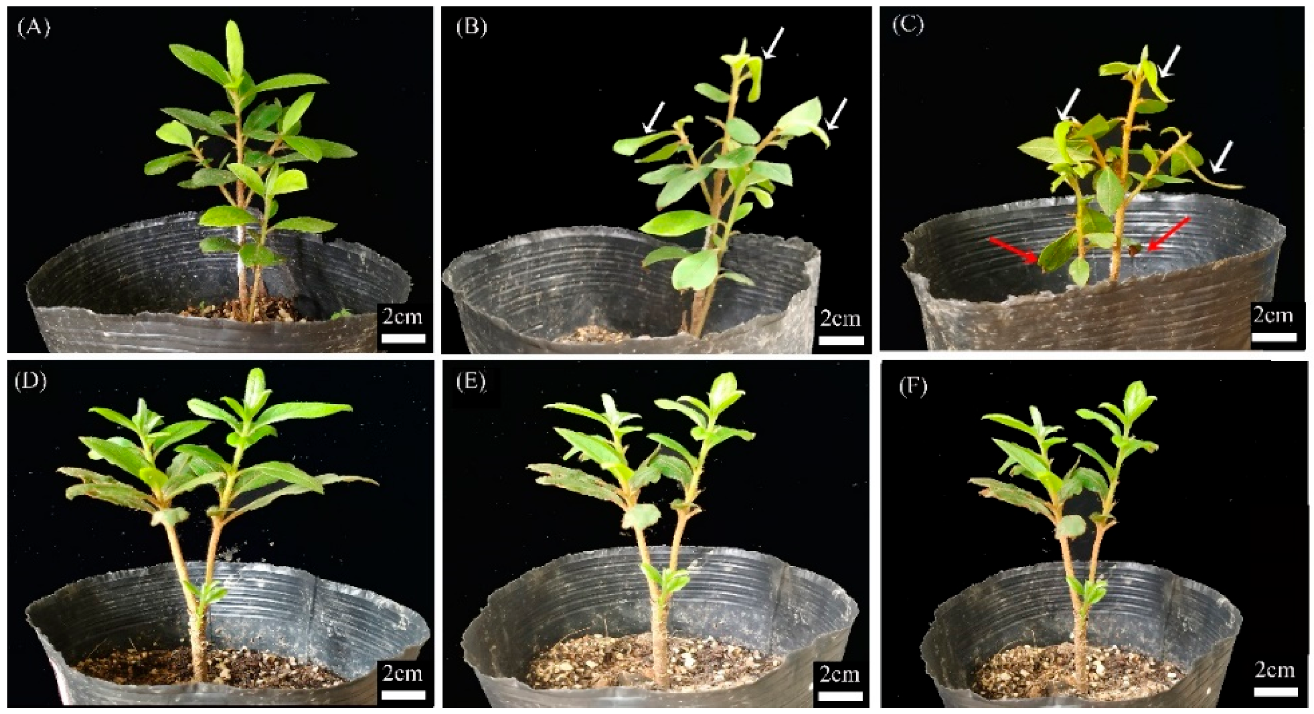
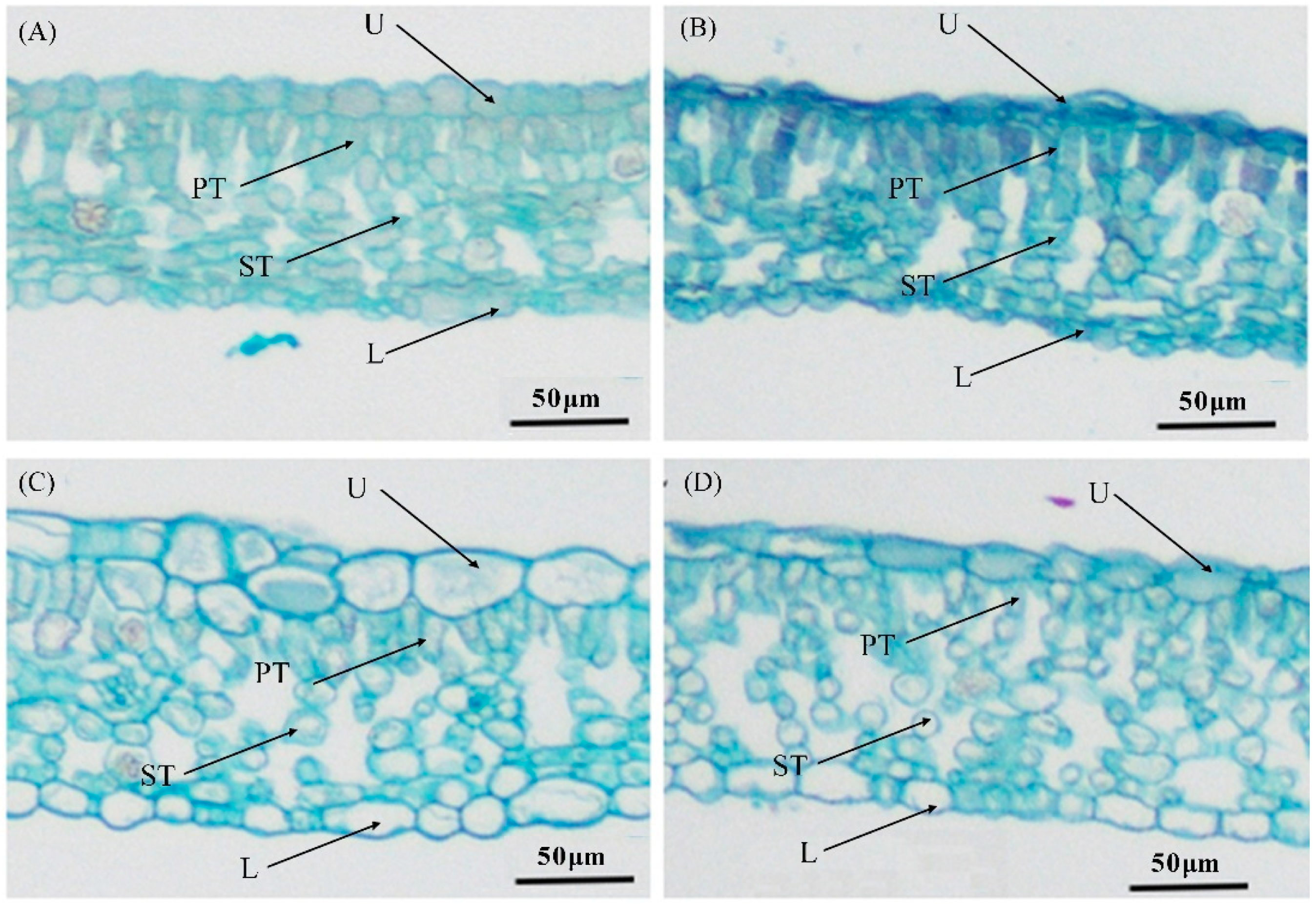
3.1. Phenotype and Microstructure of the Leaf

The phenotype of the plants under the four treatments is shown in Figure 1. There were significant differences among CK + W, CK + Ca2+, and H + Ca2+, whereas the leaves presented wilting with a burnt edge in the H + W treatment. Significant differences in the mesophyll tissue thickness were observed among the four groups in this study. As shown in Figure 2 and Table 1, in the H + W treatment, the thickness of the upper epidermis and the ratio of palisade to spongy parenchyma in the leaves were both significantly decreased when compared to CK + W, whereas the thickness of the palisade parenchyma, spongy parenchyma, lower epidermis, leaf, and ratio of palisade to leaf in the leaves were not significantly different. In the CK + Ca2+ treatment, the leaf thicknesses of the upper epidermis, spongy parenchyma, and lower epidermis were all significantly increased compared to the control treatment with distilled water. The palisade thickness was not significantly different between CK + Ca2+ and CK + W, whereas the ratio of palisade to leaf tissue and the ratio of palisade to spongy parenchyma in CK + Ca2+ were significantly lower than CK + W. In the H + Ca2+ treatment, the thickness of the upper epidermis, palisade parenchyma, lower epidermis, ratio of palisade to spongy parenchyma, and ratio of palisade to leaf were significantly decreased compared to CK + Ca2+. In CK + Ca2+ and H + Ca2+, the leaf thickness was significantly increased compared to H + W and CK + W.

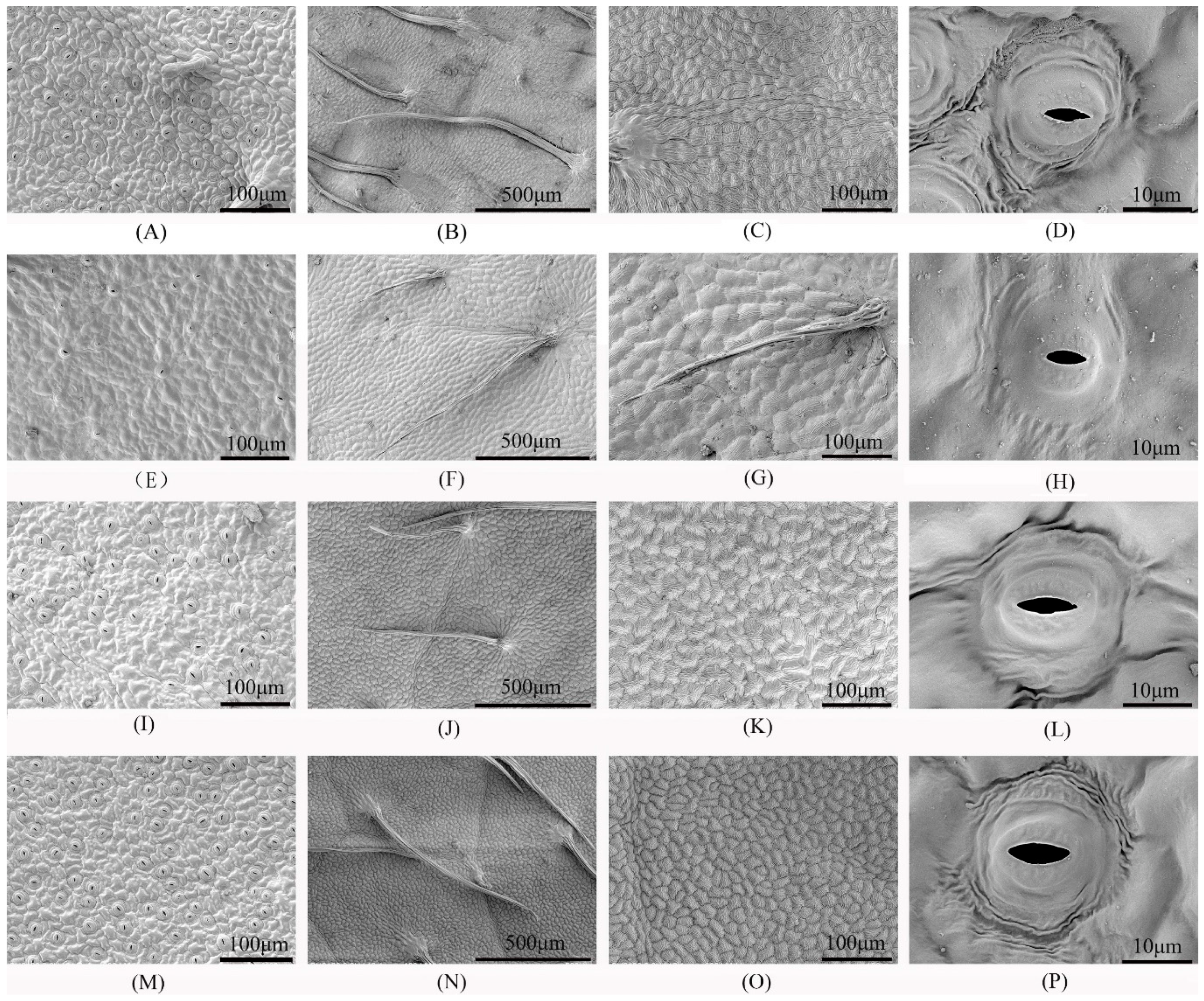
3.2. Ultrastructure of Leaf Epidermal Cells and Stomata

The ultrastructure of the leaf epidermal cells and stomata was observed and is shown in Table 2 and Figure 3. Regarding the cell density in the upper epidermis, significant differences were observed among the four treatments, with the order from high to low being (H + Ca2+) > (CK + W) > (CK + Ca2+) > (H + W). In the leaves of plants in CK + Ca2+ and H + Ca2+, the stomatal density was significantly lower than that in CK + W, whereas it was significantly higher than that in H + W. In the leaves of plants in CK + Ca2+ and H + Ca2+, the stomatal aperture was significantly increased compared to that in the other treatments with distilled water (H + W and CK + W). In the leaves of plants in the CK + Ca2+ and H + Ca2+ treatments, the closed/open stomatal ratios were significantly lower than that in the control treatment (H + W and CK + W). In H + W, the closed/open stomatal ratio in the leaves was significantly increased compared to that in CK + W.

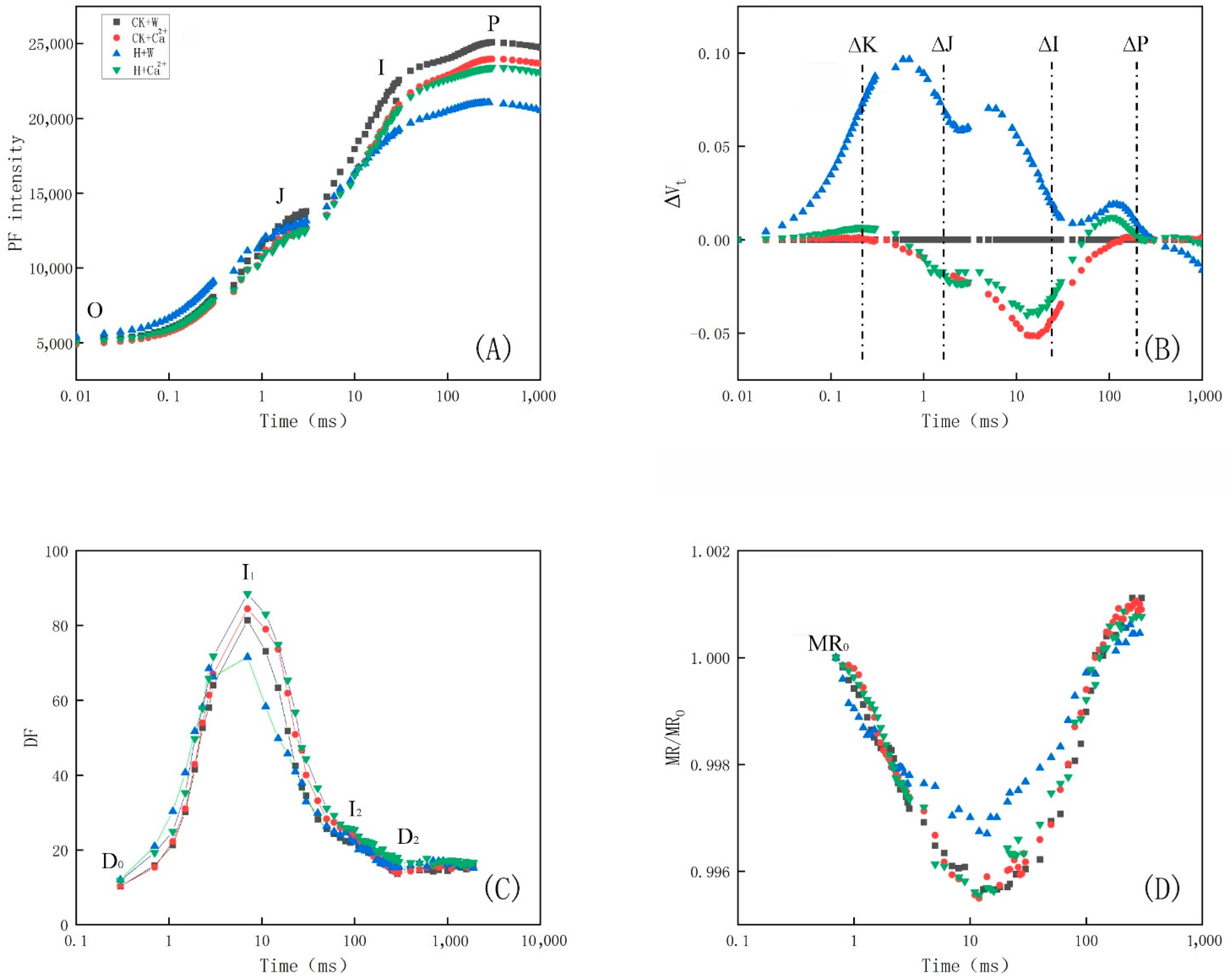
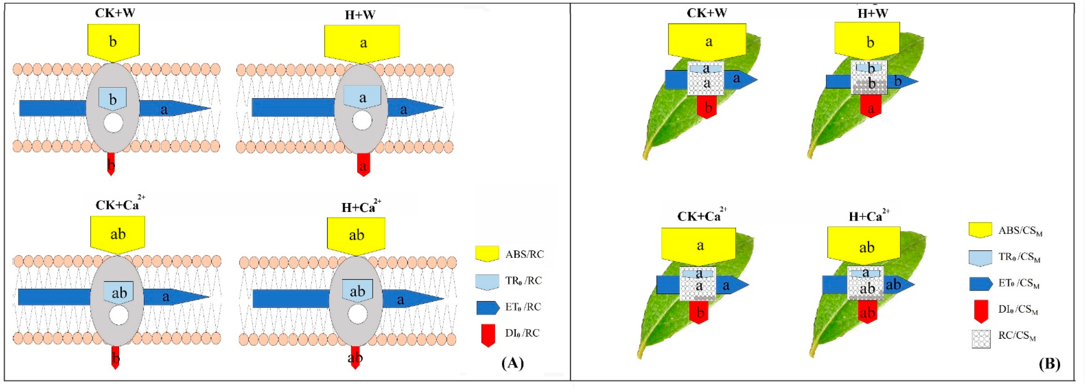
3.3. Analysis of PF, DF and MR Kinetics

Figure 4A shows that the chlorophyll-fluorescence-induced dynamic (OJIP) curve of the R. × pulchrum leaves of the H + W treatment decreased compared to those in the other three treatments at relative fluorescence intensities FI of point I and FP of point P. The ΔVt curve showed (Figure 4B) that the ΔK, ΔJ, and ΔI of the H + W treatment were higher than those of the other three treatments. In DF induction kinetics (Figure 4C), the fast phase occurred until 250 ms and only includes the I (7 ms) peak. Meanwhile, the I value decreased in the R. × pulchrum leaves of the H + W treatments, whereas it increased in the leaves of the CK + Ca2+ and H + Ca2+ treatments. The shape of the MR/MRO kinetics in the leaves of H + W was more obvious than in the other three treatments (Figure 4D).
The PF parameters for the R. × pulchrum leaves of the four treatments were influenced, as shown in Table 3. There were no significant differences in parameters ψEo, φEo, and δRo among the four treatments. However, the parameters Fv/Fm, PIABS, γRC, φRo, and φDo were obviously influenced. Among these parameters, only φRo had significant changes between the two control treatments (CK + Ca2+ and CK + W). Furthermore, the values of φRo increased in the R. × pulchrum leaves treated with CaCl2. The values of Fv/Fm, PIABS, and γRC in the R. × pulchrum leaves of the heat stress treatment with distilled water were significantly decreased compared to the other three treatments. The values of φDo significantly increased in the leaves of H + W compared to CK + W, whereas there was no significant difference between the two treatments with CaCl2 (CK + Ca2+ and H + Ca2+).
The absorbed energy fluxes of the two models are shown in Figure 5. In the membrane model (Figure 5A), the ABS/RC, TR0/RC, and DI0/RC values were all significantly increased in the leaves of H + W compared to CK + W, whereas these three parameters showed no significant difference in the leaves between the two treatments with CaCl2 (CK + Ca2+ and H + Ca2+). The values of ET0/RC were not significantly different among the four treatments. In the leaf model (Figure 5B), RC/CSM, ABS/CSM, TR0/CSM, and ET0/CSM were all decreased, and DI0/CSM was increased significantly in the leaves of H + W compared to CK + W, whereas these parameters had the same trend but were not significantly different in the leaves of H + Ca2+ compared to CK + Ca2+.

4. Discussion

4.1. Exogenous CaCl2 Influences Leaf Structure under Heat Stress

Significant differences in the ratio of palisade to spongy parenchyma in the leaves of R. × pulchrum between CK + W and H + W were found in this study (Table 1, Figure 2). This phenomenon was consistent with a previous study on grapevine [15]. After the use of exogenous CaCl2, the thickness of the epidermis and spongy tissues both significantly increased in the leaves of R. × pulchrum compared to the control under normal temperatures. In the heat stress treatment with CaCl2, the thickness of the upper epidermis, palisade parenchyma, and lower epidermis, the ratio of palisade to spongy parenchym, and the ratio of palisade to leaves were all significantly decreased when compared to the control treatment with CaCl2. Previous studies have reported that the larger the ratio of palisade to spongy tissues in plant leaves, the stronger the heat tolerance [49,50]. Thus, we predicted that exogenous CaCl2 would improve plant heat resistance, which requires further study. There may be other strategies that differ from the existing research.
In general, cell size reduction, stomatal closure, and an increased stomatal density enable plants to cope with heat stress [51]. This study found that the cell density decreased, the stomatal density decreased, the stomatal aperture decreased, and the ratio of closed/open stomata increased in the leaves of R. × pulchrum in response to heat stress (Table 2, Figure 3). The behavior of stomata in the leaves of R. × pulchrum under heat stress could curtail water loss and affect gas exchange and instantaneous water-use efficiency; this result is consistent with a previous study [16,51]. The present study found that the cell density and thickness of the upper epidermis both decreased in the leaves of R. × pulchrum under heat stress.
The mesophylic tissue consists of spongy and palisade tissue in the R. × pulchrum. The palisade tissue is usually continuous, with 1~2 rows in most plants [15,52]. Palisade tissue in the leaves of R. × pulchrum is continuous and has more than one row; however, it became discontinuous after the exogenous application of calcium chloride under heat stress treatment. Further, the morphology and thickness of the palisade and spongy tissues of R. × pulchrum were almost unaffected by heat stress. This might be a mechanism developed by R. × pulchrum to adapt to heat stress and protect mesophyll cells. However, a study of field-grown grapevines found no relationship among water-use efficiency, heat stress tolerance, and stomatal traits [53]. The larger the stomatal density in plant leaves, the stronger the heat tolerance [49,50]. Thus, we predicted that different plant species may have their own strategies to cope with heat stress, including cell and stomatal behavior.
The ability of plants to adapt to the external environment can be improved with the use of exogenous regulators, such as CaCl2, salicylic acid (SA), and putrescine. The spraying of exogenous regulators could ameliorate the plant’s adaptation of physiological responses to environmental stresses on its growth and physiological and anatomical features [3,4,45,54]. Previous studies have demonstrated that exogenous CaCl2 can improve heat resistance of plants and enable them to adapt to higher temperatures [5,6,7]. However, the effect of exogenous CaCl2 on plant leaf epidermal cells and stomatal behavior remains to be further studied. After the use of exogenous CaCl2, the cell density decreased, the stomatal density decreased, the stomatal aperture increased, and the ratio of closed/open stomata decreased in the leaves of R. × pulchrum under normal temperatures. However, the morphology and behavior of stomata in the leaves of R. × pulchrum treated with exogenous CaCl2 showed no significant change; but the cell density increased and the ratio of closed/open stomata decreased in the H + Ca2+ leaves compared with H + W, illustrating that the heat stress was moderate and not severe and most of the stomata in the H + Ca2+ leaves appear to be open (Figure 3M and Table 3). This phynomenon maybe through transpiration, accelerates water transport and increases the heat tolerance of plants. These results suggest that exogenous CaCl2 improves the heat tolerance of R. × pulchrum by regulating the behavior of epidermal cells and stomata in leaves. The mechanism that CaCl2 protect against heat stress by regulating the behavior of epidermal cells and stomata in leaves need to be further studied.

4.2. Exogenous CaCl2 Influences Leaf Chlorophyll Fluorescence in R. × pulchrum under Heat Stress

In our study, chlorophyll a fluorescence was used to study the photosynthetic energy and electron transfer in Rhododendron plants in response to heat stress. The OJIP curve of the R. × pulchrum leaves under the heat stress treatment with distilled water (H + W) decreased compared to H + Ca2+ treatment at relative fluorescence intensities FI of point I and FP of point P (Figure 4A); these results are consistent with previous studies [25,53]. Our research confirmed that OJIP transient was a reliable method for the detection of heat stress in PS II of Rhododendron [55]. The shape of the MR/MRO kinetics in the leaves of H + W was obviously different from CK + W and H + Ca2+ (Figure 4D). The fast phase and slow phase of MR/MRO kinetics in the leaves of H + W were lower than the other three treatments, and the lowest point of the MR/MRO kinetics in the leaves of H + W was higher than the other three treatments, illustrating that the oxidation and re-reduction abilities of the PS I reaction center and PC were reduced [31]. Our results indicate that the kinetics of the PF and MR of R. × pulchrum respond to heat stress, whereas these responses disappear when using exogenous CaCl2. A previous study reported that a decrease in the signal point of OJIP may be related to a reduction in the antenna pigment, a damaged PSI receptor, or denatured and degraded photosynthetic pigment–protein complexes [56,57].
Parameters extracted from the OJIP curve, such as φRo, γRC, ABS/RC, and ABS/CSM, can provide detailed information on the photosynthetic process [33,34]. The φRo and γRC values decreased in H + W and were lower than those in the other three treatments (Table 3), indicating a decrease in the quantum yield of the electron transfer from QA to the reduction of the end electron acceptors on the acceptor side of PSI under the heat stress treatment with distilled water [44,45,58]. Previous studies reported that the decrease in ABS/CSM, TR0/CSM, and ET0/CSM reflects the active reaction centers (RCs) being converted into inactive RCs, reducing the efficiency of trapping and causing a decline in PS II activity [53,59]. ABS/CSM, RC/CSM, TR0/CSM, and ET0/CSM decreased and DI0/CSM increased in the leaves of H + W compared to H + Ca2+ (Figure 5), indicating that both the size of the functional antenna and the exciton-specific rate captured by the open RCS were reduced [60,61]. The maximum photochemical quantum yield (Fv/Fm) is mainly used as a chlorophyll a fluorescence indicator of heat stress [62]. The Fv/Fm usually decreases dramatically after heat treatment [27,63]. PI refers to the energy conservation between photons absorbed by PS II and the reduction of intersystem electron acceptors (PIABS) [44]. In our study, the Fv/Fm of the H + W treatment was significantly lower than that in CK + W (Table 3); this is consistent with previous research [63]. The Fv/Fm and PIABS of the H + W treatment were both lower than those in H + Ca2+ (Table 3). However, the Fv/Fm and PIABS parameters in the two treatments with CaCl2 (CK + Ca2+ and H + Ca2+) were not different from those in the control. In addition, the ΔVt curve showed that the ΔK, ΔJ, and ΔI of H + W were higher than those in the other three treatments (Figure 4B). Previous studies found that DF is associated with the recombination of S2 or S3 states of the oxygen evolving complex (OEC) with QA or QB [63], I1 and I2 points of DF are most likely related to the electron transfer from the QB to PQH2 of PS II and re-oxidized by PS I [62]. The I1 point of DF appears in the J-I phase of PF dynamics and at the end of the fast phase of MR, whereas the I2 point of DF appears in the I-P phase of PF and the slow phase of MR. At the I1 and I2 points of DF, the H + W curve is different from all of the other curves, illustrating that the electron transfer ability of PS I and PS II was inhibited in the leaves of R. × pulchrum under heat stress. This could be alleviated by using exogenous CaCl2. These results illustrate that exogenous CaCl2 can improve the heat tolerance of R. × pulchrum [27,44,64,65], suggesting that PSII and PSI were damaged and that the ability to pass electrons downstream (electron transfer from QA to QB) was inhibited in the leaves of R. × pulchrum under heat stress whereas this damage could be alleviated by using exogenous CaCl2 [44,66].

5. Conclusions

This study aimed to explore the effect of exogenous CaCl2 on the leaf microstructure, leaf epidermal ultrastructure, and chlorophyll a fluorescence of R. × pulchrum under heat stress. The results of this study revealed that exogenous CaCl2 improves the heat tolerance ability of R. × pulchrum by regulating the behavior of its epidermal cells and stomata, which, in turn, changes the arrangement and number of palisade tissue cell layer in its leaves. Meanwhile, under heat stress, its electron transfer ability from QA to QB was inhibited which could be alleviated by using exogenous CaCl2. The results found that exogenous CaCl2 might alleviate the inhibitory impact of heat stress on the anatomical and fast chlorophyll fluorescence features of R. × pulchrum. Our study could help in the understanding of the role of exogenous CaCl2 in regulating the heat resistance of plants.

Author Contributions

Conceptualization, J.S. and S.J.; methodology, X.L., X.P. and Y.H.; software, X.P. and Y.H.; resources, S.J. and J.S. Shen; data curation, S.J. and J.S.; writing—original draft preparation and writing—review and editing, H.C., S.J. and J.S.; funding acquisition, S.J. All authors have read and agreed to the published version of the manuscript.

Funding

This research was funded by the National Key Research and Development Project (2019YFE0118900), the National Natural Science Foundation of China (31971641, 32201608), the Zhejiang Provincial Natural Science Foundation of China (LY16C160011), and the Jiyang College of Zhejiang A&F University under grant (RQ1911B07).

Data Availability Statement

Not applicable.

Conflicts of Interest

The authors declare no conflict of interest.

Abbreviations

Fv/Fm, maximal quantum efficiency of PSII; PIABS, the performance index on an absorption basis; ψEo, a trapped exciton moves an electron into the electron transport chain beyond QA at t = Fo; δRo, efficiency required for an electron to be transferred from reduced carriers between the two photosystems to the PSI end acceptors; γRC, a given chlorophyll a molecule that functions as the PSII reaction center; φEo, quantum efficiency of electron transfer at t = Fo; φRo, quantum yield for reduction of the terminal electron acceptors on the PSI acceptor side; φDo, quantum yield for energy dissipation at t = Fo; ABS/RC, absorption flux per reaction centre (RC) basis of membrane model; TR0/RC, trapped energy flux per RC (at t = 0); DI0/RC, dissipation energy flux per RC (at t = 0); ET0/RC, electron transport flux per RC (at t = 0); tFM, time (in ms) to reach the maximal fluorescence intensity FM; ABS/CSM, absorption flux per excited leaf cross-section (CS) of leaf model; RC/CSM, reaction centre per CS (at t = tFM); TR0/CSM, trapped energy flux per CS (at t = tFM); ET0/CSM, electron transport flux per CS (at t = tFM); DI0/CSM, dissipation energy flux per CS (at t = tFM).

References

  1. Lavania, D.; Dhingra, A.; Siddiqui, M.H.; Al-Whaibi, M.H.; Grover, A. Current status of the production of high temperature tolerant transgenic crops for cultivation in warmer climates. Plant Physiol. Biochem. 2015, 86, 100–108. [Google Scholar] [CrossRef] [PubMed]
  2. Grover, A.; Mittal, D.; Negi, M.; Lavania, D. Generating high temperature tolerant transgenic plants: Achievements and challenges. Plant Sci. 2013, 205, 38–47. [Google Scholar] [CrossRef] [PubMed]
  3. Laanemets, K.; Brandt, B.; Li, J.L.; Merilo, E.; Wang, Y.F.; Keshwani, M.M.; Taylor, S.S.; Kollist, H.; Schroeder, J.I. Calcium dependent and independent stomatal signaling network and compensatory feedback control of stomatal opening via Ca2+ sensitivity priming. Plant Physiol. 2013, 163, 504–513. [Google Scholar] [CrossRef] [PubMed] [Green Version]
  4. Janda, M.; Ruellan, E. Magical mystery tour: Salicylic acid signalling. Environ. Expt. Bot. 2015, 114, 117–128. [Google Scholar] [CrossRef]
  5. Gao, Y. Molecular and genetic evidence for the key role of atcam3 in heat-shock signal transduction in Arabidopsis. Plant Physiol. 2009, 149, 1773. [Google Scholar] [CrossRef] [Green Version]
  6. Li, T.; Xu, X.; Li, Y.; Wang, H.; Li, Z. Comparative transcriptome analysis reveals differential transcription in heat-susceptible and heat-tolerant pepper (Capsicum annum L.) cultivars under heat stress. J. Plant Biol. 2015, 58, 411–424. [Google Scholar] [CrossRef]
  7. Anireddy, S.N.R.; Gul, S.A.; Helena, C.; Irene, S.D. Coping with Stresses: Roles of calcium- and calcium/calmodulin-regulated gene expression. The Plant Cell 2011, 23, 2010–2032. [Google Scholar] [CrossRef] [Green Version]
  8. Snedden, W.A.; Fromm, H. Calmodulin as a versatile calcium signal transducer in plants. New Phytol. 2001, 151, 35–66. [Google Scholar] [CrossRef] [Green Version]
  9. Shen, H.; Bing, Z.; Xu, J.; Zheng, X.; Huang, W. Effects of salicylic acid and calcium chloride on heat tolerance in Rhododendron ’fen zhen zhu’. J. Am. Soc. Hortic. Sci. 2016, 141, 363–372. [Google Scholar] [CrossRef] [Green Version]
  10. Mishkind, M.; Vermeer, J.E.M.; Darwish, E.; Munnik, T. Heat stress activates phospholipase D and triggers PIP accumulation at the plasma membrane and nucleus. The Plant J. 2009, 60, 10–21. [Google Scholar] [CrossRef]
  11. Perez, D.E.; Hoyer, J.S.; Johnson, A.I.; Moody, Z.R.; Lopez, J.; Kaplinsky, N.J. BOBBER1 is a noncanonical Arabidopsis small heat shock protein required for both development and thermotolerance. Plant Physiol. 2009, 151, 241–252. [Google Scholar] [CrossRef] [PubMed] [Green Version]
  12. Chen, W.L.; Yang, W.J.; Lo, H.F.; Yeh, D.M. Physiology, anatomy, and cell membrane thermostability selection of leafy radish (Raphanus sativus var. Oleiformis Pers.) with different tolerance under heat stress. Sci. Hortic. 2014, 179, 367–375. [Google Scholar] [CrossRef]
  13. Wang, J.; Lv, J.H.; Liu, Z.B.; Liu, Y.H.; Song, J.S.; Ma, Y.Q. Integration of transcriptomics and metabolomics for Pepper (Capsicum annuum L.) in response to heat stress. Int. J. Mol. Sci. 2020, 20, 5042. [Google Scholar] [CrossRef] [PubMed] [Green Version]
  14. Zha, Q.; Xi, X.; He, Y.; Jiang, A. Transcriptomic analysis of the leaves of two grapevine cultivars under high-temperature stress. Sci. Hortic. 2020, 265, 109265. [Google Scholar] [CrossRef]
  15. Salem-Fnayou, A.B.; Bouamama, B.; Ghorbel, A.; Mliki, A. Investigations on the leaf anatomy and ultrastructure of grapevine (Vitis vinifera) under heat stress. Microsc. Res. Tech. 2011, 74, 756–762. [Google Scholar] [CrossRef]
  16. Shen, H.F.; Zhao, B.; Xu, J.J.; Liang, W.; Huang, W.M.; Li, H.H. Effects of heat stress on changes in physiology and anatomy in two cultivars of Rhododendron. S. Afr. J. Bot. 2017, 112, 338–345. [Google Scholar] [CrossRef]
  17. Feng, J.Q.; Wang, J.H.; Zhang, S.B. Leaf physiological and anatomical responses of two sympatric paphiopedilum species to temperature. Plant Divers. 2022, 44, 101e108. [Google Scholar] [CrossRef]
  18. Mathur, S.; Jajoo, A. Photosynthesis: Limitations in response to high temperature stress. J. Photochem. Photobiol. B Biol. 2014, 137, 116–126. [Google Scholar] [CrossRef]
  19. Oba, E.G.; Hu, J.; Zhang, Z.; Li, S.; Xie, W. Physiological response of three micropropagated plant lets of Rhododendron hybrids for heat stress. Eur. J. Hortic. Sci. 2021, 86, 300–307. [Google Scholar] [CrossRef]
  20. Ishikawa, T.; Shigeoka, S. Recent advances in ascorbate biosynthesis and the physiological significance of ascorbate peroxidase in photosynthesizing organisms. Biosci. Biotechnol. Biochem. 2008, 72, 1143–1154. [Google Scholar] [CrossRef]
  21. Hu, Y.; Zhang, Y.; Yu, W.; Hänninen, H.; Song, L.; Du, X.; Zhang, R.; Wu, J. Novel insights into the influence of seed sarcotesta photosynthesis on accumulation of seed dry matter and oil content in Torreya grandis cv. ‘Merrillii’. Front. Plant Sci. 2018, 8, 2179. [Google Scholar] [CrossRef] [Green Version]
  22. Oukarroum, A.; Goltsev, V.; Strasser, R.J. Temperature effects on pea plants probed by simultaneous measurements of the kinetics of prompt fluorescence, delayed fluorescence and modulated 820 nm reflection. PLoS ONE 2013, 8, e59433. [Google Scholar] [CrossRef] [PubMed]
  23. Murata, N.; Takahashi, S.; Nishiyama, Y.; Allakhverdiev, S.I. Photoinhibition of photosystem II under environmental stress. Biochim. Biophys. Acta 2007, 1767, 414–421. [Google Scholar] [CrossRef] [PubMed] [Green Version]
  24. Zushi, K.; Kajiwara, S.; Matsuzoe, N. Chlorophyll a fluorescence OJIP transient as a tool to characterize and evaluate response to heat and chilling stress in tomato leaf and fruit. Sci. Hortic. 2012, 148, 39–46. [Google Scholar] [CrossRef]
  25. Sharma, D.K.; Fernández, J.O.; Rosenqvist, E.; Ottosen, C.O.; Andersen, S.B. Genotypic response of detached leaves versus intact plants for chlorophyll fluorescence parameters under high temperature stress in wheat. Plant Physiol. 2014, 171, 576–586. [Google Scholar] [CrossRef] [PubMed]
  26. Kumari, S.D.; Maria, T.A.; Eva, R.; Carl-Otto, O.; Andersen, S.B. QTLs and potential candidate genes for heat stress tolerance identified from the mapping populations specifically segregating for Fv/Fm in wheat. Front. Plant Sci. 2017, 8, 1668. [Google Scholar] [CrossRef] [Green Version]
  27. Jedmowski, C.; Brüggemann, W. Imaging of fast chlorophyll fluorescence induction curve (OJIP) parameters, applied in a screening study with wild barley (Hordeum spontaneum) genotypes under heat stress. J. Photochem. Photobiol. B Biol. 2015, 151, 153–160. [Google Scholar] [CrossRef]
  28. Sohn, S.I.; Yong, H.L.; Sun, H.H.; Chang, S.K.; Oh, Y.J. Characterization and evaluation of response to heat and chilling stress in exotic weeds using chlorophyll a fluorescence OJIP transient. Environ. Biol. Res. 2020, 38, 450–460. [Google Scholar] [CrossRef]
  29. Ma, G.; Zhang, M.; Xu, J.; Zhou, W.; Cao, L. Transcriptomic analysis of short-term heat stress response in Pinellia ternata provided novel insights into the improved thermotolerance by spermidine and melatonin. Ecotoxicol. Environ. Safe 2020, 202, 110877. [Google Scholar] [CrossRef]
  30. Essemine, J.; Qu, M.; Lyu, M.; Song, Q.; Zhu, X.G. Photosynthetic and transcriptomic responses of two C4 grass species with different nacl tolerance. J. Plant Physiol. 2020, 253, 153244. [Google Scholar] [CrossRef]
  31. Wang, X.; Dingxuan, Q.; Shi, M. Calcium amendment for improved germination, plant growth, and leaf photosynthetic electron transport in oat (Avena sativa) under NaCl stress. PLoS ONE 2021, 16, e0256529. [Google Scholar] [CrossRef] [PubMed]
  32. Strasser, R.J.; Srivastava, A.; Govindjee. Polyphasic chlorophyll a fluorescence transient in plants and cyanobacteria. Photochem. Photobiol. 1995, 61, 32–42. [Google Scholar] [CrossRef]
  33. Strasser, R.J.; Tsimilli-Michael, M.; Srivastava, A. Analysis of the Chlorophyll a Fluorescence Transient. In Chlorophyll Fluorescence: A Signature of Photosynthesis; Papageorgiou, G.C., Govindjee, Eds.; Kluwer Academic Publishers Press: Dordrecht, The Netherlands, 2004; pp. 321–362. [Google Scholar]
  34. Wang, J.W.; Liu, Y.; Xu, Y.X.; Chen, W.J.; Han, Y.N.; Wang, G.G.; Jin, S. Sexual diferences in gas exchange and chlorophyll fuorescence of Torreya grandis under drought stress. Trees 2022, 36, 283–294. [Google Scholar] [CrossRef]
  35. Ballantyne, D.J.; Argent, G.; Mcfarlane, M. Dormancy, flowering and chlorophyll fluorescence of a rhododendron and azaleas in the greenhouse. In Rhododendrons in Horticulture & Science Papers Presented at the International Rhododendron Conference, Proceedings of the International Rhododendron Conference, Edinburgh, UK, 17–19 May 2002; Royal Botanic Garden Edinburgh: Edinburgh, UK, 2003. [Google Scholar]
  36. Fang, L.; Tong, J.; Dong, Y.; Xu, D.; Jing, M.; Yuan, Z. De novo RNA sequencing transcriptome of Rhododendron obtusum identified the early heat response genes involved in the transcriptional regulation of photosynthesis. PLoS ONE 2017, 12, e0186376. [Google Scholar] [CrossRef] [PubMed] [Green Version]
  37. Cullen, J. Hardy Rhododendron Species: A Guide to Identification; Timber Press Inc.: Portland, OR, USA, 2005. [Google Scholar]
  38. Krebs, S.L. Rhododendron. In Ornamental Crops, Handbook of Plant Breeding; Van Huylenbroeck, J., Ed.; Springer: Berlin/Heidelberg, Germany, 2018. [Google Scholar]
  39. Xia, X.M.; Yang, M.Q.; Li, C.L.; Huang, S.X.; Jin, W.T.; Shen, T.T. Spatiotemporal evolution of the global species diversity of rhododendron. Mol. Biol. Evol. 2021, 39, msab314. [Google Scholar] [CrossRef] [PubMed]
  40. Ma, Y.; Nielsen, J.; Chamberlain, D.F.; Li, X.; Sun, W. The conservation of Rhododendrons is of greater urgency than has been previously acknowledged in China. Biodivers. Conserv. 2014, 23, 3149–3154. [Google Scholar] [CrossRef]
  41. Rennenberg, H.; Loreto, F.; Polle, A.; Brilli, F.; Fares, S.; Beniwal, R.S.; Gessler, A.J. Physiological responses of forest trees to heat and drought. Plant Biol. 2006, 8, 556–571. [Google Scholar] [CrossRef]
  42. Zhang, S.; Jiang, H.; Peng, S.M.; Korpelainen, H.; Li, C.Y. Sex-related differences in morphological, physiological, and ultrastructural responses of Populus cathayana to chilling. J. Exp. Bot. 2011, 62, 675–686. [Google Scholar] [CrossRef] [Green Version]
  43. Ruppel, N.J.; Logsdon, C.A.; Whippo, C.W. A mutation in Arabidopsis seedling plastid development affects plastid differentiation in embryo-derived tissues during seedling growth. Plant Physiol. 2011, 155, 342–353. [Google Scholar] [CrossRef] [Green Version]
  44. Kong, D.X.; Li, Y.Q.; Wang, M.L.; Bai, M.; Zou, R.; Tang, H.; Wu, H. Effects of light intensity on leaf photosynthetic characteristics, chloroplast structure, and alkaloid content of Mahonia bodinieri (Gagnep.) Laferr. Acta Physiol. Plant. 2016, 38, 120. [Google Scholar] [CrossRef]
  45. Zhu, X.; Wang, L.; Yang, R.; Han, Y.Y.; Hao, J.H.; Liu, C.J.; Fan, S. Effects of exogenous putrescine on the ultrastructure of and calcium ion flow rate in lettuce leaf epidermal cells under drought stress. Hortic. Environ. Biotechnol. 2019, 60, 479–490. [Google Scholar] [CrossRef]
  46. Strasser, R.J.; Tsimilli-Michael, M.; Qiang, S.; Goltsev, V. Simultaneous in vivo recording of prompt and delayed fluorescence and 820 nm reflection changes during drying and after rehydration of the resurrection plant Haberlea rhodopensis. Biochim. Biophys. Acta 2010, 1797, 1313–1326. [Google Scholar] [CrossRef] [PubMed] [Green Version]
  47. Goltsev, V.; Zaharieva, I.; Chernev, P.; Kouzmanova, M.; Kalaji, H.M.; Yordanov, I.; Krasteva, V.; Alexandrov, V.; Stefanov, D.; Allakhverdiev, S.I.; et al. Drought induced modifications of photosynthetic electron transport in intact leaves: Analysis and use of neural networks as a tool for a rapid non-invasive estimation. Biochim. Biophys. Acta 2012, 1817, 1490–1498. [Google Scholar] [CrossRef] [PubMed] [Green Version]
  48. Shen, J.; Li, X.; Zhu, X.; Ding, Z.; Huang, X.; Chen, X.; Jin, S. Molecular and photosynthetic performance in the yellow leaf mutant of Torreya grandis according to transcriptome sequencing, chlorophyll a fluorescence, and modulated 820 nm reflection. Cells 2022, 11, 431. [Google Scholar] [CrossRef] [PubMed]
  49. Hu, W.J.; Zhang, Q.X.; Pan, H.T.; Dong, L.L. Preliminary study on physiological indexes of heat-resistance of Primula. Chin. Agric. Sci. Bull. 2010, 26, 158–163. [Google Scholar]
  50. Bian, A.N.; Lu, Y.L.; Pan, D.M. Preliminary study on heat resistant physiological indexes of daffodils. J. Minnan Norm. Univ. (Nat. Sci.) 2016, 29, 70–75. [Google Scholar] [CrossRef]
  51. Banon, S.; Fernandez, J.A.; Franco, J.A.; Torrecillas, A.; Alarcón, J.J.; Sánchez-Blanco, M.J. Effects of water stress and night temperature preconditioning on water relations and morphological and anatomical changes of Lotus creticusplants. Sci. Hortic. 2004, 101, 333–342. [Google Scholar] [CrossRef]
  52. El-Fiki, M.A.; El-Taher, A.M.; EL-Gendy, A.G.; Lila, M.I. Morphological and anatomical studies on some taxa of family Apocynaceae. Al-Azhar J. Agric. Res. 2019, 44, 136–147. [Google Scholar]
  53. Faralli, M.; Bontempo, L.; Bianchedi, P.L.; Moser, C.; Bertamini, M.; Lawson, T.; Camin, F.; Stefanini, M.; Varotto, C. Natural variation in stomatal dynamics drives divergence in heat stress tolerance and contributes to seasonal intrinsic water-use efficiency in Vitis vinifera (subsp. sativa and sylvestris). J. Exp. Bot. 2022, 73, 3238–3250. [Google Scholar] [CrossRef]
  54. El-Taher, A.M.; Abd El-Raouf, H.S.; Osman, N.A.; Azoz, S.N.; Omar, M.A.; Elkelish, A.; Abd El-Hady, M.A. Effect of salt stress and foliar application of salicylic acid on morphological, biochemical, anatomical, and productivity characteristics of Cowpea (Vigna unguiculata L.) Plants. Plants 2021, 11, 115. [Google Scholar] [CrossRef]
  55. Strauss, A.J.; Krüger, G.H.J.; Strasser, R.J.; Heerden, P.D.R.V. Ranking of dark chilling tolerance in soybean genotypes probed by the chlorophyll a fluorescence transient O-J-I-P. Environ. Exp. Bot. 2006, 56, 147–157. [Google Scholar] [CrossRef]
  56. Oster, U.; Tanaka, R.; Tanaka, A.; Rüdigger, W. Cloning and functional expression of the gene encoding the key enzyme for chlorophyll b biosynthesis (CAO) from Arabidopsis thaliana. The Plant J. 2000, 21, 305–310. [Google Scholar] [CrossRef] [PubMed]
  57. Momchil, P.; Lyubka, K.; Andon, V.; Jaco, V.; Vasilij, G. Effects of different metals on photosynthesis: Cadmium and zinc affect chlorophyll fluorescence in durum wheat. Int. J. Mol. Sci. 2018, 19, 787. [Google Scholar] [CrossRef] [Green Version]
  58. Li, X.; Brestic, M.; Tan, D.X.; Zivcak, M.; Zhu, X.; Liu, S.; Song, F.; Reiter, R.J.; Liu, F. Melatonin alleviates low PS I-limited carbon assimilation under elevated CO2 and enhances the cold tolerance of offspring in chlorophyll b-deficient mutant wheat. J. Pineal Res. 2018, 64, e12453. [Google Scholar] [CrossRef]
  59. Tongra, T.; Mehta, P.; Mathur, S.; Agrawal, D.; Bharti, S.; Los, D.A.; Allakhverdiev, S.I.; Jajoo, A. Computational analysis of fluorescence induction curves in intact spinach leaves treated at different pH. BioSystems 2011, 103, 158–163. [Google Scholar] [CrossRef]
  60. Mathur, S.; Allakhverdiev, S.I.; Jajoo, A. Analysis of high temperature stress on the dynamics of antenna size and reducing side heterogeneity of photosystem ii in wheat leaves (Triticum aestivum). Biochim. Biophys. Acta 2011, 1807, 22–29. [Google Scholar] [CrossRef] [Green Version]
  61. Ahammed, G.J.; Xu, W.; Liu, A.; Chen, S. Comt1 silencing aggravates heat stress-induced reduction in photosynthesis by decreasing chlorophyll content, photosystem ii activity and electron transport efficiency in tomato. Front. Plant Sci. 2018, 9, 998. [Google Scholar] [CrossRef]
  62. Gao, J.; Li, P.; Ma, F.; Goltsev, V. Photosynthetic performance during leaf expansion in malus micromalus probed by chlorophyll a fluorescence and modulated 820 nm reflection. J. Photochem. Photobiol. B Biol. 2014, 137, 144–150. [Google Scholar] [CrossRef]
  63. Dąbrowski, P.; Kalaji, M.H.; Baczewska, A.H.; Pawluśkiewicz, B.; Mastalerczuk, G.; Borawska-Jarmułowicz, B.; Paunov, M.; Goltsev, V. Delayed chlorophyll a fluorescence, mr 820, and gas exchange changes in perennial ryegrass under salt stress. J. Lumin. 2017, 183, 322–333. [Google Scholar] [CrossRef]
  64. Adams, W.W.; Demming-Adams, B. Chlorophyll Fluorescence as a Tool to Monitor Plant Response to the Environment; Springer: Berlin/Heidelberg, Germany, 2004. [Google Scholar] [CrossRef]
  65. Song, Y.; Chen, Q.; Ci, D.; Shao, X.; Zhang, D. Effects of high temperature on photosynthesis and related gene expression in poplar. BMC Plant Biol. 2014, 14, 111. [Google Scholar] [CrossRef] [PubMed] [Green Version]
  66. Sitko, K.; Rusinowski, S.; Pogrzeba, M.; Daszkowska-Golec, A.; Makowski, E. Development and aging of photosynthetic apparatus of Vitis vinifera L. during growing season. Photosynthetica 2020, 58, 186–193. [Google Scholar] [CrossRef]
Figure 1. The phenotypes of the plants under the four treatments. (A) CK + W represents the control treatment with distilled water; (B) H + W represents the heat stress treatment after 1 day with distilled water; (C) H + W represents the heat stress treatment after 2 days with distilled water; (D) CK + Ca2+ represents the control treatment with 10 mM CaCl2; (E) H + Ca2+ represents the heat stress treatment after 1 day with 10 mM CaCl2; (F) H + Ca2+ represents the heat stress treatment after 2 days with 10 mM CaCl2. Red arrow points to the leaves with burnt edge; white arrows point to the wilting leaves.
Agronomy 12 03226 g001
Figure 2. Leaf anatomical structure of Rhododendron × pulchrum leaves under four treatments. (A) CK + W represents the control treatment with distilled water; (B) H + W represents the heat stress treatment with distilled water; (C) CK + Ca2+ represents the control treatment with 10 mM CaCl2; (D) H + Ca2+ represents the heat stress treatment with 10 mM CaCl2. L = lower epidermis; PT = palisade tissue; ST = spongy tissue; and U = upper epidermis.
Agronomy 12 03226 g002
Figure 3. Ultrastructure of leaf epidermal cells of Rhododendron × pulchrum leaves under four treatments. (AD) CK + W represents the control treatment with distilled water; (EH) H + W represents the heat stress treatment with distilled water; (IL) CK + Ca2+ represents the control treatment with 10 mM CaCl2; (MP) H + Ca2+ represents the heat stress treatment with 10 mM CaCl2; (A,E,I,M) show the stomatal density in the lower epidermis; (B,C,F,G,J,K,N,O) show the cell density in the upper epidermis; (D,H,L,P) show the stomatal aperture in the lower epidermis.
Agronomy 12 03226 g003
Figure 4. PF, DF, and MR kinetics in the leaves of Rhododendron × pulchrum under four treatments. Each curve is the average of three replicates. (A) Prompt chlorophyll a fluorescence (PF). The signals were plotted on a logarithmic time scale. The signals were fluorescence intensity at 20 µs ≡ FO, 3 ms ≡ FJ, and 30 ms ≡ FI; maximum fluorescence intensity, FP = FM. (B) Effect of the development of PSII on the relative variable fluorescence [ΔVt = (Ft − F0)/Fv − Vt (CK + W)]; for ΔVt analysis, the fluorescence of the leaves of the control treatment with distilled water (CK + W) was used as the reference and equaled 0. (C) Delayed chlorophyll a fluorescence (DF). The signals were plotted on a logarithmic time scale. (D) Modulated 820 nm reflection (MR/MRO). The signals were plotted on a logarithmic time scale.
Agronomy 12 03226 g004
Figure 5. Energy flux models in photosystem II for the leaf samples of Rhododendron × pulchrum under four treatments. The relative value of each parameter is proportional to the width of the arrows. (A) Membrane model represents the specific activity on a single RC basis. The average size of the antenna is represented by the ABS/RC parameter. The fluxes of absorbed energy that were trapped by nonreducing PSII centers are shown as diagonally hatched areas in ABS/RC and TR/RC contours. (B) Leaf model represents the phenomenological parameters normalized per unit cross-section (CS) of a photosynthesizing object. The QA-nonreducing reaction centers are shown with black circles, and the QA-reducing active centers are depicted with white circles. These were determined at minimum (index 0) or maximum (index m) levels of chlorophyll fluorescence; for example, ET0 or ETm. The fluxes presented in the scheme were normalized to the maximal fluorescence level (m). The same letters indicate no significant difference at the 0.05 level based on Duncan’s test.
Agronomy 12 03226 g005
Table 1. Leaf anatomical structure of Rhododendron × pulchrum leaves under the four treatments.
GroupUpper Epidermis Thickness (μm)Palisade Thickness (μm)Spongy Thickness (μm)Lower Epidermis (μm)Leaf Thickness (μm)Ratio of Palisade Thickness to Spongy ThicknessRatio of Palisade Thickness to Leaf Thickness
CK + W14.96 ± 1.79 b 133.47 ± 2.51 a39.58 ± 1.76 c12.27 ± 1.39 b100.28 ± 3.23 b0.85 ± 0.09 a0.33 ± 0.02 a
H + W11.82 ± 1.70 c32.53 ± 3.53 a43.69 ± 6.36 c10.78 ± 2.25 b98.82 ± 9.84 b0.75 ± 0.08 b0.33 ± 0.02 a
CK + Ca2+22.62 ± 3.43 a31.81 ± 4.25 a53.78 ± 3.32 b17.29 ± 3.31 a125.53 ± 5.36 a0.59 ± 0.10 c0.25 ± 0.03 b
H + Ca2+15.40 ± 1.53 b22.67 ± 6.17 b65.91 ± 3.13 a13.25 ± 2.90 b117.23 ± 6.80 a0.34 ± 0.09 d0.19 ± 0.04 c
1 Mean ± SE followed by the same letter in the same column indicates no significant difference at the 0.05 level based on Duncan’s test.
Table 2. Ultrastructure of leaf epidermal cells of Rhododendron × pulchrum leaves under the four treatments.
GroupCell Density (No. of Upper Epidermises/mm2)Stomatal Density (No. of Stomata/mm2)Stomatal Transverse
Diameter/μm
Stomatal Longitudinal
Diameter/μm
Stomatal Aperture/μm2Open Stomatal Density (No. of Opened Stomata/mm2)Closed Stomatal Density (No. of Closed Stomata/mm2) Ratio of Closed/Open Stomata
CK + W217 ± 5 b 171 ± 3 a5.56 ± 0.46 b1.43 ± 0.21 b6.23 ± 0.99 b57 ± 5 a14 ± 3 a0.25 ± 0.06 b
H + W81 ± 6 d27 ± 3 c4.44 ± 0.71 c1.44 ± 0.27 b4.95 ± 0.78 b19 ± 3 c8 ± 1 b0.43 ± 0.07 a
CK + Ca2+104 ± 8 c46 ± 6 b7.38 ± 1.23 a2.31 ± 0.37 a13.55 ± 3.85 a45 ± 5 b1 ± 1 c0.03 ± 0.01 c
H + Ca2+251 ± 8 a57 ± 5 b8.02 ± 0.56 a2.39 ± 0.24 a15.03 ± 1.71 a57 ± 4 a1 ± 0 c0.02 ± 0.00 c
1 Mean ± SE followed by the same letter in the same column indicates no significant difference at the 0.05 level based on Duncan’s test.
Table 3. Parameters derived from prompt chlorophyll a fluorescence transients in the leaves of Rhododendron × pulchrum under the four treatments.
GroupFv/Fm 1PIABS 2ΨEo 3σRo 4γRC 5φEo 6φRo 7φDo 8
CK + W0.80 ± 0.01 a 95.16 ± 0.44 a0.61 ± 0.06 a0.20 ± 0.04 a0.38 ± 0.03 a0.49 ± 0.06 a0.10 ± 0.02 bc0.20 ± 0.01 b
CK + Ca2+0.80 ± 0.02 a5.44 ± 0.16 a0.63 ± 0.07 a0.25 ± 0.02 a0.37 ± 0.03 ab0.50 ± 0.06 a0.13 ± 0.01 a0.20 ± 0.02 b
H + W0.75 ± 0.03 b2.57 ± 0.47 b0.55 ± 0.12 a0.22 ± 0.09 a0.30 ± 0.04 b0.41 ± 0.10 a0.08 ± 0.02 c0.25 ± 0.03 a
H + Ca2+0.79 ± 0.02 ab4.72 ± 0.97 a0.63 ± 0.04 a0.24 ± 0.02 a0.36 ± 0.04 ab0.50 ± 0.05 a0.12 ± 0.00 ab0.21 ± 0.02 ab
1Fv/Fm, maximal quantum efficiency of PSII; 2 PIABS, the performance index on an absorption basis; 3 ψEo, a trapped exciton moves an electron into the electron transport chain beyond QA at t = Fo; 4 δRo, efficiency required for an electron to be transferred from reduced carriers between the two photosystems to the PSI end acceptors. 5 γRC, a given chlorophyll a molecule that functions as the PSII reaction center; 6 φEo, quantum efficiency of electron transfer at t = Fo; 7 φRo, quantum yield for reduction of the terminal electron acceptors on the PSI acceptor side; 8 φDo, quantum yield for energy dissipation at t = Fo; 9 mean ± SE followed by the same letter in the same column indicates no significant difference at the 0.05 level, based on Duncan’s test.
Publisher’s Note: MDPI stays neutral with regard to jurisdictional claims in published maps and institutional affiliations.

