Article

Applied Selenium as a Powerful Antioxidant to Mitigate the Harmful Effects of Salinity Stress in Snap Bean Seedlings

by
Hoda A. S. Farag
1,
Mohamed F. M. Ibrahim
2,
Ahmed Abou El-Yazied
3,
Hossam S. El-Beltagi
4,5,*,
Hany G. Abd El-Gawad
3,
Mohammed Alqurashi
6,
Tarek A. Shalaby
7,8,
Abdallah Tageldein Mansour
9,10,
Abdulmalik A. Alkhateeb
4 and
Reham Farag
2,*
1
Food and Nutrition Science Department, College of Agricultural Science and Food, King Faisal University, Al-Ahsa 31982, Saudi Arabia
2
Department of Agricultural Botany, Faculty of Agriculture, Ain Shams University, Cairo 11566, Egypt
3
Department of Horticulture, Faculty of Agriculture, Ain Shams University, Cairo 11566, Egypt
4
Agricultural Biotechnology Department, College of Agriculture and Food Sciences, King Faisal University, Al-Ahsa 31982, Saudi Arabia
5
Biochemistry Department, Faculty of Agriculture, Cairo University, Giza 12613, Egypt
6
Department of Biotechnology, Faculty of Science, Taif University, Taif 21974, Saudi Arabia
7
Department of Arid Land Agriculture, College of Agricultural and Food Science, King Faisal University, Al-Ahsa 31982, Saudi Arabia
8
Horticulture Department, Faculty of Agriculture, Kafrelsheikh University, Kafr El-Sheikh 33516, Egypt
9
Department of Aquaculture and Animal Production, College of Agriculture and Food Sciences, King Faisal University, Al-Ahsa 31982, Saudi Arabia
10
Department of Fish and Animal Production, Faculty of Agriculture (Saba Basha), Alexandria University, Alexandria 21526, Egypt
*
Authors to whom correspondence should be addressed.
Agronomy 2022, 12(12), 3215; https://doi.org/10.3390/agronomy12123215
Submission received: 1 December 2022 / Revised: 13 December 2022 / Accepted: 15 December 2022 / Published: 18 December 2022
(This article belongs to the Special Issue Antioxidant Defenses in Crop Plants)

Abstract

:
Selenium (Se) plays several significant roles in regulating growth, development and plant responses to various abiotic stresses. However, its influence on sulfate transporters (SULTRS) and achieving the harmony with other salt-tolerance features is still limited in the previous literatures. This study elucidated the effect of Se supplementation (5, 10 and 20 µM) on salt-stressed (50 mM NaCl) snap bean seedlings. Generally, the results indicated that Se had dual effects on the salt stressed seedlings according to its concentration. At a low level (5 µM), plants demonstrated a significant improvement in shoot (13.8%) and root (22.8%) fresh weight, chlorophyll a (7.4%), chlorophyll b (14.7%), carotenoids (23.2%), leaf relative water content (RWC; 8.5%), proline (17.2%), total soluble sugars (34.3%), free amino acids (FAA; 18.4%), K (36.7%), Ca (33.4%), K/Na ratio (77.9%), superoxide dismutase (SOD; 18%), ascorbate peroxidase (APX;12.8%) and guaiacol peroxidase (G-POX; 27.1%) compared to the untreated plants. Meanwhile, most of these responses as well as sulfur (S), Se and catalase (CAT) were obviously decreased in parallel with increasing the applied Se up to 20 µM. The molecular study revealed that three membrane sulfate transporters (SULTR1, SULTR2 and SULTR 3) in the root and leaves and salinity responsive genes (SOS1, NHX1 and Osmotin) in leaves displayed different expression patterns under various Se treatments. Conclusively, Se at low doses can be beneficial in mitigating salinity-mediated damage and achieving the functioning homeostasis to tolerance features.

1. Introduction

Climate change represents a serious threat to several agricultural areas worldwide, leading to increased risk of soil salinity and several challenges for sustainable agriculture and food security [1,2]. It has been predicted that 50% of the arable lands in the world will be affected by salinity stress within the next few decades [3]. Accumulation of salts in soil can affect its pore connectivity and hydraulic conductivity [4,5]. These effects lead to difficulties in the growth and productivity of several crops. After exposure to salinity stress, most plant species lose their ability to maximize the rate of photosynthesis [6,7,8]. This response is due to its harmful effect on the photosynthetic pigments [9,10], stomatal conductance [11] and electron transport chain [7]. Furthermore, under saline conditions, plants may be risked by the desiccation due to the osmotic stress [12,13]. Osmotic stress is the direct secondary effect to salinity stress on plants; it occurs due to the decrease in the solute potential of the soil solution leading to hindering of the water uptake by roots [14]. The ionic toxicity is considered the second direct effect of salinity stress on plants due to the hyper-accumulation of toxic ions such as Na+ and Cl leading to various deleterious effects on cytosolic enzymes and metabolic activities [15,16]. In addition, the cytotoxicity of these ions includes the efflux of cytosolic K+ or Ca2+ leading to the imbalance in their cellular homeostasis [17]. Oxidative stress induced by salinity stress is another main reason to restrict different developmental, physiological, biochemical and molecular aspects in plants [9,10,13]. Salinity stress can cause an excessive release to reactive oxygen species (ROS) leading to several changes in the histochemistry and gene expression and prevents the normal functioning of plants [9,10,13,18]. These changes include the damages to chloroplast structure and function [19]; alternation of the movement of ions across membranes through affecting ion transporters and channel proteins [13,20]; and regulating of osmolytes and antioxidant machinery, including the enzymatic and non-enzymatic antioxidants [9].
Antioxidants supplementation could protect the metabolism and cellular functioning of plants under various abiotic stresses [21,22,23,24,25,26,27]. In this context, selenium (Se) has been found to enhance the antioxidant capacity and protect plants against adverse conditions [28,29]. Selenium (Se) is a vital micronutrient for human health and animals through its significance as anticancer and regulating the metabolism of the thyroid hormone [30,31,32]. As well as this, it plays an important role in the metabolism of microorganisms [33] and in protecting plants against diverse abiotic stresses [34]. In nature, several factors in the soil such as soil texture, pH, organic matter, sulfur content and microbial activity alter the accessibility and allocation of Se [35]. Moreover, the effect of Se on plants depends on its concentration. At low doses, Se can delay senescence, stimulate plant growth, regulate water balance and increase the antioxidant capacity, leading to the protection of plants against a wide array of the stressful factors [17,34,36,37]. Under saline conditions, it prompts photosynthesis, leaf pigments and ion homeostasis and induces progression of downstream signals that help plants to mitigate the accumulation of salts [38]. In addition to its benefits in protecting plants against salt stress, it has been confirmed that Se biofortification can enhance the nutritional value and quality of several crops. In addition, it may compensate its global deficiency in the human diet [39,40]. In contrast, Se at higher concentrations can be toxic (selenosis) due to the ionic radius of Se, and its chemical properties are similar to sulfur (S) [28]. These properties may lead to serious changes in the three-dimensional structure of proteins and the activities of cytosolic enzymes as a result of uptake of Se instead of S and affecting the sulfur-containing amino acids [37,38]. In the soil, Se has been found in different forms such as selenide, elemental Se, selenite or selenate [41]. However, selenate is the most ubiquitous form found in agricultural soils. It is more water-soluble and taken up more readily than selenite [38]. Furthermore, selenate can be absorbed and transported through a wide spectrum of sulfate transporters (SULTRs) and channels in the root cell membranes [42,43]. Recently, SULTRs have been found to play a key role in plant growth, development or abiotic stress responses [43].
Plasma membrane (SOS1) and vacuolar (NHX1) Na+/H+ antiporters are two proteins that are responsible for excluding Na+ ions from the cytosol to outside the plasma membrane or inside the vacuole, respectively [6,10,44]. Osmotin is a cysteine-rich protein synthesized in vacuoles to function as an osmoregulator under low water potential [45]. It can also control the oxidative damage induced by ROS, specifically, H2O2 and isolate Na+, in the vacuoles during salt stress [46]. Furthermore, overexpression of osmotin has been found to reduce lipid peroxidation and increase the proline content under different stresses [47]. These responses enable plants to survive under salt stress by avoiding Na+ toxicity on different plant metabolisms and the maintenance of ion homeostasis in the cytoplasmic matrix.
Snap bean (Phaseolus vulgaris L.) is considered the most consumed legume crop worldwide [48]. It possess high content of phyto-protein, vitamins, micronutrients and fibers [49,50]. Moreover, using it as a primary food source can reduce the risk for many types of cancers [51,52]. As a glycophyte, the snap bean is considered a salt-sensitive crop with a threshold salinity level of 1 dS m−1 [53]. Therefore, enhancing its tolerance to salinity stress has become one of the most important tasks for plant scientists around the world.
Despite the well-known benefits of Se as a powerful antioxidant in mitigating the damage caused by abiotic stresses in several plant species, there are no data in the literature regarding the role of Se in regulating the uptake of sulfate and excluding Na+ ions in the salt-stressed snap bean seedlings. This study tried to explore the role of Se in enhancing plant growth, photosynthetic pigments, accumulation of osmolytes and modulating the activities of antioxidant enzymes in snap bean seedlings under saline conditions. Moreover, it can be hypothesized that applied Se at optimum concentration improved the tolerance of salt-stressed snap bean seedlings by affecting the function of several membrane sulfate transporters (SULTR1, SULTR2 and SULTR3) and a number of salt stress responsive genes (SOS1, NHX1 and Osmotin).

2. Materials and Methods

2.1. Plant Material, Growth Conditions and Treatments

Snap bean (Phaseolus vulgaris L.) seeds (Colter HMX 2117 cv. Clause Company; Medchal; Telangana; India) were sterilized with 0.5% NaOCl (w/v) for 4 min and washed with distilled water 5 times. Seeds were sown in black plastic pots (13 cm diameter & 700 cm3 volume) with an equal quantity of pre-washed sand. Each pot containing three seeds was thinned to one homogenous seedling in size and form after the full germination and seedling growth for 10 days. During this period, each pot was irrigated with 250 mL of ½ strength Hoagland’s solution every 2 days [54]. Starting from the twelfth day, pots were supplemented every 2 days with (a) ½ strength Hoagland’s solution (control), (b) ½ strength Hoagland’s solution + 5 µM Se, (c) ½ strength Hoagland’s solution + 10 µM Se, (d) ½ strength Hoagland’s solution + 20 µM Se, (e) ½ strength Hoagland’s solution modified by adding 50 mM NaCl, (f) ½ strength Hoagland’s solution modified by adding 50 mM NaCl + 5 µM Se, (g) ½ strength Hoagland’s solution modified by adding 50 mM NaCl + 10 µM Se and (h) ½ strength Hoagland’s solution modified by adding 50 mM NaCl + 20 µM Se for an additional 2 weeks. Selenium (Se) was added as sodium selenate (Na2SeO4) which is considered the most ubiquitous form found in the agricultural soils [38]. All pots were kept under greenhouse conditions (Faculty of Agriculture, Ain Shams University, Cairo, Egypt; Latitude: 30.113636 and longitude: 31.2470226). The average air temperature (24.3 ± 5.3) and relative humidity (73.4 ± 2.6) were recorded using a digital Thermo/hygrometer Art placed in the middle of greenhouse (No. 30.5000/30.5002, TFA, Wertheim, Baden-Württemberg, Germany). The experimental layout was complete randomized design (CRD) with three replicates. Twenty-eight old seedlings were collected to determine the various traits. The total number of pots was 192, which equaled 8 treatments × 8 pots × 3 replicates.

2.2. Determination of Growth Parameters and Leaf Pigments

At 28 days after sowing, seedlings were collected to determine the different traits. The root and shoot fresh weights were recorded immediately using a digital balance. Meanwhile, dry weight was determined according to Alsamadany, Mansour, Elkelish and Ibrahim [9]. Chlorophyll a, b and total chlorophyll were determined in the acetone extract using two specific wavelengths at 645 and 662 nm as described by Costache et al. [55]. Carotenoids were quantified using the acetone or petroleum ether method as described by de Carvalho et al. [56] utilizing the following formula: Carotenoids (mg/g FW) = A450 × V (mL) × 10/(A1%1cm × W (g)), where A450 = Absorbance at 450 nm, V = Total extract volume, W = sample weight, A1%1cm = 2592 (β-carotene coefficient in petroleum ether).

2.3. Quantification of Relative Water Content and Osmolytes

Relative water content (RWC) was estimated according to Abd Elbar et al. [57]. The fresh leaf sample (0.2 g) was incubated in 50 mL of distilled water for 4 h. Then, turgid weights of leaf samples were measured. Leaf samples were oven dried to calculate dry weight at 70 °C for 48 h. The RWC was determined by the following equation:
RWC   ( % ) = FW DW TW DW × 100
Proline was determined according to the method of Bates et al. [58] with some modifications. Fresh leaves (0.5 g) were ground and homogenized with 4 mL of 100 mM potassium phosphate buffer (pH 6.0). The samples were centrifuged for 10 min at 10,000 rpm. The reaction mixture contained 200 µL of ortho-phosphoric acid, acetic acid and water (15:60:25; V.V.V.). The reaction proceeded for 1 h in a boiling water bath and the developed red dye was extracted with 1 mL of toluene and measured by spectrophotometer at 515 nm. Total soluble sugars were extracted by homogenizing a known weight (0.5 g) of leaves in 10 mL of 80% ethanol for at least 24 h at 0 °C; the alcoholic extract was collected and the remained tissue re-extracted using 10 mL 80% twice. Finally, the collected extract was completed to 50 mL using 80% ethanol. Soluble sugars were estimated according to Chow and Landhäusser [59], which can be summarized as follows: 0.5 mL of extracted solution was mixed with 1 mL of 2% phenol solution followed by rapid addition of 2.5 mL of concentrated sulfuric acid (H2SO4). After 10 min of yellow color development in the dark and an additional 30 min of cooling in a water bath at 22 °C, absorbance was measured at wavelength 490 nm. Free amino acids (FAA) were determined by ninhydrin reagent by reading the developed bluish purple color at 570 nm as glycine according to the method of Yemm et al. [60].

2.4. Measurements of Lipid Peroxidation and H2O2

For evaluating the level of malondialdehyde (MDA), leaf samples (0.1 g) were boiled with 50 mM phosphate buffer. After cooling and centrifuging, 0.5% thiobarbituric acid was added to supernatant and the mixture was homogenized before being analyzed colorimetrically at 532 and 600 nm [61]. The concentration of the MDA/TBA complex was calculated using the following equation:
MDA (nmol·g−1 FW) = (A535 − A600)/ε
where ε is the extinction coefficient = 155 mM−1 cm−1.
Concentration of H2O2 was extracted with 5% (w/v) trichloroacetic acid and determined based on the absorbance change at 415 nm [62].

2.5. Determination of Antioxidant Enzyme Activities

Crude enzyme was extracted by homogenizing 0.2 g of fresh leaf sample in 50 mM potassium phosphate buffer (pH 7.0) containing 0.1 mM EDTA and 1% polyvinylpyrrolidone (w/v). Superoxide dismutase (SOD) assay was based on the method described by Beyer and Fridovich [63]. Reaction mixture with a total volume of 3 mL contained 100 μL crude enzyme, 50 mM phosphate buffer (pH 7.8), 75 μM NBT, 13 mM L-methionine, 0.1 mM EDTA and 0.5 mM riboflavin. The reaction was initiated by addition of riboflavin, then the reaction mixture was illuminated for 20 min with a 20 W fluorescent lamp. One unit of enzyme activity was defined as the amount of enzyme required to result in a 50% inhibition in the rate of nitro blue tetrazolium (NBT) reduction at 560 nm. The activity of ascorbate peroxidase (APX) was determined in the supernatant by mixing 500 mM potassium phosphate buffer, pH 6.0, 0.8 mM ascorbic acid, 1.0 mM hydrogen peroxide. The APX activity was determined by monitoring the ascorbate oxidation rate at 290 nm every 15 s for 3 min, as described by Nakano and Asada [64]. The molar extinction coefficient used was 2.8 mM−1 cm−1. Guaiacol peroxidase (G-POX) activity was determined at 25 °C according to Dias and Costa [65] with some modifications. The reaction mixture contained 2.25 mM guaiacol, 11 mM H2O2 in 0.1 M phosphate buffer (pH 6.0) and 100 mL of enzyme extract in a total volume of 2 mL. G-POX activity was determined by following the increase in absorbance of guaiacol at 470 nm. Catalase (CAT) activity was measured by observing the rate of H2O2 decomposition through monitoring the decrease in absorbance at 240 nm [66]. The enzyme assay mixture contained 18 mM H2O2 in 0.1 M phosphate buffer (pH 7.0) or 100 µL of enzyme extract in a total volume of 2 mL.

2.6. Quantification of Minerals Content

Total Na, K, Ca, S and Se were determined using an atomic absorption spectrometer (AAS-Hitachi, Tokyo, Japan). A measure of 10 g of samples were crushed and weighed in crucible porcelain. The samples were then dried for 5 h in an oven, charred on a hot plate, then ashed for 3 h at an initial temperature of 100 °C automatically rising to a final temperature of 500 °C. Destruction results were allowed to cooled in the desiccator, a few drops of demineralized water spilled through the wall of the crucible porcelain until wet; they were dissolved in 5 mL of nitric acid 5 N, put in a 100-mL volumetric flask, rinsed in crucible porcelain 3 times, each time flushing with 10 mL demineralized water, put in the same volumetric flask, diluted with demineralized water until the marking line, and shaken until homogeneous. The mixture was filtered with filter paper, the first 10 mL of filtrate discarded, the subsequently filtrate accommodated in amber glass bottles, stored, and used for quantitative analysis [67,68,69]. All the standard and sample solutions of Na, K, Ca, S and Se were further measured by atomic absorption spectrometer using sodium, potassium, calcium, Selenium and a sulfur hollow cathode lamp at a wavelength, respectively, of 589.0 nm, 766.5 nm, 422.7 nm, 196.0 nm and 180.7 nm using air acetylene flame; and the measurement results had to be within the concentration range of the series solution of standard sodium, potassium, calcium, selenium and sulfur.

2.7. Genes Relative Expression by qRT-PCR

The primer sequences that were used in this study are listed in Table 1. RNA was extracted using an RNA extraction kit (Sigma-Aldrich, St. Louis, MO, USA). After the reverse transcription of RNA and cDNA, the concentration was adjusted using a NanoDrop™ 2000/2000c spectrophotometer according to manufacturer’s protocol (Promega, Walldorf, Germany). For each assay, 20 μL of total volume with gene specific primers were used, with 4 μL SYBR® Green, 1 μL reverse or 1 μL forward primers, 1 μL cDNA sample and 13 μL nuclease-free water added into each well. The analysis was performed on a Rotor-Gene 6000 (Hilden, Germany). Briefly, the protocol included 95 °C for 12 min, 45 cycles of 95 °C for 15 s, 60 °C for 30 s and 72 °C for 30 s, and the melt curve was held in 0.5 °C increments from 60 °C to 95 °C. The results (3 replicates) were normalized according to the glyceraldehyde-3-phosphate dehydrogenase (GAPDH) housekeeping gene, and relative gene expression was presented according to the 2∆DDCt method [65].

2.8. Statistical Analysis and Figures Preparation

The statistical analysis was conducted utilizing Duncan’s Multiple Comparison test (One-way ANOVA) using SAS software 9.1 for windows; p = 0.05 [70]. All measurements were presented as means ± standard error (SE). All figures were prepared using Microsoft Excel 2010.

3. Results

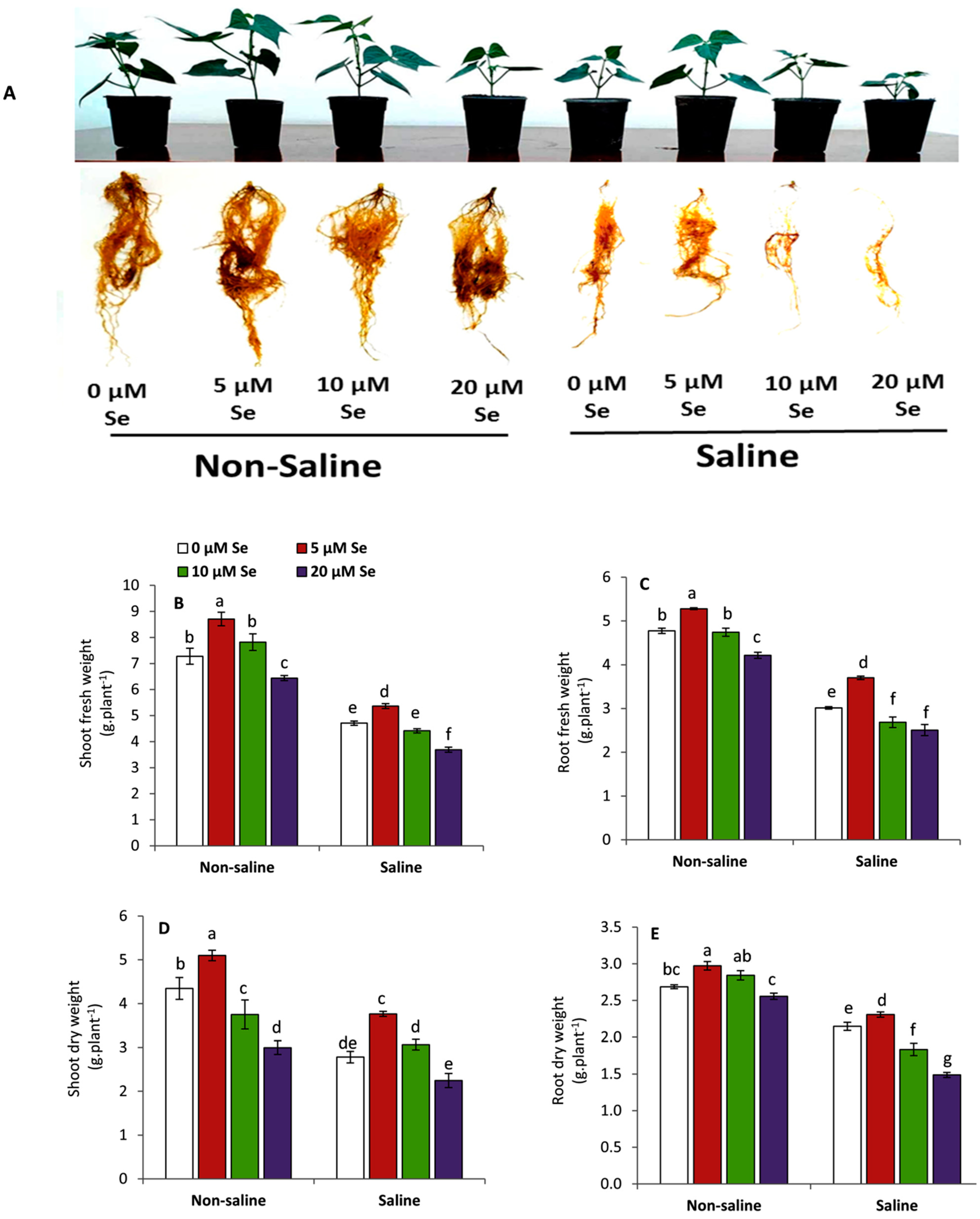
3.1. Effect of Applied-Se on Vegetative Growth

Snap bean seedlings exposed to salinity stress demonstrated an obvious and significant decrease in the biomass accumulation compared to the unstressed conditions (Figure 1). This reduction included the fresh and dry weights for the shoot and root systems compared to those of non-saline conditions. Applied Se displayed varying results according to its concentration. Generally, the growth parameters of seedlings treated with 5 µM Se were improved significantly compared to the untreated plants either under non-saline or saline conditions. In this context, the highest significant results in shoot fresh weight (13.8%), shoot dry weight (35.4%), root fresh weigh (22.8%) and root dry weight (7.5%) were obtained by the treatment of 5 µM Se compared to the untreated plants under saline conditions. In contrast, all examined growth parameters were progressively inhibited with increasing the concentration of applied Se up to 20 µM.

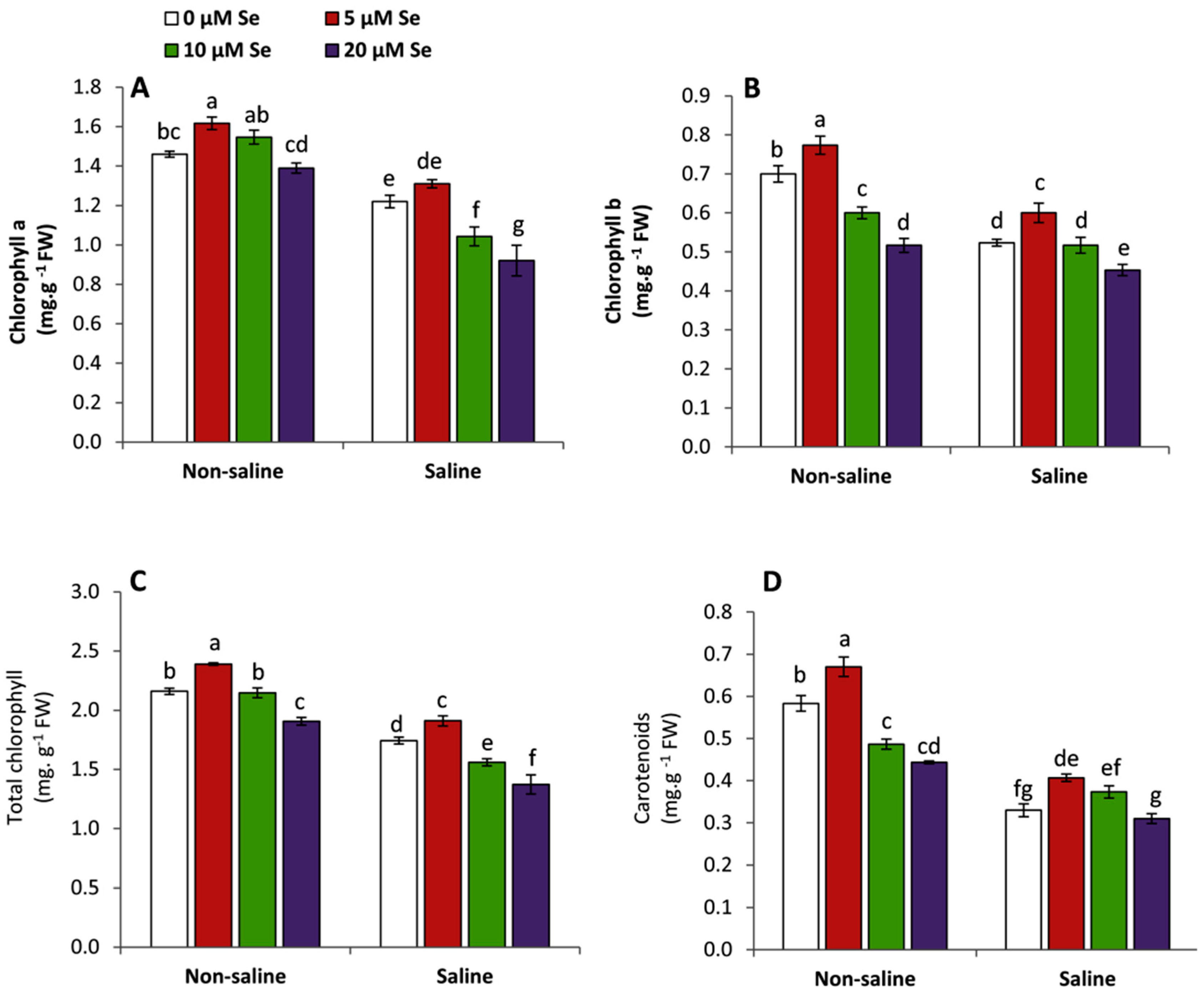
3.2. Effect of Applied-Se on Leaf Pigments

A similar trend to growth was observed in the content of photosynthetic pigments including Chl a, Chl b, Chl a + b or carotenoids (Figure 2), since applied Se displayed dual effects on the photosynthetic pigments according to its concentration. Plants treated with 5 µM Se exhibited a significant increase in Chl a (7.4%), Chl b (14.7 %), Chl a + b (9.6 %) and carotenoids (23.2%) compared to the untreated plants under saline conditions. However, the treatments of Se at 10 and 20 µM obviously reduced all these attributes. Generally, the lowest significant findings were achieved by Se at 20 µM under saline conditions.

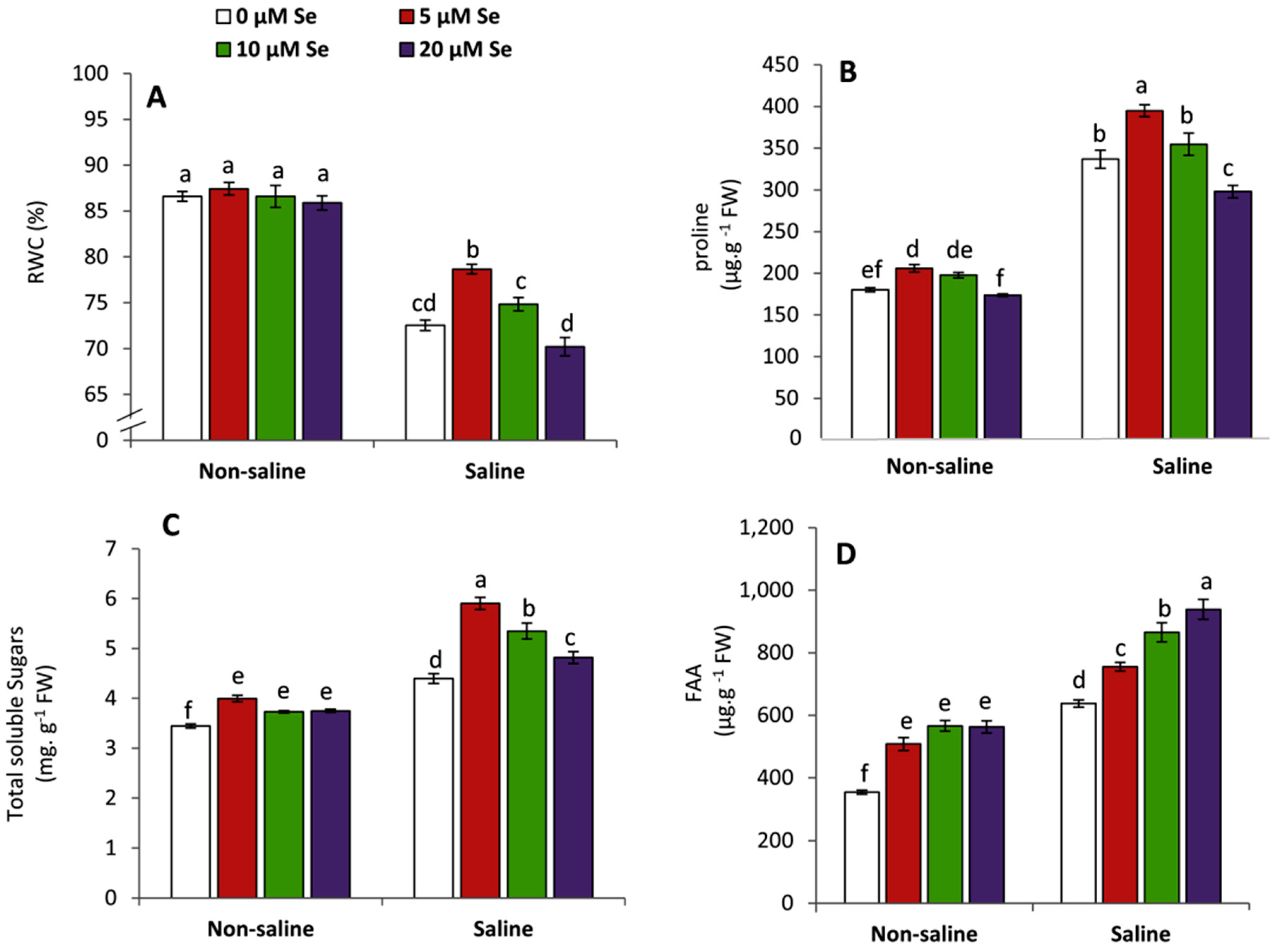
3.3. Effect of Applied Se on RWC and Osmolytes

Under saline conditions, all Se-treated and non-treated plants exhibited a significant decrease in RWC compared to the unstressed plants (Figure 3A). Meanwhile, no significant changes were observed under non-saline conditions. The results indicated that applied Se at 5 or 10 µM significantly enhanced RWC by 8.5 and 3.2%, respectively, compared to the untreated plants under saline conditions. In contrast, the treatment of 20 µM Se significantly reduced RWC compared to the untreated plants. On the other hand, there was a significant accumulation in proline, total soluble sugars and free amino acids in salt-stressed plants compared to those of unstressed conditions (Figure 3B–D). The highest significant results in proline (17.2%) and total soluble sugars (34.3%) were obtained by the treatment of 5 µM Se compared to the untreated plants under saline conditions. However, the maximum accumulation of free amino acids (47.2%) was achieved by the treatment of 20 µM Se under saline conditions. Under non-saline conditions, all Se treatments exhibited a significant improvement in total soluble sugars and free amino acids compared to the untreated plants, but proline was enhanced by the treatment of Se at 5 µM.

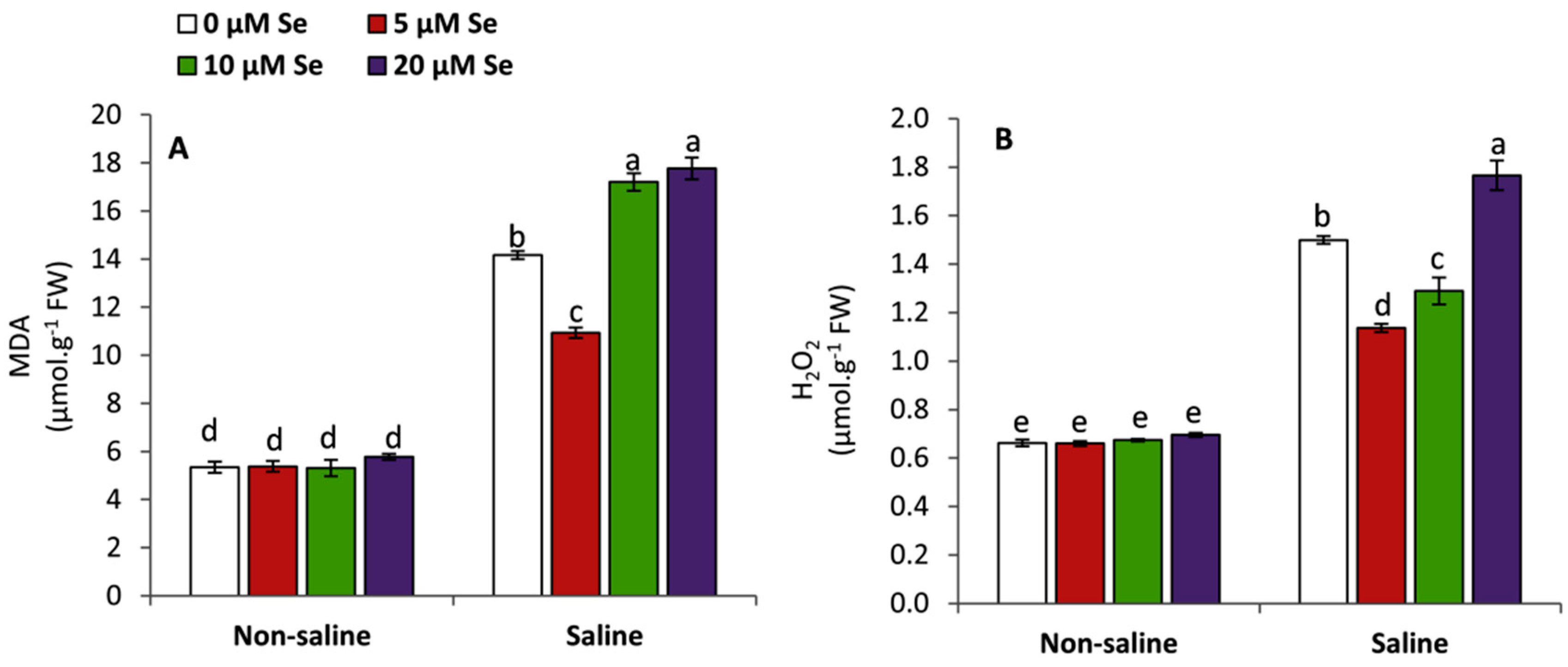
3.4. Effect of Applied-Se on the Accumulation of MDA and H2O2

Salt-stressed plants demonstrated higher oxidative stress as a result to increase the rate of lipid peroxidation (MDA) and accumulation of H2O2 (Figure 4). Plants treated with 5 µM Se showed a significant decrease in MDA (22.8%) and H2O2 (24.2%) compared to the untreated plants under saline conditions. A similar trend was observed in H2O2 by Se at 10 µM. Meanwhile, Se at 20 µM aggravated the oxidative damage by increasing the accumulation of MDA (25.4%) and H2O2 (17.8%) over the untreated plants under saline conditions. On the other hand, no significant changes were observed in MDA and H2O2 under non-saline conditions.

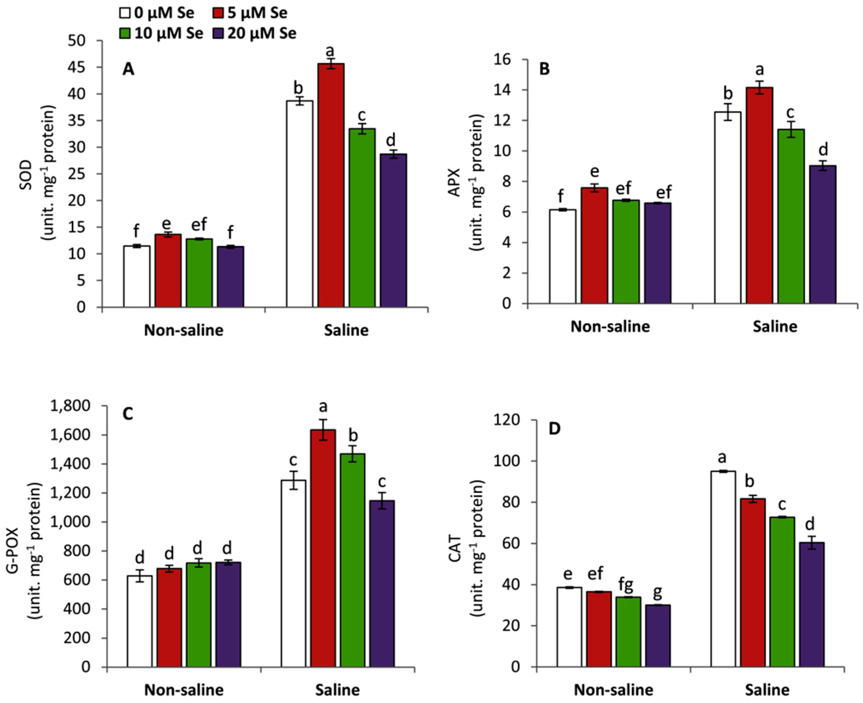
3.5. Effect of Applied Se on the Activities of Antioxidant Enzymes

Under saline conditions, snap bean seedlings demonstrated a significant increase in the activities of antioxidant enzymes including SOD, CAT, G-POX and APX compared to those grown under non-saline conditions (Figure 5). Plants treated with 5 µM Se showed a significant and greater activity in SOD (18.1%), APX (12.8%) and G-POX (27.1%) compared to the untreated plants under saline conditions. However, plants treated with 5 µM Se showed a significant decrease in CAT by 14.1% compared to the untreated plants under saline conditions. Conversely, increasing the dose of applied Se to 20 µM negatively affected all investigated antioxidant enzymes.

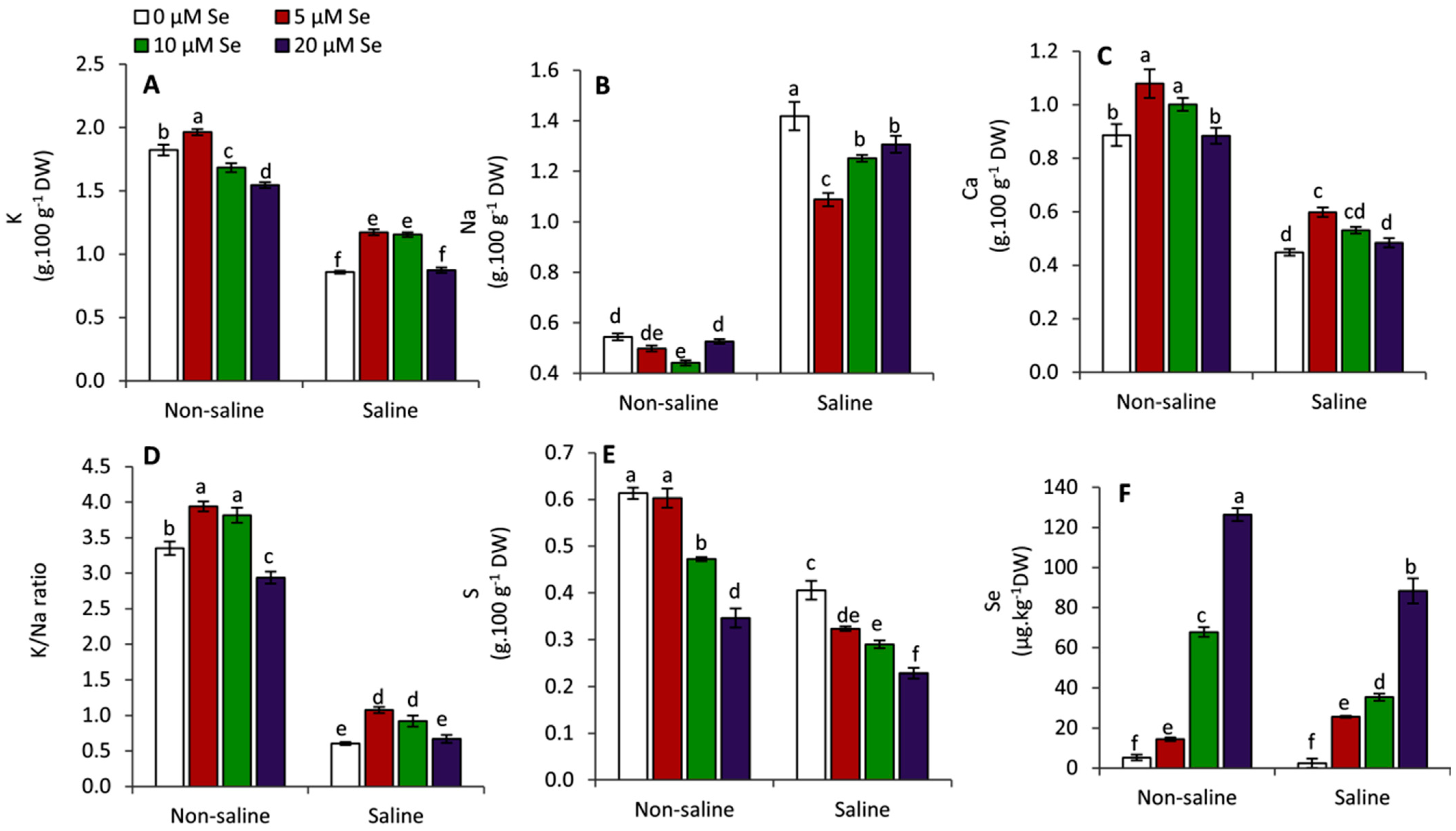
3.6. Effect of Applied Se on the Mineral Contents

Snap bean seedlings exposed to salinity stress demonstrated several changes in K, Na, Ca, K/Na ratio, S and Se compared to those grown under non-saline conditions (Figure 6). It was observed that salinity stress without Se treatments negatively and significantly affected K, Ca, K/Na ratio and S compared to the unstressed conditions. However, Na exhibited greater accumulation in the salt-stressed plants but no changes in Se. On the other hand, plants treated with 5 µM Se showed an improvement in K (36.7%; 7.8%), Ca (33.4%; 21.7%), K/Na ratio (77.9%; 17.6%) and Se (952%; 175.6%) compared to the untreated plants in both saline and non-saline conditions, respectively. Conversely, applied Se at 5 µM significantly decreased Na (23.3%) and S (20.3%) compared to untreated plants under saline conditions. This decrease was aggravated with increasing concentration of applied Se up to 20 µM.

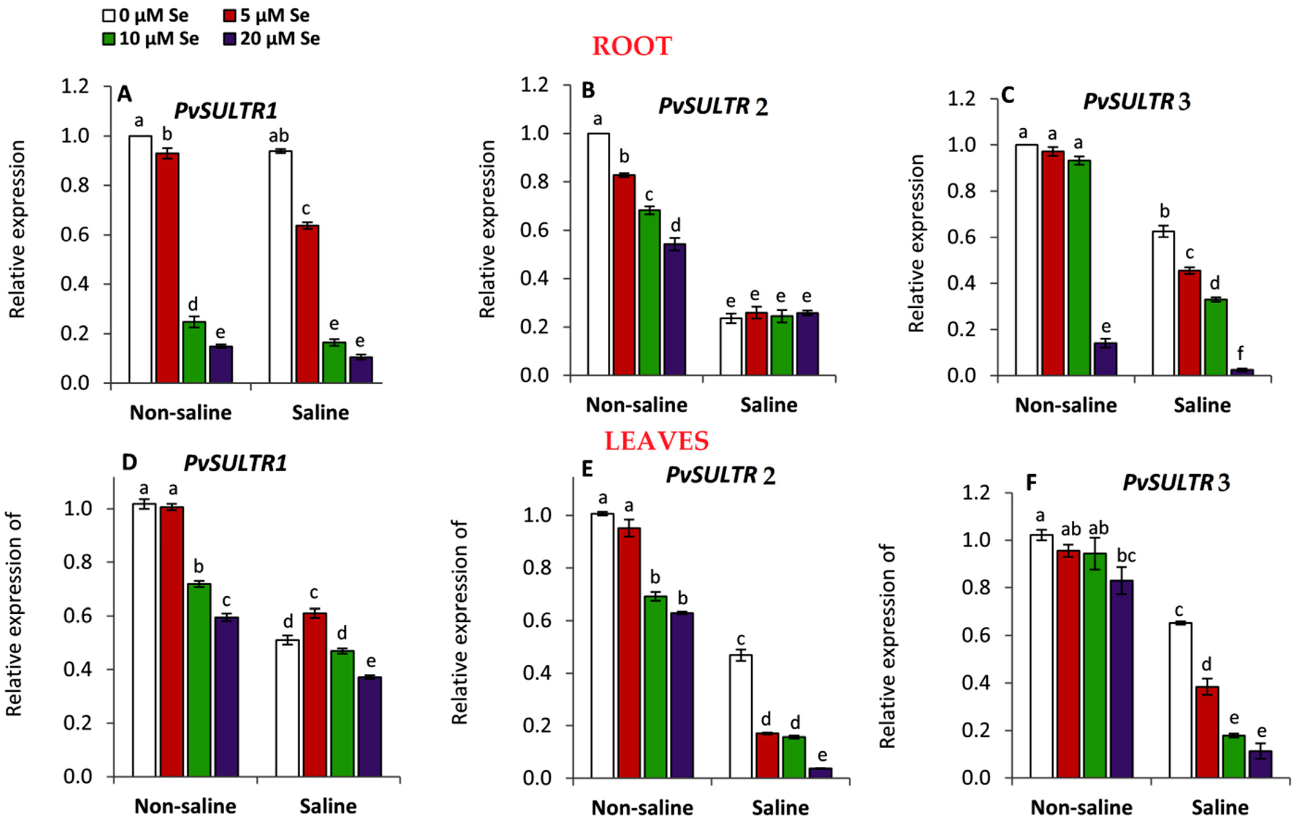
3.7. Effect of Applied Se on the Expression of Sulfate Transporter (SULTRs) Genes

Three sulfate (SO42−) transporter genes including SULTR1, SULTR2 and SULTR3 were investigated for their expression patterns in snap bean seedlings under saline and non-saline conditions (Figure 7). It was observed that SULTR1 in the root was significantly downregulated compared to the untreated plants under saline and non-saline conditions. This response was more obvious by the treatments of Se at 10 and 20 µM relative to 5 µM. A similar trend was observed in the expression of root SULTR2 and SULTR3 under non-saline and saline conditions, respectively. On the other hand, SULTR2 in roots did not reveal any significant changes under saline conditions. Meanwhile, SULTR3 in the root was dramatically declined by Se at 20 µM. Similarly, the relative expression of SULTR2 and SULTR3 in leaves was significantly downregulated with increasing the concentration of Se applications either under saline or non-saline conditions. As for SULTR1 in leaves, it showed a significant up-regulation with the treatment of Se at 5 µM under saline conditions, while it followed a similar pattern to SULTR2 and SULTR3 under non-saline conditions.

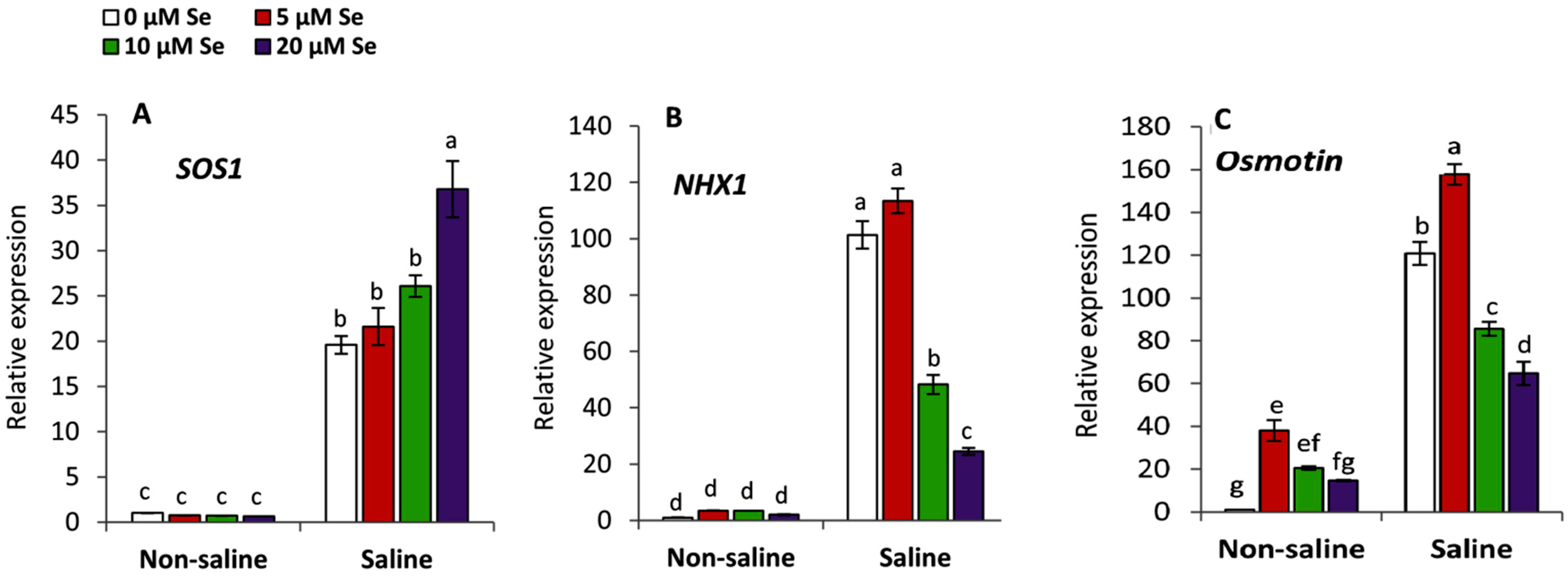
3.8. Effect of Applied Se on the Salt Stress Responsive Genes

Snap bean seedlings treated with various Se applications under saline or non-saline conditions displayed different patterns of expression to the plasma membrane Na+/H+ antiporter protein (SOS1), vacuolar Na+/H+ antiporter protein (NHX1) and Osmotin as a defensive protein against a wide array of biotic and abiotic stresses (Figure 8). The results indicated that the expression of SOS1, NHX1 and Osmotin was significantly upregulated under saline stress compared to the unstressed conditions. The highest upregulation in NHX1 and Osmotin under saline conditions was achieved by the treatment of Se at 5 µM compared to the other treatments. However, the highest significant upregulation in SOS1 was shown by Se at 20 µM under saline conditions. These findings imply that NHX1 and Osmotin were downregulated in parallel with an increase of the concentration of Se under saline conditions, but SOS1 was upregulated under the same conditions. On the other hand, no changes in SOS1, NHX1 were detected between different Se-treated and non-treated plants under non-saline conditions. Meanwhile, Osmotin was obviously and significantly upregulated with applied Se at 5 µM under non-saline conditions.

4. Discussion

Salinity stress is considered one of the most adverse environmental factors that can restrict plant growth or development. This effect could be attributed as hindering the rate of cell division, elongation and cytogenetic activities [71,72,73]. Furthermore, the toxic effect of salts ions (Na+ & Cl) can negatively affect photosynthesis [6], phytohormones [74], the balance of plant water status [75] and nutrients homeostasis [76,77]. In this study, applied Se at 5 µM exhibited the highest significant improvement in growth parameters compared to the other treatments under non-saline and saline conditions, whereas raising Se concentration up to 20 µM showed an obvious and significant decrease. Several lines of evidence proved that Se in plants has a narrow range between essentiality and toxicity [17,37,38]. At low doses, it can enhance plant growth through promoting the efficiency of photosynthesis and enhancing the chloroplast antioxidant defense system [38]. Many previous studies have found that applied Se at optimal levels can stimulate plant growth and confer salt tolerance in many plant species, i.e., canola [78], tomato [79] lettuce [80], maize [36] and wheat [17]. Conversely, the high levels of Se can cause a significant inhibition to plant growth and various biochemical attributes like photosynthesis and ion homeostasis. In this respect, it has been found that exogenous Se at 100 µM severely inhibited the growth parameters of potato plants under drought stress compared to 10 µM which demonstrated a protective role and achieved many benefits to plants under such conditions [37]. Moreover, the treatment of Se at 5 µM was more effective in enhancing wheat tolerance to salinity stress compared with 10 µM [17]. Meanwhile, applied Se at 40 µM strongly inhibited the growth of the root system in Arabidopsis thaliana L [81].
In this study, photosynthetic pigments including Chl a, Chl b, Chl a + b and carotenoids followed the similar trend of growth parameters. Under saline conditions, applied Se at low concentrations has been found to enhance the leaf content of photosynthetic pigments in many plant species [17,79,82,83]. For instance, 1 µM Se significantly improved the chlorophyll content of salt-stressed maize seedlings, but 25 µM Se showed an inhibitory effect on chlorophyll [83]. Furthermore, applied 5 µM Se was more effective in enhancing the chlorophyll and carotenoids of salt-stressed wheat seedlings than 10 µM Se [17]. This improvement could be attributed to the ability of Se at low doses to reduce the damage of chloroplast and maintain its ultrastructure under salinity stress [83], since most photosynthetic pigments are synthesized and localized in the chloroplast membranes [84].
Under saline conditions, plants accumulate a high concentration of low molecular-mass organic solutes such as proline, soluble sugars and free amino acids to regulate the osmotic potential of cells aiming at improving water absorption under such conditions [6,10,13,22]. In this study, applied Se specifically at 5 µM improved RWC, while Se at 20 µM reduced this response under saline conditions. Se supplementation at an optimal level can regulate the plant water status under desiccation induced by salinity [17] and drought stress [37]. These effects could be attributed to the ability of Se at low concentrations to protect the integrity of cell membranes from the salt stress-induced oxidative damage [38,82]. In contrast, Se at high levels could cause a replacement to sulfur ions with selenium leading to severe changes to protein which contributes to cell membranes structure and functioning [29,38]. As for the positive effect of applied Se at 5 µM on proline, this effect could be attributed to the ability of Se to increase the activity of γ-glutamyl kinase and reduce the activity of proline oxidase leading to an increase in proline biosynthesis and a reduction of its degradation [17,85]. In addition, enhancing the content of soluble sugars by Se at 5 µM under saline conditions could be related to the improvement of photosynthesis and maintaining RWC. On the other hand, increasing free amino acids by Se at 20 µM may be attributed to the inhibition of protein synthesis or stimulation of its degradation under saline conditions.
It has been documented that salt stress can induce oxidative damage to plant tissues as a result of the excessive production of reactive oxygen species [6,9,10,13]. These molecules at high levels can impair plant cell components from nucleic acids, proteins and lipids, and at severe levels may lead to cell death [83,86]. In this context, salinity stress was reported to mediate a decline in plant cell membrane stability due to increasing the activity of lipoxygenase activity and consequently reducing the rate of polyunsaturated fatty acids [87,88]. In this study, the plant exposed to salt stress revealed a significant increase in the rate of lipid peroxidation (MDA) and H2O2 compared to the non-saline conditions. Plants treated with Se at 5 µM exhibited an obvious decrease in MDA and H2O2, while the treatment of Se at 20 µM aggravated the oxidative damage induced by salinity stress. These results may imply the protective or harmful effects of Se on salt-stressed snap bean seedlings according to its applied concentration.
Applied Se can be involved in plant tolerance to salt stress by affecting the activities of several antioxidant enzymes. In this context, it has been confirmed that Se is involved in the formation of glutathione peroxidase (GPx) [89]. Furthermore, several lines of evidence indicated that applied Se at a low concentration can enhance the activities of antioxidant enzymes such as SOD, APX, CAT, G-POX, GR and GST under salinity stress [17,36,79,83]. In this study, plants treated with 5 µM Se exhibited higher activities in SOD, APX and G-POX compared to the untreated plants. However, an opposite trend was observed in CAT with an increase of the concentration of applied Se. SOD is considered the first defensive line against salt-induced oxidative damage by converting the produced superoxide radicals to H2O2 [90]. After that, it is important to eliminate the hyperaccumulation of H2O2 using another enzymatic mechanism [12,21,57,91]. Here, G-POX, CAT and APX play a key role in reducing the generated H2O2 by different pathways. This integration between different antioxidant enzymes was observed between SOD, APX and G-POX under the circumstances of this study. However, the opposite trend of CAT may imply that CAT might be sensitive to the concentration of applied Se in the salt-stressed snap bean seedlings.
In this study, applied Se at low concentration (5 µM) was found to improve the leaf content of K, Ca, K/Na ratio and Se either under saline or non-saline conditions. In contrast, Na was markedly decreased in Se-treated plants at 5 µM, in particular, under saline conditions. Applied Se at the optimum concentration can increase the activities of tonoplast proteins like H+ ATPase and Na+/H+ antiporters in the roots leading to preventing the uptake of Na+ and increasing the K/Na ratio [92]. These responses could maintain the osmotic balance and protect the different vital processes in Se-treated plants under saline conditions [93]. On the other hand, S was dramatically decreased in parallel with increasing the concentration of applied Se either under saline or non-saline conditions. The ionic radius of Se is approximately similar to S and has the same chemical properties [28]. Therefore, in the presence of Se, several sulfate transporters (SULTRs) can be involved in the absorption and transporting of Se instead of S [42,43].
Not much is known in the previous literature about the role of Se in regulating the function of sulfate transporters (SULTRs) in snap bean plants under saline conditions. In this study, the relative expression of three sulfate transporter genes (SULTR1, SULTR2 and SULTR3) using qRT-PCR were investigated in the roots and leaves of snap bean seedlings grown under saline and non-saline conditions. The results indicated that there were different expression patterns for all investigated SULTRs between the root and leaf tissues. Although SULTR1, SULTR2 were down-regulated in the roots with applied-Se at 5 µM under non-saline conditions, SULTR3 was not affected under the same conditions. This response was followed by a relative stability in the expression of all studied SULTRs genes between the non-treated and Se-treated plants at 5 µM in the leaves, leading to achieving S homeostasis under non-saline conditions (Figure 6E). In contrast, all examined SULTRs showed an obvious decline in their expression under saline conditions, leading to a decrease in the uptake of sulfate and consequently, a reduction in the concentration of S with an increase in the applied doses of Se (Figure 6E). In addition, it was obvious that the distinct inhibition of SULTR2 in roots under salinity stress may be attributed to NaCl stress and not to Se applications (Figure 7B).
Besides SULTRs genes, the salt-stress responsive genes, including salt overly sensitive gene (SOS1), vacuolar-localized Na+/H+ antiporter protein (NHX1) and the multifunctional osmotic protective protein (Osmotin), were studied (Figure 8). The results indicated that under saline conditions, there is an overexpression in all studied genes compared to plants grown under non-saline conditions. This response enables plants to maintain their osmotic balance and greater K+/Na+ ratio leading to an improvement in their tolerance to salt stress [6,94]. On the other hand, there was an overexpression in SOS1 followed by the decline of NHX1 with Se at 20 µM under saline conditions (Figure 8A,B). This response could refer to an increase in the toxicity of Na+ in the cytosol and a decrease in its transport to the vacuole with a raise in the concentration of applied Se. Conversely, the improvement in the expression of NHX1 and Osmotin with the treatment of 5 µM could explain the protective effect of this optimal concentration in enhancing the growth, osmotic status and tolerance to salinity stress under the circumstances of this study.

5. Conclusions

The present study provided the first evidence that elucidates the importance of applied Se at a low concentration in activating sulfate transporters (SULTRS) and achieving several beneficial roles that can help snap bean plants to mitigate the deleterious effects of salinity stress. Applied Se at a low concentration (5 µM) improved growth, photosynthetic pigments, osmolytes and nutrients’ homeostasis and antioxidant enzymes. Moreover, it regulated the balance between the uptake of Se and S as sulfate through mediating the function of SULTRS, alternation the expression of Na+/H+ antiporters (SOS1 and NHX1) and up-regulation of Osmotin, leading to enhanced plant tolerance under salinity stress. Generally, the different possible protective effects of Se on the salt-stressed snap bean plants can be summarized as shown in Figure 9. Further molecular studies are required in the future to explore the role of Se in regulating different membrane transporter systems and its influences on plant tolerance to salinity stress.

Author Contributions

Conceptualization, H.S.E.-B., M.F.M.I., A.A.E.-Y. and R.F.; methodology, M.F.M.I., H.G.A.E.-G., H.A.S.F., A.A.A. and R.F.; software, A.A.E.-Y. and M.A.; validation, H.S.E.-B., H.G.A.E.-G. and M.A.; T.A.S., A.A.A. and A.T.M.; formal analysis, M.F.M.I., H.A.S.F. and A.T.M.; investigation, H.S.E.-B., M.F.M.I., A.T.M. and R.F.; resources, M.A., H.A.S.F., M.A., T.A.S., A.A.A. and A.T.M.; data curation, H.S.E.-B., H.A.S.F., T.A.S., A.A.A. and A.T.M.; writing—original draft preparation, R.F., A.A.E.-Y. and H.G.A.E.-G.; writing—review and editing, H.S.E.-B., M.F.M.I., M.A., H.A.S.F., T.A.S., A.A.A. and A.T.M.; visualization, A.A.E.-Y. and M.A.; supervision, H.S.E.-B. and H.A.S.F.; project administration, H.S.E.-B. and H.A.S.F.; funding acquisition, H.S.E.-B., H.A.S.F., M.A., T.A.S., A.A.A. and A.T.M. All authors have read and agreed to the published version of the manuscript.

Funding

This work was supported through the annual funding track by the Deanship of Scientific Research, Vice Presidency for Graduate Studies and Scientific Research, King Faisal University, Saudi Arabia (Grant No. 1823).

Informed Consent Statement

Not applicable.

Data Availability Statement

Not applicable.

Acknowledgments

The authors thank the Deanship of Scientific Research, Vice Presidency for Graduate Studies and Scientific Research, King Faisal University, Saudi Arabia, for financial support to conduct and publish this research.

Conflicts of Interest

The authors declare no conflict of interest.

References

  1. Corwin, D.L. Climate change impacts on soil salinity in agricultural areas. Eur. J. Soil Sci. 2021, 72, 842–862. [Google Scholar] [CrossRef]
  2. Mukhopadhyay, R.; Sarkar, B.; Jat, H.S.; Sharma, P.C.; Bolan, N.S. Soil salinity under climate change: Challenges for sustainable agriculture and food security. J. Environ. Manag. 2021, 280, 111736. [Google Scholar] [CrossRef] [PubMed]
  3. Miceli, A.; Moncada, A.; Vetrano, F. Use of microbial biostimulants to increase the salinity tolerance of vegetable transplants. Agronomy 2021, 11, 1143. [Google Scholar] [CrossRef]
  4. Adeyemo, T.; Kramer, I.; Levy, G.J.; Mau, Y. Salinity and sodicity can cause hysteresis in soil hydraulic conductivity. Geoderma 2022, 413, 115765. [Google Scholar] [CrossRef]
  5. Alizadeh, A.; Akbarabadi, M.; Barsotti, E.; Piri, M.; Fishman, N.; Nagarajan, N. Salt precipitation in ultratight porous media and its impact on pore connectivity and hydraulic conductivity. Water Resour. Res. 2018, 54, 2768–2780. [Google Scholar] [CrossRef]
  6. Alnusairi, G.S.; Mazrou, Y.S.; Qari, S.H.; Elkelish, A.A.; Soliman, M.H.; Eweis, M.; Abdelaal, K.; El-Samad, G.A.; Ibrahim, M.F.; ElNahhas, N. Exogenous nitric oxide reinforces photosynthetic efficiency, osmolyte, mineral uptake, antioxidant, expression of stress-responsive genes and ameliorates the effects of salinity stress in wheat. Plants 2021, 10, 1693. [Google Scholar] [CrossRef]
  7. Çiçek, N.; Oukarroum, A.; Strasser, R.J.; Schansker, G. Salt stress effects on the photosynthetic electron transport chain in two chickpea lines differing in their salt stress tolerance. Photosynth. Res. 2018, 136, 291–301. [Google Scholar] [CrossRef]
  8. Manaa, A.; Goussi, R.; Derbali, W.; Cantamessa, S.; Abdelly, C.; Barbato, R. Salinity tolerance of quinoa (Chenopodium quinoa Willd) as assessed by chloroplast ultrastructure and photosynthetic performance. Environ. Exp. Bot. 2019, 162, 103–114. [Google Scholar] [CrossRef]
  9. Alsamadany, H.; Mansour, H.; Elkelish, A.; Ibrahim, M.F. Folic Acid Confers Tolerance against Salt Stress-Induced Oxidative Damages in Snap Beans through Regulation Growth, Metabolites, Antioxidant Machinery and Gene Expression. Plants 2022, 11, 1459. [Google Scholar] [CrossRef]
  10. Gong, B.; Wen, D.; Bloszies, S.; Li, X.; Wei, M.; Yang, F.; Shi, Q.; Wang, X. Comparative effects of NaCl and NaHCO3 stresses on respiratory metabolism, antioxidant system, nutritional status, and organic acid metabolism in tomato roots. Acta Physiolog. Plant. 2014, 36, 2167–2181. [Google Scholar] [CrossRef]
  11. Lotfi, R.; Ghassemi-Golezani, K.; Pessarakli, M. Salicylic acid regulates photosynthetic electron transfer and stomatal conductance of mung bean (Vigna radiata L.) under salinity stress. Biocatal. Agric. Biotechnol. 2020, 26, 101635. [Google Scholar] [CrossRef]
  12. El Nahhas, N.; AlKahtani, M.D.; Abdelaal, K.A.; Al Husnain, L.; AlGwaiz, H.I.; Hafez, Y.M.; Attia, K.A.; El-Esawi, M.A.; Ibrahim, M.F.; Elkelish, A. Biochar and jasmonic acid application attenuates antioxidative systems and improves growth, physiology, nutrient uptake and productivity of faba bean (Vicia faba L.) irrigated with saline water. Plant Physiol. Biochem. 2021, 166, 807–817. [Google Scholar] [CrossRef] [PubMed]
  13. Ramadan, K.M.A.; Alharbi, M.M.; Alenzi, A.M.; El-Beltagi, H.S.; Darwish, D.B.; Aldaej, M.I.; Shalaby, T.A.; Mansour, A.T.; El-Gabry, Y.A.; Ibrahim, M.F.M. Alpha Lipoic Acid as a Protective Mediator for Regulating the Defensive Responses of Wheat Plants against Sodic Alkaline Stress: Physiological, Biochemical and Molecular Aspects. Plants 2022, 11, 787. [Google Scholar] [CrossRef] [PubMed]
  14. Yan, G.; Fan, X.; Peng, M.; Yin, C.; Xiao, Z.; Liang, Y. Silicon improves rice salinity resistance by alleviating ionic toxicity and osmotic constraint in an organ-specific pattern. Front. Plant Sci. 2020, 11, 260. [Google Scholar] [CrossRef] [PubMed]
  15. Geilfus, C.-M. Chloride: From nutrient to toxicant. Plant Cell Physiol. 2018, 59, 877–886. [Google Scholar] [CrossRef]
  16. Yang, L.; Wang, X.; Chang, N.; Nan, W.; Wang, S.; Ruan, M.; Sun, L.; Li, S.; Bi, Y. Cytosolic glucose-6-phosphate dehydrogenase is involved in seed germination and root growth under salinity in Arabidopsis. Front. Plant Sci. 2019, 10, 182. [Google Scholar] [CrossRef] [Green Version]
  17. Elkelish, A.A.; Soliman, M.H.; Alhaithloul, H.A.; El-Esawi, M.A. Selenium protects wheat seedlings against salt stress-mediated oxidative damage by up-regulating antioxidants and osmolytes metabolism. Plant Physiol. Biochem. 2019, 137, 144–153. [Google Scholar] [CrossRef]
  18. De Micco, V.; Arena, C.; Amitrano, C.; Rouphael, Y.; De Pascale, S.; Cirillo, C. Effects of NaCl and CaCl2 Salinization on Morpho-Anatomical and Physiological Traits of Potted Callistemon citrinus Plants. Forests 2021, 12, 1666. [Google Scholar] [CrossRef]
  19. Hameed, A.; Ahmed, M.Z.; Hussain, T.; Aziz, I.; Ahmad, N.; Gul, B.; Nielsen, B.L. Effects of salinity stress on chloroplast structure and function. Cells 2021, 10, 2023. [Google Scholar] [CrossRef]
  20. Wang, C.-F.; Han, G.-L.; Yang, Z.-R.; Li, Y.-X.; Wang, B.-S. Plant salinity sensors: Current understanding and future directions. Front. Plant Sci. 2022, 13, 859224. [Google Scholar] [CrossRef]
  21. Abd Elhady, S.A.; El-Gawad, H.G.A.; Ibrahim, M.F.; Mukherjee, S.; Elkelish, A.; Azab, E.; Gobouri, A.A.; Farag, R.; Ibrahim, H.A.; El-Azm, N.A. Hydrogen peroxide supplementation in irrigation water alleviates drought stress and boosts growth and productivity of potato plants. Sustainability 2021, 13, 899. [Google Scholar] [CrossRef]
  22. El-Beltagi, H.S.; Ahmad, I.; Basit, A.; Shehata, W.F.; Hassan, U.; Shah, S.T.; Haleema, B.; Jalal, A.; Amin, R.; Khalid, M.A. Ascorbic acid enhances growth and yield of sweet peppers (Capsicum annum) by mitigating salinity stress. Gesunde Pflanz. 2022, 74, 423–433. [Google Scholar] [CrossRef]
  23. El-Beltagi, H.S.; Mohamed, M.E.M.; Ullah, S.; Shah, S. Effects of Ascorbic Acid and/or α-Tocopherol on Agronomic and Physio-Biochemical Traits of Oat (Avena sativa L.) under Drought Condition. Agronomy 2022, 12, 2296. [Google Scholar] [CrossRef]
  24. Elkelish, A.; Ibrahim, M.F.; Ashour, H.; Bondok, A.; Mukherjee, S.; Aftab, T.; Hikal, M.; El-Yazied, A.A.; Azab, E.; Gobouri, A.A. Exogenous Application of Nitric Oxide Mitigates Water Stress and Reduces Natural Viral Disease Incidence of Tomato Plants Subjected to Deficit Irrigation. Agronomy 2021, 11, 87. [Google Scholar] [CrossRef]
  25. El-Yazied, A.A.; Ibrahim, M.F.; Ibrahim, M.A.; Nasef, I.N.; Al-Qahtani, S.M.; Al-Harbi, N.A.; Alzuaibr, F.M.; Alaklabi, A.; Dessoky, E.S.; Alabdallah, N.M. Melatonin Mitigates Drought Induced Oxidative Stress in Potato Plants through Modulation of Osmolytes, Sugar Metabolism, ABA Homeostasis and Antioxidant Enzymes. Plants 2022, 11, 1151. [Google Scholar] [CrossRef]
  26. Iwaniuk, P.; Borusiewicz, A.; Lozowicka, B. Fluazinam and its mixtures induce diversified changes of crucial biochemical and antioxidant profile in leafy vegetable. Sci. Hortic. 2022, 298, 110988. [Google Scholar] [CrossRef]
  27. Jahan, M.S.; Hasan, M.M.; Alotaibi, F.S.; Alabdallah, N.M.; Alharbi, B.M.; Ramadan, K.M.; Bendary, E.S.; Alshehri, D.; Jabborova, D.; Al-Balawi, D.A. Exogenous Putrescine Increases Heat Tolerance in Tomato Seedlings by Regulating Chlorophyll Metabolism and Enhancing Antioxidant Defense Efficiency. Plants 2022, 11, 1038. [Google Scholar] [CrossRef]
  28. Bodnar, M.; Konieczka, P.; Namiesnik, J. The properties, functions, and use of selenium compounds in living organisms. J. Environ. Sci. Health Part C 2012, 30, 225–252. [Google Scholar] [CrossRef]
  29. Terry, N.; Zayed, A.; De Souza, M.; Tarun, A. Selenium in higher plants. Annu. Rev. Plant Biol. 2000, 51, 401–432. [Google Scholar] [CrossRef] [Green Version]
  30. Brigelius-Flohé, R. Selenium in human health and disease: An overview. In Selenium; Springer: Berlin/Heidelberg, Germany, 2018; pp. 3–26. [Google Scholar]
  31. Kieliszek, M.; Bano, I.; Zare, H. A comprehensive review on selenium and its effects on human health and distribution in Middle Eastern countries. Biol. Trace Elem. Res. 2022, 200, 971–987. [Google Scholar] [CrossRef]
  32. Surai, P.F.; Kochish, I.I.; Fisinin, V.I.; Juniper, D.T. Revisiting oxidative stress and the use of organic selenium in dairy cow nutrition. Animals 2019, 9, 462. [Google Scholar] [CrossRef] [Green Version]
  33. Heider, J.; Bock, A. Selenium metabolism in micro-organisms. Adv. Microb. Physiol. 1993, 35, 71–109. [Google Scholar]
  34. Feng, R.; Wei, C.; Tu, S. The roles of selenium in protecting plants against abiotic stresses. Environ. Exp. Bot. 2013, 87, 58–68. [Google Scholar] [CrossRef]
  35. Sager, M. Selenium in agriculture, food, and nutrition. Pure Appl. Chem. 2006, 78, 111–133. [Google Scholar] [CrossRef]
  36. Ashraf, M.A.; Akbar, A.; Parveen, A.; Rasheed, R.; Hussain, I.; Iqbal, M. Phenological application of selenium differentially improves growth, oxidative defense and ion homeostasis in maize under salinity stress. Plant Physiol. Biochem. 2018, 123, 268–280. [Google Scholar] [CrossRef]
  37. Ibrahim, M.; Ibrahim, H.A. Assessment of selenium role in promoting or inhibiting potato plants under water stress. J. Hortic. Sci. Ornam. Plants 2016, 8, 125–139. [Google Scholar]
  38. Rasool, A.; Shah, W.H.; Mushtaq, N.U.; Saleem, S.; Hakeem, K.R.; ul Rehman, R. Amelioration of salinity induced damage in plants by selenium application: A review. South Afr. J. Bot. 2022, 147, 98–105. [Google Scholar] [CrossRef]
  39. Bañuelos, G.S.; Lin, Z.-Q.; Broadley, M. Selenium biofortification. In Selenium in Plants; Springer: Berlin/Heidelberg, Germany, 2017; pp. 231–255. [Google Scholar]
  40. Malagoli, M.; Schiavon, M.; dall’Acqua, S.; Pilon-Smits, E.A. Effects of selenium biofortification on crop nutritional quality. Front. Plant Sci. 2015, 6, 280. [Google Scholar] [CrossRef] [Green Version]
  41. Pyrzyńska, K. Determination of selenium species in environmental samples. Microchim. Acta 2002, 140, 55–62. [Google Scholar] [CrossRef]
  42. Feist, L.J.; Parker, D.R. Ecotypic variation in selenium accumulation among populations of Stanleya pinnata. New Phytol. 2001, 149, 61–69. [Google Scholar] [CrossRef]
  43. Zhao, Q.; Geng, J.; Du, Y.; Li, S.; Yuan, X.; Zhu, J.; Zhou, Z.; Wang, Q.; Du, J. The common bean (Phaseolus vulgaris) SULTR gene family: Genome-wide identification, phylogeny, evolutionary expansion and expression patterns. Biotechnol. Biotechnol. Equip. 2022, 36, 724–736. [Google Scholar] [CrossRef]
  44. Zhang, W.-D.; Wang, P.; Bao, Z.; Ma, Q.; Duan, L.-J.; Bao, A.-K.; Zhang, J.-L.; Wang, S.-M. SOS1, HKT1; 5, and NHX1 synergistically modulate Na+ homeostasis in the halophytic grass Puccinellia tenuiflora. Front. Plant Sci. 2017, 8, 576. [Google Scholar]
  45. Ullah, A.; Hussain, A.; Shaban, M.; Khan, A.H.; Alariqi, M.; Gul, S.; Jun, Z.; Lin, S.; Li, J.; Jin, S. Osmotin: A plant defense tool against biotic and abiotic stresses. Plant Physiol. Biochem. 2018, 123, 149–159. [Google Scholar]
  46. Wan, Q.; Hongbo, S.; Zhaolong, X.; Jia, L.; Dayong, Z.; Yihong, H. Salinity tolerance mechanism of osmotin and osmotin-like proteins: A promising candidate for enhancing plant salt tolerance. Curr. Genom. 2017, 18, 553–556. [Google Scholar] [CrossRef]
  47. Bashir, M.A.; Silvestri, C.; Ahmad, T.; Hafiz, I.A.; Abbasi, N.A.; Manzoor, A.; Cristofori, V.; Rugini, E. Osmotin: A cationic protein leads to improve biotic and abiotic stress tolerance in plants. Plants 2020, 9, 992. [Google Scholar] [CrossRef]
  48. Câmara, C.R.; Urrea, C.A.; Schlegel, V. Pinto beans (Phaseolus vulgaris L.) as a functional food: Implications on human health. Agriculture 2013, 3, 90–111. [Google Scholar] [CrossRef]
  49. Celmeli, T.; Sari, H.; Canci, H.; Sari, D.; Adak, A.; Eker, T.; Toker, C. The nutritional content of common bean (Phaseolus vulgaris L.) landraces in comparison to modern varieties. Agronomy 2018, 8, 166. [Google Scholar] [CrossRef] [Green Version]
  50. Ibrahim, M.; Ibrahim, H.A.; Abd El-Gawad, H. Folic acid as a protective agent in snap bean plants under water deficit conditions. J. Hortic. Sci. Biotechnol. 2021, 96, 94–109. [Google Scholar] [CrossRef]
  51. Didinger, C.; Foster, M.T.; Bunning, M.; Thompson, H.J. Nutrition and Human Health Benefits of Dry Beans and Other Pulses. In Dry Beans and Pulses: Production, Processing, and Nutrition, 2nd ed.; John Wiley & Sons Ltd.: Hoboken, NJ, USA, 2022; pp. 481–504. [Google Scholar]
  52. Sangaramoorthy, M.; Koo, J.; John, E.M. Intake of bean fiber, beans, and grains and reduced risk of hormone receptor-negative breast cancer: The San Francisco Bay Area Breast Cancer Study. Cancer Med. 2018, 7, 2131–2144. [Google Scholar] [CrossRef]
  53. Garcia, C.L.; Dattamudi, S.; Chanda, S.; Jayachandran, K. Effect of salinity stress and microbial inoculations on glomalin production and plant growth parameters of snap bean (Phaseolus vulgaris). Agronomy 2019, 9, 545. [Google Scholar] [CrossRef] [Green Version]
  54. Hoagland, D.R.; Arnon, D.I. The water-culture method for growing plants without soil. Circ. Calif. Agric. Exp. Stn. 1950, 347, 32. [Google Scholar]
  55. Costache, M.A.; Campeanu, G.; Neata, G. Studies concerning the extraction of chlorophyll and total carotenoids from vegetables. Rom. Biotechnol. Lett. 2012, 17, 7702–7708. [Google Scholar]
  56. de Carvalho, L.M.J.; Gomes, P.B.; de Oliveira Godoy, R.L.; Pacheco, S.; do Monte, P.H.F.; de Carvalho, J.L.V.; Nutti, M.R.; Neves, A.C.L.; Vieira, A.C.R.A.; Ramos, S.R.R. Total carotenoid content, α-carotene and β-carotene, of landrace pumpkins (Cucurbita moschata Duch): A preliminary study. Food Res. Int. 2012, 47, 337–340. [Google Scholar] [CrossRef] [Green Version]
  57. Abd Elbar, O.H.; Elkelish, A.; Niedbała, G.; Farag, R.; Wojciechowski, T.; Mukherjee, S.; Abou-Hadid, A.F.; El-Hennawy, H.M.; Abou El-Yazied, A.; Abd El-Gawad, H.G. Protective Effect of γ-Aminobutyric Acid Against Chilling Stress during Reproductive Stage in Tomato Plants Through Modulation of Sugar Metabolism, Chloroplast Integrity, and Antioxidative Defense Systems. Front. Plant Sci. 2021, 12, 663750. [Google Scholar] [CrossRef]
  58. Bates, L.; Waldren, R.; Teare, I. Rapid determination of free proline for water-stress studies. Plant Soil 1973, 39, 205–207. [Google Scholar] [CrossRef]
  59. Chow, P.S.; Landhäusser, S.M. A method for routine measurements of total sugar and starch content in woody plant tissues. Tree Physiol. 2004, 24, 1129–1136. [Google Scholar] [CrossRef]
  60. Yemm, E.W.; Cocking, E.C.; Ricketts, R.E. The determination of amino-acids with ninhydrin. Analyst 1955, 80, 209–214. [Google Scholar] [CrossRef]
  61. Heath, R.L.; Packer, L. Photoperoxidation in isolated chloroplasts: I. Kinetics and stoichiometry of fatty acid peroxidation. Arch. Biochem. Biophys. 1968, 125, 189–198. [Google Scholar] [CrossRef]
  62. Patterson, B.D.; MacRae, E.A.; Ferguson, I.B. Estimation of hydrogen peroxide in plant extracts using titanium (IV). Anal. Biochem. 1984, 139, 487–492. [Google Scholar] [CrossRef]
  63. Beyer, W.F., Jr.; Fridovich, I. Assaying for superoxide dismutase activity: Some large consequences of minor changes in conditions. Anal. Biochem. 1987, 161, 559–566. [Google Scholar] [CrossRef]
  64. Nakano, Y.; Asada, K. Hydrogen peroxide is scavenged by ascorbate-specific peroxidase in spinach chloroplasts. Plant Cell Physiol. 1981, 22, 867–880. [Google Scholar]
  65. Dias, M.A.; Costa, M.M. Effect of low salt concentrations on nitrate reductase and peroxidase of sugar beet leaves. J. Exp. Bot. 1983, 34, 537–543. [Google Scholar] [CrossRef]
  66. Cakmak, I.; Strbac, D.; Marschner, H. Activities of hydrogen peroxide-scavenging enzymes in germinating wheat seeds. J. Exp. Bot. 1993, 44, 127–132. [Google Scholar] [CrossRef]
  67. Carrero, P.E.; Tyson, J.F. Determination of Selenium by Atomic Absorption Spectrometry WithSimultaneous Retention of Selenium (IV) and Tetrahydroborate (III) on anAnion-exchange Resin Followed by Flow Injection Hydride Generation Fromthe Solid Phase. Analyst 1997, 122, 915–919. [Google Scholar] [CrossRef] [Green Version]
  68. Kirkbright, G.; Marshall, M. Direct determination of sulfur by atomic absorption spectrometry in a nitrogen separated nitorus oxide-acetylene flame. Anal. Chem. 1972, 44, 1288–1290. [Google Scholar] [CrossRef]
  69. Momen, A.A.; Zachariadis, G.A.; Anthemidis, A.N.; Stratis, J.A. Investigation of four digestion procedures for multi-element determination of toxic and nutrient elements in legumes by inductively coupled plasma-optical emission spectrometry. Anal. Chim. Acta 2006, 565, 81–88. [Google Scholar] [CrossRef]
  70. SAS. SAS/STAT User’s Guide: Release, 6.03 ed.; SAS Institute Ltd.: Cary, NC, USA, 1988. [Google Scholar]
  71. Barakat, H. Interactive effects of salinity and certain vitamins on gene expression and cell division. Int. J. Agric. Biol. 2003, 3, 219–225. [Google Scholar]
  72. Tabur, S.; Demir, K. Role of some growth regulators on cytogenetic activity of barley under salt stress. Plant Growth Regul. 2010, 60, 99–104. [Google Scholar] [CrossRef]
  73. Valenzuela, C.E.; Acevedo-Acevedo, O.; Miranda, G.S.; Vergara-Barros, P.; Holuigue, L.; Figueroa, C.R.; Figueroa, P.M. Salt stress response triggers activation of the jasmonate signaling pathway leading to inhibition of cell elongation in Arabidopsis primary root. J. Exp. Bot. 2016, 67, 4209–4220. [Google Scholar] [CrossRef] [Green Version]
  74. Yu, Z.; Duan, X.; Luo, L.; Dai, S.; Ding, Z.; Xia, G. How plant hormones mediate salt stress responses. Trends Plant Sci. 2020, 25, 1117–1130. [Google Scholar] [CrossRef]
  75. Stępień, P.; Kłbus, G. Water relations and photosynthesis in Cucumis sativus L. leaves under salt stress. Biol. Plant. 2006, 50, 610–616. [Google Scholar] [CrossRef]
  76. Abdelaal, K.; Alsubeie, M.S.; Hafez, Y.; Emeran, A.; Moghanm, F.; Okasha, S.; Omara, R.; Basahi, M.A.; Darwish, D.B.E.; Ibrahim, M.F.M.; et al. Physiological and Biochemical Changes in Vegetable and Field Crops under Drought, Salinity and Weeds Stresses: Control Strategies and Management. Agriculture 2022, 12, 2084. [Google Scholar] [CrossRef]
  77. Youssef, M.H.; Raafat, A.; El-Yazied, A.A.; Selim, S.; Azab, E.; Khojah, E.; El Nahhas, N.; Ibrahim, M.F. Exogenous Application of Alpha-Lipoic Acid Mitigates Salt-Induced Oxidative Damage in Sorghum Plants through Regulation Growth, Leaf Pigments, Ionic Homeostasis, Antioxidant Enzymes, and Expression of Salt Stress Responsive Genes. Plants 2021, 10, 2519. [Google Scholar] [CrossRef] [PubMed]
  78. Hashem, H.A.; Hassanein, R.A.; Bekheta, M.A.; El-Kady, F.A. Protective role of selenium in canola (Brassica napus L.) plant subjected to salt stress. Egypt. J. Exp. Biol. 2013, 9, 199–211. [Google Scholar]
  79. Diao, M.; Ma, L.; Wang, J.; Cui, J.; Fu, A.; Liu, H.-y. Selenium promotes the growth and photosynthesis of tomato seedlings under salt stress by enhancing chloroplast antioxidant defense system. J. Plant Growth Regul. 2014, 33, 671–682. [Google Scholar] [CrossRef]
  80. Khalifa, G.; Abdelrassoul, M.; Hegazi, A.; Elsherif, M. Mitigation of saline stress adverse effects in lettuce plant using selenium and silicon. Middle East J. Agric. Res. 2016, 5, 347–361. [Google Scholar]
  81. Lehotai, N.; Kolbert, Z.; Pető, A.; Feigl, G.; Ördög, A.; Kumar, D.; Tari, I.; Erdei, L. Selenite-induced hormonal and signalling mechanisms during root growth of Arabidopsis thaliana L. J. Exp. Bot. 2012, 63, 5677–5687. [Google Scholar] [CrossRef] [Green Version]
  82. Hawrylak-Nowak, B. Beneficial effects of exogenous selenium in cucumber seedlings subjected to salt stress. Biol. Trace Elem. Res. 2009, 132, 259–269. [Google Scholar] [CrossRef] [PubMed]
  83. Jiang, C.; Zu, C.; Lu, D.; Zheng, Q.; Shen, J.; Wang, H.; Li, D. Effect of exogenous selenium supply on photosynthesis, Na+ accumulation and antioxidative capacity of maize (Zea mays L.) under salinity stress. Sci. Rep. 2017, 7, 42039. [Google Scholar] [CrossRef] [Green Version]
  84. Matile, P.; Schellenberg, M.; Vicentini, F. Localization of chlorophyllase in the chloroplast envelope. Planta 1997, 201, 96–99. [Google Scholar] [CrossRef]
  85. Khan, M.I.R.; Nazir, F.; Asgher, M.; Per, T.S.; Khan, N.A. Selenium and sulfur influence ethylene formation and alleviate cadmium-induced oxidative stress by improving proline and glutathione production in wheat. J. Plant Physiol. 2015, 173, 9–18. [Google Scholar] [CrossRef]
  86. Akyol, T.Y.; Yilmaz, O.; Uzilday, B.; Uzilday, R.Ö.; TÜrkan, İ. Plant response to salinity: An analysis of ROS formation, signaling, and antioxidant defense. Turk. J. Bot. 2020, 44, 1–13. [Google Scholar]
  87. Heidari, M.; Tafazoli, E. Effect of sodium chloride on lipoxygenase activity, hydrogen peroxide content and lipid peroxidation rate in the seedlings of three Pistacia rootstocks. Isfahan Univ. Technol.-J. Crop Prod. Process. 2005, 9, 41–50. [Google Scholar]
  88. Xu, X.-Q.; Beardall, J. Effect of salinity on fatty acid composition of a green microalga from an antarctic hypersaline lake. Phytochemistry 1997, 45, 655–658. [Google Scholar] [CrossRef]
  89. Rotruck, J.T.; Pope, A.L.; Ganther, H.E.; Swanson, A.; Hafeman, D.G.; Hoekstra, W. Selenium: Biochemical role as a component of glutathione peroxidase. Science 1973, 179, 588–590. [Google Scholar] [CrossRef] [PubMed]
  90. Berwal, M.; Ram, C. Superoxide dismutase: A stable biochemical marker for abiotic stress tolerance in higher plants. In Abiotic and Biotic Stress in Plants; IntechOpen: London, UK, 2018; pp. 1–10. [Google Scholar]
  91. Abd El-Gawad, H.G.; Mukherjee, S.; Farag, R.; Abd Elbar, O.H.; Hikal, M.; Abou El-Yazied, A.; Abd Elhady, S.A.; Helal, N.; ElKelish, A.; El Nahhas, N. Exogenous γ-aminobutyric acid (GABA)-induced signaling events and field performance associated with mitigation of drought stress in Phaseolus vulgaris L. Plant Signal. Behav. 2021, 16, 1853384. [Google Scholar] [CrossRef]
  92. Zhang, Y.; Wang, L.; Liu, Y.; Zhang, Q.; Wei, Q.; Zhang, W. Nitric oxide enhances salt tolerance in maize seedlings through increasing activities of proton-pump and Na+/H+ antiport in the tonoplast. Planta 2006, 224, 545–555. [Google Scholar] [CrossRef]
  93. Gupta, M.; Gupta, S. An overview of selenium uptake, metabolism, and toxicity in plants. Front. Plant Sci. 2017, 7, 2074. [Google Scholar] [CrossRef] [Green Version]
  94. Yue, Y.; Zhang, M.; Zhang, J.; Duan, L.; Li, Z. SOS1 gene overexpression increased salt tolerance in transgenic tobacco by maintaining a higher K+/Na+ ratio. J. Plant Physiol. 2012, 169, 255–261. [Google Scholar] [CrossRef]
Figure 1. Effect of Se supplementation at 5, 10 and 20 µM on growth performance (A), shoot fresh weight (B), shoot fresh weight (C), shoot dry weight (D) and root dry weight (E) of non-stressed and salt-stressed (50 mM NaCl) snap bean seedlings (28 days after sowing). The results are expressed as mean values of three measurements ± SE using Duncan’s multiple range test (p = 0.05). Different letters indicate significant differences among the treatments.
Agronomy 12 03215 g001
Figure 2. Effect of Se supplementation at 5, 10 and 20 µM on chlorophyll a (A), chlorophyll b (B), total chlorophyll (C) and carotenoids (D) of non-stressed and salt-stressed (50 mM NaCl) snap bean seedlings (28 days after sowing). The results are expressed as mean values of three measurements ± SE using Duncan’s multiple range test (p = 0.05). Different letters indicate significant differences among the treatments.
Agronomy 12 03215 g002
Figure 3. Effect of Se supplementation at 5, 10 and 20 µM on relative water content; RWC (A), proline (B), total soluble sugars (C) or free amino acids; FAA (D) of non-stressed and salt-stressed (50 mM NaCl) snap bean seedlings (28 days after sowing). The results are expressed as mean values of three measurements ± SE using Duncan’s multiple range test (p = 0.05). Different letters indicate significant differences among the treatments.
Agronomy 12 03215 g003
Figure 4. Effect of Se supplementation at 5, 10 and 20 µM on relative water content; malondialdehyde; MDA (A) and hydrogen peroxide; H2O2 (B) in the leaves of non-stressed and salt-stressed (50 mM NaCl) snap bean seedlings (28 days after sowing). The results are expressed as mean values of three measurements ± SE using Duncan’s multiple range test (p = 0.05). Different letters indicate significant differences among the treatments.
Agronomy 12 03215 g004
Figure 5. Effect of Se supplementation at 5, 10 and 20 µM on the activity of superoxide dismutase; SOD (A), ascorbate peroxidase; APX (B), guiacol peroxidase; G-POx (C) and catalase; CAT (D) of non-stressed and salt-stressed (50 mM NaCl) snap bean seedlings (28 days after sowing). The results are expressed as mean values of three measurements ± SE using Duncan’s multiple range test (p = 0.05). Different letters indicate significant differences among the treatments.
Agronomy 12 03215 g005
Figure 6. Effect of Se supplementation at 5, 10 and 20 µM on the leaf content of K (A), Na (B), Ca (C), K/Na ratio (D), S (E) and Se (F) of non-stressed and salt-stressed (50 mM NaCl) snap bean seedlings (28 days after sowing). The results are expressed as mean values of three measurements ± SE using Duncan’s multiple range test (p = 0.05). Different letters indicate significant differences among the treatments.
Agronomy 12 03215 g006
Figure 7. Effect of Se supplementation at 5, 10 and 20 µM on the relative expression of sulfate transporter genes. Root-PvSULTR1 (A), Root-PvSULTR2 (B), Root-PvSULTR3 (C), Shoot-PvSULTR1 (D) Shoot-PvSULTR2 (E) and Shoot-PvSULTR3 (F) of non-stressed and salt-stressed (50 mM NaCl) snap bean seedlings (28 days after sowing). The results are expressed as mean values of three measurements ± SE using Duncan’s multiple range test (p = 0.05). Different letters indicate significant differences among the treatments.
Agronomy 12 03215 g007
Figure 8. Effect of Se supplementation at 5, 10 and 20 µM on relative expression of salt stress responsive genes. SOS1 (A), NHX1 (B) and Osmotin (C) of non-stressed and salt-stressed (50 mM NaCl) snap bean seedlings (28 days after sowing). The results are expressed as mean values of three measurements ± SE using Duncan’s multiple range test (p = 0.05). Different letters indicate significant differences among the treatments.
Agronomy 12 03215 g008
Figure 9. Simplified model for the suggested effects of applied Se on snap bean plants grown under salinity stress. RWC, relative water content; MDA, malondialdehyde; FAA, free amino acids; G-POX, Guaiacol peroxidases; CAT, catalase; APX; ascorbate peroxidase; SOD, superoxide dismutase; blue solid lines indicate the positive effects of applied Se at low concentration; red dotted lines indicate the negative effects of applied Se at high level; green up arrow, increase; red down arrow.
Agronomy 12 03215 g009
Table 1. Oligonucleotide primer pairs used for quantitative RT-PCR analysis.
Gene NameSequence
SULTR1F5′-CGCAGACTATGAATACCCGA-3′
R5′-TTCCTAAACGGGTCATCTGG-3′
SULTR2F5′-CAGAAGGAATAGCAATAGGA-3′
R5′-CAAGTAGCAGGAAGTAAAAG-3′
SULTR3F5′-TCTTTCTCACGGTCAGCAGT-3′
R5′-TAGCATTTGGAGTGTATTCG-3′
SOS1F5′-ACTTGCAGGAGGAATACAAC-3′
R5′-CGAGAAGAGAAGACCACATC-3′
OsmotinF5′-GAACGGAGGGTGTCACAAAATC-3′
R5′-CGTAGTGGGTCCACAAGTTCCT-3′
NHX1F5′-CGTGATGTCGCATTACACCT-3′
R5′-CTGGCAAACTCCCACTTCTC-3′
GAPDHF5′-TGACGACATCAAGAAGGTGGTG-3′
R5′-GAAGGTGGAGGAGTGGGTGTC-3′
Publisher’s Note: MDPI stays neutral with regard to jurisdictional claims in published maps and institutional affiliations.

