Article

Fabrication of Copper Oxide Nanoparticles Using Passiflora edulis Extract for the Estimation of Antioxidant Potential and Photocatalytic Methylene Blue Dye Degradation

1
Department of Chemistry, School of Science, University of Management and Technology, Lahore 54770, Pakistan
2
Department of Chemistry, School of Natural Sciences (SNS), National University of Science and Technology (NUST), H-12, Islamabad 46000, Pakistan
3
Pharmaceutics and Pharmaceutical Technology Department, College of Pharmacy, Taibah University, Medina 42353, Saudi Arabia
4
Research Center for Advanced Materials Science (RCAMS), King Khalid University, Abha 61413, Saudi Arabia
5
Department of Physics, Faculty of Science, King Khalid University, Abha 61421, Saudi Arabia
6
Department of Chemistry, Faculty of Applied Science, Umm Al-Qura University, Makkah 24230, Saudi Arabia
7
Department of Chemistry, College of Science, Taif University, Taif 21944, Saudi Arabia
8
Department of Biotechnology, College of Science, Taif University, Taif 21944, Saudi Arabia
*
Authors to whom correspondence should be addressed.
Agronomy 2022, 12(10), 2315; https://doi.org/10.3390/agronomy12102315
Submission received: 6 September 2022 / Revised: 20 September 2022 / Accepted: 21 September 2022 / Published: 27 September 2022
(This article belongs to the Special Issue Phytochemicals of Edible Plants—Volume II)

Abstract

:
In the present work, copper oxide nanoparticles have been fabricated by using a biological method. Copper oxide nanoparticles (CuO NPs) have received more attention than other metal oxides due to their distinctive properties and applications. Plant-mediated synthesis of nanoparticles has gained the attention of researchers because of its simple and ecologically sustainable approach. The biosynthesis of CuO NPs included the use of Passiflora edulis leaf extract that acts as a stabilizing and reducing agent. A non-toxic, cost-effective, and ecologically acceptable method was the use of plant leaf extract in the biogenesis of nanoscale materials. UV-vis, SEM, FTIR, and XRD techniques were used to examine the biologically produced copper oxide nanoparticles. The findings of the SEM examination, which gives morphological information, demonstrate that the synthesized NPs have a spherical shape and have an average particle size of between 60 and 65 nm. CuO has been further investigated in the current study as a photo-catalyst in the methylene blue (MB) dye degradation and as an antioxidant in free radical scavenging activities. The decolorization efficiency was approximately 93% after 160 min. Furthermore, CuO nanoparticles were tested for antioxidant performance by scavenging 2, 2-diphenyl-1-picrylhydrazyl hydrate free radicals (DPPH) and evaluated by UV-Vis spectroscopy. The result showed that biologically synthesized CuO NPs can be used as an effective antioxidant. The half maximal inhibitory concentration IC50 of copper oxide nanoparticles was found to be in the range of 0.13–0.20.

1. Introduction

In recent decades, the environmental hazards particularly caused by environmental pollution have been increasing day by day because of various industrial activities [1,2]. Human and aquatic life has been badly affected. According to an estimated amount, 900 million people have been denied access to fresh water supplies, and almost 50% of the world’s population has been experiencing water scarcity owing to contaminated water. Diarrhea and other water-borne illnesses claim the lives of about 6 million people each year. Furthermore, every year, various water-borne diseases may lead to the death of 1.8 million children. Due to its inadequate sanitation system, industrialization, and urbanization, Pakistan also has a problem with water contamination [3,4,5]. Industrial effluents, such as dyes, that are considered to be a major class of synthetic organic compounds, when released in aquatic environments, may cause harm to both aquatic organisms and humans.
Numerous chemically stable macromolecules may be found in dye effluents [6]. Various industries, including paper, cosmetics, textile, plastic, and leather, release a variety of organic compounds, thus causing serious water contamination [7,8,9]. Nature still struggles to purge itself of various color compounds as a consequence. Therefore, there is a lot of interest in the treatment of polluted water. Metal nanoparticles act as a photo-catalyst in the breakdown of tough dye molecules into aquatic minerals. One of them is the well-known dye methylene blue (MB), which is used as a model water pollutant to assess the effectiveness of photo-catalytic degradation. [10,11,12].
Synthetic technologies that are environmentally benign attract a lot of study interest since they provide solutions to global environmental degradation issues [13]. Due to their ability to address the global challenges caused by environmental contamination, eco-friendly synthetic technologies are receiving significant study interest [14,15]. Metallic nanoparticles can be synthesized by using a range of chemical and physical processes. However, these methods have several drawbacks, such as the use of harmful solvents, the production of dangerous byproducts, and excessive energy usage. Therefore, it is imperative to create environmentally friendly processes for the fabrication of metallic nanoparticles [16]. Metallic nanoparticles may be utilized for a wide range of applications and are widely employed in a wide range of industries and medical specializations, including medication delivery, cancer therapy, DNA analysis, wastewater treatment, as antibacterial agents, biosensors, and solar power production [17,18,19,20,21,22,23,24]. Alternatives to chemical and physical processes that are both affordable and ecologically benign include the green production of metallic oxide nanoparticles [25,26,27]. CuO NPs are endowed with unique features, including optical, antibacterial, anticancer, sensors, catalysts, and electrical attributes [28,29,30,31,32]. Using chemical reduction, these NPs provide excellent, cost-efficient protection against hazardous substances [33].
The substances used to reduce the damage caused by free radicals are known as antioxidants. The electrons have been supplied by the antioxidants to the damaged cells, which may lead to the stabilization of free radicals into stable compounds [34,35]. Fruits and vegetables are naturally enriched with antioxidants, thus adequate intake of an antioxidant-rich diet reduces the risk of diseases caused by free radicals [36]. Phytochemicals present in plants, including polyphenols, carotenoids, vitamins C and E, and flavonoids, are mainly responsible for health benefits [37]. Analyzing the antioxidant activity of nanomaterials has emerged as one of the key fundamental research areas in nanoscience and technology. Food undergoes an oxidation process when chemical constituents are exposed to oxygen in the air, which results in the loss of nutritional value, developing rancidity, and causes food to become discolored [38,39]. Foods that have undergone oxidation may lead to significant illnesses like hepatomegaly or epithelial tissue necrosis. These disorders are caused by lipid peroxides and low molecular weight molecules, which are the result of oxidative reactions. By scavenging the free radicals produced during the oxidation process, antioxidants play a critical part in stopping oxidative rancidity in food [40,41].
In this study, Passiflora edulis leaf extract will be used to suggest a low-cost, environmentally friendly technique for the production of CuO NPs [42]. There have not been any prior reports on the use of Passiflora edulis leaf extract to speed up the green synthesis of CuO NPs and to assess their photocatalytic and antioxidant properties. The main goal of the current study is to investigate a low-cost, environmentally friendly method of producing CuO NPs using Passiflora edulis leaf extract, and examine the applications of these nanoparticles, including their ability to degrade MB dye and act as antioxidants.

2. Experimental Work

2.1. Chemicals

All of the chemicals used in this investigation, including the MB dye, 2,2-diphenyl-1-picrylhydrazyl, methanol, and Cu(NO3)2. 3H2O were of analytical quality and were purchased from Sigma Aldrich Germany. Deionized H2O was used as an aqueous solution

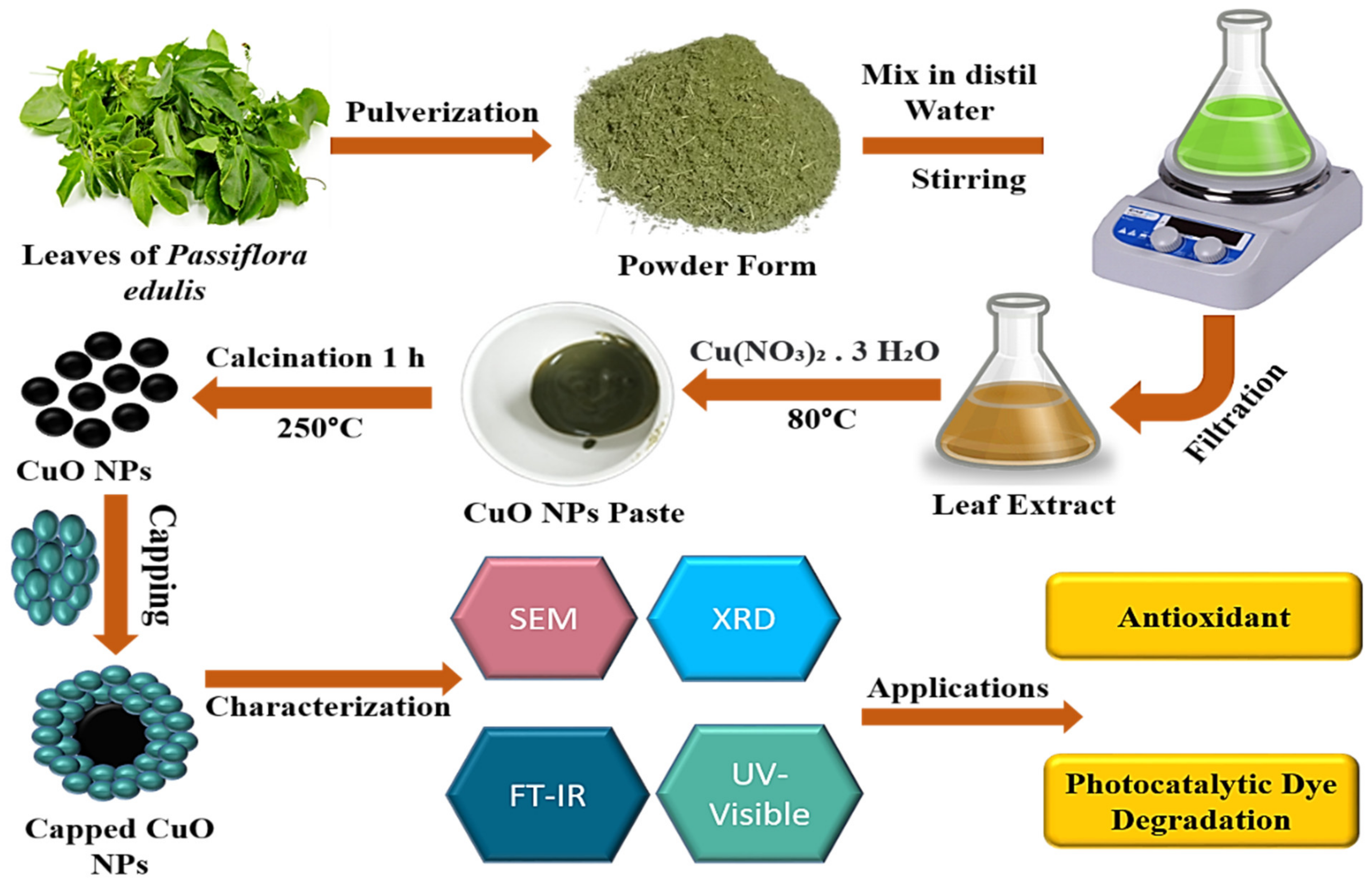
2.2. Preparation of Leaf Extract

Fresh Passiflora edulis leaves were firstly rinsed with water to remove dirt. The leaves were then dried in the shade for a week before being pulverized. 10 g of pulverized leaves were mixed in 300 mL of deionized water and stirred for an hour at 80 °C. After about an hour, the resulting mixture was filtered, and the leaf extract was stored at 4 °C for further use.

2.3. Copper Oxide (CuO) Nanoparticle Synthesis

In a beaker, unify 60 mL of leaf extract with 2 g copper nitrate tri hydrate while stirring and heating at 80 °C. Stirring and heating remains continue until the mixture changes to green paste, followed by filtration and washing twice or three times with distilled water. The paste was then placed in a silica crucible and calcinated for one hour at 250 °C in a muffle furnace. The subsequent dark color powder was CuO nanoparticles [43,44,45]. The biomolecules found in the leaf extract of Passiflora edulis were then used to cap the synthetic NPs. The plant’s phytochemicals acted as stabilizing and decreasing capping agents to help the nanoparticles develop. The capping agents help in providing stability and reducing agglomeration and can aid in adjusting nanoparticles’ size and shape. Figure 1 depicts the synthesis process for the production of CuO NPs.

2.4. Photocatalytic Activity

Methylene blue dye was used to test the photocatalytic activity of green fabricated Copper oxide nanoparticles. Prepare a 1000 mL MB dye (5 mg L−1) stock solution in distilled water. Add 50 mL of MB solution and 100 mg CuO NPs in a separate beaker. The solution was then agitated at room temperature in the dark for one hour to achieve absorption desorption equilibrium. The sample was exposed to sunlight. At specified intervals, the sample was pipetted out, and it was centrifuged for 15 min at 4000 rpm. The concentration of dye molecules in the solution was evaluated by measuring the supernatant with a UV-Vis spectrophotometer between 400 and 800 nm. Equation (1) was used to compute the percent of MB dye degradation [46].
Degradation   ( % ) = Ci C Ci × 100
where Ci is the control’s concentration and C is the sample’s concentration after a certain amount of time has passed.

2.5. Antioxidant Activity

The scavenging potential of CuO nanoparticles on stable DPPH free radical activity was investigated to determine their antioxidant activity. The DPPH solution was prepared and kept at 4 °C in the dark. A 3 mL methanolic DPPH (0.1 mM) solution was added to 600 µL CuO NPs solution with various concentrations (1, 0.5, 0.1, and 0.02 mL L−1). Each solution received 3 mL of methanol before being given a jarring shaking. The mixes were evaluated for absorbance at 517 nm after standing for 60 min. Methanol was used to set the absorbance to zero. The same amount of DPPH and methanol were also used to create a blank sample. Investigated was the DPPH free radical scavenging capability of CuO NPs at varying concentrations (1, 0.5, 0.1-, and 0.02-mL L−1) ppm). According to the findings, adding more CuO NPs improved the radical scavenging activity. The radical scavenging activities of the samples were computed and represented as a percentage of inhibition using the following equation.
Percentage   ( % )   inhibition = AB     AA AB × 100  
The absorbance levels of the test and blank samples are represented by AA and AB in the given equation. A percent inhibition versus concentration curve was drawn, and the sample concentration necessary for 50% inhibition (IC50) was determined.

2.6. Mechanism of Antioxidant Activity

The antioxidant potential of biologically synthesized CuO NPs has been investigated by DPPH assay. The DPPH radical is quite stable due to its remarkable stability. It may lead to the delocalization of radicals in aromatic rings. Initially, it exhibits deep purple color. The radical accepts a hydrogen atom or an electron from the antioxidant species and thus gets neutralized. The color changes from intense purple to pale yellow when it is in reduced form (DPPH-H). The unpaired electron of DPPH radicals shows strong absorbance at 517 nm, exhibiting deep purple color. An odd electron pair up with another electron to make it stable, and the color decolorizes to pale yellow. The mechanism of antioxidant activity is shown in Figure 2.

2.7. Stabilizing and Reducing Effects of Plant Extract

In the creation of nanoparticles, plants function as reducing and stabilizing agents. When nanoparticles are being synthesized, the OH and COOH groups serve as stabilizing and reducing agents. As capping agents, phenolic and alkaloids are essential and may cause decreased NPs to stabilize. The development of a stable bond between metallic NPs and phyto-constituents found in leaf extracts is the reason why stable NPs arise. According to reports in the literature, the compounds found in Passiflora edulis leaf extract [47]. Figure 3 demonstrates how plant extract contributes to the stability and production of nanoparticles.

3. Result

SEM, UV-vis, XRD, and FTIR were used to characterize copper oxide nanoparticles.

3.1. UV-Visible Analysis of CuO NPs

This study investigated the UV-Vis spectra of CuO nanoparticles at 200–400 nm wavelengths. Figure 4 displays the UV-Vis spectrum of CuO nanoparticles produced by biosynthesis. The surface Plasmon absorption of copper oxide nanoparticles led to an optimal absorbance in the region of 249.9 nm.

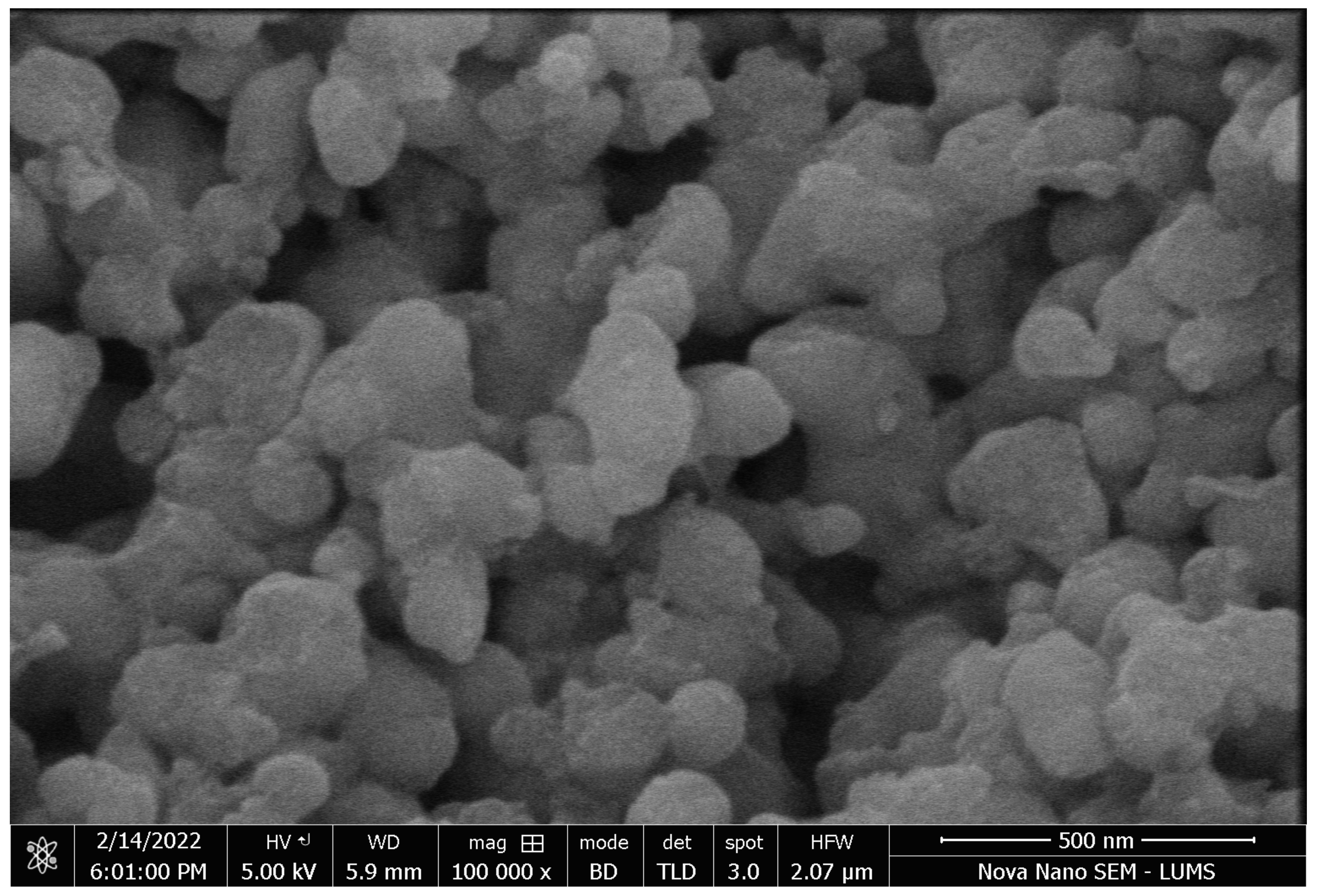
3.2. SEM Images of Biosynthesized CuO NPs

Scanning electron microscope provides information about surface morphology. SEM images of copper oxide nanoparticles were taken at 500 nm. Figure 5 shows the spherical morphology of CuO NPs with some agglomeration. CuO NPs that were produced were found to have an average particle size between 60 and 65 nm.

3.3. FTIR Analysis of Copper Oxide Nanoparticles

Figure 6 shows the FTIR spectra of biologically synthesized CuO nanoparticles. An absorption peak at 3260 cm–1 in copper oxide nanoparticle FTIR spectra corresponds to the symmetric vibrations of the hydroxyl (OH) functional group found in alcohols and phenols. The presence of hydroxyl groups on the surface of biogenic CuO NPs is thought to serve as a stabilizing factor throughout the synthesis process. C-H asymmetric stretching vibrations seem to be responsible for the peak at 2953 cm−1, whereas the peak at 1662 cm−1 is caused by C=C, and the peak at 1461 cm−1 is due to aliphatic C-H stretching. Passiflora edulis leaf extract is the primary source of these chemical groups, and the bands on the surface of NPs caused by protein residues that remain after the NPs have been fabricated. According to the current research, the proteins in the leaf extract function as stabilizing and capping agents for CuO nanoparticles, as well as reducing the metal salt to nanoparticles (NPs). It is suspected that the peak that is reflected in the range of 763 to 577 cm−1 is caused by stretching vibrations of Cu–O in the sample. The FTIR examination indicated that the nanoparticles produced are not bare CuO but rather CuO coated with organic compounds derived from Passiflora edulis.

3.4. X-ray Diffraction Spectroscopy

CuO NPs that have been produced are seen in Figure 7 XRD diffraction patterns. The XRD pattern is plotted between 2 theta (θ) and intensity. 2θ = 32.34, 35.64, 38.69, 48.70, 53.47, 58.35, and 66.12 are assigned to (110), (111), (200), (−202), (020), (202), and (022) reflection lines, respectively of crystalline CuO nanoparticles that are in correct agreement with the results reported (Sarker et. al., 2020) [48].

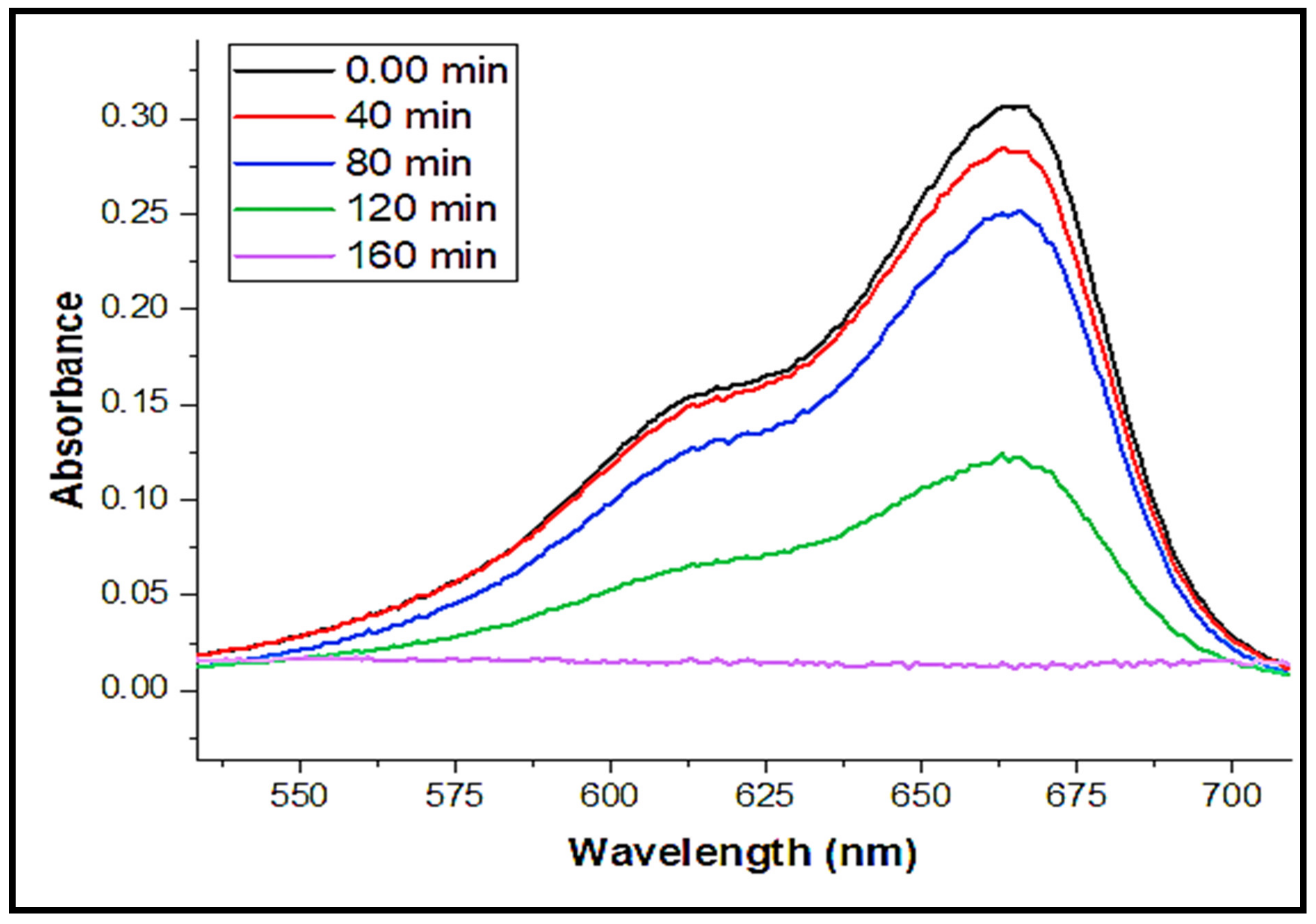
3.5. Photocatalytic Degradation

By monitoring the methylene blue degradation under sunlight irradiation, the photocatalytic activity of biosynthesized CuO nanoparticles was assessed.
Figure 8 shows how dye degradation may be distinguished by the dye solution gradually turning from a deep blue to a colorless state. Dye solution with biogenic CuO nanoparticles was exposed to sunlight for 2 h and 40 min shown to have reduced peak intensity at 662 nm.
Following Equation (1), a percentage of dye degradation was computed, and its fluctuation over time is shown in Figure 9. All dye concentrations were calculated using the UV-Vis spectrum absorbance value at 662 nm. [49,50,51]. Dye was efficiently degraded by the sunlight (Figure 10).
The following has been hypothesized as a mechanism for MB degradation during solar irradiation: For the most part, the process of photocatalytic dye degradation involves the generation of reactive radicals, particularly hydroxyl (HO) and superoxide (O2) radicals, which break down the dyes into less toxic compounds. According to the suggested method, CuO NPs are exposed to a photon with energy (hv) larger than or equal to the band gap energy, causing VB electrons to be promoted to the CB and leaving holes (h+) in the VB.VB holes oxidize H2O, H2O2, or HO- to HO, whereas CB electrons break down dissolved oxygen in solution into O−2 or H2O2, resulting in hydroperoxyl radicals. The dyes may absorb radiation, causing them to switch from HOMO to LUMO. Figure 11 shows the mechanism of photocatalytic dye degradation.

3.6. CuO Nanoparticles Have Antioxidant Properties

In biological systems, the interaction of biomolecules with molecular oxygen results in the production of free radicals. DPPH scavenging tests are the most extensively used methods for determining the antioxidant capabilities of various materials and compounds. CuO nanoparticles were tested for their antioxidant properties using a DPPH radical assay [46,52,53,54]. Percentage Scavenging and IC50 value of the standard (ascorbic acid) are shown in Table 1. The sample’s antioxidants weaken the stable nitrogen radical DPPH, which lowers the absorbance at 517 nm in the DPPH test [55].
Percentage scavenging values for standard at the concentration of 0.02, 0.1, 0.5, and 1 mg/mL were 11.940, 20.880, 33.313, and 49.253%, respectively. IC50 values for standard at concentration 0.02, 0.1, 0.5 and 1 mg/mL were 0.18, 0.19, 0.21 and 0.25 mg/mL. Figure 12 shows the graphical representation of the percentage RSA of ascorbic acid and CuO nanoparticles.
The percentage of scavenging and IC50 value of CuO Nanoparticles is shown in Table 2. Percentage scavenging values for CuO nanoparticles at the concentration of 0.02, 0.1, 0.5 and 1 mg/mL were 16.417, 26.866, 44.776 and 64.170%. IC50 values for CuO nanoparticles at concentration 0.02, 0.1, 0.5 and 1 mg/mL were 0.13, 0.14, 0.17 and 0.20 mg/mL.
IC50 values of CuO nanoparticles were compared with ascorbic acid (standard), as shown in Figure 13. It was found that better scavenging potential was shown by CuO nanoparticles than ascorbic acid (standard) as its IC50 was less. CuO nanoparticles synthesized using the leaf extract of Passiflora edulis used in this investigation were found to have better antioxidant properties than standard ascorbic acid.

4. Discussion

Because of their distinctive properties and diverse range of uses as catalysts, resistors, superconducting materials, biologically significant objects, and sensors, metal oxide nanoparticles have attracted the interest of many researchers [55]. The current study focuses on the production of CuO NPs utilizing a green synthesis method and a leaf extract of Passiflora edulis, taking into account the significance of metal oxide nanoparticles (NPs). The extract works as a stabilizing, capping, and reducing agent. The specific method of how plant extract works as a stabilizer and reducing agent has previously been covered. The crystalline makeup of the produced nanoparticles was examined using XRD analysis. The findings show that the crystalline CuO nanoparticles accord correctly with the findings from past studies [47]. The average particle size and shape of the CuO NPs produced by plants are determined by SEM examinations. It was discovered that the typical particle size ranges from 60 to 65 nm, which is in the nanoscale. The peaks of different functional groups or biomolecules found in the plant extract were identified using FT-IR analysis. UV-Visible spectra of CuO NPs result in the optimum absorption in the range of 249.9 nm.
In the present work, antioxidant potential of CuO NPs has been investigated by DPPH method. DPPH scavenging tests are the most extensively used methods for determining the antioxidant capabilities of various materials and compounds. The efficiency of biologically synthesized CuO NPs as a potentized antioxidant material was estimated and then compared with the standard drug (ascorbic acid). The half maximum inhibitory concentration IC50 values were calculated and then compared. The radical scavenging potential was also estimated, and results showed that CuO NPs is a more potent antioxidant material than standard ascorbic acid and thus can be used to synthesize antioxidant medicines. Plants are rich in antioxidants, that is why biologically synthesized CuO NPs are beneficial and show antioxidant potential.
Environmental pollution is going to increase day by day, and discharge of industrial effluents without treatment is the leading cause of pollution. Dyes are mostly used in the textile and dyeing industries and contain various toxic organic compounds. Bright color dyes are more problematic as they are soluble in water and thus polluting aquatic ecosystems when discharged directly. The efficacy of photocatalytic dye degradation by CuO NPs has been examined in the present study, and efficient degradation of MB Dye in 160 min has been noted. A 93 percent degrading efficiency for CuO NPs was discovered. Reactive radicals attack organic pollutants, causing decomposition.

5. Conclusions

MB dye has the ability to be degraded efficiently, and a low-cost biogenic chemical that also has antioxidant potential to stabilize free radicals was used to create copper oxide nanoparticles. The biologically fabricated CuO NPs were of spherical morphology with a size range between 60 and 65 nm. The decolorization efficiency of biosynthesized CuO NPs was found to be 93 percent after 160 min of exposure to sunshine when their photocatalytic activity was measured by the photo-degradation of MB. According to the findings, the biosynthesized CuO NPs displayed good photocatalytic efficacy and may have prospective uses in the treatment of industrial wastewater. The DPPH technique was used to assess antioxidant activity. IC50 values were compared with sample (CuO NPs) and standard (ascorbic acid) to check efficiency. Low IC50 showed that the efficiency of nanoparticles was high. The percentage of radical scavenging for the standard ascorbic acid was found to be 11–49%, while for the biosynthesized CuO NPs the value of percentage radical scavenging was found to be in the range of 16–64%.

Author Contributions

A.Y.: conceptualization and data curation; U.F.: formal analysis, supervision, writing—original draft preparation; S.S.: investigation, methodology; S.M.: project administration; H.I.: writing—original draft preparation; M.J.: writing—review and editing; S.I.: reviewing data, funding acquisition; H.A.: resources, writing—original draft preparation; H.H.S.: visualization, software; R.A.P.: writing—review and editing; E.A.: writing—review and editing; A.-E.F.: writing—review and editing. All authors have read and agreed to the published version of the manuscript.

Funding

The authors would like to thank the Deanship of Scientific Research at Umm Al-Qura University for supporting this work by Grant Code: (22UQU4320141DSR40). Email: rapa-sha@uqu.edu.sa. This work was supported by King Khalid University through a grant (KKU/RCAMS/22) under the Research Center for Advanced Materials Science (RCAMS) at King Khalid University, Saudi Arabia.

Institutional Review Board Statement

Not Applicable.

Informed Consent Statement

Not Applicable.

Data Availability Statement

The data will be available on request.

Acknowledgments

The authors would like to thank the Deanship of Scientific Research at Umm Al-Qura University for supporting this work by Grant Code: (22UQU4320141DSR40). Email: rapasha@uqu.edu.sa. This work was supported by King Khalid University through a grant (KKU/RCAMS/22) under the Research Center for Advanced Materials Science (RCAMS) at King Khalid University, Saudi Arabia.

Conflicts of Interest

The authors declare no conflict of interest.

References

  1. Chaudhry, F.N.; Malik, M.F. Factors affecting water pollution: A review. J. Ecosyst. Ecography 2017, 7, 225. [Google Scholar]
  2. Lee, J.; Mahendra, S.; Alvarez, P.J. Nanomaterials in the construction industry: A review of their applications and environmental health and safety considerations. ACS Nano 2010, 4, 3580–3590. [Google Scholar] [CrossRef] [PubMed]
  3. Mahmood, N.; Wang, Z.; Zhang, B. The role of nuclear energy in the correction of environmental pollution: Evidence from Pakistan. Nucl. Eng. Technol. 2020, 52, 1327–1333. [Google Scholar] [CrossRef]
  4. López-Carrillo, L.; González-González, L.; Piña-Pozas, M.; Mérida-Ortega, Á.; Gamboa-Loira, B.; Blanco-Muñoz, J.; Torres-Sánchez, L.E.; Hurtado-Díaz, M.; Cortez-Lugo, M.; Guerra, G.; et al. State of children environmental health research in Latin America. Ann. Glob. Health 2018, 84, 204. [Google Scholar] [CrossRef] [PubMed]
  5. Ramírez-Hernández, H.; Perera-Rios, J.; May-Euán, F.; Uicab-Pool, G.; Peniche-Lara, G.; Pérez-Herrera, N. Environmental risks and children’s health in a Mayan community from southeast of Mexico. Ann. Glob. Health 2018, 84, 292. [Google Scholar] [CrossRef]
  6. Vaidehi, D.; Bhuvaneshwari, V.; Bharathi, D.; Sheetal, B.P. Antibacterial and photocatalytic activity of copper oxide nanoparticles synthesized using Solanum lycopersicum leaf extract. Mater. Res. Express 2018, 5, 085403. [Google Scholar] [CrossRef]
  7. Marimuthu, S.; Antonisamy, A.J.; Malayandi, S.; Rajendran, K.; Tsai, P.C.; Pugazhendhi, A.; Ponnusamy, V.K. Silver nanoparticles in dye effluent treatment: A review on synthesis, treatment methods, mechanisms, photocatalytic degradation, toxic effects and mitigation of toxicity. J. Photochem. Photobiol. B Biol. 2020, 205, 111823. [Google Scholar] [CrossRef]
  8. Varjani, S.; Rakholiya, P.; Ng, H.Y.; You, S.; Teixeira, J.A. Microbial degradation of dyes: An overview. Bioresour. Technol. 2020, 314, 123728. [Google Scholar] [CrossRef]
  9. Shah, K. Biodegradation of azo dye compounds. Int. Res. J. Biochem. Biotechnol. 2014, 1, 5–13. [Google Scholar]
  10. Moosavi, S.; Li, R.Y.M.; Lai, C.W.; Yusof, Y.; Gan, S.; Akbarzadeh, O.; Chowhury, Z.Z.; Yue, X.G.; Johan, M.R. Methylene blue dye photocatalytic degradation over synthesised Fe3O4/AC/TiO2 nano-catalyst: Degradation and reusability studies. Nanomaterials 2020, 10, 2360. [Google Scholar] [CrossRef]
  11. Javed, M.; Qamar, M.A.; Iqbal, S.; Aljazzar, S.O.; Iqbal, S.; Khan, H.; Abourehab, M.A.; Elkaeed, E.B.; Alharthi, A.I.; Awwad, N.S.; et al. Synergistic Influences of Doping Techniques and Well-Defined Heterointerface Formation to Improve the Photocatalytic Ability of the S-ZnO/GO Nanocomposite. ChemistrySelect 2022, 7, e202201913. [Google Scholar] [CrossRef]
  12. Qamar, M.A.; Javed, M.; Shahid, S.; Sher, M. Fabrication of g-C3N4/transition metal (Fe, Co, Ni, Mn and Cr)-doped ZnO ternary composites: Excellent visible light active photocatalysts for the degradation of organic pollutants from wastewater. Mater. Res. Bull. 2022, 147, 111630. [Google Scholar] [CrossRef]
  13. Ijaz, F.; Shahid, S.; Khan, S.A.; Ahmad, W.; Zaman, S. Green synthesis of copper oxide nanoparticles using Abutilon indicum leaf extract: Antimicrobial, antioxidant and photocatalytic dye degradation activitie. Trop. J. Pharm. Res. 2017, 16, 743–753. [Google Scholar] [CrossRef]
  14. Sreekala, G.; Beevi, A.F.; Beena, B. Adsorption of lead (II) Ions by ecofriendly copper oxide nanoparticles. Orient. J. Chem. 2019, 35, 1731–1736. [Google Scholar] [CrossRef]
  15. Thangamani, N.; Bhuvaneshwari, N. Green synthesis of gold nanoparticles using Simarouba glauca leaf extract and their biological activity of micro-organism. Chem. Phys. Lett. 2019, 732, 136587. [Google Scholar] [CrossRef]
  16. Thakkar, K.N.; Mhatre, S.S.; Parikh, R.Y. Biological synthesis of metallic nanoparticles. Nanomed. Nanotechnol. Biol. Med. 2010, 6, 257–262. [Google Scholar] [CrossRef] [PubMed]
  17. Gupta, A.; Tandon, M.; Kaur, A. Role of metallic nanoparticles in water remediation with special emphasis on sustainable synthesis: A review. Nanotechnol. Environ. Eng. 2020, 5, 27. [Google Scholar] [CrossRef]
  18. Da Silva, B.F.; Pérez, S.; Gardinalli, P.; Singhal, R.K.; Mozeto, A.A.; Barceló, D. Analytical chemistry of metallic nanoparticles in natural environments. TrAC Trends Anal. Chem. 2011, 30, 528–540. [Google Scholar] [CrossRef]
  19. Durán, N.; Marcato, P.D. Biotechnological routes to metallic nanoparticles production: Mechanistic aspects, antimicrobial activity, toxicity and industrial applications. In Nano-Antimicrobials; Springer: Berlin/Heidelberg, Germany, 2012; pp. 337–374. [Google Scholar]
  20. Adelere, I.A.; Lateef, A. A novel approach to the green synthesis of metallic nanoparticles: The use of agro-wastes, enzymes, and pigments. Nanotechnol. Rev. 2016, 5, 567–587. [Google Scholar] [CrossRef]
  21. Singla, R.; Guliani, A.; Kumari, A.; Yadav, S.K. Metallic nanoparticles, toxicity issues and applications in medicine. In Nanoscale Materials in Targeted Drug Delivery, Theragnosis and Tissue Regeneration; Springer: Singapore, 2016; pp. 41–80. [Google Scholar]
  22. Shahzadi, S.; Zafar, N.; Sharif, R. Antibacterial activity of metallic nanoparticles. In Bacterial Pathogenesis and Antibacterial Control; IntechOpen: London, UK, 2018. [Google Scholar]
  23. Ahmad, M.Z.; Akhter, S.; Jain, G.K.; Rahman, M.; Pathan, S.A.; Ahmad, F.J.; Khar, R.K. Metallic nanoparticles: Technology overview & drug delivery applications in oncology. Expert Opin. Drug Deliv. 2010, 7, 927–942. [Google Scholar] [PubMed]
  24. Rahman, M.; Alam, K.; Hafeez, A.; Ilyas, R.; Beg, S. Metallic nanoparticles in drug delivery and cancer treatment. In Nanoformulation Strategies for Cancer Treatment; Elsevier: Amsterdam, The Netherlands, 2021; pp. 107–119. [Google Scholar]
  25. Chari, N.; Felix, L.; Davoodbasha, M.; Ali, A.S.; Nooruddin, T. In vitro and in vivo antibiofilm effect of copper nanoparticles against aquaculture pathogens. Biocatal. Agric. Biotechnol. 2017, 10, 336–341. [Google Scholar] [CrossRef]
  26. Dong, Y.; Jiang, X.; Mo, J.; Zhou, Y.; Zhou, J. Hollow CuO nanoparticles in carbon microspheres prepared from cellulose-cuprammonium solution as anode materials for Li-ion batteries. Chem. Eng. J. 2020, 381, 122614. [Google Scholar] [CrossRef]
  27. Fuku, X.; Modibedi, M.; Mathe, M. Green synthesis of Cu/Cu2O/CuO nanostructures and the analysis of their electrochemical properties. SN Appl. Sci. 2020, 2, 902. [Google Scholar] [CrossRef]
  28. Tshireletso, P.; Ateba, C.N.; Fayemi, O.E. Spectroscopic and antibacterial properties of CuONPs from orange, lemon and tangerine peel extracts: Potential for combating bacterial resistance. Molecules 2021, 26, 586. [Google Scholar] [CrossRef]
  29. Hajipour, M.J.; Fromm, K.M.; Ashkarran, A.A.; de Aberasturi, D.J.; de Larramendi, I.R.; Rojo, T.; Serpooshan, V.; Parak, W.J.; Mahmoudi, M. Antibacterial properties of nanoparticles. Trends Biotechnol. 2012, 30, 499–511. [Google Scholar] [CrossRef] [PubMed]
  30. Shwetha, U.R.; Latha, M.S.; Kumar, C.R.; Kiran, M.S.; Onkarappa, H.S.; Betageri, V.S. Potential antidiabetic and anticancer activity of copper oxide nanoparticles synthesized using Areca catechu leaf extract. Adv. Nat. Sci. Nanosci. Nanotechnol. 2021, 2, 025008. [Google Scholar] [CrossRef]
  31. Avinash, B.; Ravikumar, C.R.; Kumar, M.A.; Nagaswarupa, H.P.; Santosh, M.S.; Bhatt, A.S.; Kuznetsov, D. Nano CuO: Electrochemical sensor for the determination of paracetamol and D-glucose. J. Phys. Chem. Solids 2019, 134, 193–200. [Google Scholar] [CrossRef]
  32. Khan, M.A.; Nayan, N.; Ahmad, M.K.; Soon, C.F. Surface study of CuO nanopetals by advanced nanocharacterization techniques with enhanced optical and catalytic properties. Nanomaterials 2020, 107, 1298. [Google Scholar] [CrossRef]
  33. Aminuzzaman, M.; Kei, L.M.; Liang, W.H. Green synthesis of copper oxide (CuO) nanoparticles using banana peel extract and their photocatalytic activities. In AIP Conference Proceedings; AIP Publishing LLC: Long Island, NY, USA, 2017; Volume 1828, p. 020016. [Google Scholar]
  34. Rahman, M.; Islam, M.; Biswas, M.; Khurshid Alam, A.H.M. In vitro antioxidant and free radical scavenging activity of different parts of Tabebuia pallida growing in Bangladesh. BMC Res. Notes 2015, 8, 621. [Google Scholar] [CrossRef] [PubMed]
  35. Bautista-Diaz, J.; Cruz-Alvarez, O.; Hernández-Rodríguez, O.A.; Sánchez-Chávez, E.; Jacobo-Cuellar, J.L.; Preciado-Rangel, P.; Avila-Quezada, G.D.; Ojeda-Barrios, D.L. Zinc sulphate or zinc nanoparticle applications to leaves of green beans. Folia Hortic. 2021, 33, 365–375. [Google Scholar] [CrossRef]
  36. Hamid, K.; Saha, M.R.; Urmi, K.F.; Habib, M.R.; Rahman, M.M. Screening of different parts of the plant Pandanus odorus for its antioxidant activity. Int. J. Appl. Biol. Pharm. 2010, 1, 1364–1368. [Google Scholar]
  37. Steinmetz, K.A.; Potter, J.D. Vegetables, fruit, and cancer prevention: A review. J. Am. Diet. Assoc. 1996, 96, 1027–1039. [Google Scholar] [CrossRef]
  38. Frankel, E.N. In search of better methods to evaluate natural antioxidants and oxidative stability in food lipids. Trends Food Sci. Technol. 1993, 4, 220–225. [Google Scholar] [CrossRef]
  39. Fernández, J.; Pérez-Álvarez, J.A.; Fernández-López, J.A. Thiobarbituric acid test for monitoring lipid oxidation in meat. Food Chem. 1997, 59, 345–353. [Google Scholar] [CrossRef]
  40. Coppin, E.A.; Pike, O.A. Oil stability index correlated with a sensory determination of oxidative stability in light-exposed soybean oil. J. Am. Oil Chem. Soc. 2001, 78, 13–18. [Google Scholar] [CrossRef]
  41. Beltran, E.; Pla, R.; Yuste, J.; Mor-Mur, M. Use of antioxidants to minimize rancidity in pressurized and cooked chicken slurries. Meat Sci. 2004, 66, 719–725. [Google Scholar] [CrossRef]
  42. Thomas, B.; Vithiya, B.; Prasad, T.; Mohamed, S.B.; Magdalane, C.M.; Kaviyarasu, K.; Maaza, M. Antioxidant and photocatalytic activity of aqueous leaf extract mediated green synthesis of silver nanoparticles using Passiflora edulis f. flavicarpa. J. Nanosci. Nanotechnol. 2019, 19, 2640–2648. [Google Scholar] [CrossRef]
  43. Cheirmadurai, K.; Biswas, S.; Murali, R.; Thanikaivelan, P. Green synthesis of copper nanoparticles and conducting nanobiocomposites using plant and animal sources. RSC Adv. 2014, 4, 19507–19511. [Google Scholar] [CrossRef]
  44. Gonçalves Martins, T.A.; Botelho Junior, A.B.; Moraes, V.T.D.; Espinosa, D.C.R. Study of pH Influence in the Synthesis of Copper Nanoparticles Using Ascorbic Acid as Reducing and Stabilizing Agent. In TMS 2020 149th Annual Meeting & Exhibition Supplemental Proceedings; Springer: Cham, Switzerland, 2020; pp. 1547–1557. [Google Scholar]
  45. Jain, M.; Yadav, M.; Chaudhry, S. Copper oxide nanoparticles for the removal of divalent nickel ions from aqueous solution. Toxin Rev. 2021, 40, 872–885. [Google Scholar] [CrossRef]
  46. Muthuvel, A.; Jothibas, M.; Manoharan, C. Synthesis of copper oxide nanoparticles by chemical and biogenic methods: Photocatalytic degradation and in vitro antioxidant activity. Nanotechnol. Environ. Eng. 2020, 5, 14. [Google Scholar] [CrossRef]
  47. De Araújo Esteves Duarte, I.; Milenkovic, D.; Borges, T.K.; de Lacerda de Oliveira, L.; Costa, A.M. Brazilian passion fruit as a new healthy food: From its composition to health properties and mechanisms of action. Food Funct. 2021, 12, 11106–11120. [Google Scholar] [CrossRef] [PubMed]
  48. Sarkar, J.; Chakraborty, N.; Chatterjee, A.; Bhattacharjee, A.; Dasgupta, D.; Acharya, K. Green synthesized copper oxide nanoparticles ameliorate defence and antioxidant enzymes in Lens culinaris. Nanomaterials 2020, 10, 312. [Google Scholar] [CrossRef] [PubMed]
  49. Qamar, M.A.; Shahid, S.; Javed, M.; Iqbal, S.; Sher, M.; Bahadur, A.; AL-Anazy, M.M.; Laref, A.; Li, D. Designing of highly active g-C3N4/Ni-ZnO photocatalyst nanocomposite for the disinfection and degradation of the organic dye under sunlight radiations. Colloids Surf A Physicochem. Eng. Asp. 2021, 614, 126176. [Google Scholar] [CrossRef]
  50. Singh, J.; Kumar, V.; Kim, K.H.; Rawat, M. Biogenic synthesis of copper oxide nanoparticles using plant extract and its prodigious potential for photocatalytic degradation of dyes. Environ. Res. 2019, 177, 108569. [Google Scholar] [CrossRef] [PubMed]
  51. Sher, M.; Javed, M.; Shahid, S.; Iqbal, S.; Qamar, M.A.; Bahadur, A.; Qayyum, M.A. The controlled synthesis of g-C3N4/Cd-doped ZnO nanocomposites as potential photocatalysts for the disinfection and degradation of organic pollutants under visible light irradiation. RSC. Adv. 2021, 11, 2025–2039. [Google Scholar] [CrossRef] [PubMed]
  52. Mansoor, S.; Shahid, S.; Javed, M.; Saad, M.; Iqbal, S.; Alsaab, H.O.; Awwad, N.S.; Ibrahium, H.A.; Zaman, S.; Sarwar, M.N.; et al. Green synthesis of a MnO-GO-Ag nanocomposite using leaf extract of Fagonia arabica and its antioxidant and anti-inflammatory performance. Nano Struct. Nano Objects 2022, 29, 100835. [Google Scholar] [CrossRef]
  53. Shahid, S.; Ejaz, A.; Javed, M.; Mansoor, S.; Iqbal, S.; Elkaeed, E.B.; Alzhrani, R.M.; Alsaab, H.O.; Awwad, N.S.; Ibrahium, H.A.; et al. The Anti-Inflammatory and Free Radical Scavenging Activities of Bio-Inspired Nano Magnesium Oxide. Front. Mater. 2022, 9, 875163. [Google Scholar] [CrossRef]
  54. Shahid, S.; Mansoor, S.; Javed, M.; Iqbal, S.; Yousaf, U.; Alsaab, H.O.; Awwad, N.S.; Ibrahium, H.A.; Alzhrani, R.M.; Alqahtani, M.D.; et al. CuO-GO-Ag; Green Synthesis with Fagonia Arabica and Biomedical Potential is a Bioinspired Nano Theranostics Composite. Front. Mater. 2022, 9, 875148. [Google Scholar] [CrossRef]
  55. Hosny, M.; Eltaweil, A.S.; Mostafa, M.; El-Badry, Y.A.; Hussein, E.E.; Omer, A.M.; Fawzy, M. Facile synthesis of gold nanoparticles for anticancer, antioxidant applications, and photocatalytic degradation of toxic organic pollutants. ACS Omega 2022, 7, 3121–3133. [Google Scholar] [CrossRef] [PubMed]
Figure 1. Copper oxide nanoparticle green synthesis.
Agronomy 12 02315 g001
Figure 2. Mechanism of antioxidant activity.
Agronomy 12 02315 g002
Figure 3. Role of plant extract.
Agronomy 12 02315 g003
Figure 4. UV-Visible spectra of biosynthesized CuO nanoparticles.
Agronomy 12 02315 g004
Figure 5. SEM images of CuO NPs.
Agronomy 12 02315 g005
Figure 6. (a) FTIR spectra of plant extract (b) FTIR spectra of CuO NPs.
Agronomy 12 02315 g006
Figure 7. XRD spectra of CuO nanoparticles.
Agronomy 12 02315 g007
Figure 8. Change in color depth during dye degradation.
Agronomy 12 02315 g008
Figure 9. UV-Vis spectra show the gradual photocatalytic deterioration of MB dye.
Agronomy 12 02315 g009
Figure 10. Graphical representation of percentage dye degradation.
Agronomy 12 02315 g010
Figure 11. Process of MB dye degradation with exposure to sunlight.
Agronomy 12 02315 g011
Figure 12. (a) Graphical representation of percentage RSA of ascorbic acid, (b) graphical representation of RSA of CuO nanoparticles.
Agronomy 12 02315 g012
Figure 13. Comparison between IC50 of ascorbic acid and CuO nanoparticles.
Agronomy 12 02315 g013
Table 1. Percentage (%) scavenging and IC50 of standard (ascorbic acid).
Concentrations (mg L−1)Absorbance of ControlAbsorbance of SamplePercentage RSAIC50
0.02 mg L−10.670.5911.90.18
0.1 mg L−10.670.5320.80.19
0.5 mg L−10.670.4237.30.21
1 mg L−10.670.3449.20.25
Table 2. Percentage Scavenging and IC50 value of CuO.
Concentrations (mg L−1)Absorbance of ControlAbsorbance of SamplePercentage RSAIC50
0.02 mg L−10.670.5616.40.13
0.1 mg L−10.670.4926.80.14
0.5 mg L−10.670.3544.70.17
1 mg L−10.670.2464.10.20
Publisher’s Note: MDPI stays neutral with regard to jurisdictional claims in published maps and institutional affiliations.

