Article

Investigating the Potential of Streptomyces spp. in Suppression of Rhizoctonia solani (AG1-IA) Causing Rice Sheath Blight Disease in Northern Iran

by
Meysam Soltani Nejad
1,*,
Neda Samandari Najafabadi
2,
Sonia Aghighi
3,
Amir Hashem Shahidi Bonjar
4,
Kheda Magomed-Salihovna Murtazova
5,
Magomed Ramzanovich Nakhaev
6 and
Meisam Zargar
7,*
1
Department of Plant Protection, Faculty of Agriculture, Shahid Bahonar University of Kerman, Kerman 7616914111, Iran
2
Department of Plant Protection, Faculty of Agriculture, Ferdowsi University of Mashhad, Mashhad 9177948978, Iran
3
Research and Technology Institute of Plant Production, Shahid Bahonar University of Kerman, Kerman 7616914111, Iran
4
Clinician Scientist of Dental Materials and Restorative Dentistry, School of Dentistry, Shahid Beheshty University of Medical Sciences, Tehran 1983963113, Iran
5
Engineering Center of Carbon, Kadyrov Chechen State University, 364024 Grozny, Russia
6
Applied Mathematics and Computer Technology, Kadyrov Chechen State University, 364024 Grozny, Russia
7
Department of Agrobiotechnology, Agricultural-Technological Institute, RUDN University, 117198 Moscow, Russia
*
Authors to whom correspondence should be addressed.
Agronomy 2022, 12(10), 2292; https://doi.org/10.3390/agronomy12102292
Submission received: 13 August 2022 / Revised: 17 September 2022 / Accepted: 21 September 2022 / Published: 24 September 2022
(This article belongs to the Special Issue Crop Productivity and Energy Balance in Large-Scale Fields)

Abstract

:
A study was conducted in the Guilan Province of Iran, using a variety of Actinomycetes species isolated from the rice fields, with the intention of identifying useful biocontrol agents to lessen rice sheath blight disease. The antagonistic effects against the rice pathogen agent were also assessed both in vitro and in vivo. The antifungal abilities of more than 30 Actinomycetes isolates against the Rhizoctonia solani Kühn (AG1-IA) were used. The biocontrol abilities of the most active isolates were studied in a greenhouse. The size of the inhibition zone against pathogen development and the most potent antagonist Actinomycetes isolates were determined based on the dual culture screening test findings. The ability to create hydrolytic enzymes including amylase, chitinase, protease, and lipase were shown by hydrolytic enzyme assays on the putative antagonists. Antifungal activities of Streptomyces isolates against fungus mycelia were also studied using SEM since, compared to the control grown mycelia and mycelia adjacent to the inhibition zone in the plate, tests revealed an unusual and deformed structure; in our opinion, the chitinase secreted can destroy fungal mycelium. Chloroform test showed that its antifungal effect persists upon exposure to chloroform. All possible isolates belonged to the Streptomyces species, according to the 16S rDNA molecular analysis of the majority of active isolates. Comparing isolates, G had the highest impact in reducing sheath blight disease. The Iranian strain of the Streptomyces has antifungal capabilities, highlighting its potential as a viable biocontrol agent to be used in an Integrated Disease Management (IDM) program to control the rice sheath blight disease.

1. Introduction

For a substantial portion of the world’s population, rice (Oryza sativa L.) is an essential cereal crop, used as a staple diet in many countries [1,2]. Rice farmers all over the globe suffer severe losses due to rice diseases such as Rhizoctonia solani Kühn AG1 (Teleomorph: Thanatephorus cucumeris; Anastomosis Group 1- IA), which not only harms the crop but also eventually hurts the economy [3,4,5]. The use of chemical fungicides is the primary technique of disease control, yet doing so not only pollutes the environment, but also resistance of the pathogens to the current used chemical fungicides started to be observed [6]. Therefore, it is essential to continually develop techniques that emphasize the simultaneous and integrated use of chemicals and biological agents that have been shown to be successful in overcoming pathogen resistance to chemicals [7,8]. Application of biocontrol agents is primarily intended to restore a natural balance in an ecological niche rather than to eliminate the disease [9,10]. Strong antagonists with inhibitory effects against a variety of plant diseases are known to exist in Actinobacteria [11]. Actinomycetes have the capacity to colonize the surface of plant roots and generate a variety of secondary metabolites that function as biocontrol agents [12,13]. Streptomyces species are well-known Actinobacteria taxon and are often found in agricultural soils where they actively contribute to the creation of a microbial balance by inhibiting phytopathogens, making them one of the most effective instruments in suppressive soils against plant infections [14,15]. For the control of plant infections, certain Streptomyces species have been successfully developed into commercial biocontrol agents. Like the soil-borne pathogenic fungi Fusarium oxysporum f. sp. lycopersici and Verticillium dahlia, which cause black scorch on date palms, and Streptomyces griseoviridis strain K61 (Mycostop®), which has been used for the biocontrol of Ceratocystis radicicola [16]. Streptomycetes are capable of producing a large number of secondary metabolites that are used as antiseptics, antibiotics, herbicides, and pesticides [17,18,19]. For example, Streptomyces philanthi RM-1-138, which was isolated from the rhizosphere soil, was effective in controlling rice sheath blight disease. Red rust disease in tea plants was effectively controlled, using Streptomyces species as biocontrol [20]. In both in vitro and in vivo settings, Streptomyces globisporus JK-1 shows great potential for controlling tomato fruits’ postharvest grey mold [21]. Magnaporthe oryzae is the common causative agent of rice blast disease, and Li et al. have shown that the antifungal component in the culture filtrate of Streptomyces globisporus JK-1 greatly inhibited the plant diseases [22]. Streptomyces sp. PM5 produced two antifungal aliphatic compounds, SPM5C-1 and SPM5C-2, having a lactone and ketone carbonyl unit, respectively. These compounds are known to be very efficient against rice diseases such as blast and sheath blight both in vitro and in vivo [23]. According to in vitro studies, Streptomyces has inhibitory capabilities against Magnaporthe oryzae and Rhizoctonia solani. Furthermore, Streptomyces treatment of infected rice seedlings under in vivo conditions reduced rice blast by up to 88.3 percent in numerous investigations [24]. The creation of a wide array of antibiotics and antifungal volatile organic compounds, a range of fungal cell-wall-degrading enzymes, and the establishment of systemic resistance in plants against plant diseases comprise the method of action of Streptomycetes’ metabolites against plant disease infections [25]. Rice is a major food for a large proportion of Iran’s population and is an important primary crop in muddy farmlands. Sheath blight disease in rice plant has caused major crop losses in northern Iran [26]. Managing the causal agent of disease is difficult because of its broad host range and formation of sclerotia, which can survive in harsh environmental conditions; therefore, developing biological control for disease management methods without use of hazardous chemicals has been considered as the main concern to maintain sustainable agriculture. In the current study, local Actinobacteria from Guilan province in northern Iran were identified and subjected to both in vitro and in vivo tests for resistance to rice sheath blight disease. This pathogen is one of the biggest threats to rice fields in Iran.

2. Materials and Methods

2.1. Seed Source, Rice Pathogen and Actinomycetes Isolation

Rice Research Institute of Iran (RRII), Rasht, provided the rice seeds of Oryza sativa L. var. Hashemi and pure culture of R. solani AG-1 IA, the disease-causing agent for rice sheath blight. The Actinomycetes were first discovered in the soil of rice fields, in the Guilan Province in Iran’s northern area. Briefly, our Actinomycetes samples were collected from upper 25–35 cm of soil, air-dried at room temperature for 20 days and passed through a 0.8 mm mesh sieve. About 10 g of dried soils samples were mixed with 100 milliliter of sterile water, and 1 mL of diluted soil suspensions (10−1) was transferred to nine milliliters of sterile distilled water and diluted to 10−2, 10−3, 10−4, 10−5, and 10−6. About 1 mL of soil dilutions (10−3–10−6) was added to Casein Glycerol Agar (CGA) before pouring the plates [27]. The isolates were transferred in a fresh CGA media for a week at 29 ± 1 °C and the CGA was provided by mixing the following contents: 0.3 g of casein, 2 g of NaCl, 2 g of KNO3, 2 g of K2HPO4, 0.5 g of MgSO4, 0.2 g of CaCO3, 10 g of glycerin, 18 g of agar—in 1 L of distilled water [28].

2.2. In Vitro Screening, Antifungal Bioassays and Scanning Electron Microscopy (SEM)

According to Aghighi et al.’s descriptions of the agar disk, dual culture, and well diffusion bioassay procedures, tests were conducted to assess the antifungal properties of Actinomycetes isolates against the rice sheath blight disease [29]. By measuring the size of the inhibition zones around the Actinomycetes agar disks, antagonistic characteristics were evaluated. A modified version of Lee, Hwang, and El-rating Tarabily’s approach was used to assess the inhibition in dual cultures [30,31]. A plug from each antagonist’s inhibition zone was cut, and the effects of active antagonists on R. solani’s mycelia were evaluated to analyze the interaction between the pathogen and antagonists by scanning electron microscopy (SEM) observation.

2.3. Chloroform Sensitivity Assay of Bioactive Compounds

The following procedure was used to assess the bioactive components’ susceptibility to chloroform in a non-polar solvent: subcultures of each Streptomyces antagonist isolate were grown on CGA. Colonies were exposed to the solvent by uncovering plates for five hours over watch glasses containing 5 mL of chloroform after plates had been cultured for three days at 29 ± 1 °C. Glasses were taken off, and plates were aerated for two hours in a fume hood [32]. Ten milliliters of agar containing 1 percent water was then added and spread over the surface of the plates. Fungi evenly colonized the surface of the agar after 30 min, and the plates were incubated at 30 °C for a week. The presence or absence of zones inhibiting fungal development revealed whether the bioactive chemicals produced by the examined bacteria were susceptible to chloroform or resistant to it. Three replicates were used to evaluate each isolate [33].

2.4. Monitoring Antifungal Activity in Submerged Cultures and Preparation of Crude Extract

Agar was not used in the preparation of CGA; therefore, bacteria were cultivated in submerged cultures of CG medium before being put on a shaker and incubated in a chamber. Small volumes of culture filtrate were obtained aseptically at 24 h intervals for 30 continuous days to track the antifungal activity in submerged cultures over time. Their activity was then assessed using the well diffusion technique [29]. The pathogen’s highest level of antifungal activity was measured one day after inoculation when the cultures, spores, and other components were filtered through three layers of cheesecloth. Three replicates were considered for each Streptomyces isolate.

2.5. Determination of Minimum Inhibitory Concentrations (MIC) and Thermal Inactivation Point (TIP)

The crude extract was produced in Dimethyl Sulphoxide (DM)—Methanol (1/1: v/v) and DM solvent at two-fold serial doses of 10, 5, 2.5, 1.25, 0.625, 0.312, and 0.156 mg/L to test MIC—and antifungal activity was assessed using the well diffusion method [34]. Small aliquots of soluble crude (50 mg/mL) were subjected to temperatures of 30, 40, 50, 60, 70, 80, and 90 °C for 10 min each, and then they were chilled on ice to observe the impact of temperature on bioactivity. To evaluate temperatures over 90 °C, cooking oil was utilized. The diffusion technique was used to examine the bioactivity of treated materials. An untreated sample was incubated at ambient temperature as a control [35]. All data represent the average of four replicated tests for each active isolate.

2.6. Detection of Fungicidal and/or Fungistatic Activities

Small blocks of inhibition zones (1 mm3) from active isolates tested against fungi were transferred to PDA plates and cultured for five days at 30 °C to determine if they had fungicidal or fungistatic capabilities. The fungus’s development or absence during incubation was examined. Growth rejuvenation was a sign of fungistatic activity, while the absence of growth indicated fungicidal characteristics of the tested antagonist [36]. The experiments were performed three times with three replicates for each Streptomyces isolate.

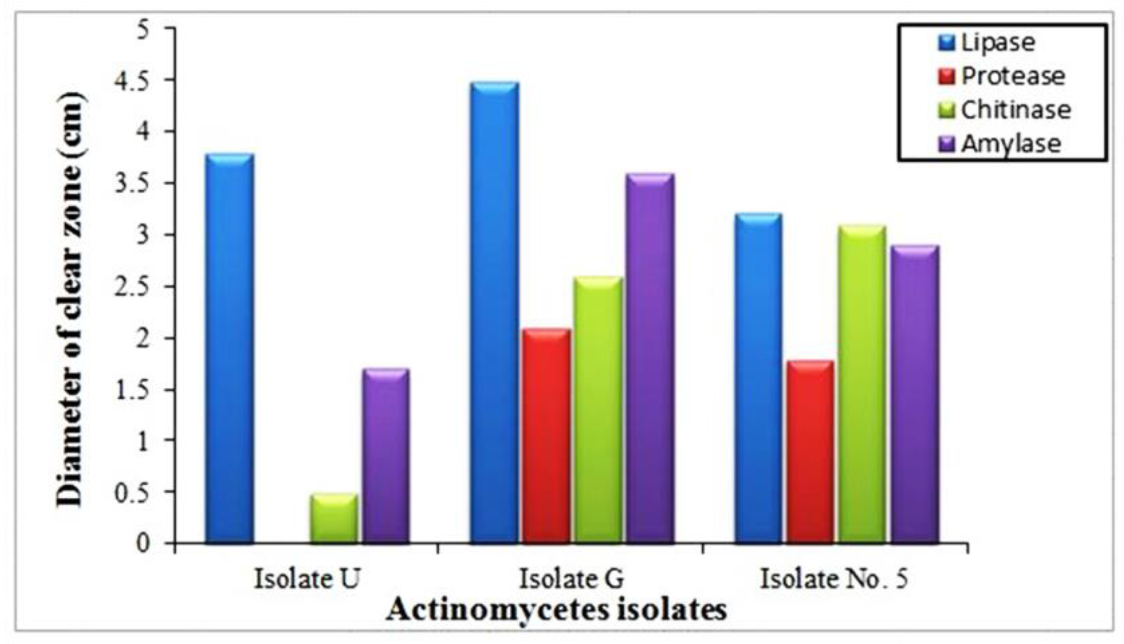
2.7. Production of Extracellular Lytic Enzymes (Proteases, Lipases, Amylases, and Chitinase)

Casein hydrolysis on agar Petri plates containing 0.3 percent casein, 0.1 percent glucose, and 2 percent agar was used to test the proteolytic activity. From each antagonist’s fully developed colony, a 3 mm disk was removed and added to the medium. For a week, plates were incubated at 28 °C and developing colonies were checked for hollow zones, which indicate proteolytic activity [37,38]. Adversaries’ lipolytic activity was assessed using a technique developed by Sadeghian et al. [32]. A 3 mm disk was added to the medium, which also included 10 mL of autoclaved Tween-80, 1 percent peptone, 0.5 percent NaCl, 0.01 percent CaCl2, and 1.5 percent agar. When a powder-like substance developed surrounding the colonies, it showed that the antagonists were active in lipolysis. Approximately 0.3 percent of soluble starch was added to the nutritional agar base medium for the amylase test, and a few isolates were cultured at 28 °C for five days. The amount of starch hydrolysis was then determined by dousing the plate with diluted Lugol’s iodine. The starch–iodine mixture lacked the bluish–purple tint, which is a sign of amylase (starch hydrolysis) capabilities. The candidate isolates were inoculated on a minimum 2 percent agar medium that included 0.5 percent colloidal chitin to ascertain their chitinase activity. According to Sadeghian et al.’s instructions, a colloidal chitin was created [32]. Chitinolytic characteristics were observed for those antagonists a week after incubation at 28 °C, exhibiting a hallow zone around the expanding colony. The taste values of three replicates were recorded.

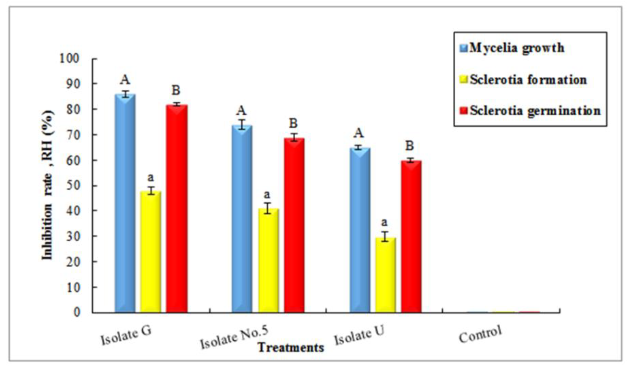
2.8. Investigation of Inhibitory Effects of Antagonists on Mycelia and Sclerotia of Pathogen

Before filling plates with PDA medium, 100 g/mL suspensions of antagonist crude extract were put into Petri dishes to assess the in vitro antifungal activities of crude extract against rice pathogen (potato dextrose agar). Each Petri plate containing crude extract had uniform agar plugs of 6 mm in diameter concurrently seeded in the center, followed by incubation at 29 ± 1 °C for a week. The mycelia growth inhibition rate was calculated using Equation (1) [38].
Inhibition   rate   ( RH )   % = ( R r ) R × 100
RH stands for the rate of inhibition, R for the inhibition of mycelium development is the increase in diameter of the mycelial fungus in the control, and R for the inhibition of sclerotia formation growth is the weight of the sclerotia in the control dish. When treated with crude extract, the fungus’ mycelium expands in width, and when sclerotia form and proliferate under inhibition, the parameter r is the weight of the sclerotia after being treated with crude extract. After adding crude extract concentration to the PDA medium material, the antifungal impact of crude extract against the development of fungus sclerotia was determined. To observe sclerotia development, inoculated pathogen plates were kept at 29 ± 1 °C for 20 days. The sclerotia formation inhibition rate was estimated using (1). Three copies of each test were used on each sample. Using the following technique, the impact of crud extract on the germination of sclerotia was evaluated [39]. The R. solani sclerotia developed on PDA at 15 °C after 20 days of incubation with the infected plates. From PDA plates, uniform sclerotia were recovered, and the surface was sanitized for three minutes in a 1.5 percent sodium hypochlorite solution. In a Petri dish, three surface-sterilized sclerotia were then treated with crude extract and cultured for a week at 25 °C in the dark. Measured sclerotia germination rates were compared with the control (without antagonist crud extract). Utilizing the following formula (Eq1), the percentage of inhibition against sclerotia was obtained. All data represent the average of three replicates for each of the three antagonists.

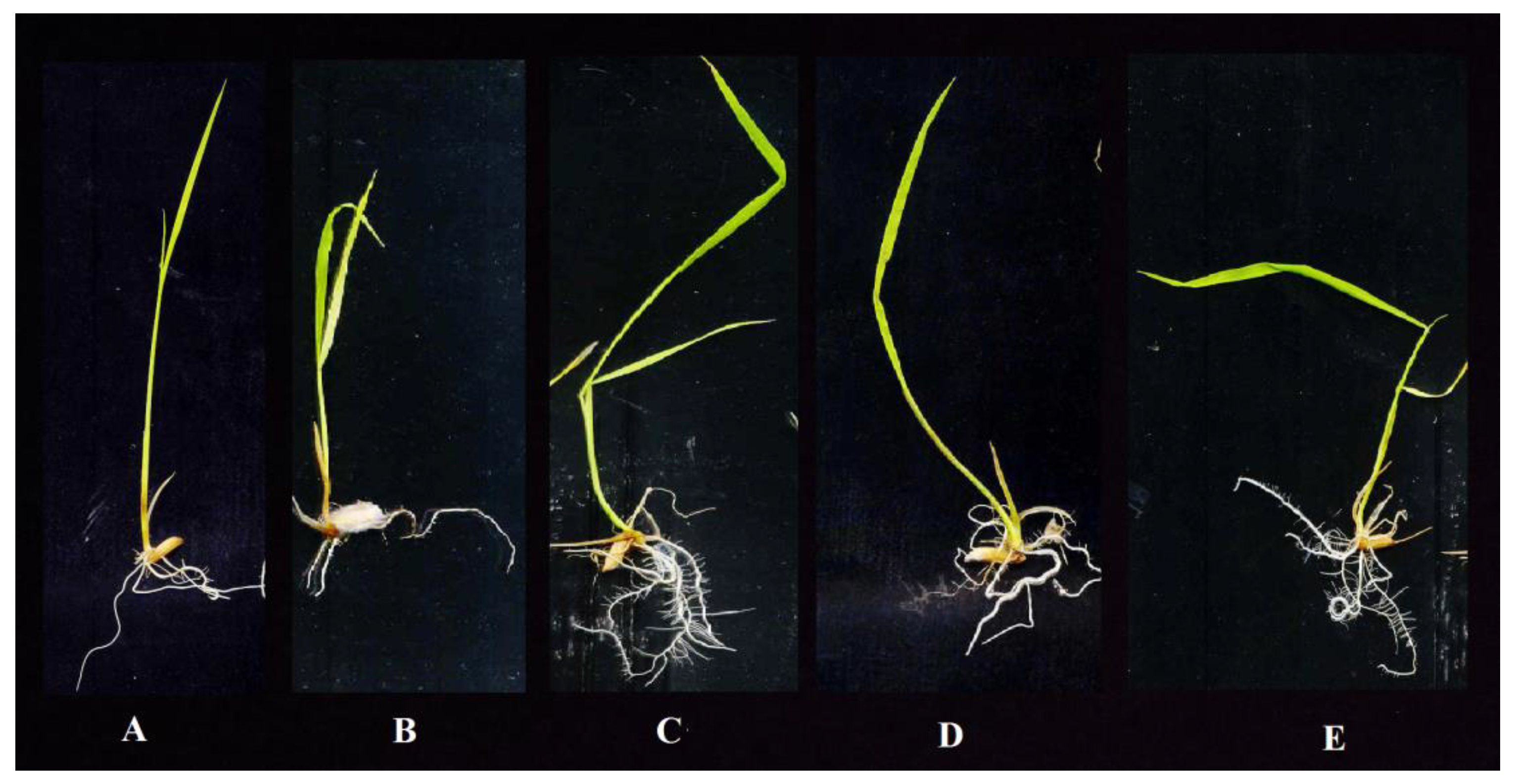
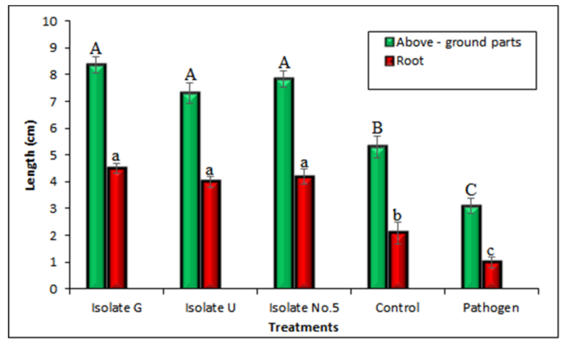
2.9. In Vivo Experiments to Assess the Effects of Potential Antagonists on Rice Seedlings Growth

Rice plant sterile seeds were incubated for 60 min in a solution of bacteria with 2 × 10⁸ CFU/mL. To a solid 50% Murashige–Skoog Basal medium (MS medium), dried seeds were added (Sigma-Aldrich, Merck, Germany). The length of the growing seedlings was measured after 15 days. Distilled water was used to soak the control seedlings. For each isolate, three duplicates were consider account.

2.10. Morphological and Molecular Identifications of Potential Antagonists

Selected isolates were grown on CGA at 30 °C for a week, and the morphological characteristics of isolates G, U, and No. 5 were noted. Scanning electron microscopy (SEM) was used to examine surface ornamentation on cultures that had been cultivated on CGA for 10 days. Genomic DNA was taken to perform molecular identification. Following the procedure outlined by Soltani Nejad et al. [27], the 16S rRNA gene of three isolates was amplified by polymerase chain reaction (PCR) using universal bacterial primers FD1 and RP2. Three active isolates were subjected to a 5 L PCR product electrophoresis on a 1 percent agarose gel (UltraPureTM Agarose, Invitrogen) containing ethidium bromide, and the results were seen under UV light. As a molecular weight marker, a 100 bp ladder (Gene Ruler, mTMDNA Ladder Mix, Fermentas) was used. By Macrogen Co, Seoul, South Korea, PCR products were purified and sequenced. After that, the sequences were read and modified using BioEdit Sequence Alignment Editor. The MEGA 7.0 program’s Clustal W tool were used to align all the sequences of the individual gene sets (Molecular Evolutionary Genetics Analysis, Biodesign Institute, Kent, OH, USA). Using the maximum parsimony (MP) technique, a phylogenetic tree was rebuilt. As the outgroup taxon, Bacillus cereus (MK855405) was employed.

2.11. In Vivo Examination of Potential Antagonists against Rhizoctonia Solani under Glasshouse Conditions

To segregate the rice seeds into eight groups, four pots (as duplicates) were inserted in each group, 3–4 cm under the soil surface of the 1L pots and four replicates for each treatment was considered. Control, pathogen (R. solani), pathogen + Streptomyces isolate No. 5, pathogen + Streptomyces isolate U, pathogen + Streptomyces isolate G, isolate No. 5, isolate G, and isolate U are shown in (a), (b), (c), and (d), respectively. Rice plants were grown in pots in a glasshouse at a temperature of 30 °C and relative humidity of 85–95%, with daily, routine hand watering. Fertilizer was not given to them. The inoculation procedure with R. solani was used to treat the plants when they entered their late tiller stage. To do this, R. Solani’s mycelia suspension was applied to the rice plants using a sprayer bottle [40]. After seven days, disease severity was determined by measuring the relative lesion height (RLH), fresh weight, and dry weight for the root and shoot, shoot length, and root length in accordance with the RRII standard. Using Eq. 2 [41], the relative lesion height of each tiller was computed as follows: RLH % = [Lesion height]/[Plant height] × 100.

2.12. Statistical Analysis

Using SAS software, analysis of variance was performed on all data (SAS Institute, Cary, NC, USA). To compare means, Duncan’s Multiple Range Test was used for inhibition rate and in vivo tests.

3. Results

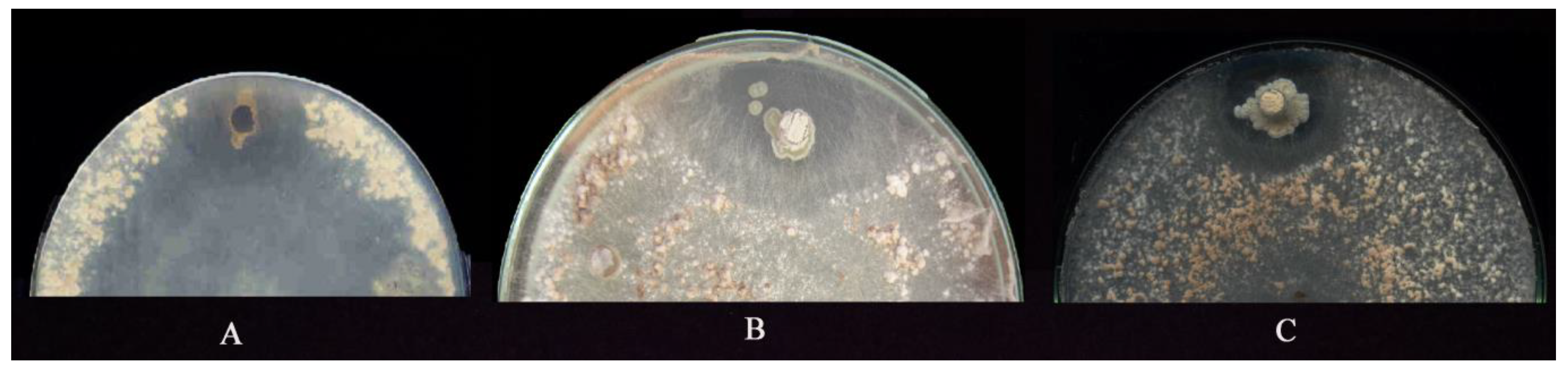
3.1. Isolation and SCREENING the Actinobacteria for Suppressiveness Activity

A dual culture technique was applied for screening and three of the thirty Actinomycetes isolates were examined; isolates No. 5 and U had the most antagonistic responses to R. solani, the causative agent of sheath blight (ShB), shown in Figure 1. Scanning electron microscopy (SEM) analysis was used to examine the antagonistic relationship between three Actinomycetes isolates and the fungus Rhizoctonia solani, as shown in Figure 2. In the micrograph findings of the SEM test, the interaction of antagonistic Actinomycetes clearly showed the inhibition of mycelia’s apical development, irregular distortion in the fungal hyphae followed by diminutive growth, and lyses of the mycelium.

3.2. Chloroform Sensitivity Assay

All three isolates preserved their antifungal activities against R. solani after exposure to the chloroform.

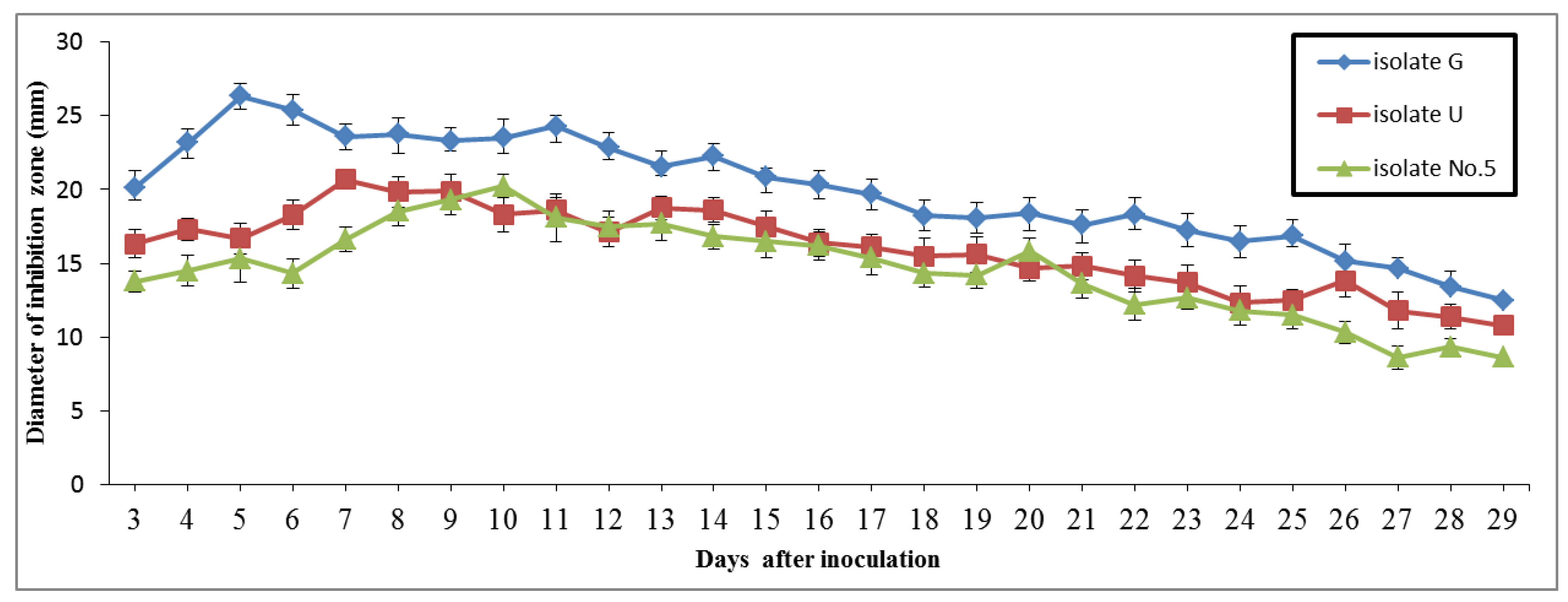
3.3. Antifungal Properties of Submerged Cultures

Three isolates were subjected to daily serial testing for 30 days. The results showed that isolate G achieved its maximal antifungal activity six days after inoculation, whilst isolates U and No. 5 reached their peak inhibitory activity in the 8th and 11th days after inoculation, respectively, shown in Figure 3 and Figure 4.

3.4. Fungicidal and/or Fungistatic Properties of Antagonists

Fungistatic activity of isolates G and U was determined after observing re-growth of the fungus mycelia but isolate No. 5 indicated the fungicidal effect on R. solani.

3.5. Minimum Inhibitory Concentration and Thermal Inactivation Point

Minimum inhibitory concentrations of tested crude extracts of isolates No. 5, G, and U were measured as 0.625 mg/L, 0.625 mg/L, and 1.25 mg/L, respectively.
Thermal inactivation points were recorded as 160 °C, 100 °C, and 60 °C for isolates G, U, and No. 5, respectively.

3.6. Production of Extracellular Lytic Enzymes

All three tested isolates could produce hydrolytic enzymes including amylases, chitinases, and lipases. Isolates G and 5 could produce protease (Figure 5).

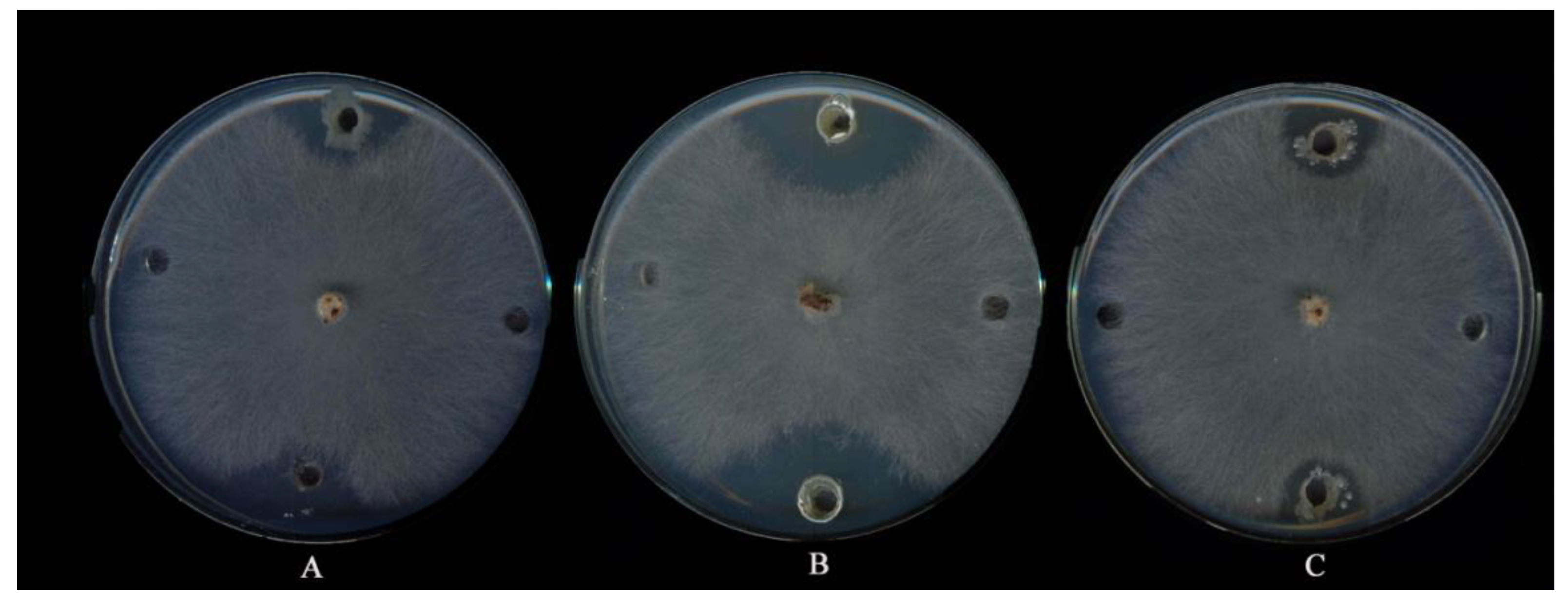
3.7. Examination of Inhibitory Effects of Antagonists on Mycelia and Sclerotia of Fungus

Figure 6 shows how each antagonist’s crude extract affected mycelium development, sclerotia production, and germination. The least amount of sclerotia were seen on the plates treated with isolate G. For isolates U, No. 5, and G, the RH percent values were 30, 41, and 48, respectively. Regarding mycelia development, isolates U, No. 5, and G had RH values of 65, 74, and 86 percent, respectively. Regarding sclerotia germination, isolates U, No. 5, and G, respectively, had RH values of 60, 69, and 82 percent. These findings show that fungus was severely reduced by crude extracts of antagonists in an in vitro condition.

3.8. Effect of Antagonistic Isolates on Rice Seedlings Grown in MS Media

When compared with control, rice seedlings treated with Actinomycetes isolates had significantly longer above-ground lengths (8.35, 7.82, and 7.30 cm for isolates G, No. 5, and U, respectively), as well as significantly longer roots (4.50, 4.20, and 4.0 cm for isolates G, No. 5, and U, respectively). The quantity of lateral roots has also grown due to antagonists (Figure 7 and Figure 8).

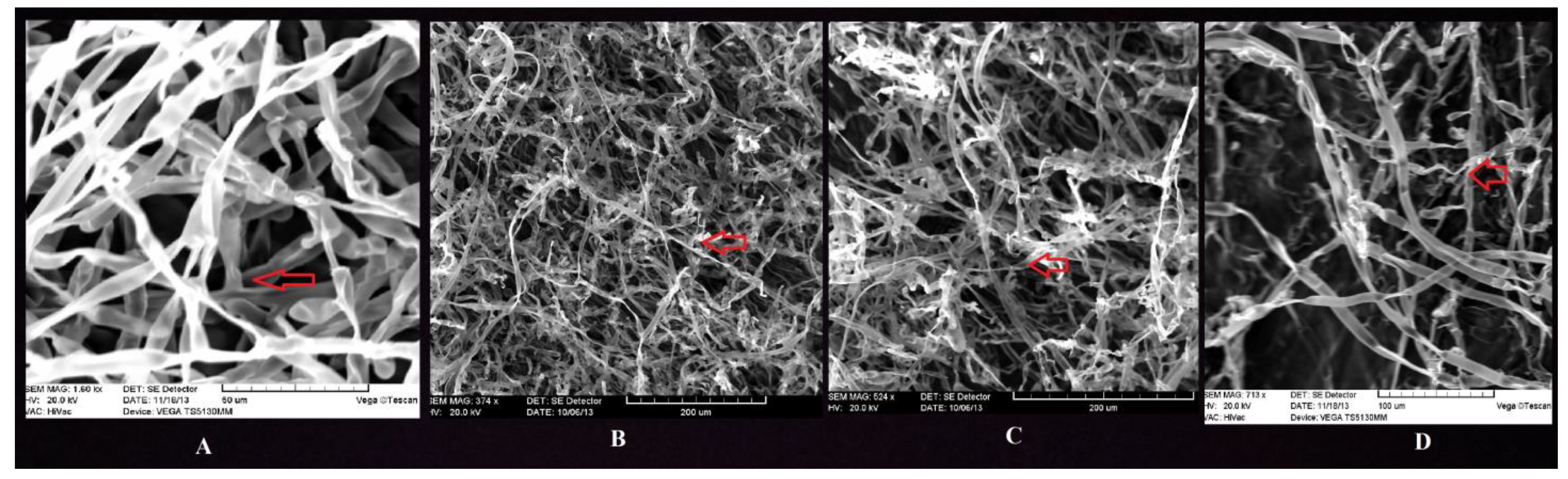
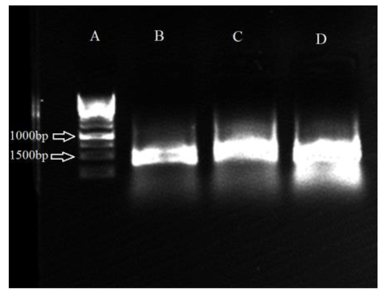
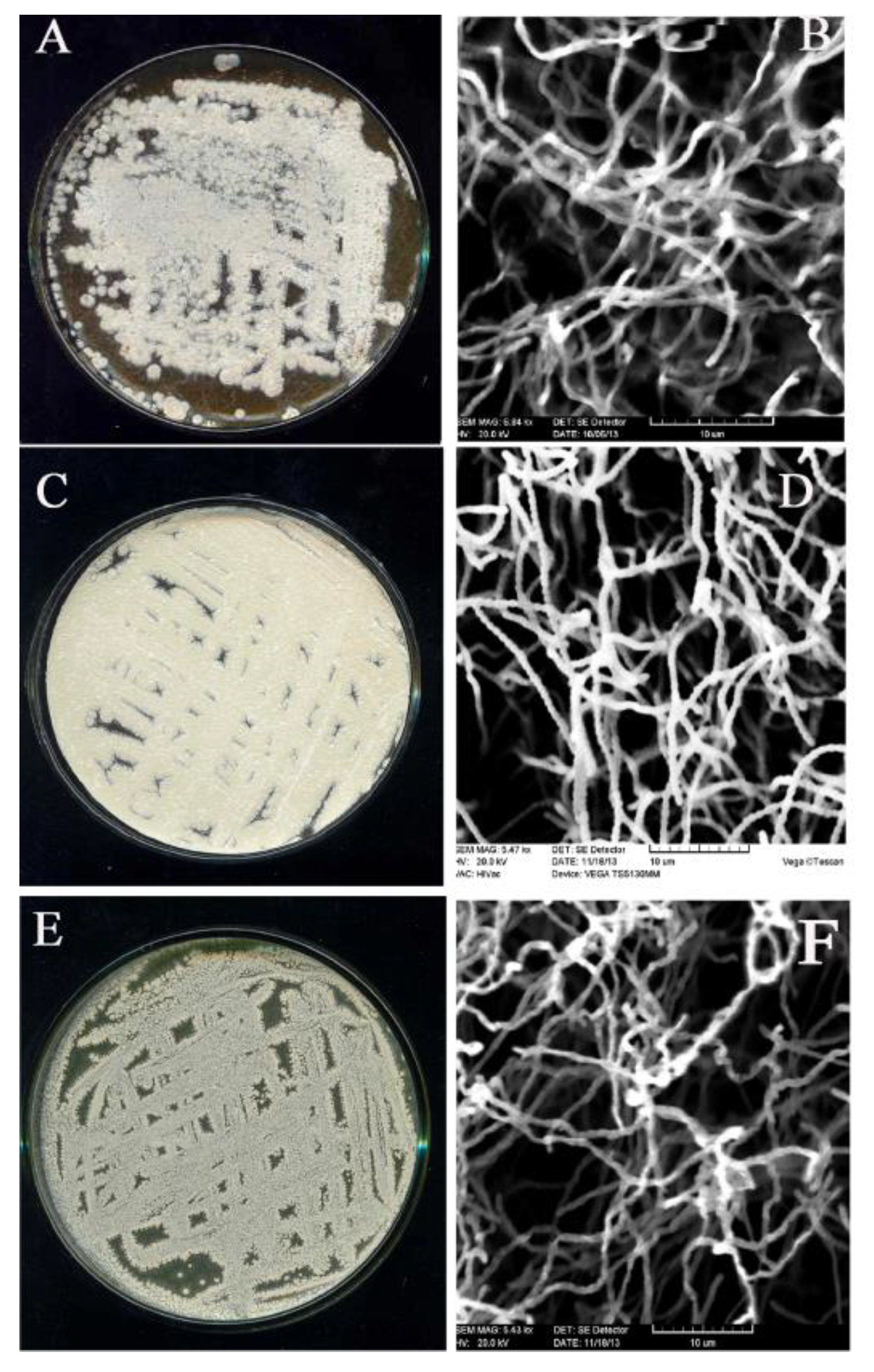
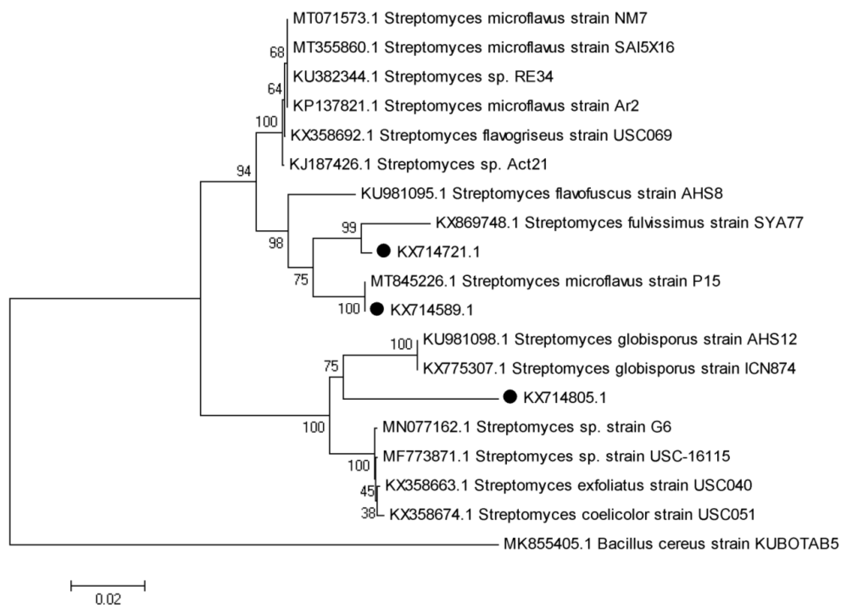
3.9. Molecular Identification of Promising Antagonists and SEM Studies

16S rRNA gene of isolates G, No. 5, and U were amplified by PCR as indicated in Figure 9. Comparison of the near-full-length 16S rDNA sequences of three isolates to GenBank sequences (submitted to GenBank by the following accession numbers: isolate 5, KX714.589; isolate G, KX714805; isolate U, KX714721) indicated that the isolate No. 5 had the most similarity to Streptomyces microflavus (E-value = 0.0 and maximum identity = 100%), isolate U to Streptomyces fulvissimus (E-value = 0.0 and maximum identity = 99%), and isolate G to Streptomyces somaliensis (E-value = 0.0 and max. identity = 100%). For SEM studies, spore chain morphology and spore surface ornamentation were observed (Figure 10). The phylogenetic analysis is illustrated in Figure 11. A phylogenetic tree was constructed by applying software.

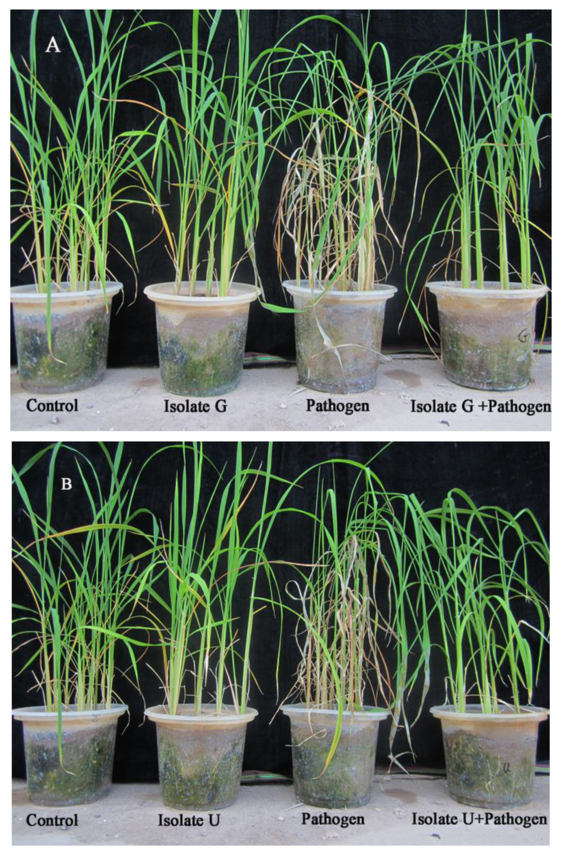
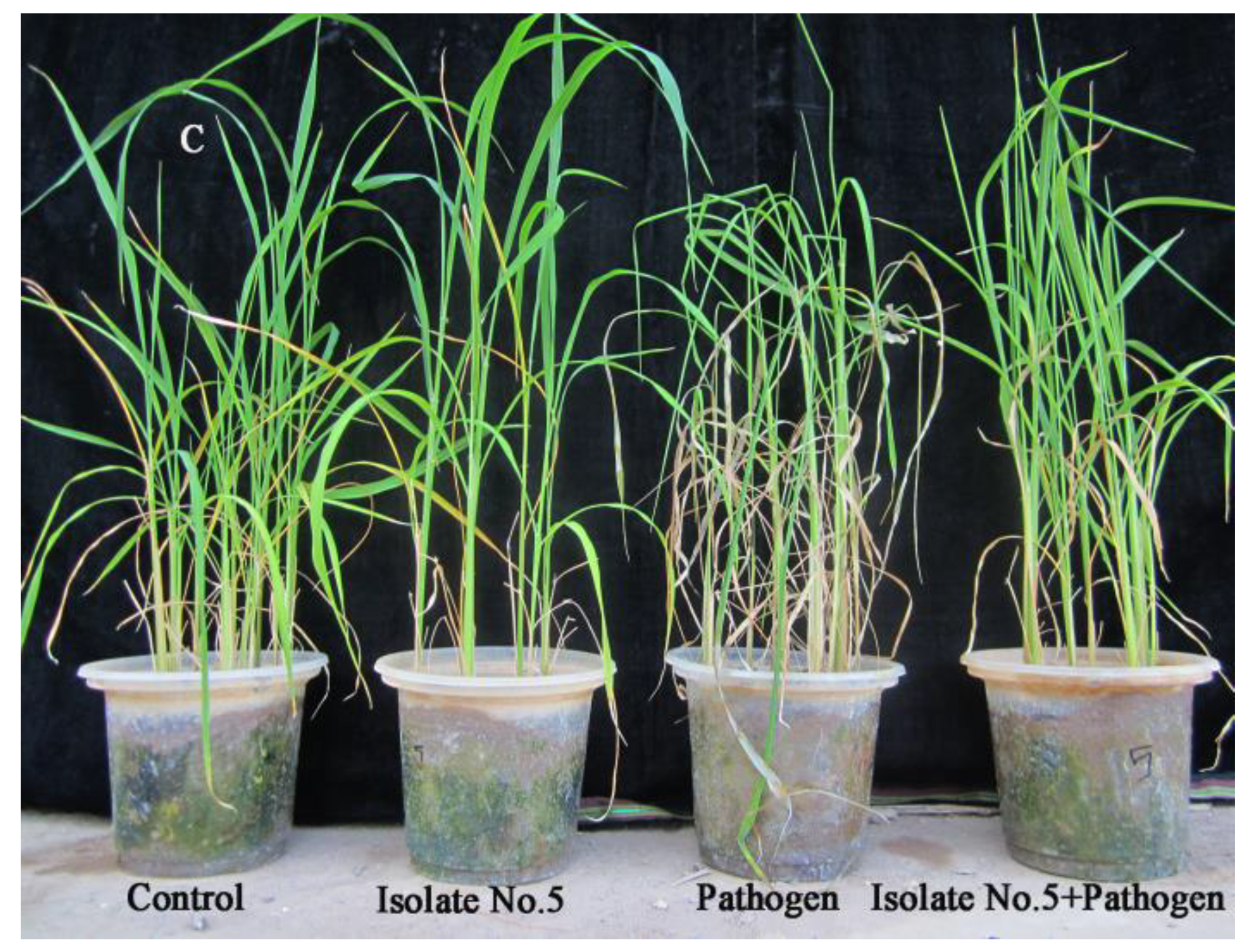
3.10. Examination of Potential Antagonists against Rhizoctonia Solani under Glasshouse Condition

The glasshouse experiment revealed that treating plants with R. solani in the absence of antagonists caused the usual signs of sheath blight, whereas treating plants with R. solani with antagonists resulted in varying degrees of suppression. In pots treated with antagonists, R. solani symptoms were significantly reduced. In treatments using isolates No. 5 and G, the fresh weight and dry weight greatly increased. Isolate U demonstrated a smaller impact on the fresh and dry weight than the other two isolates (Table 1). The RLH of rice tillers dropped, suggesting that antagonists significantly reduced the severity of the disease and the amount of lesions in rice tillers. Significantly less the number of lesions on rice plant were found after comparing the inhibitory potential of tested antagonists against R. solani (Figure 12. The most effective antagonist against R. solani was listed as isolate No. 5.

4. Discussion

In the study that is being presented, Actinomycetes isolates from the Iranian region of Guilan province were gathered and tested for the presence of R. solani, the pathogen that causes rice sheath blight. The top three active isolates that significantly reduced the disease under the glasshouse testing were identified as S. microflavus (isolate No. 5), S. somaliensis (isolate G), and S. fulvissimus based on both morphological and molecular criteria (isolate U). Streptomycetes are widely recognized for their use as biofertilizers in agricultural soils, biocontrol agents for the treatment of plant diseases, and bioproducts in the medical field [42]. Antibiotics and insecticides made from synthetic chemicals have caused many health problems and microbiological resistance to these biohazards. Consequently, it is essential to hunt for natural alternatives [43,44]. Additionally, the capacity of Streptomyces species to manufacture antibiotics and secondary extracellular cell wall disintegrating enzymes enabled them to function as potent biocontrol agents against fungi, either alone or in concert with other agents by dissolving the fungal cell walls [45,46]. The impacts of helpful microorganisms and secondary plant metabolites have been studied extensively as a bio alternative to reduce plant pathogenic bacterial and fungal diseases. These bioactive substances are environmentally safe and biodegradable [47]. Plant infections can be controlled biologically without harming the environment, although farmers use it less often and find it less acceptable than chemical approaches [48,49]. Researchers have shown that the Streptomyces species may prevent the spread of Penicillium italicum, the culprit behind citrus blue mold, by creating a variety of volatile chemicals on the surface of wheat seeds [50]. Penicillium purpurogenum and Alternaria alternata are two phytopathogens that Streptomyces microflavus may effectively inhibit [51,52]. Species of Streptomyces may enter the cell wall and membrane of fungi. In another study, Streptomyces ahygroscopicus var. wuyiensis was used as a biofungicide against Botrytis cinerea on tomato leaves. Its significant effect on disease suppression was comparable to that of other Streptomyces species antibiotics, and this property was referred to as the production of extracellular hydrolytic enzymes such as protease, lipase, b-1,3-glucanase, and chitinase [53]. The rhizosphere may be effectively colonized by the Streptomyces species. They may also be endophytes that invade host plants’ interior tissues [54]. Replication rate; antibiotics; quorum sensing of regulated gene expression; the creation of cellulases, chitinases, lipases, and 1,3-glucanases; and the manufacture of siderophores, phytohormones, or amino acids may all contribute to these features. The chemotactic migration of these bacteria is what attracts Streptomyces’ exudate to the rhizosphere [55]. Three out of the thirty Actinomycetes isolates U, G, and No. 5 showed significant antagonistic activity as potential antagonists to create a biocontrol tool against Rhizoctonia solani as an alternative to fungicides. Different degrees of extracellular hydrolytic enzyme production were observed in Streptomycetes isolates. The tested antagonists’ generation of extracellular enzymes such as chitinase and protease may be one of the underlying mechanisms behind their antifungal action. The proteases enzymes generated by antagonistic Streptomycetes break down the extracellular position to secretory enzyme location and stop plant pathogen infection processes. The ability for production of extracellular enzymes, especially chitinase, by the evaluated antagonists may be one of the principles of their antifungal properties. Antifungal properties of Streptomyces isolates against fungus mycelia were also studied using SEM since, compared with the control grown mycelia and mycelia adjacent to the inhibition zone in the plate, tests revealed an unusual and deformed mycelium; in our opinion, the chitinase secreted can destroy fungal mycelium. Chloroform experiment showed that its antifungal effect persists upon exposure to chloroform. At the present situation, we do not consider the active Streptomycetes as biocontrol agents; however, we consider that active Streptomycetes need further investigation. As the thermal inactivation points of all three isolates were above 60 °C, the active principles are not so vulnerable at ambient temperature while being processed in future purification attempts and molecular characterizations. Although we obtained promising results under greenhouse conditions, it is necessary to evaluate the antagonists under field conditions in several locations having the disease. The disease symptoms on the leaves of the rice plants were dramatically reduced by spraying antagonist spores on the plants. Even though we have obtained encouraging findings in a glasshouse setting, the antagonists still need to be tested in the field at various sites where the disease is prevalent. The field experiments must be statistically evaluated over a period of three to five years. Results of in vivo studies were statistically significant. The percentage of infected rice plants in Streptomyces-isolate-containing treatments, in comparison with controls (without Streptomyces), showed RLH% was decreased. In treatments receiving antagonists, severity of symptoms and lesions was significantly lower than pathogen treatments, especially for isolate G. Effect of antagonistic isolates on rice seedlings growth showed that rice seedlings treated with active Streptomycetes isolates had significantly longer above-ground lengths and significantly longer roots compared with control. We believe that developed and used Streptomycetes inoculants are beneficial to seeds and increased rice plant resistance against rice sheath blight disease, thereby increasing the yields. According to this study, Streptomycetes have a strong potential to be consider account in integrated disease management (IDM) systems and may significantly reduce the need to use toxic chemical pesticides on a regular basis to control various rice diseases, particularly sheath blight disease [56]. The fundamental objectives of an IPM program are to create sustainable agricultural yields while minimizing the financial and environmental costs associated with managing plant pests.

5. Conclusions

Since Actinobacteria produce several antibiotics and enzymes that break down extracellular cell walls, their practical usage and commercialization in agriculture need to be given greater thought and attention. Bacterial antagonism, which is responsible for biological control, can work by inhibiting the pathogen with antimicrobial compounds (antibiosis), inducing plant defense mechanisms, competing for iron by producing siderophores, competing for colonization sites and nutrients provided by seeds and roots, inactivating pathogen germination factors present in seed or root exudates, degrading pathogenicity factors of the pathogen such toxins, and parasitism. This study offered information for the first stages of developing biological control agents against the Rhizoctonia solani AG1-IA-caused rice sheath blight disease in Iran. Three of the thirty native Actinomycete isolates that were identified displayed antagonistic traits. Isolates U, No. 5, and G showed greater levels of disease suppression based on in vitro bioassays and under glasshouse conditions. The active isolates were able to generate extracellular lytic enzymes in bioassays for those enzymes. To determine the primary mechanism of action involved in the putative antagonists’ bioactivities, further research on these compounds is required. The results of this study may serve as a valuable starting point for other researchers who are looking to commercialize biocontrol technologies and advance sustainable agriculture

Author Contributions

All author contributed different sections to this research article, Data collection, M.S.N.; formal analysis, N.S.N., M.S.N. and A.H.S.B.; methodology, A.H.S.B., M.S.N. and M.R.N.; project administration, M.S.N. and S.A.; supervision, S.A. and M.Z.; validation, S.A. and K.M.-S.M.; writing—original draft, M.S.N. and N.S.N.; writing—review and editing, A.H.S.B. and N.S.N. All authors have read and agreed to the published version of the manuscript.

Funding

This research received no external funding.

Data Availability Statement

Not applicable.

Acknowledgments

This paper has been supported by the Kadyrov Chechen state university development program 2021–2030.

Conflicts of Interest

We declare that there is no conflict of interest.

References

  1. Ali, M.; Ahmad, A.; Sultana, S.; Mir, S.R. Chemical constituents from the seed husks of Oryza sativa L. Nat. Prod. Res. 2022, 1–9. [Google Scholar] [CrossRef] [PubMed]
  2. Li, S.; Shen, L.; Hu, P.; Liu, Q.; Zhu, X.; Qian, Q.; Wang, K.; Wang, Y. Developing disease-resistant thermo sensitive male sterile rice by multiplex gene editing. J. Integr. Plant Biol. 2019, 61, 1201–1205. [Google Scholar] [CrossRef] [PubMed]
  3. Boukaew, S.; Prasertsan, P. Suppression of rice sheath blight disease using a heat stable culture filtrate from Streptomyces philanthi RM-1-138. Crop Prot. 2014, 61, 1–10. [Google Scholar] [CrossRef]
  4. Yu, Y.-Y.; Jiang, C.-H.; Wang, C.; Chen, L.-J.; Li, H.-Y.; Xu, Q.; Guo, J.-H. An improved strategy for stable biocontrol agents selecting to control rice sheath blight caused by Rhizoctonia solani. Microbiol. Res. 2017, 203, 1–9. [Google Scholar] [CrossRef]
  5. Nookongbut, P.; Kantachote, D.; Khuong, N.Q.; Tantirungkij, M. The biocontrol potential of acid-resistant Rhodopseudomonas palustris KTSSR54 and its exopolymeric substances against rice fungal pathogens to enhance rice growth and yield. Biol. Control 2020, 150, 104354. [Google Scholar] [CrossRef]
  6. Daryaei, A.; Jones, E.E.; Alizadeh, H.; Glare, T.R.; Falloon, R.E. Biochemical characteristics of Trichoderma atroviride associated with conidium fitness for biological control. Biocontrol Sci. Technol. 2016, 26, 189–205. [Google Scholar] [CrossRef]
  7. Kumar, R.; Khatri, A.; Acharya, V. Deep learning uncovers distinct behavior of rice network to pathogens response. iScience 2022, 25, 104546. [Google Scholar] [CrossRef]
  8. Gnanamanickam, S. An Overview of Progress in Biological Control. In Biological Control of Rice Diseases; Progress in Biological Control; Springer: Amsterdam, The Netherlands, 2009; Volume 8, pp. 43–51. [Google Scholar] [CrossRef]
  9. Cuong, N.; Nicolaisen, M.; Sørensen, J.; Olsson, S. Hyphae-Colonizing Burkholderia sp. A New Source of Biological Control Agents Against Sheath Blight Disease (Rhizoctonia solani AG1-IA) in Rice. Microb. Ecol. 2011, 62, 425–434. [Google Scholar] [CrossRef]
  10. Boubekri, K.; Soumare, A.; Mardad, I.; Lyamlouli, K.; Ouhdouch, Y.; Hafidi, M.; Kouisni, L. Multifunctional role of Actinobacteria in agricultural production sustainability: A review. Microbiol. Res. 2022, 261, 127059. [Google Scholar] [CrossRef] [PubMed]
  11. Ebrahimi-Zarandi, M.; Saberi Riseh, R.; Tarkka, M.T. Actinobacteria as Effective Biocontrol Agents against Plant Pathogens, an Overview on Their Role in Eliciting Plant Defense. Microorganisms 2022, 10, 1739. [Google Scholar] [CrossRef]
  12. Chukwuneme, C.F.; Babalola, O.O.; Kutu, F.R.; Ojuederie, O.B. Characterization of Actinomycetes isolates for plant growth promoting traits and their effects on drought tolerance in maize. J. Plant Interact. 2020, 15, 93–105. [Google Scholar] [CrossRef]
  13. Cui, H.; Shaaban, K.; Schiebel, M.; Qin, S.; Laatsch, H. New antibiotic with typical plant anthraquinone structure obtained studying terrestrial and marine Streptomycetes. World J. Microbiol. Biotechnol. 2008, 24, 419–421. [Google Scholar] [CrossRef]
  14. Xu, Z.; Ji, L.; Tang, W.; Guo, L.; Gao, C.; Chen, X.; Liu, J.; Hu, G.; Liu, L. Metabolic engineering of Streptomyces to enhance the synthesis of valuable natural products. Eng. Microbiol. 2022, 2, 100022. [Google Scholar] [CrossRef]
  15. Minuto, A.; Spadaro, D.; Garibaldi, A.; Gullino, M.L. Control of soilborne pathogens of tomato using a commercial formulation of Streptomyces griseoviridis and solarization. Crop. Prot. 2006, 25, 468–475. [Google Scholar] [CrossRef]
  16. Kang, J.E.; Han, J.W.; Jeon, B.J.; Kim, B.S. Efficacies of quorum sensing inhibitors, piericidin A and glucopiericidin A, produced by Streptomyces xanthocidicus KPP01532 for the control of potato soft rot caused by Erwinia carotovora subsp. atroseptica. Microbiol. Res. 2016, 184, 32–41. [Google Scholar] [CrossRef] [PubMed]
  17. Rambabu, V.; Suba, S.; Vijayakumar, S. Antimicrobial and antiproliferative prospective of kosinostatin a secondary metabolite isolated from Streptomyces sp. J. Pharm. Anal. 2015, 5, 378–382. [Google Scholar] [CrossRef] [PubMed]
  18. Lyu, M.; Cheng, Y.; Han, X.; Wen, Y.; Song, Y.; Li, J.; Chen, Z. AccR, a TetR family transcriptional repressor, coordinates short-chain acyl coenzyme A homeostasis in Streptomyces avermitilis. Appl. Environ. Microbiol. 2020, 86, e00508–e00520. [Google Scholar] [CrossRef]
  19. John, K.N.; Valentin, V.; Abdullah, B.; Bayat, M.; Kargar, M.H.; Zargar, M. Weed mapping technologies in discerning and managing weed infestation levels of farming systems. Res. on Crops. 2020, 21, 93–98. [Google Scholar]
  20. Li, Q.; Jiang, Y.; Ning, P.; Zheng, L.; Huang, J.; Li, G.; Jiang, D.; Hsiang, T. Suppression of Magnaporthe oryzae by culture filtrates of Streptomyces globisporus JK-1. Biol. Control 2011, 58, 139–148. [Google Scholar] [CrossRef]
  21. Li, Q.; Ning, P.; Zheng, L.; Huang, J.; Li, G.; Hsiang, T. Effects of volatile substances of Streptomyces globisporus JK-1 on control of Botrytis cinerea on tomato fruit. Biol. Control 2012, 61, 113–120. [Google Scholar] [CrossRef]
  22. Prabavathy, V.R.; Mathivanan, N.; Murugesan, K. Control of blast and sheath blight diseases of rice using antifungal metabolites produced by Streptomyces sp. PM5. Biol. Control 2006, 39, 313–319. [Google Scholar] [CrossRef]
  23. Law, J.W.-F.; Ser, H.-L.; Khan, T.M.; Chuah, L.-H.; Pusparajah, P.; Chan, K.-G.; Goh, B.-H.; Lee, L.-H. The potential of Streptomyces as biocontrol agents against the rice blast fungus, Magnaporthe oryzae (Pyricularia oryzae). Front. Microbiol. 2017, 8, 3. [Google Scholar] [CrossRef] [PubMed]
  24. Zhao, J.; Xue, Q.-h.; Niu, G.-g.; Xue, L.; Shen, G.-h.; Du, J.-z. Extracellular enzyme production and fungal mycelia degradation of antagonistic Streptomyces induced by fungal mycelia preparation of cucurbit plant pathogens. Ann. Microbiol. 2013, 63, 809–812. [Google Scholar] [CrossRef]
  25. Soltani Nejad, M.; Samandari Najafabadi, N.; Aghighi, S.; Pakina, E.; Zargar, M. Evaluation of phoma sp. biomass as an endophytic fungus for synthesis of extracellular gold nanoparticles with antibacterial and antifungal properties. Molecules 2022, 27, 1181. [Google Scholar] [CrossRef] [PubMed]
  26. Firouzi, S.; Nikkhah, A.; Aminpanah, H. Rice single cropping or ratooning agro-system: Which one is more environment-friendly? Environ. Sci. Pollut. Res. 2018, 25, 32246–32256. [Google Scholar] [CrossRef]
  27. Soltani Nejad, M.; Khatami, M.; Shahidi Bonjar, G.H. Extracellular synthesis gold nanotriangles using biomass of Streptomyces microflavus. IET Nanobiotechnol. 2016, 10, 33–38. [Google Scholar] [CrossRef]
  28. Soltani Nejad, M.; Shahidi Bonjar, G.H.; Khaleghi, N. Biosynthesis of gold nanoparticles using Streptomyces fulvissimus isolate. Nanomed. J. 2015, 2, 153–159. [Google Scholar] [CrossRef]
  29. Aghighi, S.; Shahidi Bonjar, G.; Rawashdeh, R.; Batayneh, S.; Saadoun, I. First report of antifungal spectra of activity of Iranian Actinomycetes strains against Alternaria solani, Alternaria alternate, Fusarium solani, Phytophthora megasperma, Verticillium dahliae and Saccharomyces cerevisiae. Asian J. Plant Sci. 2004, 3, 463–471. [Google Scholar] [CrossRef]
  30. Lee, J.Y.; Hwang, B.K. Diversity of antifungal Actinomycetes in various vegetative soils of Korea. Can. J. Microbiol. 2002, 48, 407–417. [Google Scholar] [CrossRef]
  31. El-Tarabily, K.A.; Sivasithamparam, K. Non-streptomycete actinomycetes as biocontrol agents of soil-borne fungal plant pathogens and as plant growth promoters. Soil Biol. Biochem. 2006, 38, 1505–1520. [Google Scholar] [CrossRef]
  32. Sadeghian, M.; Shahidi Bonjar, G.H.; Sharifi Sirchi, G.R. Post harvest biological control of apple bitter rot by soil-borne Actinomycetes and molecular identification of the active antagonist. Postharvest. Biol. Technol. 2016, 112, 46–54. [Google Scholar] [CrossRef]
  33. Davelos, A.L.; Kinkel, L.L.; Samac, D.A. Spatial variation in frequency and intensity of antibiotic interactions among Streptomycetes from prairie soil. Appl. Environ. Microbiol. 2004, 70, 1051–1058. [Google Scholar] [CrossRef] [PubMed]
  34. Jorjandi, M.; Bonjar, G.; Baghizadeh, A.; Sirchi, G.; Massumi, H.; Baniasadi, F.; Aghighi, S.; Farokhi, P. Biocontrol of Botrytis allii Munn the causal agent of neck rot, the postharvest disease in onion, by use of a new Iranian isolate of Streptomyces. Am. J. Agric. Biol. Sci. 2009, 4, 72–78. [Google Scholar] [CrossRef]
  35. Hassanisaadi, M.; Shahidi Bonjar, G.H.; Hosseinipour, A.; Abdolshahi, R.; Ait Barka, E.; Saadoun, I. Biological control of Pythium aphanidermatum, the causal agent of tomato root rot by two Streptomyces root symbionts. Agronomy 2021, 11, 846. [Google Scholar] [CrossRef]
  36. Dunne, C.; Moënne-Loccoz, Y.; de Bruijn, F.J.; O’Gara, F. Overproduction of an inducible extracellular serine protease improves biological control of Pythium ultimum by Stenotrophomonas maltophilia strain W81. Microbiology 2000, 146, 2069–2078. [Google Scholar] [CrossRef] [PubMed]
  37. Rodarte, M.P.; Dias, D.R.; Vilela, D.M.; Schwan, R.F. Proteolytic activities of bacteria, yeasts and filamentous fungi isolated from coffee fruit (Coffea arabica L.). Acta Sci. Agron. 2011, 33, 457–464. [Google Scholar] [CrossRef]
  38. Soltani Nejad, M.; Bonjar, G.H.S.; Khatami, M.; Amini, A.; Aghighi, S. In vitro and in vivo antifungal properties of silver nanoparticles against Rhizoctonia solani, a common agent of rice sheath blight disease. IET Nanobiotechnol. 2017, 11, 236–240. [Google Scholar] [CrossRef]
  39. Kazemi, H.; Kamkar, B.; Lakzaei, S.; Badsar, M.; Shahbyki, M. Energy flow analysis for rice production in different geographical regions of Iran. Energy 2015, 84, 390–396. [Google Scholar] [CrossRef]
  40. Park, D.-S.; Sayler, R.J.; Hong, Y.-G.; Nam, M.-H.; Yang, Y. A method for inoculation and evaluation of rice sheath blight disease. Plant Dis. 2007, 92, 25–29. [Google Scholar] [CrossRef]
  41. Yadav, S.; Anuradha, G.; Kumar, R.; Vemireddy, L.; Sudhakar, R.; Donempudi, K.; Venkata, D.; Jabeen, F.; Narasimhan, Y.; Marathi, B.; et al. Identification of QTLs and possible candidate genes conferring sheath blight resistance in rice (Oryza sativa L.). SpringerPlus 2015, 4, 175. [Google Scholar] [CrossRef] [Green Version]
  42. Pereira, P.M.; Santana, F.M.; Van Der Sand, S. Evaluation of Streptomyces spp. strains as potential biocontrol agents for Pyrenophora tritici-repentis. Biocontrol Sci. Technol. 2022, 32, 1095–1106. [Google Scholar] [CrossRef]
  43. Salla, T.D.; Ramos da Silva, T.; Astarita, L.V.; Santarém, E.R. Streptomyces rhizobacteria modulate the secondary metabolism of Eucalyptus plants. Plant Physiol. Biochem. 2014, 85, 14–20. [Google Scholar] [CrossRef] [PubMed]
  44. Pacios-Michelena, S.; Aguilar González, C.N.; Alvarez-Perez, O.B.; Rodriguez-Herrera, R.; Chávez-González, M.; Arredondo Valdés, R.; Ascacio Valdés, J.A.; Govea Salas, M.; Ilyina, A. Application of Streptomyces antimicrobial compounds for the control of hpytopathogens. Front. Sustain. Food Syst. 2021, 5, 696518. [Google Scholar] [CrossRef]
  45. Santos-Beneit, F.; Ceniceros, A.; Nikolaou, A.; Salas, J.A.; Gutierrez-Merino, J. Identification of antimicrobial compounds in two Streptomyces sp. strains isolated from beehives. Front. Microbiol. 2022, 13, 742168. [Google Scholar] [CrossRef]
  46. Zhang, L.; Liu, Z.; Wang, Y.; Zhang, J.; Wan, S.; Huang, Y.; Yun, T.; Xie, J.; Wang, W. Biocontrol potential of endophytic Streptomyces malaysiensis 8ZJF-21 from medicinal plant against banana Fusarium wilt caused by Fusarium oxysporum f. sp. cubense tropical race 4. Front. Plant Sci. 2022, 13, 874819. [Google Scholar] [CrossRef]
  47. Dziggel, C.; Schäfer, H.; Wink, M. Tools of pathway reconstruction and production of economically relevant plant secondary metabolites in recombinant microorganisms. Biotechnol. J. 2017, 12, 1600145. [Google Scholar] [CrossRef]
  48. Peng, D.; Li, S.; Wang, J.; Chen, C.; Zhou, M. Integrated biological and chemical control of rice sheath blight by Bacillus subtilis NJ-18 and jinggangmycin. Pest Manag. Sci. 2014, 70, 258–263. [Google Scholar] [CrossRef]
  49. Santiago, T.R.; Grabowski, C.; Rossato, M.; Romeiro, R.S.; Mizubuti, E.S.G. Biological control of eucalyptus bacterial wilt with rhizobacteria. Biol. Control 2015, 80, 14–22. [Google Scholar] [CrossRef]
  50. Li, Q.; Ning, P.; Zheng, L.; Huang, J.; Li, G.; Hsiang, T. Fumigant activity of volatiles of Streptomyces globisporus JK-1 against Penicillium italicum on Citrus microcarpa. Postharvest. Biol. Technol. 2010, 58, 157–165. [Google Scholar] [CrossRef]
  51. Gao, F.; Wu, Y.; Wang, M. Identification and antifungal activity of an Actinomycetes strain against Alternaria spp. Span. J. Agric. Res. 2014, 12, 1158–1165. [Google Scholar] [CrossRef] [Green Version]
  52. Gao, Y.; Zeng, X.D.; Ren, B.; Zeng, J.R.; Xu, T.; Yang, Y.Z.; Hu, X.C.; Zhu, Z.Y.; Shi, L.M.; Zhou, G.Y.; et al. Antagonistic activity against rice blast disease and elicitation of host-defence response capability of an endophytic Streptomyces albidoflavus OsiLf-2. Plant Pathol. 2020, 69, 259–271. [Google Scholar] [CrossRef]
  53. AlAli, H.A.; Khalifa, A.; Almalki, M. Plant growth-promoting bacterium from non-agricultural soil Improves okra plant growth. Agriculture 2022, 12, 873. [Google Scholar] [CrossRef]
  54. Olanrewaju, O.S.; Babalola, O.O. Streptomyces: Implications and interactions in plant growth promotion. Appl. Microbiol. Biotechnol. 2019, 103, 1179–1188. [Google Scholar] [CrossRef] [PubMed]
  55. Rachid, L. Biological Control of Plant Pathogens: A Global Perspective. Microorganisms 2022, 10, 596. [Google Scholar] [CrossRef]
  56. Hu, D.; Li, S.; Li, Y.; Peng, J.; Wei, X.; Ma, J.; Zhang, C.; Jia, N.; Wang, E.; Wang, Z. Streptomyces sp. strain TOR3209: A rhizosphere bacterium promoting growth of tomato by affecting the rhizosphere microbial community. Sci. Rep. 2020, 10, 20132. [Google Scholar] [CrossRef] [PubMed]
Figure 1. Inhibition zone of Actinomycetes isolates against Rhizoctonia solani: (A) isolate U, (B) isolate G, and (C) isolate No. 5.
Agronomy 12 02292 g001
Figure 2. SEM images from the inhibition zone demonstrating the antagonistic behavior of Actinomycetes isolates against Rhizoctonia solani. The arrows in the control image depict healthy, intact mycelia, whereas those in the other images reveal malformed, damaged R. solani hyphae. Control, isolate U, isolate No. 5, and isolate G, respectively, are shown in (AD).
Agronomy 12 02292 g002
Figure 3. Diameter of inhibition zone (mm) of antagonist’s culture filtrates against Rhizoctonia solani versus post seeding time using well diffusion method.
Agronomy 12 02292 g003
Figure 4. Inhibitory impacts of aqueous culture filtrate of antagonist isolates in the well diffusion method against Rhizoctonia solani. Control (left and right: CG medium), (A) culture filtrate (top and bottom) of isolate No. 5 at 11th day, (B) isolate G at 6th day, and (C) isolate U at 8th day post seeding time.
Agronomy 12 02292 g004
Figure 5. Enzymatic activities (lipase, protease, chitinase, and amylase) of tested potential antagonists (isolates U, G, and No. 5). Isolate U lacks protease properties.
Agronomy 12 02292 g005
Figure 6. In vitro inhibition rate (RH%) of three antagonist effects on mycelia growth, sclerotia germination, and sclerotia formation against Rhizoctonia solani. The presented data are the means of three replications and the Duncan’s Multiple Range test (p ≤ 0.05).
Agronomy 12 02292 g006
Figure 7. Longitudinal root growth of rice plants after being inoculated with antagonistic Actinomycetes including (C) isolate G, (D) isolate U, (E) isolate No. 5, (A) control, and (B) Rhizoctonia solani.
Agronomy 12 02292 g007
Figure 8. Rice plants’ longitudinal root growth ratios after in vivo inoculation. The standard deviation is shown by error bars. Duncan’s Multiple Range Test reveals a significant difference between the means denoted by various letters (p ≤ 0.05). Different letters represent significant differences.
Agronomy 12 02292 g008
Figure 9. Molecular identification of Streptomycete isolates: (A) Ladder, amplification of 16S rDNA of Streptomyces sp. Isolates by PCR: (B) isolate G, (C) isolate No. 5, and (D) isolate U.
Agronomy 12 02292 g009
Figure 10. Pure culture of potential antagonists and findings of scanning electron micrographs. (A), (B) isolate 5, (C,D) isolate U, and (E,F) isolate G.
Agronomy 12 02292 g010
Figure 11. Phylogeny of Streptomyces isolates. BLAST search for the three isolates indicated 100% homology for isolate No. 5 as Streptomyces microflavus and isolate Gas Streptomyces somaliensis, and isolate U showed 99% homology with Streptomyces fulvissimus.
Agronomy 12 02292 g011
Figure 12. Suppression of sheath blight symptoms in rice leaves by application of Actinomycetes isolates, (A) G; (B) U, and (C) No. 5.
Agronomy 12 02292 g012aAgronomy 12 02292 g012b
Table 1. The inhibitory effects of Actinomycetes isolates against Rhizoctonia solani (AG1-IA) causing rice sheath blight disease under glasshouse condition. Isolate U showed smaller effects on the fresh and dry weight than the other two isolates. According to the results, isolate G was the most effective isolate against rice pathogen. Different letters represent significant changes.
TreatmentsRLH%Shoot Fresh Weight (g)Shoot Dry Weight (g)Root Fresh Weight (g)Root Dry Wight (g)Shoot Length (cm)Root Length
(cm)
Control_11.83 * A5.57 B6.53 AB3.51 AB30.52 AB12.40 BC
Pathogen95%4.57 D2.48 DC2.28 D0.96 D10.18 D5.37 D
Isolate No. 5_11.23 AB7.43 ABC6.84 BC3.54 AB28.54 B14.41 BC
Isolate G_11.76 A8.75 AB7.89 AB4.61 A32.27 A20.26 A
Isolate U_11.19 ABC6.24 AB6.48 CD3.18 ABC22.11 BC10.61 BC
Pathogen+ G42%11.12 BC6.89 AB7.61 D4.35 CD21.83 BC18.20 AC
Pathogen + No. 553%11.07 BC6.17 AB6.73 AD3.21 BC19.69 BC19.12 A
Pathogen+ U75%8.43 CD4.26 BC5.39 D2.64 CD18.18 BCD9.76 CD
SE± 0.910.830.860.571.681.25
* The in vivo condition’s presented data are the means of four replications; for each trait, different letters indicate significant differences at significance level a = 0.05 according to Duncan’s Multiple Range Test.
Publisher’s Note: MDPI stays neutral with regard to jurisdictional claims in published maps and institutional affiliations.

