


Metal–Polymer Nanocomposites: A Promising Approach to Antibacterial Materials


Abstract
:1. Introduction
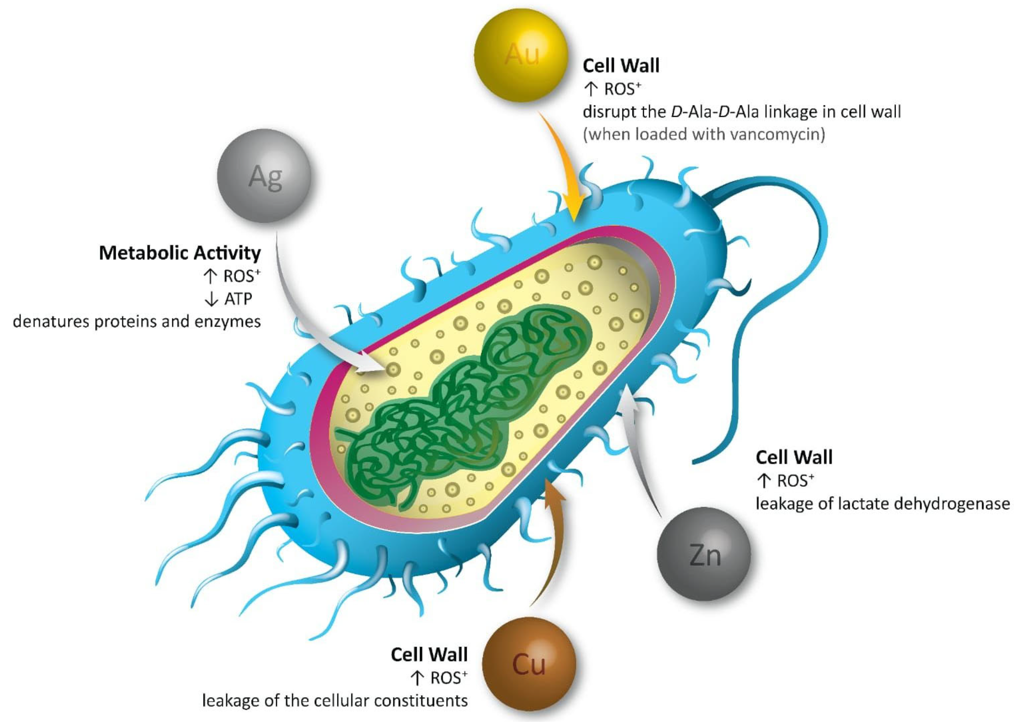
2. Antibacterial Mechanism of Action of Metal–Polymer Nanocomposites
2.1. Antibacterial Mechanism of Action of Silver Nanocomposites
2.2. Antibacterial Mechanism of Action of Copper Nanocomposites
2.3. Antibacterial Mechanism of Action of Zinc Nanocomposites
2.4. Antibacterial Mechanism of Action of Gold Nanocomposites
3. Types of Metal–Polymer Nanocomposites with Antibacterial Properties Based on Metals (Silver, Copper, Zinc, and Gold)
3.1. Silver-Based Nanocomposites
3.2. Copper-Based Nanocomposites
3.3. Zinc-Based Nanocomposites

| No. | Polymer Name | Source | Properties | Uses | Ref. |
|---|---|---|---|---|---|
| 1 | Chitosan hydrogel | Synthetic | 30 nm | Antimicrobial | [110] |
| 2 | Cellulose | Synthetic | 65 nm | Photocatalytic and antibacterial | [111] |
| 3 | Agar biopolymer | Synthetic | 20 nm | Antibacterial and anticancer | [112] |
| 4 | Gelatin/tragacanth | Synthetic | 10.6 nm | Antimicrobial biomaterials for food packaging | [113] |
| 5 | Chitosan | Natural | 25 to 70 nm | Antimicrobial | [114] |
| 6 | Chitosan | Synthetic | 20–150 nm | Antibacterial and photocatalytic | [115] |
| 7 | Low density polyethylene (LDPE) | Synthetic | ~17 nm | Antimicrobial | [116] |
| 8 | Chitosan | Natural | 24 nm | Antibacterial and photocatalytic | [117] |
| 9 | Alginate beads | Natural | 20–100 nm | Antimicrobial agent for water disinfection | [118] |
| 10 | Polyvinyl alcohol | Synthetic | - | Antimicrobial coating | [119] |
| 11 | Polyaniline | Synthetic | 61.6 nm | Antibacterial | [120] |
| 12 | Poly(3-hydroxybutyrate-co-3-hydroxyvalerate) | Synthetic | 3.5, 25 nm | Food packaging and food contact surface applications | [121] |
| 13 | Multiwalled carbon nanotubes (MWCNTs) | Synthetic | - | Food packaging | [122] |
| 14 | Hydroxyethyl cellulose, carboxymethyl chitosan (CMCS) composite/film | Synthetic | - | Antibacterial, food packaging | [123] |
| 15 | Graphene oxide, composite resins | Synthetic | - | Antimicrobial | [124] |
| 16 | Mahua-oil-based polyurethane/chitosan/nano ZnO composite | Synthetic | 30 nm | Food packaging | [125] |
| 17 | Polyvinyl (alcohol)/chitosan/nano zinc oxide hydrogels | Synthetic | 30 nm | Wound healing | [126] |
| 18 | AZO-Np in Guar gum/polyvinyl alcohol composite fiber mats | Semisynthetic | Less than 50 nm | Antibacterial | [127] |
| 19 | Chitosan–alginate–gelatin and chitosan–bentonite–gelatin films with ZnO | Natural | Nanoscaled | Skin burn healing | [128] |
| 20 | Aminoalkylsilane-grafted bacterial nanocellulose | Synthetic | 30 nm | Multifunctional wound dressing, antibacterial | [129] |
| 21 | PVA and xylan, nanoscaled ZnO | Synthetic | - | Bacteriostatic films | [130] |
| 22 | Oxidized sodium alginate and its electrospun bio-hybrids | Synthetic | - | Wound healing | [130] |
3.4. Gold-Based Nanocomposites
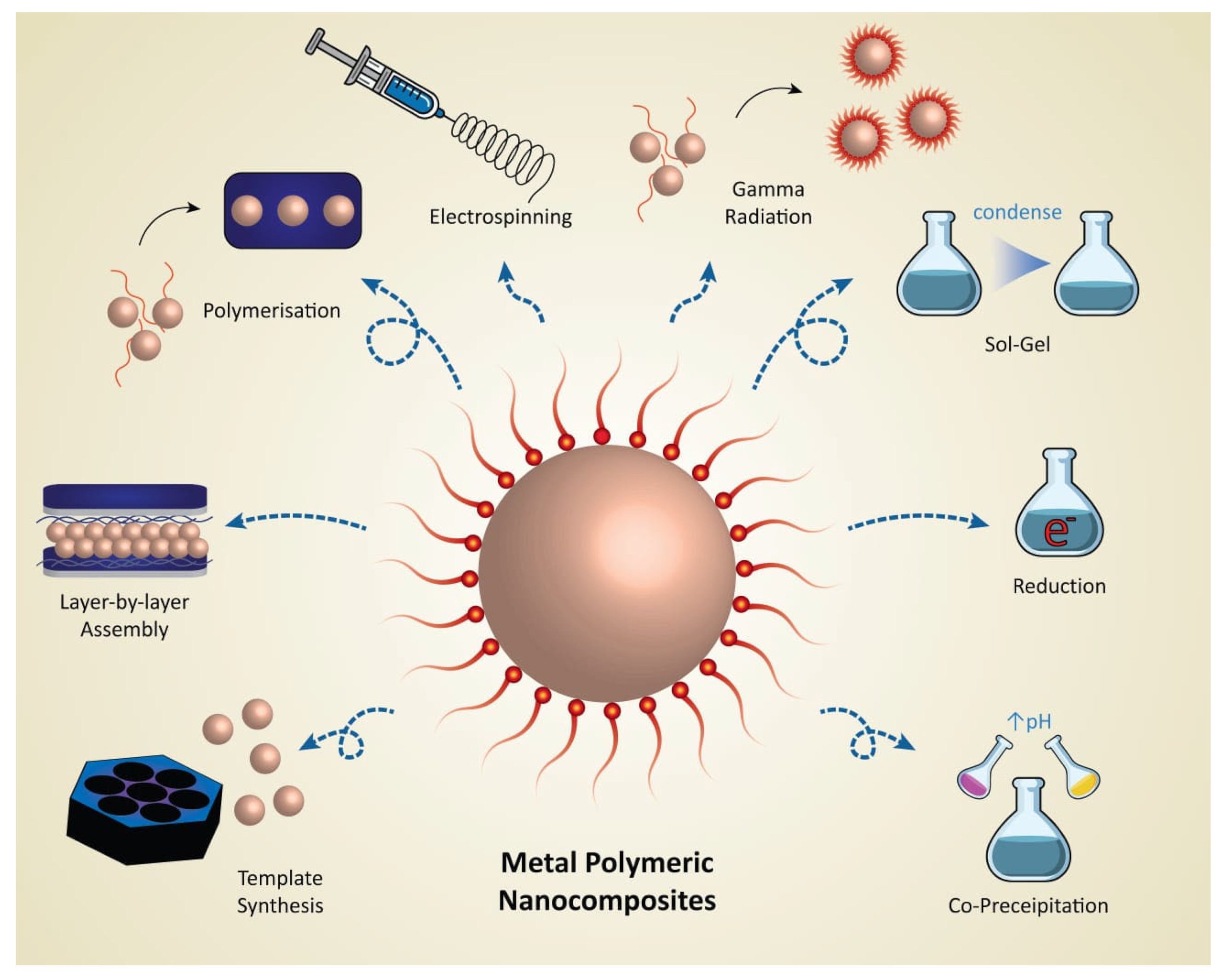
4. Synthesis of Metal–Polymeric Nanocomposites
4.1. In Situ Polymerization
4.2. Chemical Reduction
4.2.1. Engineering Polymers
4.2.2. Carbohydrates and Biopolymers
4.2.3. Dendrimers as Templates and Hosts for Metal Nanoparticles
4.3. Electrospinning
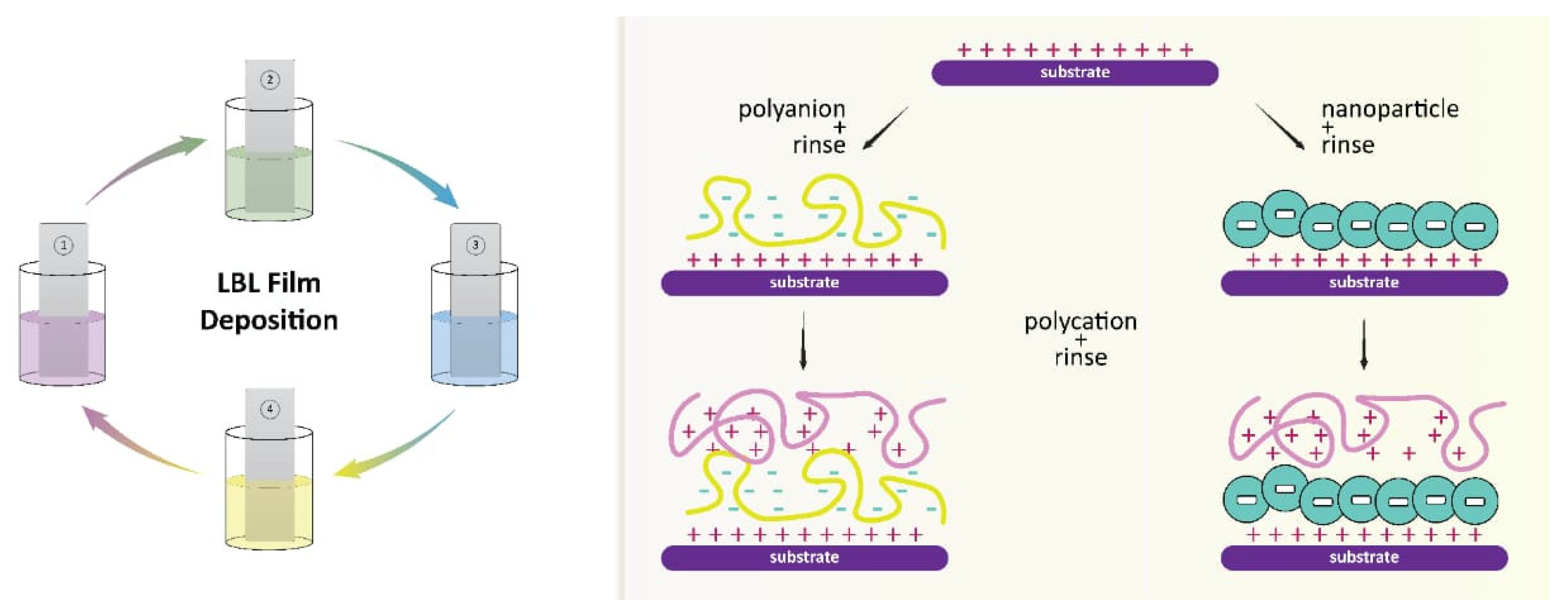
4.4. Layer-by-Layer Assembly
4.5. Template Synthesis
4.5.1. Hard Template Approach
4.5.2. Soft Template Approach
4.6. Coprecipitation
4.7. Sol–Gel Process
4.8. Gamma Radiation
5. Challenges of Synthetic Approaches
6. Characterization of Metal–Polymeric Nanocomposites
7. Applications and Limitations
8. Conclusions
Author Contributions
Funding
Institutional Review Board Statement
Data Availability Statement
Acknowledgments
Conflicts of Interest
References
- Kapoor, G.; Saigal, S.; Elongavan, A. Action and resistance mechanisms of antibiotics: A guide for clinicians. J. Anaesthesiol. Clin. Pharmacol. 2017, 33, 300–305. [Google Scholar] [CrossRef]
- Bao, Q.; Zhang, D.; Qi, P. Synthesis and characterization of silver nanoparticle and graphene oxide nanosheet composites as a bactericidal agent for water disinfection. J. Colloid Interface Sci. 2011, 360, 463–470. [Google Scholar] [CrossRef] [PubMed]
- Chandraker, K.; Nagwanshi, R.; Jadhav, S.K.; Ghosh, K.K.; Satnami, M.L. Antibacterial properties of amino acid functionalized silver nanoparticles decorated on graphene oxide sheets. Spectrochim. Acta Part A Mol. Biomol. Spectrosc. 2017, 181, 47–54. [Google Scholar] [CrossRef]
- Prasad, K.; Lekshmi, G.S.; Ostrikov, K.; Lussini, V.; Blinco, J.; Mohandas, M.; Vasilev, K.; Bottle, S. Synergic bactericidal effects of reduced graphene oxide and silver nanoparticles against Gram-positive and Gram-negative bacteria. Sci. Rep. 2017, 7, 1591. [Google Scholar] [CrossRef] [PubMed]
- Yadollahi, M.; Gholamali, I.; Namazi, H.; Aghazadeh, M. Synthesis and characterization of antibacterial carboxymethyl cellulose/ZnO nanocomposite hydrogels. Int. J. Biol. Macromol. 2015, 74, 136–141. [Google Scholar] [CrossRef]
- Alswat, A.A.; Ahmad, M.B.; Saleh, T.A. Preparation and Characterization of Zeolite\Zinc Oxide-Copper Oxide Nanocomposite: Antibacterial Activities. Colloid Interface Sci. Commun. 2017, 16, 19–24. [Google Scholar] [CrossRef]
- Ghosh, T.; Das, A.B.; Jena, B.; Pradhan, C. Antimicrobial effect of silver zinc oxide (Ag-ZnO) nanocomposite particles. Front. Life Sci. 2015, 8, 47–54. [Google Scholar] [CrossRef]
- Prema, D.; Prakash, J.; Vignesh, S.; Veluchamy, P.; Ramachandran, C.; Samal, D.B.; Oh, D.-H.; Sahabudeen, S.; Devanand Venkatasubbu, G. Mechanism of inhibition of graphene oxide/zinc oxide nanocomposite against wound infection causing pathogens. Appl. Nanosci. 2020, 10, 827–849. [Google Scholar] [CrossRef]
- Thambidurai, S.; Gowthaman, P.; Venkatachalam, M.; Suresh, S. Enhanced bactericidal performance of nickel oxide-zinc oxide nanocomposites synthesized by facile chemical co-precipitation method. J. Alloys Compd. 2020, 830, 154642. [Google Scholar] [CrossRef]
- Huang, W.C.; Tsai, P.J.; Chen, Y.C. Functional gold nanoparticles as photothermal agents for selective-killing of pathogenic bacteria. Nanomedicine 2007, 2, 777–787. [Google Scholar] [CrossRef]
- Perni, S.; Piccirillo, C.; Pratten, J.; Prokopovich, P.; Chrzanowski, W.; Parkin, I.P.; Wilson, M. The antimicrobial properties of light-activated polymers containing methylene blue and gold nanoparticles. Biomaterials 2009, 30, 89–93. [Google Scholar] [CrossRef] [PubMed]
- Nirmala, R.; Park, H.M.; Kalpana, D.; Kang, H.S.; Navamathavan, R.; Lee, Y.S.; Kim, H.Y. Bactericidal activity and in vitro cytotoxicity assessment of hydroxyapatite containing gold nanoparticles. J. Biomed. Nanotechnol. 2011, 7, 342–350. [Google Scholar] [CrossRef] [PubMed]
- Regiel-Futyra, A.; Kus-Liśkiewicz, M.; Sebastian, V.; Irusta, S.; Arruebo, M.; Stochel, G.; Kyzioł, A. Development of noncytotoxic chitosan-gold nanocomposites as efficient antibacterial materials. ACS Appl. Mater. Interfaces 2015, 7, 1087–1099. [Google Scholar] [CrossRef] [PubMed]
- Mendoza, G.; Regiel-Futyra, A.; Andreu, V.; Sebastián, V.; Kyzioł, A.; Stochel, G.; Arruebo, M. Bactericidal Effect of Gold–Chitosan Nanocomposites in Coculture Models of Pathogenic Bacteria and Human Macrophages. ACS Appl. Mater. Interfaces 2017, 9, 17693–17701. [Google Scholar] [CrossRef]
- Chan, J.F.; Lau, S.K.; To, K.K.; Cheng, V.C.; Woo, P.C.; Yuen, K.Y. Middle East respiratory syndrome coronavirus: Another zoonotic betacoronavirus causing SARS-like disease. Clin. Microbiol. Rev. 2015, 28, 465–522. [Google Scholar] [CrossRef]
- Ellis, B.R.; Barrett, A.D. The enigma of yellow fever in East Africa. Rev. Med. Virol. 2008, 18, 331–346. [Google Scholar] [CrossRef]
- Andersen, K.G.; Rambaut, A.; Lipkin, W.I.; Holmes, E.C.; Garry, R.F. The proximal origin of SARS-CoV-2. Nat. Med. 2020, 26, 450–452. [Google Scholar] [CrossRef]
- Jiang, S.; Shi, Z.; Shu, Y.; Song, J.; Gao, G.F.; Tan, W.; Guo, D. A distinct name is needed for the new coronavirus. Lancet 2020, 395, 949. [Google Scholar] [CrossRef]
- Moritz, M.; Geszke-Moritz, M. The newest achievements in synthesis, immobilization and practical applications of antibacterial nanoparticles. Chem. Eng. J. 2013, 228, 596–613. [Google Scholar] [CrossRef]
- Lara, H.H.; Ayala-Nuñez, N.V.; Ixtepan-Turrent, L.; Rodriguez-Padilla, C. Mode of antiviral action of silver nanoparticles against HIV-1. J. Nanobiotechnol. 2010, 8, 1. [Google Scholar] [CrossRef]
- Lara, H.H.; Garza-Treviño, E.N.; Ixtepan-Turrent, L.; Singh, D.K. Silver nanoparticles are broad-spectrum bactericidal and virucidal compounds. J. Nanobiotechnol. 2011, 9, 30. [Google Scholar] [CrossRef] [PubMed]
- Kumar, R.; Nayak, M.; Sahoo, G.C.; Pandey, K.; Sarkar, M.C.; Ansari, Y.; Das, V.N.; Topno, R.K.; Madhukar, M.; Das, P. Iron oxide nanoparticles based antiviral activity of H1N1 influenza A virus. J. Infect. Chemother. 2019, 25, 325–329. [Google Scholar] [CrossRef]
- Simoncic, B.; Tomsic, B. Structures of Novel Antimicrobial Agents for Textiles—A Review. Text. Res. J. 2010, 80, 1721–1737. [Google Scholar] [CrossRef]
- Ye, S.; Shao, K.; Li, Z.; Guo, N.; Zuo, Y.; Li, Q.; Lu, Z.; Chen, L.; He, Q.; Han, H. Antiviral Activity of Graphene Oxide: How Sharp Edged Structure and Charge Matter. ACS Appl. Mater. Interfaces 2015, 7, 21571–21579. [Google Scholar] [CrossRef]
- Lishchynskyi, O.; Shymborska, Y.; Stetsyshyn, Y.; Raczkowska, J.; Skirtach, A.G.; Peretiatko, T.; Budkowski, A. Passive antifouling and active self-disinfecting antiviral surfaces. Chem. Eng. J. 2022, 446, 137048. [Google Scholar] [CrossRef]
- Dallas, P.; Sharma, V.K.; Zboril, R. Silver polymeric nanocomposites as advanced antimicrobial agents: Classification, synthetic paths, applications, and perspectives. Adv. Colloid Interface Sci. 2011, 166, 119–135. [Google Scholar]
- Hoseini-Alfatemi, S.M.; Karimi, A.; Armin, S.; Fakharzadeh, S.; Fallah, F.; Kalanaky, S. Antibacterial and antibiofilm activity of nanochelating based silver nanoparticles against several nosocomial pathogens. Appl. Organomet. Chem. 2018, 32, e4327. [Google Scholar]
- Al-Ramamneh, E.A.M.; Ghrair, A.M.; Shakya, A.K. Efficacy of Sterculia diversifolia Leaf Extracts: Volatile Compounds, Antioxidant and Anti-Inflammatory Activity, and Green Synthesis of Potential Antibacterial Silver Nanoparticles. Plants 2022, 11, 2492. [Google Scholar] [CrossRef] [PubMed]
- Tang, S.; Zheng, J. Antibacterial activity of silver nanoparticles: Structural effects. Adv. Healthc. Mater. 2018, 7, 1701503. [Google Scholar] [CrossRef]
- Innes, M.E.; Umraw, N.; Fish, J.S.; Gomez, M.; Cartotto, R.C. The use of silver coated dressings on donor site wounds: A prospective, controlled matched pair study. Burns 2001, 27, 621–627. [Google Scholar] [CrossRef]
- Zheng, Z.; Liu, P.; Zhang, X.; Zou, X.; Mei, X.; Zhang, S.; Zhang, S. Strategies to improve bioactive and antibacterial properties of polyetheretherketone (PEEK) for use as orthopedic implants. Mater. Today Bio 2022, 16, 100402. [Google Scholar] [CrossRef] [PubMed]
- Thokala, N.; Kealey, C.; Kennedy, J.; Brady, D.B.; Farrell, J. Comparative activity of silver-based antimicrobial composites for urinary catheters. Int. J. Antimicrob. Agents 2018, 52, 166–171. [Google Scholar] [CrossRef]
- Priyadarshini, S.; Gopinath, V.; Meera Priyadharsshini, N.; MubarakAli, D.; Velusamy, P. Synthesis of anisotropic silver nanoparticles using novel strain, Bacillus flexus and its biomedical application. Colloids Surf. B Biointerfaces 2013, 102, 232–237. [Google Scholar] [CrossRef]
- Yamanaka, M.; Hara, K.; Kudo, J. Bactericidal actions of a silver ion solution on Escherichia coli, studied by energy-filtering transmission electron microscopy and proteomic analysis. Appl. Environ. Microbiol. 2005, 71, 7589–7593. [Google Scholar] [CrossRef]
- Jung, W.K.; Koo, H.C.; Kim, K.W.; Shin, S.; Kim, S.H.; Park, Y.H. Antibacterial activity and mechanism of action of the silver ion in Staphylococcus aureus and Escherichia coli. Appl. Environ. Microbiol. 2008, 74, 2171–2178. [Google Scholar] [CrossRef] [PubMed]
- Slavin, Y.N.; Asnis, J.; Häfeli, U.O.; Bach, H. Metal nanoparticles: Understanding the mechanisms behind antibacterial activity. J. Nanobiotechnol. 2017, 15, 65. [Google Scholar] [CrossRef] [PubMed]
- Maurya, P.; Singh, S.; Naik, R.R.; Shakya, A.K. Biohazards of Nanomaterials. In Integrative Nanomedicine for New Therapies; Krishnan, A., Chuturgoon, A., Eds.; Springer International Publishing: Cham, Switzerland, 2020; pp. 39–70. [Google Scholar]
- Dakal, T.C.; Kumar, A.; Majumdar, R.S.; Yadav, V. Mechanistic Basis of Antimicrobial Actions of Silver Nanoparticles. Front. Microbiol. 2016, 7, 1831. [Google Scholar] [CrossRef] [PubMed]
- Yang, W.; Shen, C.; Ji, Q.; An, H.; Wang, J.; Liu, Q.; Zhang, Z. Food storage material silver nanoparticles interfere with DNA replication fidelity and bind with DNA. Nanotechnology 2009, 20, 085102. [Google Scholar] [CrossRef]
- Zarnegar, Z.; Safari, J.; Zahraei, Z. Design, synthesis and antimicrobial evaluation of silver decorated magnetic polymeric nanocomposites. Nano-Struct. Nano-Objects 2019, 19, 100368. [Google Scholar] [CrossRef]
- Hasan, N.; Cao, J.; Lee, J.; Hlaing, S.P.; Oshi, M.A.; Naeem, M.; Ki, M.H.; Lee, B.L.; Jung, Y.; Yoo, J.W. Bacteria-Targeted Clindamycin Loaded Polymeric Nanoparticles: Effect of Surface Charge on Nanoparticle Adhesion to MRSA, Antibacterial Activity, and Wound Healing. Pharmaceutics 2019, 11, 236. [Google Scholar] [CrossRef]
- Oliani, W.L.; Parra, D.F.; Komatsu, L.G.H.; Lincopan, N.; Rangari, V.K.; Lugao, A.B. Fabrication of polypropylene/silver nanocomposites for biocidal applications. Mater. Sci. Eng. C 2017, 75, 845–853. [Google Scholar] [CrossRef] [PubMed]
- Rehan, M.; Nada, A.A.; Khattab, T.A.; Abdelwahed, N.A.; Abou El-Kheir, A.A. Development of multifunctional polyacrylonitrile/silver nanocomposite films: Antimicrobial activity, catalytic activity, electrical conductivity, UV protection and SERS-active sensor. J. Mater. Res. Technol. 2020, 9, 9380–9394. [Google Scholar]
- Narayanan, K.B.; Han, S.S. Dual-crosslinked poly (vinyl alcohol)/sodium alginate/silver nanocomposite beads–A promising antimicrobial material. Food Chem. 2017, 234, 103–110. [Google Scholar] [CrossRef]
- Spagnol, C.; Fragal, E.H.; Pereira, A.G.; Nakamura, C.V.; Muniz, E.C.; Follmann, H.D.; Silva, R.; Rubira, A.F. Cellulose nanowhiskers decorated with silver nanoparticles as an additive to antibacterial polymers membranes fabricated by electrospinning. J. Colloid Interface Sci. 2018, 531, 705–715. [Google Scholar] [CrossRef]
- Xie, Y.; Liao, X.; Zhang, J.; Yang, F.; Fan, Z. Novel chitosan hydrogels reinforced by silver nanoparticles with ultrahigh mechanical and high antibacterial properties for accelerating wound healing. Int. J. Biol. Macromol. 2018, 119, 402–412. [Google Scholar] [CrossRef]
- Hajji, S.; Khedir, S.B.; Hamza-Mnif, I.; Hamdi, M.; Jedidi, I.; Kallel, R.; Boufi, S.; Nasri, M. Biomedical potential of chitosan-silver nanoparticles with special reference to antioxidant, antibacterial, hemolytic and in vivo cutaneous wound healing effects. Biochim. Et Biophys. Acta (BBA)-Gen. Subj. 2019, 1863, 241–254. [Google Scholar] [CrossRef]
- Ye, H.; Cheng, J.; Yu, K. In situ reduction of silver nanoparticles by gelatin to obtain porous silver nanoparticle/chitosan composites with enhanced antimicrobial and wound-healing activity. Int. J. Biol. Macromol. 2019, 121, 633–642. [Google Scholar] [CrossRef]
- Hernández-Rangel, A.; Silva-Bermudez, P.; Espana-Sanchez, B.; Luna-Hernández, E.; Almaguer-Flores, A.; Ibarra, C.; Garcia-Perez, V.; Velasquillo, C.; Luna-Barcenas, G. Fabrication and in vitro behavior of dual-function chitosan/silver nanocomposites for potential wound dressing applications. Mater. Sci. Eng. C 2019, 94, 750–765. [Google Scholar]
- Suteewong, T.; Wongpreecha, J.; Polpanich, D.; Jangpatarapongsa, K.; Kaewsaneha, C.; Tangboriboonrat, P. PMMA particles coated with chitosan-silver nanoparticles as a dual antibacterial modifier for natural rubber latex films. Colloids Surf. B Biointerfaces 2019, 174, 544–552. [Google Scholar] [CrossRef]
- Lee, D.; Lee, S.J.; Moon, J.-H.; Kim, J.H.; Heo, D.N.; Bang, J.B.; Lim, H.-N.; Kwon, I.K. Preparation of antibacterial chitosan membranes containing silver nanoparticles for dental barrier membrane applications. J. Ind. Eng. Chem. 2018, 66, 196–202. [Google Scholar] [CrossRef]
- Bahrami, A.; Rezaei Mokarram, R.; Sowti Khiabani, M.; Ghanbarzadeh, B.; Salehi, R. Physico-mechanical and antimicrobial properties of tragacanth/hydroxypropyl methylcellulose/beeswax edible films reinforced with silver nanoparticles. Int. J. Biol. Macromol. 2019, 129, 1103–1112. [Google Scholar] [CrossRef]
- Haider, M.K.; Ullah, A. Fabricating Antibacterial and Antioxidant Electrospun Hydrophilic Polyacrylonitrile Nanofibers Loaded with AgNPs by Lignin-Induced In-Situ Method. Polymers 2021, 13, 748. [Google Scholar] [CrossRef]
- Hsueh, Y.H.; Hsieh, C.T.; Chiu, S.T.; Tsai, P.H.; Liu, C.Y.; Ke, W.J. Antibacterial Property of Composites of Reduced Graphene Oxide with Nano-Silver and Zinc Oxide Nanoparticles Synthesized Using a Microwave-Assisted Approach. Int. J. Mol. Sci. 2019, 20, 5394. [Google Scholar] [CrossRef]
- Liu, C.; Ling, J.; Yang, L.Y.; Ouyang, X.K.; Wang, N. Chitosan-based carbon nitride-polydopamine-silver composite dressing with antibacterial properties for wound healing. Carbohydr. Polym. 2023, 303, 120436. [Google Scholar] [CrossRef]
- Massey, S.; Iqbal, F.; Rehman, A.U.; Iqbal, M.S. Preparation, characterization and biological evaluation of silver nanoparticles and drug loaded composites for wound dressings formed from Lallemantia royleana seeds’ mucilage. J. Biomater. Sci. Polym. Ed. 2022, 33, 481–498. [Google Scholar] [CrossRef] [PubMed]
- Rather, A.H.; Khan, R.S.; Wani, T.U.; Rafiq, M.; Jadhav, A.H.; Srinivasappa, P.M.; Abdal-Hay, A.; Sultan, P.; Rather, S.U.; Macossay, J.; et al. Polyurethane and cellulose acetate micro-nanofibers containing rosemary essential oil, and decorated with silver nanoparticles for wound healing application. Int. J. Biol. Macromol. 2023, 226, 690–705. [Google Scholar] [CrossRef] [PubMed]
- Khan, M.U.A.; Abd Razak, S.I.; Mehboob, H.; Abdul Kadir, M.R.; Anand, T.J.S.; Inam, F.; Shah, S.A.; Abdel-Haliem, M.E.F.; Amin, R. Synthesis and Characterization of Silver-Coated Polymeric Scaffolds for Bone Tissue Engineering: Antibacterial and In Vitro Evaluation of Cytotoxicity and Biocompatibility. ACS Omega 2021, 6, 4335–4346. [Google Scholar] [CrossRef] [PubMed]
- Zhou, M.; Lin, F.; Li, W.; Shi, L.; Li, Y.; Shan, G. Development of nanosilver doped carboxymethyl chitosan-polyamideamine alginate composite dressing for wound treatment. Int. J. Biol. Macromol. 2021, 166, 1335–1351. [Google Scholar] [CrossRef]
- Stetsyshyn, Y.; Awsiuk, K.; Kusnezh, V.; Raczkowska, J.; Jany, B.R.; Kostruba, A.; Harhay, K.; Ohar, H.; Lishchynskyi, O.; Shymborska, Y.; et al. Shape-Controlled synthesis of silver nanoparticles in temperature-responsive grafted polymer brushes for optical applications. Appl. Surf. Sci. 2019, 463, 1124–1133. [Google Scholar] [CrossRef]
- Wichai, S.; Chuysinuan, P.; Chaiarwut, S.; Ekabutr, P.; Supaphol, P. Development of bacterial cellulose/alginate/chitosan composites incorporating copper (II) sulfate as an antibacterial wound dressing. J. Drug Deliv. Sci. Technol. 2019, 51, 662–671. [Google Scholar] [CrossRef]
- Nisar, P.; Ali, N.; Rahman, L.; Ali, M.; Shinwari, Z.K. Antimicrobial activities of biologically synthesized metal nanoparticles: An insight into the mechanism of action. JBIC J. Biol. Inorg. Chem. 2019, 24, 929–941. [Google Scholar] [CrossRef]
- Halbus, A.F.; Horozov, T.S.; Paunov, V.N. Strongly enhanced antibacterial action of copper oxide nanoparticles with boronic acid surface functionality. ACS Appl. Mater. Interfaces 2019, 11, 12232–12243. [Google Scholar] [CrossRef]
- Bezza, F.A.; Tichapondwa, S.M.; Chirwa, E.M. Fabrication of monodispersed copper oxide nanoparticles with potential application as antimicrobial agents. Sci. Rep. 2020, 10, 16680. [Google Scholar] [CrossRef]
- Palza, H. Antimicrobial polymers with metal nanoparticles. Int. J. Mol. Sci. 2015, 16, 2099–2116. [Google Scholar] [PubMed]
- Arjunan, N.; Singaravelu, C.M.; Kulanthaivel, J.; Kandasamy, J. A potential photocatalytic, antimicrobial and anticancer activity of chitosan-copper nanocomposite. Int. J. Biol. Macromol. 2017, 104, 1774–1782. [Google Scholar] [CrossRef] [PubMed]
- Jayaramudu, T.; Varaprasad, K.; Reddy, K.K.; Pyarasani, R.D.; Akbari-Fakhrabadi, A.; Amalraj, J. Chitosan-pluronic based Cu nanocomposite hydrogels for prototype antimicrobial applications. Int. J. Biol. Macromol. 2020, 143, 825–832. [Google Scholar] [CrossRef] [PubMed]
- Hasanin, M.; Al Abboud, M.A.; Alawlaqi, M.M.; Abdelghany, T.M.; Hashem, A.H. Ecofriendly synthesis of biosynthesized copper nanoparticles with starch-based nanocomposite: Antimicrobial, antioxidant, and anticancer activities. Biol. Trace Elem. Res. 2021, 200, 2099–2112. [Google Scholar] [CrossRef]
- Phan, D.-N.; Dorjjugder, N.; Saito, Y.; Khan, M.Q.; Ullah, A.; Bie, X.; Taguchi, G.; Kim, I.-S. Antibacterial mechanisms of various copper species incorporated in polymeric nanofibers against bacteria. Mater. Today Commun. 2020, 25, 101377. [Google Scholar]
- Castro Mayorga, J.L.; Fabra Rovira, M.J.; Cabedo Mas, L.; Sánchez Moragas, G.; Lagarón Cabello, J.M. Antimicrobial nanocomposites and electrospun coatings based on poly (3-hydroxybutyrate-co-3-hydroxyvalerate) and copper oxide nanoparticles for active packaging and coating applications. J. Appl. Polym. Sci. 2018, 135, 45673. [Google Scholar] [CrossRef]
- Ashjari, H.R.; Dorraji, M.S.S.; Fakhrzadeh, V.; Eslami, H.; Rasoulifard, M.H.; Rastgouy-Houjaghan, M.; Gholizadeh, P.; Kafil, H.S. Starch-based polyurethane/CuO nanocomposite foam: Antibacterial effects for infection control. Int. J. Biol. Macromol. 2018, 111, 1076–1082. [Google Scholar] [CrossRef]
- El Nahrawy, A.M.; Hammad, A.B.A.; Youssef, A.M.; Mansour, A.M.; Othman, A.M. Thermal, dielectric and antimicrobial properties of polystyrene-assisted/ITO:Cu nanocomposites. Appl. Phys. A 2019, 125, 46. [Google Scholar] [CrossRef]
- Araújo, I.M.S.; Silva, R.R.; Pacheco, G.; Lustri, W.R.; Tercjak, A.; Gutierrez, J.; Júnior, J.R.S.; Azevedo, F.H.C.; Figuêredo, G.S.; Vega, M.L.; et al. Hydrothermal synthesis of bacterial cellulose–copper oxide nanocomposites and evaluation of their antimicrobial activity. Carbohydr. Polym. 2018, 179, 341–349. [Google Scholar] [CrossRef]
- Marković, D.; Deeks, C.; Nunney, T.; Radovanović, Ž.; Radoičić, M.; Šaponjić, Z.; Radetić, M. Antibacterial activity of Cu-based nanoparticles synthesized on the cotton fabrics modified with polycarboxylic acids. Carbohydr. Polym. 2018, 200, 173–182. [Google Scholar] [CrossRef]
- Nouri, A.; Yaraki, M.T.; Ghorbanpour, M.; Agarwal, S.; Gupta, V.K. Enhanced Antibacterial effect of chitosan film using Montmorillonite/CuO nanocomposite. Int. J. Biol. Macromol. 2018, 109, 1219–1231. [Google Scholar] [CrossRef] [PubMed]
- Chen, M.; Li, Z.; Chen, L. Highly antibacterial rGO/Cu2O nanocomposite from a biomass precursor: Synthesis, performance, and mechanism. Nano Mater. Sci. 2020, 2, 172–179. [Google Scholar] [CrossRef]
- Logpriya, S.; Bhuvaneshwari, V.; Vaidehi, D.; SenthilKumar, R.P.; Nithya Malar, R.S.; Pavithra Sheetal, B.; Amsaveni, R.; Kalaiselvi, M. Preparation and characterization of ascorbic acid-mediated chitosan–copper oxide nanocomposite for anti-microbial, sporicidal and biofilm-inhibitory activity. J. Nanostruct. Chem. 2018, 8, 301–309. [Google Scholar] [CrossRef]
- Sportelli, M.C.; Izzi, M.; Volpe, A.; Lacivita, V.; Clemente, M.; Di Franco, C.; Conte, A.; Del Nobile, M.A.; Ancona, A.; Cioffi, N. A new nanocomposite based on LASiS-generated CuNPs as a preservation system for fruit salads. Food Packag. Shelf Life 2019, 22, 100422. [Google Scholar] [CrossRef]
- Solairaj, D.; Rameshthangam, P.; Arunachalam, G. Anticancer activity of silver and copper embedded chitin nanocomposites against human breast cancer (MCF-7) cells. Int. J. Biol. Macromol. 2017, 105, 608–619. [Google Scholar] [CrossRef]
- Tabesh, E.; Salimijazi, H.R.; Kharaziha, M.; Mahmoudi, M.; Hejazi, M. Development of an in-situ chitosan-copper nanoparticle coating by electrophoretic deposition. Surf. Coat. Technol. 2019, 364, 239–247. [Google Scholar] [CrossRef]
- Muthulakshmi, L.; Rajini, N.; Nellaiah, H.; Kathiresan, T.; Jawaid, M.; Rajulu, A.V. Preparation and properties of cellulose nanocomposite films with in situ generated copper nanoparticles using Terminalia catappa leaf extract. Int. J. Biol. Macromol. 2017, 95, 1064–1071. [Google Scholar] [CrossRef]
- Abdollahi, Z.; Zare, E.N.; Salimi, F.; Goudarzi, I.; Tay, F.R.; Makvandi, P. Bioactive Carboxymethyl Starch-Based Hydrogels Decorated with CuO Nanoparticles: Antioxidant and Antimicrobial Properties and Accelerated Wound Healing In Vivo. Int. J. Mol. Sci. 2021, 22, 2531. [Google Scholar] [CrossRef] [PubMed]
- Abu-Elala, N.M.; AbuBakr, H.O.; Khattab, M.S.; Mohamed, S.H.; El-Hady, M.A.; Ghandour, R.A.; Morsi, R.E. Aquatic environmental risk assessment of chitosan/silver, copper and carbon nanotube nanocomposites as antimicrobial agents. Int. J. Biol. Macromol. 2018, 113, 1105–1115. [Google Scholar] [CrossRef] [PubMed]
- Ahmed, S.B.; Mohamed, H.I.; Al-Subaie, A.M.; Al-Ohali, A.I.; Mahmoud, N.M.R. Investigation of the antimicrobial activity and hematological pattern of nano-chitosan and its nano-copper composite. Sci. Rep. 2021, 11, 9540. [Google Scholar] [CrossRef]
- Al-Enizi, A.M.; Ahamad, T.; Al-Hajji, A.B.; Ahmed, J.; Chaudhary, A.A.; Alshehri, S.M. Cellulose gum and copper nanoparticles based hydrogel as antimicrobial agents against urinary tract infection (UTI) pathogens. Int. J. Biol. Macromol. 2018, 109, 803–809. [Google Scholar] [CrossRef] [PubMed]
- Al-Saeedi, S.I.; Al-Kadhi, N.S.; Al-Senani, G.M.; Almaghrabi, O.A.; Nafady, A. Antibacterial potency, cell viability and morphological implications of copper oxide nanoparticles encapsulated into cellulose acetate nanofibrous scaffolds. Int. J. Biol. Macromol. 2021, 182, 464–471. [Google Scholar] [CrossRef]
- Bagchi, B.; Salvadores Fernandez, C.; Bhatti, M.; Ciric, L.; Lovat, L.; Tiwari, M.K. Copper nanowire embedded hypromellose: An antibacterial nanocomposite film. J. Colloid Interface Sci. 2022, 608, 30–39. [Google Scholar] [CrossRef]
- des Ligneris, E.; Dumée, L.F.; Al-Attabi, R.; Castanet, E.; Schütz, J.; Kong, L. Mixed Matrix Poly(Vinyl Alcohol)-Copper Nanofibrous Anti-Microbial Air-Microfilters. Membranes 2019, 9, 87. [Google Scholar] [CrossRef]
- He, W.; Huang, X.; Zheng, Y.; Sun, Y.; Xie, Y.; Wang, Y.; Yue, L. In situ synthesis of bacterial cellulose/copper nanoparticles composite membranes with long-term antibacterial property. J. Biomater. Sci. Polym. Ed. 2018, 29, 2137–2153. [Google Scholar] [CrossRef]
- Jayaramudu, T.; Varaprasad, K.; Pyarasani, R.D.; Reddy, K.K.; Akbari-Fakhrabadi, A.; Carrasco-Sánchez, V.; Amalraj, J. Hydroxypropyl methylcellulose-copper nanoparticle and its nanocomposite hydrogel films for antibacterial application. Carbohydr. Polym. 2021, 254, 117302. [Google Scholar] [CrossRef]
- Jayaramudu, T.; Varaprasad, K.; Pyarasani, R.D.; Reddy, K.K.; Kumar, K.D.; Akbari-Fakhrabadi, A.; Mangalaraja, R.V.; Amalraj, J. Chitosan capped copper oxide/copper nanoparticles encapsulated microbial resistant nanocomposite films. Int. J. Biol. Macromol. 2019, 128, 499–508. [Google Scholar] [CrossRef]
- Kruk, T.; Gołda-Cępa, M.; Szczepanowicz, K.; Szyk-Warszyńska, L.; Brzychczy-Włoch, M.; Kotarba, A.; Warszyński, P. Nanocomposite multifunctional polyelectrolyte thin films with copper nanoparticles as the antimicrobial coatings. Colloids Surf. B Biointerfaces 2019, 181, 112–118. [Google Scholar] [CrossRef]
- Li, M.; Liu, X.; Tan, L.; Cui, Z.; Yang, X.; Li, Z.; Zheng, Y.; Yeung, K.W.K.; Chu, P.K.; Wu, S. Noninvasive rapid bacteria-killing and acceleration of wound healing through photothermal/photodynamic/copper ion synergistic action of a hybrid hydrogel. Biomater. Sci. 2018, 6, 2110–2121. [Google Scholar] [CrossRef] [PubMed]
- Mehta, R.; Brahmbhatt, H.; Bhojani, G.; Mukherjee, M.; Bhattacharya, A. Poly(piperizinamide) with copper ion composite membranes: Application for mitigation of Hexaconazole from water and combat microbial contamination. J. Hazard. Mater. 2019, 376, 102–111. [Google Scholar] [CrossRef]
- Mugesh, S.; Arun, R.; Arunkumar, K.; Murugan, M. Synthesis of Biogenic Copper Nanoparticles Embedded in Graphene Oxide-Chitosan Composite and Its Anti-Bacterial and Cytotoxic Activities. J. Nanosci. Nanotechnol. 2019, 19, 2625–2632. [Google Scholar] [CrossRef]
- Prokhorov, E.; España-Sánchez, B.L.; Luna-Bárcenas, G.; Padilla-Vaca, F.; Cruz-Soto, M.E.; Vázquez-Lepe, M.O.; Kovalenko, Y.; Elizalde-Peña, E.A. Chitosan/copper nanocomposites: Correlation between electrical and antibacterial properties. Colloids Surf. B Biointerfaces 2019, 180, 186–192. [Google Scholar] [CrossRef] [PubMed]
- Pulit-Prociak, J.; Staroń, A.; Staroń, P.; Chmielowiec-Korzeniowska, A.; Drabik, A.; Tymczyna, L.; Banach, M. Preparation and of PVA-based compositions with embedded silver, copper and zinc oxide nanoparticles and assessment of their antibacterial properties. J. Nanobiotechnol. 2020, 18, 148. [Google Scholar] [CrossRef]
- Sathiyavimal, S.; Vasantharaj, S.; Kaliannan, T.; Pugazhendhi, A. Eco-biocompatibility of chitosan coated biosynthesized copper oxide nanocomposite for enhanced industrial (Azo) dye removal from aqueous solution and antibacterial properties. Carbohydr. Polym. 2020, 241, 116243. [Google Scholar] [CrossRef] [PubMed]
- Sathiyavimal, S.; Vasantharaj, S.; Kaliannan, T.; Garalleh, H.A.; Garaleh, M.; Brindhadevi, K.; Chi, N.T.L.; Sharma, A.; Pugazhendhi, A. Bio-functionalized copper oxide/chitosan nanocomposite using Sida cordifolia and their efficient properties of antibacterial, anticancer activity against on breast and lung cancer cell lines. Environ. Res. 2023, 218, 114986. [Google Scholar] [CrossRef]
- Sun, X.; Dong, M.; Guo, Z.; Zhang, H.; Wang, J.; Jia, P.; Bu, T.; Liu, Y.; Li, L.; Wang, L. Multifunctional chitosan-copper-gallic acid based antibacterial nanocomposite wound dressing. Int. J. Biol. Macromol. 2021, 167, 10–22. [Google Scholar] [CrossRef]
- Wang, W.B.; Clapper, J.C. Antibacterial Activity of Electrospun Polyacrylonitrile Copper Nanoparticle Nanofibers on Antibiotic Resistant Pathogens and Methicillin Resistant. Nanomaterials 2022, 12, 2139. [Google Scholar] [CrossRef]
- Yang, Y.; Dong, Z.; Li, M.; Liu, L.; Luo, H.; Wang, P.; Zhang, D.; Yang, X.; Zhou, K.; Lei, S. Graphene Oxide/Copper Nanoderivatives-Modified Chitosan/Hyaluronic Acid Dressings for Facilitating Wound Healing in Infected Full-Thickness Skin Defects. Int. J. Nanomed. 2020, 15, 8231–8247. [Google Scholar] [CrossRef] [PubMed]
- Kumar, V.; Menon, S.; Agarwal, H.; Gopalakrishnan, D. Characterization and optimization of bacterium isolated from soil samples for the production of siderophores. Resour. Effic. Technol. 2017, 3, 434–439. [Google Scholar] [CrossRef]
- Derewacz, D.K.; Goodwin, C.R.; McNees, C.R.; McLean, J.A.; Bachmann, B.O. Antimicrobial drug resistance affects broad changes in metabolomic phenotype in addition to secondary metabolism. Proc. Natl. Acad. Sci. USA 2013, 110, 2336–2341. [Google Scholar] [CrossRef]
- Landini, P.; Antoniani, D.; Burgess, J.G.; Nijland, R. Molecular mechanisms of compounds affecting bacterial biofilm formation and dispersal. Appl. Microbiol. Biotechnol. 2010, 86, 813–823. [Google Scholar] [CrossRef]
- Rai, M.; Yadav, A.; Gade, A. Silver nanoparticles as a new generation of antimicrobials. Biotechnol. Adv. 2009, 27, 76–83. [Google Scholar] [CrossRef]
- Tiwari, V.; Mishra, N.; Gadani, K.; Solanki, P.S.; Shah, N.A.; Tiwari, M. Mechanism of Anti-bacterial Activity of Zinc Oxide Nanoparticle Against Carbapenem-Resistant Acinetobacter baumannii. Front. Microbiol. 2018, 9, 1218. [Google Scholar] [CrossRef]
- Singh, T.A.; Sharma, A.; Tejwan, N.; Ghosh, N.; Das, J.; Sil, P.C. A state of the art review on the synthesis, antibacterial, antioxidant, antidiabetic and tissue regeneration activities of zinc oxide nanoparticles. Adv. Colloid Interface Sci. 2021, 295, 102495. [Google Scholar] [CrossRef]
- Rajabi, H.R.; Khani, O.; Shamsipur, M.; Vatanpour, V. High-performance pure and Fe3+-ion doped ZnS quantum dots as green nanophotocatalysts for the removal of malachite green under UV-light irradiation. J. Hazard. Mater. 2013, 250–251, 370–378. [Google Scholar] [CrossRef] [PubMed]
- Afrasiabi, S.; Bahador, A.; Partoazar, A. Combinatorial therapy of chitosan hydrogel-based zinc oxide nanocomposite attenuates the virulence of Streptococcus mutans. BMC Microbiol. 2021, 21, 62. [Google Scholar] [CrossRef]
- Lefatshe, K.; Muiva, C.M.; Kebaabetswe, L.P. Extraction of nanocellulose and in-situ casting of ZnO/cellulose nanocomposite with enhanced photocatalytic and antibacterial activity. Carbohydr. Polym. 2017, 164, 301–308. [Google Scholar] [CrossRef] [PubMed]
- Magesh, G.; Bhoopathi, G.; Nithya, N.; Arun, A.P.; Ranjith Kumar, E. Structural, morphological, optical and biological properties of pure ZnO and agar/zinc oxide nanocomposites. Int. J. Biol. Macromol. 2018, 117, 959–966. [Google Scholar] [CrossRef] [PubMed]
- Shahvalizadeh, R.; Ahmadi, R.; Davandeh, I.; Pezeshki, A.; Seyed Moslemi, S.A.; Karimi, S.; Rahimi, M.; Hamishehkar, H.; Mohammadi, M. Antimicrobial bio-nanocomposite films based on gelatin, tragacanth, and zinc oxide nanoparticles—Microstructural, mechanical, thermo-physical, and barrier properties. Food Chem. 2021, 354, 129492. [Google Scholar] [CrossRef]
- Preethi, S.; Abarna, K.; Nithyasri, M.; Kishore, P.; Deepika, K.; Ranjithkumar, R.; Bhuvaneshwari, V.; Bharathi, D. Synthesis and characterization of chitosan/zinc oxide nanocomposite for antibacterial activity onto cotton fabrics and dye degradation applications. Int. J. Biol. Macromol. 2020, 164, 2779–2787. [Google Scholar] [CrossRef] [PubMed]
- Bharathi, D.; Ranjithkumar, R.; Chandarshekar, B.; Bhuvaneshwari, V. Preparation of chitosan coated zinc oxide nanocomposite for enhanced antibacterial and photocatalytic activity: As a bionanocomposite. Int. J. Biol. Macromol. 2019, 129, 989–996. [Google Scholar] [CrossRef]
- Rojas, K.; Canales, D.; Amigo, N.; Montoille, L.; Cament, A.; Rivas, L.M.; Gil-Castell, O.; Reyes, P.; Ulloa, M.T.; Ribes-Greus, A.; et al. Effective antimicrobial materials based on low-density polyethylene (LDPE) with zinc oxide (ZnO) nanoparticles. Compos. Part B Eng. 2019, 172, 173–178. [Google Scholar] [CrossRef]
- Madhan, G.; Begam, A.A.; Varsha, L.V.; Ranjithkumar, R.; Bharathi, D. Facile synthesis and characterization of chitosan/zinc oxide nanocomposite for enhanced antibacterial and photocatalytic activity. Int. J. Biol. Macromol. 2021, 190, 259–269. [Google Scholar] [CrossRef] [PubMed]
- Motshekga, S.C.; Sinha Ray, S.; Maity, A. Synthesis and characterization of alginate beads encapsulated zinc oxide nanoparticles for bacteria disinfection in water. J. Colloid Interface Sci. 2018, 512, 686–692. [Google Scholar] [CrossRef] [PubMed]
- Jayakumar, A.; Radoor, S.; Nair, I.C.; Siengchin, S.; Parameswaranpillai, J.; Radhakrishnan, E.K. Lipopeptide and zinc oxide nanoparticles blended polyvinyl alcohol-based nanocomposite films as antimicrobial coating for biomedical applications. Process Biochem. 2021, 102, 220–228. [Google Scholar] [CrossRef]
- Mohsen, R.M.; Morsi, S.M.M.; Selim, M.M.; Ghoneim, A.M.; El-Sherif, H.M. Electrical, thermal, morphological, and antibacterial studies of synthesized polyaniline/zinc oxide nanocomposites. Polym. Bull. 2019, 76, 1–21. [Google Scholar] [CrossRef]
- Castro-Mayorga, J.L.; Fabra, M.J.; Pourrahimi, A.M.; Olsson, R.T.; Lagaron, J.M. The impact of zinc oxide particle morphology as an antimicrobial and when incorporated in poly(3-hydroxybutyrate-co-3-hydroxyvalerate) films for food packaging and food contact surfaces applications. Food Bioprod. Process. 2017, 101, 32–44. [Google Scholar] [CrossRef]
- Wen, Y.-H.; Tsou, C.-H.; de Guzman, M.R.; Huang, D.; Yu, Y.-Q.; Gao, C.; Zhang, X.-M.; Du, J.; Zheng, Y.-T.; Zhu, H.; et al. Antibacterial nanocomposite films of poly(vinyl alcohol) modified with zinc oxide-doped multiwalled carbon nanotubes as food packaging. Polym. Bull. 2022, 79, 3847–3866. [Google Scholar] [CrossRef]
- Cen, C.; Wang, F.; Wang, Y.; Li, H.; Fu, L.; Li, Y.; Chen, J.; Wang, Y. Design and characterization of an antibacterial film composited by hydroxyethyl cellulose (HEC), carboxymethyl chitosan (CMCS), and nano ZnO for food packaging. Int. J. Biol. Macromol. 2023, 231, 123203. [Google Scholar] [CrossRef] [PubMed]
- Farhangian, Z.; Alaghehmand, H.; Tashakkorian, H.; Mokhtarpour, F.; Davoodabadi, A. Antimicrobial effect of different physical and chemical compounds of zinc oxide and graphene oxide added to composite resins. Dent. Res. J. 2022, 19, 81. [Google Scholar]
- Sarojini, S.; Indumathi, M.P.; Rajarajeswari, G.R. Mahua oil-based polyurethane/chitosan/nano ZnO composite films for biodegradable food packaging applications. Int. J. Biol. Macromol. 2019, 124, 163–174. [Google Scholar] [CrossRef]
- Khorasani, M.T.; Joorabloo, A.; Adeli, H.; Mansoori-Moghadam, Z.; Moghaddam, A. Design and optimization of process parameters of polyvinyl (alcohol)/chitosan/nano zinc oxide hydrogels as wound healing materials. Carbohydr. Polym. 2019, 207, 542–554. [Google Scholar] [CrossRef]
- Lubambo, A.F.; Mattoso, N. In Situ Synthesis of AZO-Np in Guar Gum/PVOH Composite Fiber Mats for Potential Bactericidal Release. Polymers 2022, 14, 4983. [Google Scholar] [CrossRef]
- Nozari, M.; Gholizadeh, M.; Zahiri Oghani, F.; Tahvildari, K. Studies on novel chitosan/alginate and chitosan/bentonite flexible films incorporated with ZnO nano particles for accelerating dermal burn healing: In vivo and in vitro evaluation. Int. J. Biol. Macromol. 2021, 184, 235–249. [Google Scholar] [CrossRef]
- Shahriari-Khalaji, M.; Hu, G.; Chen, L.; Cao, Z.; Andreeva, T.; Xiong, X.; Krastev, R.; Hong, F.F. Functionalization of Aminoalkylsilane-Grafted Bacterial Nanocellulose with ZnO-NPs-Doped Pullulan Electrospun Nanofibers for Multifunctional Wound Dressing. ACS Biomater. Sci. Eng. 2021, 7, 3933–3946. [Google Scholar] [CrossRef] [PubMed]
- Wang, H.; Xue, T.; Wang, S.; Jia, X.; Cao, S.; Niu, B.; Guo, R.; Yan, H. Preparation, characterization and food packaging application of nano ZnO@Xylan/quaternized xylan/polyvinyl alcohol composite films. Int. J. Biol. Macromol. 2022, 215, 635–645. [Google Scholar] [CrossRef]
- von Maltzahn, G.; Park, J.H.; Agrawal, A.; Bandaru, N.K.; Das, S.K.; Sailor, M.J.; Bhatia, S.N. Computationally guided photothermal tumor therapy using long-circulating gold nanorod antennas. Cancer Res. 2009, 69, 3892–3900. [Google Scholar] [CrossRef]
- Gupta, A.; Pandey, S.; Yadav, J.S. A Review on Recent Trends in Green Synthesis of Gold Nanoparticles for Tuberculosis. Adv. Pharm. Bull. 2021, 11, 10–27. [Google Scholar] [CrossRef]
- Krawinkel, J.; Richter, U.; Torres-Mapa, M.L.; Westermann, M.; Gamrad, L.; Rehbock, C.; Barcikowski, S.; Heisterkamp, A. Optical and electron microscopy study of laser-based intracellular molecule delivery using peptide-conjugated photodispersible gold nanoparticle agglomerates. J. Nanobiotechnol. 2016, 14, 2. [Google Scholar] [CrossRef]
- Lin, C.A.; Yang, T.Y.; Lee, C.H.; Huang, S.H.; Sperling, R.A.; Zanella, M.; Li, J.K.; Shen, J.L.; Wang, H.H.; Yeh, H.I.; et al. Synthesis, characterization, and bioconjugation of fluorescent gold nanoclusters toward biological labeling applications. ACS Nano 2009, 3, 395–401. [Google Scholar] [CrossRef] [PubMed]
- Wang, Z.L. Functional oxide nanobelts: Materials, properties and potential applications in nanosystems and biotechnology. Annu. Rev. Phys. Chem. 2004, 55, 159–196. [Google Scholar] [CrossRef] [PubMed]
- Sikdar, D.; Rukhlenko, I.D.; Cheng, W.; Premaratne, M. Optimized gold nanoshell ensembles for biomedical applications. Nanoscale Res. Lett. 2013, 8, 142. [Google Scholar] [CrossRef]
- Shah, M.; Badwaik, V.; Kherde, Y.; Waghwani, H.K.; Modi, T.; Aguilar, Z.P.; Rodgers, H.; Hamilton, W.; Marutharaj, T.; Webb, C.; et al. Gold nanoparticles: Various methods of synthesis and antibacterial applications. Front. Biosci. 2014, 19, 1320–1344. [Google Scholar] [CrossRef] [PubMed]
- Dheyab, M.A.; Aziz, A.A.; Khaniabadi, P.M.; Jameel, M.S.; Ahmed, N.M.; Ali, A.T. Distinct advantages of using sonochemical over laser ablation methods for a rapid-high quality gold nanoparticles production. Mater. Res. Express 2021, 8, 015009. [Google Scholar] [CrossRef]
- Mobed, A.; Hasanzadeh, M.; Seidi, F. Anti-bacterial activity of gold nanocomposites as a new nanomaterial weapon to combat photogenic agents: Recent advances and challenges. RSC Adv. 2021, 11, 34688–34698. [Google Scholar] [CrossRef]
- Virgili, A.H.; Laranja, D.C.; Malheiros, P.S.; Pereira, M.B.; Costa, T.M.H.; de Menezes, E.W. Nanocomposite film with antimicrobial activity based on gold nanoparticles, chitosan and aminopropylsilane. Surf. Coat. Technol. 2021, 415, 127086. [Google Scholar] [CrossRef]
- Hashem, A.H.; Shehabeldine, A.M.; Ali, O.M.; Salem, S.S. Synthesis of chitosan-based gold nanoparticles: Antimicrobial and wound-healing activities. Polymers 2022, 14, 2293. [Google Scholar] [CrossRef]
- Hussein, M.A.M.; Grinholc, M.; Dena, A.S.A.; El-Sherbiny, I.M.; Megahed, M. Boosting the antibacterial activity of chitosan–gold nanoparticles against antibiotic–resistant bacteria by Punicagranatum L. extract. Carbohydr. Polym. 2021, 256, 117498. [Google Scholar] [CrossRef] [PubMed]
- Doghish, A.S.; Hashem, A.H.; Shehabeldine, A.M.; Sallam, A.-A.M.; El-Sayyad, G.S.; Salem, S.S. Nanocomposite based on gold nanoparticles and carboxymethyl cellulose: Synthesis, characterization, antimicrobial, and anticancer activities. J. Drug Deliv. Sci. Technol. 2022, 77, 103874. [Google Scholar] [CrossRef]
- Zhang, K.; Zhao, G. An Effective Wound Healing Material Based on Gold Incorporation into a Heparin-Polyvinyl Alcohol Nanocomposite: Enhanced In Vitro and In Vivo Care of Perioperative Period. J. Clust. Sci. 2022, 33, 1655–1665. [Google Scholar] [CrossRef]
- Adil, S.F.; Shaik, M.R.; Nasr, F.A.; Alqahtani, A.S.; Ahmed, M.Z.; Qamar, W.; Kuniyil, M.; Almutairi, A.; Alwarthan, A.; Siddiqui, M.R.H.; et al. Enhanced Apoptosis by Functionalized Highly Reduced Graphene Oxide and Gold Nanocomposites in MCF-7 Breast Cancer Cells. ACS Omega 2021, 6, 15147–15155. [Google Scholar] [CrossRef] [PubMed]
- Aksoy, İ.; Küçükkeçeci, H.; Sevgi, F.; Metin, Ö.; Hatay Patir, I. Photothermal Antibacterial and Antibiofilm Activity of Black Phosphorus/Gold Nanocomposites against Pathogenic Bacteria. ACS Appl. Mater. Interfaces 2020, 12, 26822–26831. [Google Scholar] [CrossRef] [PubMed]
- Chamundeeswari, M.; Sobhana, S.S.; Jacob, J.P.; Kumar, M.G.; Devi, M.P.; Sastry, T.P.; Mandal, A.B. Preparation, characterization and evaluation of a biopolymeric gold nanocomposite with antimicrobial activity. Biotechnol. Appl. Biochem. 2010, 55, 29–35. [Google Scholar] [CrossRef]
- Choodari Gharehpapagh, A.; Farahpour, M.R.; Jafarirad, S. The biological synthesis of gold/perlite nanocomposite using Urtica dioica extract and its chitosan-capped derivative for healing wounds infected with methicillin-resistant Staphylococcus aureus. Int. J. Biol. Macromol. 2021, 183, 447–456. [Google Scholar] [CrossRef]
- Jayeoye, T.J.; Eze, F.N.; Singh, S.; Olatunde, O.O.; Benjakul, S.; Rujiralai, T. Synthesis of gold nanoparticles/polyaniline boronic acid/sodium alginate aqueous nanocomposite based on chemical oxidative polymerization for biological applications. Int. J. Biol. Macromol. 2021, 179, 196–205. [Google Scholar] [CrossRef]
- Lu, B.; Lu, F.; Ran, L.; Yu, K.; Xiao, Y.; Li, Z.; Dai, F.; Wu, D.; Lan, G. Imidazole-molecule-capped chitosan–gold nanocomposites with enhanced antimicrobial activity for treating biofilm-related infections. J. Colloid Interface Sci. 2018, 531, 269–281. [Google Scholar] [CrossRef]
- Mane, P.C.; Chaudhari, R.D.; Shinde, M.D.; Kadam, D.D.; Song, C.K.; Amalnerkar, D.P.; Lee, H. Designing Ecofriendly Bionanocomposite Assembly with Improved Antimicrobial and Potent on-site Zika Virus Vector Larvicidal Activities with its Mode of Action. Sci. Rep. 2017, 7, 15531. [Google Scholar] [CrossRef]
- Łyczek, J.; Bończak, B.; Krzymińska, I.; Giżyński, K.; Paczesny, J. Gold–oxoborate nanocomposite-coated orthodontic brackets gain antibacterial properties while remaining safe for eukaryotic cells. J. Biomed. Mater. Res. Part B Appl. Biomater. 2023, 111, 996–1004. [Google Scholar] [CrossRef] [PubMed]
- Christau, S.; Möller, T.; Brose, F.; Genzer, J.; Soltwedel, O.; von Klitzing, R. Effect of gold nanoparticle hydrophobicity on thermally induced color change of PNIPAM brush/gold nanoparticle hybrids. Polymer 2016, 98, 454–463. [Google Scholar] [CrossRef]
- Tröger, L.; Hünnefeld, H.; Nunes, S.; Oehring, M.; Fritsch, D. Structural characterization of catalytically active metal nanoclusters in poly (amide imide) films with high metal loading. J. Phys. Chem. B 1997, 101, 1279–1291. [Google Scholar] [CrossRef]
- Günther-Schade, K.; Castricum, H.; Ziegler, H.; Bakker, H.; Faupel, F. Free volume changes in mechanically milled PS and PC studied by positron annihilation lifetime spectroscopy (PALS). Polym. Eng. Sci. 2004, 44, 1351–1359. [Google Scholar] [CrossRef]
- Martinů, L.; Biederman, H.; Zemek, J. Metal doped polymer films prepared by simultaneous plasma polymerization of tetrafluoromethane and evaporation of gold. Vacuum 1985, 35, 171–176. [Google Scholar] [CrossRef]
- Hong, J.; Kay, E.; Wang, S.X. Granular magnetic cobalt metal/polymer thin film system. IEEE Trans. Magn. 1996, 32, 4475–4477. [Google Scholar] [CrossRef]
- Biederman, H. RF sputtering of polymers and its potential application. Vacuum 2000, 59, 594–599. [Google Scholar] [CrossRef]
- Khangarot, R.K.; Khandelwal, M.; Singh, R. Copper-based polymer nanocomposites: Application as sensors. In Metal Nanocomposites for Energy and Environmental Applications; Springer: Berlin/Heidelberg, Germany, 2022; pp. 489–508. [Google Scholar]
- Tamayo, L.; Azócar, M.; Kogan, M.; Riveros, A.; Páez, M. Copper-polymer nanocomposites: An excellent and cost-effective biocide for use on antibacterial surfaces. Mater. Sci. Eng. C 2016, 69, 1391–1409. [Google Scholar] [CrossRef] [PubMed]
- Solanki, J.N.; Sengupta, R.; Murthy, Z. Synthesis of copper sulphide and copper nanoparticles with microemulsion method. Solid State Sci. 2010, 12, 1560–1566. [Google Scholar] [CrossRef]
- Qian, Z.; Guye, K.N.; Masiello, D.J.; Ginger, D.S. Dynamic optical switching of polymer/plasmonic nanoparticle hybrids with sparse loading. J. Phys. Chem. B 2017, 121, 1092–1099. [Google Scholar] [CrossRef]
- Dzhardimalieva, G.I.; Uflyand, I.E. Preparation of metal-polymer nanocomposites by chemical reduction of metal ions: Functions of polymer matrices. J. Polym. Res. 2018, 25, 255. [Google Scholar] [CrossRef]
- Gün Gök, Z.; Günay, K.; Arslan, M.; Yiğitoğlu, M.; Vargel, İ. Coating of modified poly (ethylene terephthalate) fibers with sericin-capped silver nanoparticles for antimicrobial application. Polym. Bull. 2020, 77, 1649–1665. [Google Scholar] [CrossRef]
- Heydari Foroushani, P.; Rahmani, E.; Alemzadeh, I.; Vossoughi, M.; Pourmadadi, M.; Rahdar, A.; Díez-Pascual, A.M. Curcumin sustained release with a hybrid chitosan-silk fibroin nanofiber containing silver nanoparticles as a novel highly efficient antibacterial wound dressing. Nanomaterials 2022, 12, 3426. [Google Scholar] [CrossRef] [PubMed]
- Lizundia, E.; Sipponen, M.H.; Greca, L.G.; Balakshin, M.; Tardy, B.L.; Rojas, O.J.; Puglia, D. Multifunctional lignin-based nanocomposites and nanohybrids. Green Chem. 2021, 23, 6698–6760. [Google Scholar] [CrossRef] [PubMed]
- Dairi, N.; Ferfera-Harrar, H.; Ramos, M.; Garrigós, M.C. Cellulose acetate/AgNPs-organoclay and/or thymol nano-biocomposite films with combined antimicrobial/antioxidant properties for active food packaging use. Int. J. Biol. Macromol. 2019, 121, 508–523. [Google Scholar] [CrossRef]
- Li, M.; Cai, Y.-N.; Peng, C.-F.; Wei, X.-L.; Wang, Z.-P. DNA dendrimer–templated copper nanoparticles: Self-assembly, aggregation-induced emission enhancement and sensing of lead ions. Microchim. Acta 2021, 188, 346. [Google Scholar] [CrossRef]
- Najafi, F.; Salami-Kalajahi, M.; Roghani-Mamaqani, H.; Kahaie-Khosrowshahi, A. Effect of grafting ratio of poly (propylene imine) dendrimer onto gold nanoparticles on the properties of colloidal hybrids, their DOX loading and release behavior and cytotoxicity. Colloids Surf. B Biointerfaces 2019, 178, 500–507. [Google Scholar] [CrossRef]
- Kayaci, F.; Ozgit-Akgun, C.; Donmez, I.; Biyikli, N.; Uyar, T. Polymer–inorganic core–shell nanofibers by electrospinning and atomic layer deposition: Flexible nylon–ZnO core–shell nanofiber mats and their photocatalytic activity. ACS Appl. Mater. Interfaces 2012, 4, 6185–6194. [Google Scholar] [CrossRef]
- Deniz, A.E.; Vural, H.A.; Ortaç, B.; Uyar, T. Gold nanoparticle/polymer nanofibrous composites by laser ablation and electrospinning. Mater. Lett. 2011, 65, 2941–2943. [Google Scholar] [CrossRef]
- Qi, W.; Zhang, X.; Wang, H. Self-assembled polymer nanocomposites for biomedical application. Curr. Opin. Colloid Interface Sci. 2018, 35, 36–41. [Google Scholar] [CrossRef]
- Zhang, D.; Jiang, C.; Zhou, Q. Layer-by-layer self-assembly of tricobalt tetroxide-polymer nanocomposite toward high-performance humidity-sensing. J. Alloys Compd. 2017, 711, 652–658. [Google Scholar] [CrossRef]
- Qi, W.; Xue, Z.; Yuan, W.; Wang, H. Layer-by-layer assembled graphene oxide composite films for enhanced mechanical properties and fibroblast cell affinity. J. Mater. Chem. B 2014, 2, 325–331. [Google Scholar] [CrossRef] [PubMed]
- Cui, S.; Yang, L.; Wang, J.; Wang, X. Fabrication of a sensitive gas sensor based on PPy/TiO2 nanocomposites films by layer-by-layer self-assembly and its application in food storage. Sens. Actuators B Chem. 2016, 233, 337–346. [Google Scholar] [CrossRef]
- Capadona, J.R.; Van Den Berg, O.; Capadona, L.A.; Schroeter, M.; Rowan, S.J.; Tyler, D.J.; Weder, C. A versatile approach for the processing of polymer nanocomposites with self-assembled nanofibre templates. Nat. Nanotechnol. 2007, 2, 765–769. [Google Scholar] [CrossRef]
- Lengert, E.V.; Koltsov, S.I.; Li, J.; Ermakov, A.V.; Parakhonskiy, B.V.; Skorb, E.V.; Skirtach, A.G. Nanoparticles in polyelectrolyte multilayer layer-by-layer (LbL) films and capsules—Key enabling components of hybrid coatings. Coatings 2020, 10, 1131. [Google Scholar] [CrossRef]
- Abuid, N.J.; Gattás-Asfura, K.M.; Schofield, E.A.; Stabler, C.L. Layer-by-Layer Cerium Oxide Nanoparticle Coating for Antioxidant Protection of Encapsulated Beta Cells. Adv. Healthc. Mater. 2019, 8, 1801493. [Google Scholar] [CrossRef]
- Hu, H.; Pauly, M.; Felix, O.; Decher, G. Spray-assisted alignment of Layer-by-Layer assembled silver nanowires: A general approach for the preparation of highly anisotropic nano-composite films. Nanoscale 2017, 9, 1307–1314. [Google Scholar] [CrossRef] [PubMed]
- Meyer, B.; Croce, F. MATERIALS|Nanofibers. In Encyclopedia of Electrochemical Power Source; Elsevier: Amsterdam, The Netherlands, 2009; pp. 607–612. [Google Scholar] [CrossRef]
- Marcos-Hernández, M.; Villagrán, D. Mesoporous composite nanomaterials for dye removal and other applications. In Composite Nanoadsorbents; Elsevier: Amsterdam, The Netherlands, 2019; pp. 265–293. [Google Scholar]
- Poolakkandy, R.R.; Menamparambath, M.M. Soft-template-assisted synthesis: A promising approach for the fabrication of transition metal oxides. Nanoscale Adv. 2020, 2, 5015–5045. [Google Scholar] [CrossRef]
- Idumah, C.I.; Ezeani, E.; Nwuzor, I. A review: Advancements in conductive polymers nanocomposites. Polym. Plast. Technol. Mater. 2021, 60, 756–783. [Google Scholar] [CrossRef]
- Khasim, S. Polyaniline-Graphene nanoplatelet composite films with improved conductivity for high performance X-band microwave shielding applications. Results Phys. 2019, 12, 1073–1081. [Google Scholar] [CrossRef]
- Sankar, S.; Parvathi, K.; Ramesan, M. Structural characterization, electrical properties and gas sensing applications of polypyrrole/Cu-Al2O3 hybrid nanocomposites. High Perform. Polym. 2020, 32, 719–728. [Google Scholar] [CrossRef]
- Nangai, E.K.; Saravanan, S. Synthesis, fabrication and testing of polymer nanocomposites: A review. Mater. Today: Proc. 2021. [Google Scholar] [CrossRef]
- Sedighi, F.; Esmaeili-Zare, M.; Sobhani-Nasab, A.; Behpour, M. Synthesis and characterization of CuWO4 nanoparticle and CuWO4/NiO nanocomposite using co-precipitation method; application in photodegradation of organic dye in water. J. Mater. Sci. Mater. Electron. 2018, 29, 13737–13745. [Google Scholar] [CrossRef]
- Jazi, F.S.; Parvin, N.; Rabiei, M.; Tahriri, M.; Shabestari, Z.M.; Azadmehr, A.R. Effect of the synthesis route on the grain size and morphology of ZnO/Ag nanocomposite. J. Ceram. Process. Res. 2012, 13, 523–526. [Google Scholar]
- Somraksa, W.; Suwanboon, S.; Amornpitoksuk, P.; Randorn, C. Physical and photocatalytic properties of CeO2/ZnO/ZnAl2O4 ternary nanocomposite prepared by Co-precipitation method. Mater. Res. 2020, 23, e20190627. [Google Scholar] [CrossRef]
- Eslami, H.; Ehrampoush, M.H.; Esmaeili, A.; Ebrahimi, A.A.; Ghaneian, M.T.; Falahzadeh, H.; Salmani, M.H. Synthesis of mesoporous Fe-Mn bimetal oxide nanocomposite by aeration co-precipitation method: Physicochemical, structural, and optical properties. Mater. Chem. Phys. 2019, 224, 65–72. [Google Scholar] [CrossRef]
- Egizbek, K.; Kozlovskiy, A.; Ludzik, K.; Zdorovets, M.; Korolkov, I.; Marciniak, B.; Jazdzewska, M.; Chudoba, D.; Nazarova, A.; Kontek, R. Stability and cytotoxicity study of NiFe2O4 nanocomposites synthesized by co-precipitation and subsequent thermal annealing. Ceram. Int. 2020, 46, 16548–16555. [Google Scholar] [CrossRef]
- Parashar, M.; Shukla, V.K.; Singh, R. Metal oxides nanoparticles via sol–gel method: A review on synthesis, characterization and applications. J. Mater. Sci. Mater. Electron. 2020, 31, 3729–3749. [Google Scholar] [CrossRef]
- Tseng, T.K.; Lin, Y.S.; Chen, Y.J.; Chu, H. A review of photocatalysts prepared by sol-gel method for VOCs removal. Int. J. Mol. Sci. 2010, 11, 2336–2361. [Google Scholar] [CrossRef]
- Ward, D.A.; Ko, E.I. Preparing catalytic materials by the sol-gel method. Ind. Eng. Chem. Res. 1995, 34, 421–433. [Google Scholar] [CrossRef]
- Hench, L.L.; West, J.K. The sol-gel process. Chem. Rev. 1990, 90, 33–72. [Google Scholar] [CrossRef]
- Taufik, A.; Albert, A.; Saleh, R. Sol-gel synthesis of ternary CuO/TiO2/ZnO nanocomposites for enhanced photocatalytic performance under UV and visible light irradiation. J. Photochem. Photobiol. A Chem. 2017, 344, 149–162. [Google Scholar] [CrossRef]
- Ahmed, M. Synthesis and structural features of mesoporous NiO/TiO2 nanocomposites prepared by sol–gel method for photodegradation of methylene blue dye. J. Photochem. Photobiol. A Chem. 2012, 238, 63–70. [Google Scholar] [CrossRef]
- Lavin, A.; Sivasamy, R.; Mosquera, E.; Morel, M.J. High proportion ZnO/CuO nanocomposites: Synthesis, structural and optical properties, and their photocatalytic behavior. Surf. Interfaces 2019, 17, 100367. [Google Scholar] [CrossRef]
- Amin, S.A.; Pazouki, M.; Hosseinnia, A. Synthesis of TiO2–Ag nanocomposite with sol–gel method and investigation of its antibacterial activity against E. coli. Powder Technol. 2009, 196, 241–245. [Google Scholar] [CrossRef]
- Flores-Rojas, G.; López-Saucedo, F.; Bucio, E. Gamma-irradiation applied in the synthesis of metallic and organic nanoparticles: A short review. Radiat. Phys. Chem. 2020, 169, 107962. [Google Scholar] [CrossRef]
- Kokulnathan, T.; Wang, T.-J.; Ahmed, F.; Kumar, S. Deep eutectic solvents-assisted synthesis of NiFe-LDH/Mo2C nanocomposite for electrochemical determination of nitrite. J. Mol. Liq. 2023, 369, 120785. [Google Scholar] [CrossRef]
- Hashim, A.; Habeeb, M.; Jebur, Q. Structural, Dielectric and Optical properties for (Polyvinyl Alcohol–Polyethylene Oxide-Manganese Oxide) Nanocomposites. Egypt. J. Chem. 2019, 62, 735–749. [Google Scholar] [CrossRef]
- Madou, M.J. Manufacturing Techniques for Microfabrication and Nanotechnology; CRC Press: Boca Raton, FL, USA, 2011. [Google Scholar]
- Brust, M.; Walker, M.; Bethell, D.; Schiffrin, D.J.; Whyman, R. Synthesis of thiol-derivatised gold nanoparticles in a two-phase liquid–liquid system. J. Chem. Soc. Chem. Commun. 1994, 801–802. [Google Scholar] [CrossRef]
- Guisbiers, G.; Abudukelimu, G.; Hourlier, D. Size-dependent catalytic and melting properties of platinum-palladium nanoparticles. Nanoscale Res. Lett. 2011, 6, 1–5. [Google Scholar] [CrossRef] [PubMed]
- Vimala, K.; Samba Sivudu, K.; Murali Mohan, Y.; Sreedhar, B.; Mohana Raju, K. Controlled silver nanoparticles synthesis in semi-hydrogel networks of poly(acrylamide) and carbohydrates: A rational methodology for antibacterial application. Carbohydr. Polym. 2009, 75, 463–471. [Google Scholar] [CrossRef]
- Liu, H.L.; Dai, S.A.; Fu, K.Y.; Hsu, S.H. Antibacterial properties of silver nanoparticles in three different sizes and their nanocomposites with a new waterborne polyurethane. Int. J. Nanomed. 2010, 5, 1017–1028. [Google Scholar] [CrossRef]
- Liu, B.S.; Huang, T.B. Nanocomposites of genipin-crosslinked chitosan/silver nanoparticles--structural reinforcement and antimicrobial properties. Macromol. Biosci. 2008, 8, 932–941. [Google Scholar] [CrossRef]
- Fortunati, E.; Latterini, L.; Rinaldi, S.; Kenny, J.M.; Armentano, I. PLGA/Ag nanocomposites: In vitro degradation study and silver ion release. J. Mater. Sci. Mater. Med. 2011, 22, 2735–2744. [Google Scholar] [CrossRef]
- Cioffi, N.; Torsi, L.; Ditaranto, N.; Tantillo, G.; Ghibelli, L.; Sabbatini, L.; Bleve-Zacheo, T.; D’Alessio, M.; Zambonin, P.G.; Traversa, E. Copper Nanoparticle/Polymer Composites with Antifungal and Bacteriostatic Properties. Chem. Mater. 2005, 17, 5255–5262. [Google Scholar] [CrossRef]
- Shevtsova, T.; Cavallaro, G.; Lazzara, G.; Milioto, S.; Donchak, V.; Harhay, K.; Korolko, S.; Budkowski, A.; Stetsyshyn, Y. Temperature-responsive hybrid nanomaterials based on modified halloysite nanotubes uploaded with silver nanoparticles. Colloids Surf. A: Physicochem. Eng. Asp. 2022, 641, 128525. [Google Scholar] [CrossRef]
- Liu, H.F.; Liu, Z.L.; Xie, C.S.; Yu, J.; Zhu, C.H. The antifertility effectiveness of copper/low-density polyethylene nanocomposite and its influence on the endometrial environment in rats. Contraception 2007, 75, 157–161. [Google Scholar] [CrossRef] [PubMed]
- Choi, J.; Wang, N.S. Nanoparticles in Biomedical Applications and Their Safety Concerns; InTech Open: London, UK, 2011. [Google Scholar]
- Colino, C.I.; Lanao, J.M.; Gutierrez-Millan, C. Recent advances in functionalized nanomaterials for the diagnosis and treatment of bacterial infections. Mater. Sci. Eng. C Mater. Biol. Appl. 2021, 121, 111843. [Google Scholar] [CrossRef] [PubMed]


| No. | Polymer Name | Source | Properties | Uses | Ref. |
|---|---|---|---|---|---|
| 1 | Polyethylene glycol PEG | Synthetic | 5 to 20 nm | Antimicrobial | [40] |
| 2 | Chitosan | Synthetic | 20.3 ± 0.7 nm and 44.6 ± 0.3 nm | Antifungal | [41] |
| 3 | Modified polypropylene | Synthetic | 26 and 41 | Antibacterial | [42] |
| 4 | Poly-acrylonitrile (PAN) | Synthetic | 30–95 nm | Antimicrobial activity, electrical conductivity, ultraviolet blocking, and catalytic activity | [43] |
| 5 | Poly (vinyl alcohol) (PVA) | Synthetic | 40.49–44.77 | Antimicrobial | [44] |
| 6 | Cellulose nanowhiskers | Cotton fibers | 6 nm and 18 nm | Antibacterial | [45] |
| 7 | Chitosan hydrogels | Synthetic | 4.45–9.22 nm | Antibacterial | [46] |
| 8 | Chitosan | Synthetic | 190 nm | Antioxidant, antibacterial, hemolytic, and cutaneous wound healing. | [47] |
| 9 | Gelatin chitosan | Synthetic | 3–6 nm | Antimicrobial and wound healing | [48] |
| 10 | Chitosan | Synthetic | 35 nm | Antibacterial and wound healing | [49] |
| 11 | Poly (methyl methacrylate) with chitosan | Synthetic | 37.2 ± 16.4 nm | Antibacterial | [50] |
| 12 | Chitosan | Synthetic | 5.0–9.9 nm | Dental barrier membrane applications | [51] |
| 13 | Tragacanth/hydroxy-propyl methyl cellulose/beeswax films | Natural | 8–10 nm | Antimicrobial, edible films | [52] |
| 14 | Polyacrylonitrile nanofibers | Synthetic | 7–20 nm (AgNP); 450–700 nm (nanofiber) | Antibacterial, antioxidant | [53] |
| 15 | Reduced graphene oxide | Synthetic | <10 nm | Antibacterial | [54] |
| 16 | Chitosan-based carbon nitride-polydopamine silver composite | Synthetic | - | Wound healing antibacterial | [55] |
| 17 | Hemicellulose, chitosan/chitin, and glutaraldehyde | Synthetic | - | Wound healing dressings | [56] |
| 18 | Polyurethane and cellulose acetate | Synthetic | - | Wound healing | [57] |
| 19 | Arabinoxylan-co-acrylic acid, nanohydroxyapatite (nHAp), graphene oxide | Synthetic | 20–100 nm | Bone tissue engineering, antibacterial, biocompatible | [58] |
| 20 | Carboxymethyl chitosan-polyamideamine alginate composite | Synthetic | 158.0 ± 2.3 nm | Antibacterial, wound healing | [59] |
| 21 | P4VP, P (4VP-co-OEGMA246), and POEGMA246 | Synthetic | 20–60 nm | Optical application | [60] |
| No. | Polymer Name | Source | Properties | Uses | Ref. |
|---|---|---|---|---|---|
| 1 | Chitosan | Synthetic | 160 nm | Antimicrobial, photocatalyst | [66] |
| 2 | Chitosan: Pluronic F127 | Synthetic | ~8 ± 2 nm | Antimicrobial | [67] |
| 3 | Starch | Natural | 200 nm | Antimicrobial, antioxidant, and anticancer | [68] |
| 4 | Polyacrylonitrile | Synthetic | 100–200 nm long, 80 nm wide | Antibacterial | [69] |
| 5 | Poly(3-hydroxybutyrate-co-3-hydroxyvalerate) (PHBV) | Synthetic | 182.65 nm | Antibacterial | [70] |
| 6 | Polyurethane | Synthetic | 47.51 nm | Antibacterial | [71] |
| 7 | Polystyrene | Synthetic | 18–25 nm | Antimicrobial | [72] |
| 8 | Bacterial cellulose | Natural | 25 and 35 nm | Antimicrobial | [73] |
| 9 | Cotton fabrics modified with polycarboxylic acids | Synthetic | Antibacterial | [74] | |
| 10 | Montmorillonite | Synthetic | 15.29 nm | Antibacterial | [75] |
| 11 | Reduced graphene oxide | Synthetic | 5–40 nm | Antibacterial | [76] |
| 12 | Chitosan | Natural | 17 nm | Antimicrobial, sporicidal, and biofilm-inhibitory activity | [77] |
| 13 | Polyethylene oxide | Synthetic | 12 ± 6 nm | Preservation system for fruit salads | [78] |
| 14 | Chitin | Synthetic | 52.1 ± 20 nm | Anticancer | [79] |
| 15 | Chitosan | Synthetic | 11 ± 6 nm | Antibacterial | [80] |
| 16 | Cellulose | Natural | 21–30 nm and 31–40 nm | Antibacterial | [81] |
| 17 | Carboxymethyl starch | Synthetic | 30–50 nm | Antimicrobial, wound healing | [82] |
| 18 | Chitosan | Natural | 40–110 | Antimicrobial | [83] |
| 19 | Nanochitosan | Natural | 18 to 40 nm | Antimicrobial | [84] |
| 20 | Cellulose gum | Natural | 7 to 12 nm | Antimicrobial against UTI | [85] |
| 21 | Cellulose acetate | Synthetic | 143.2 to 157.1 nm, | Antimicrobial, cell viability | [86] |
| 22 | Hypromellose polymer | Synthetic | 50–60 nm, nanofibers | Antibacterial | [87] |
| 23 | poly(vinyl alcohol) | Synthetic | 300 nm | Antimicrobial-air microfilter | [88] |
| 24 | Bacterial cellulose | Natural | 10–100 nm | Antibacterial | [89] |
| 25 | Hydroxypropyl methyl cellulose hydrogel | Synthetic | 3–17 nm | Antibacterial | [90] |
| 26 | Chitosan-capped NP | Natural | 5–9 nm | Microbial-resistant nanocomposites | [91] |
| 27 | Poly(diallyldimethylammonium chloride) (PDADMAC), Poly(sodium 4-styrenesulfonate)(PSS) | Synthetic | 50 and 70 nm | Antimicrobial coating | [92] |
| 28 | 3-(Trimethoxysilyl)propyl methacrylate, mesoporous silica gel | Synthetic | ~10 nm | Bactericidal, wound healing | [93] |
| 29 | Poly(piperizinamide)/copper composite membrane | Synthetic | 1 nm pore size | Mitigation of hexaconazole from water and combat microbial contamination | [94] |
| 30 | Graphene oxide–chitosan composite | Synthetic | 2.4 to 257.6 nm | Antibacterial and cytotoxic activities | [95] |
| 31 | Chitosan nanocomposite | Natural | Polygonal 30–50 nm | Antibacterial and electrical properties | [96] |
| 32 | Polyvinyl alcohol | Synthetic | 1–100 nm | Antibacterial | [97] |
| 33 | Chitosan | Bio-synthetic | 20–100 nm | Antibacterial activity | [98] |
| 34 | Chitosan nanocomposite | Natural, nanocomposite using Sida cordifolia extract | ~ | Antibacterial, anticancer activity against on breast and lung cancer cell lines | [99] |
| 35 | Multifunctional chitosan–gallic-acid-based nanocomposite | Natural | 30 nm | Antibacterial, wound dressing | [100] |
| 36 | Polyacryonitrile | Synthetic | Nanofibers, nanoparticles, 100 nm | Antibacterial against resistant strains, MRSA | [101] |
| 37 | Nanoderivatives-modified chitosan/hyaluronic acid | Synthetic | 5–10nm | Wound healing | [102] |
| No. | Polymer name | Source | Properties | Uses | Ref. |
|---|---|---|---|---|---|
| 1 | Chitosan/aminopropylsilane | Synthetic | 3.99 nm | Antimicrobial | [140] |
| 2 | Chitosan | Synthetic | 5–100 nm | Antimicrobial and wound-healing activities | [141] |
| 3 | Imidazole-molecule-capped chitosan | Synthetic | 12.16 nm, 10.87 nm, 9.56 nm, and 8.56 nm | Antimicrobial | [70] |
| 4 | Chitosan | Natural | 16.9 ± 2.0, 25.0 ± 4.0 and 34.1 ± 5.9 nm | Antibacterial | [142] |
| 5 | Chitosan | Synthetic | 14 nm | Antibacterial | [14] |
| 6 | Carboxymethyl cellulose | Synthetic | 10–90 nm | Antimicrobial and anticancer | [143] |
| 7 | Heparin-polyvinyl alcohol | Synthetic | less than 100 nm | Wound healing | [144] |
| 8 | Highly reduced graphene oxide | Synthetic | 3.27 ± 0.02 nm | Antiproliferative | [145] |
| 9 | Black phosphorus/gold nanocomposites | Synthetic | Nanoscale | Antibacterial activity due to photothermal effect, oxidative stress, and physical membrane damage | [146] |
| 10 | Chitosan-capped gold nanoparticles coupled with ampicillin | Natural | ellipsoidal particles, 50–100 nm | Antimicrobial activity | [147] |
| 11 | Chitosan, (gold)/perlite (Au/Perl) nanocomposites | Natural | Mesoporous/spherical shape, 13–15 nm | Antibacterial, wound healing against MRSA | [148] |
| 12 | Sodium alginate nanocomposite with gold nanoparticles/polyaniline boronic acid | Synthetic | 15–20 nm | Antibacterial, biocompatible | [149] |
| 13 | Chitosan, imidazole-molecule-capped chitosan | Synthetic | 8.5–12.5 nm | Antimicrobial activity | [150] |
| 14 | Dialyzed natural polymer, fibroin, (gold) nanocomposite | Natural | 20–30 nm | Zika virus vector larvicidal activities | [151] |
| 15 | Polyoxoborate matrix/nanocomposite of gold nanoparticles | Synthetic | Nanoscaled | Antibacterial | [152] |
| 16 | Poly(N- isopropylacrylamide) (PNIPAM) brushes | Synthetic | Nanoscaled | Nanosensor | [153] |
Disclaimer/Publisher’s Note: The statements, opinions and data contained in all publications are solely those of the individual author(s) and contributor(s) and not of MDPI and/or the editor(s). MDPI and/or the editor(s) disclaim responsibility for any injury to people or property resulting from any ideas, methods, instructions or products referred to in the content. |
© 2023 by the authors. Licensee MDPI, Basel, Switzerland. This article is an open access article distributed under the terms and conditions of the Creative Commons Attribution (CC BY) license (https://creativecommons.org/licenses/by/4.0/).
Share and Cite
Ghazzy, A.; Naik, R.R.; Shakya, A.K. Metal–Polymer Nanocomposites: A Promising Approach to Antibacterial Materials. Polymers 2023, 15, 2167. https://doi.org/10.3390/polym15092167
Ghazzy A, Naik RR, Shakya AK. Metal–Polymer Nanocomposites: A Promising Approach to Antibacterial Materials. Polymers. 2023; 15(9):2167. https://doi.org/10.3390/polym15092167
Chicago/Turabian StyleGhazzy, Asma, Rajashri R. Naik, and Ashok K. Shakya. 2023. "Metal–Polymer Nanocomposites: A Promising Approach to Antibacterial Materials" Polymers 15, no. 9: 2167. https://doi.org/10.3390/polym15092167