
Black Tea Extracts/Polyvinyl Alcohol Active Nanofibers Electrospun Mats with Sustained Release of Polyphenols for Food Packaging Applications
 and
and
Abstract
:
1. Introduction
2. Materials and Methods
2.1. Materials
2.2. Black Tea Extracts
2.2.1. Black Tea Aqueous Extract
2.2.2. Black Tea Ethanolic Extract
2.2.3. Solid’s Extraction Yields
2.2.4. Total Polyphenol Content (TPC) and Antioxidant Activity
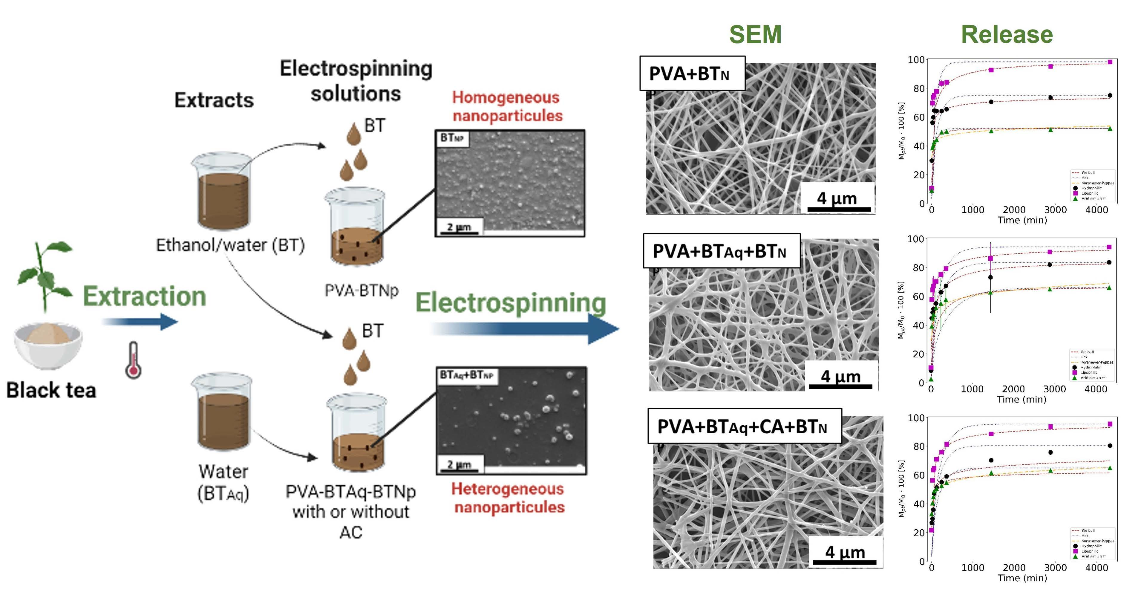
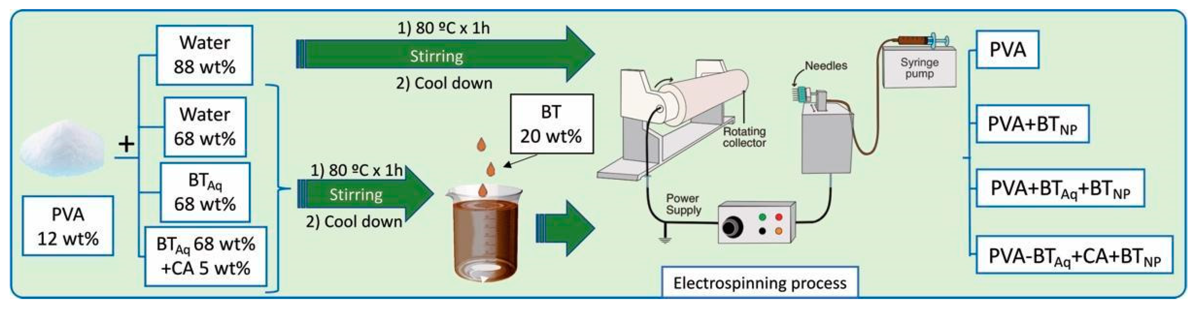
2.3. Preparation of Nanoparticles and Electrospun Mats
2.4. Characterizations of Nanoparticles and Mats
2.4.1. Nanoparticles Size and Mats Morphology
2.4.2. FTIR
2.4.3. Contact Angle (θ)
2.4.4. Water Solubility (S)
2.4.5. Total Extracted Polyphenol Content (TEPC) and Antioxidant Activity of Mats
2.4.6. Release of Polyphenols from the Mats
2.5. Data Treatment and Statistical Analysis
3. Results and Discussion
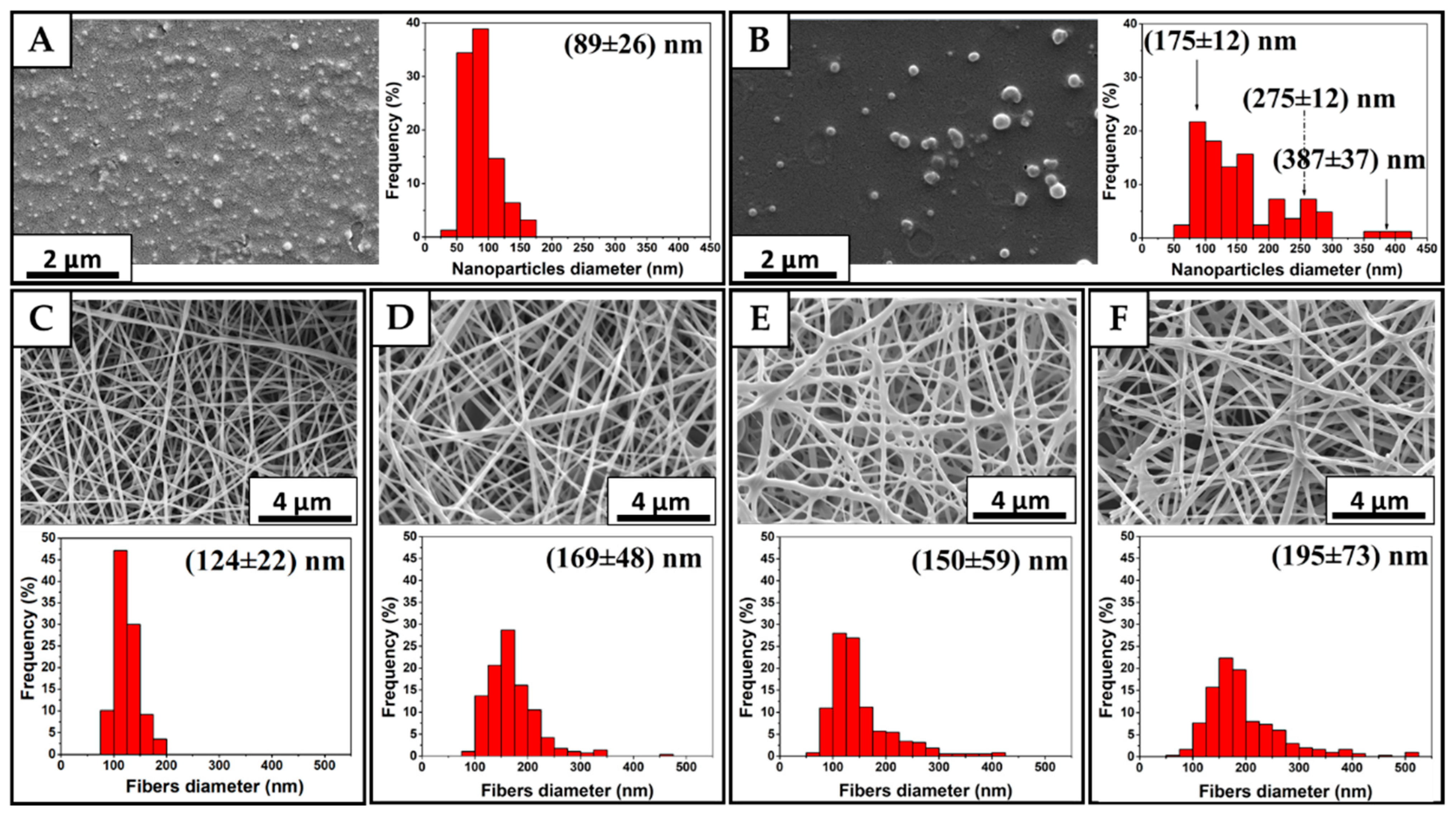
3.1. Characterizations of Nanoparticles and Mats
Nanoparticles Size and Mats Morphology
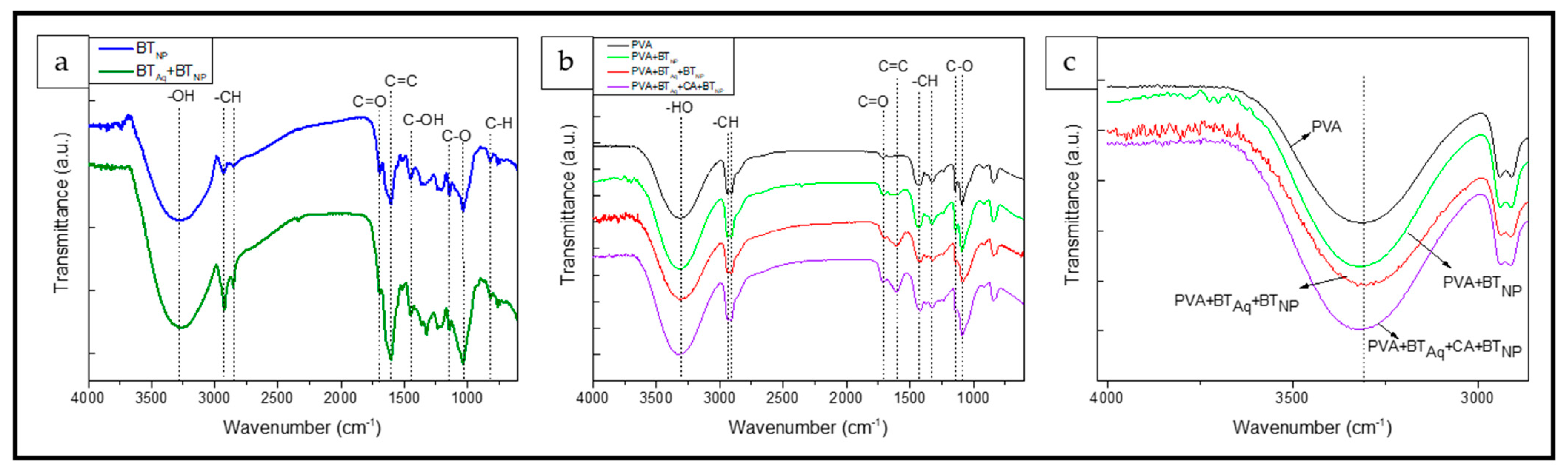
3.2. FTIR
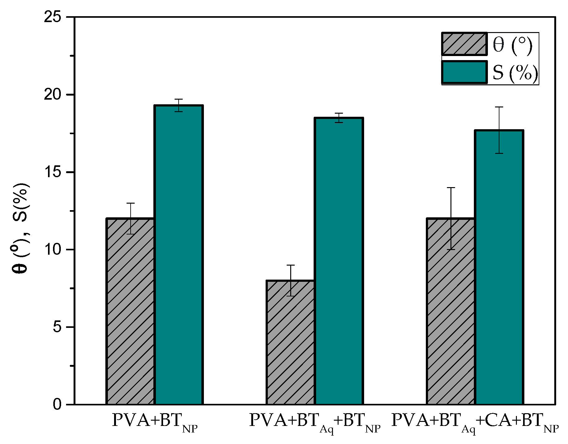
3.3. Contact Angle (θ)
3.4. Water Solubility (S)
3.5. Total Polyphenol Extracted Content (TEPC) and Antioxidant Activity of Mats
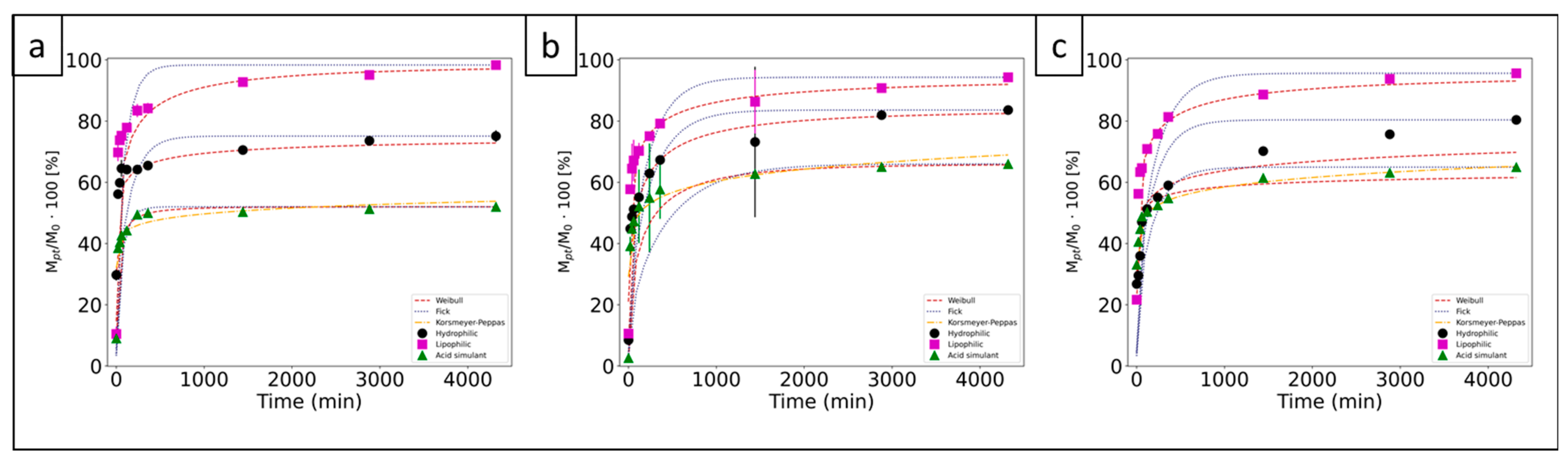
3.6. Release of Polyphenols from the Mats
4. Conclusions
Author Contributions
Funding
Institutional Review Board Statement
Data Availability Statement
Conflicts of Interest
References
- Van Schoubroeck, S.; Chacon, L.; Reynolds, A.M.; Lavoine, N.; Hakovirta, M.; Gonzalez, R.; Van Passel, S.; Venditti, R.A. Environmental sustainability perception toward obvious recovered waste content in paper—Based packaging: An online and in—Person survey best—Worst scaling experiment. Resour. Conserv. Recycl. 2023, 188, 106682. [Google Scholar] [CrossRef]
- Iacovone, C.; Yulita, F.; Cerini, D.; Peña, D.; Candal, R.; Goyanes, S.; Pietrasanta, L.; Guz, L.; Famá, L. Effect of TiO2 nanoparticles and extrusion process on the physicochemical properties of biodegradable and active cassava starch nanocomposites. Polymers 2023, 15, 535. [Google Scholar] [CrossRef] [PubMed]
- Packaging Trends Driven by Consumer Demands. Available online: https://pmg-designer.s3.amazonaws.com/FreeDownloads/PW/2021_PW_ConsumerBehavior.pdf (accessed on 26 December 2022).
- Packaging World News Roundup: A Comprehensive Guide of Sustainability Best Practices and Case Studies. Sustainability. Available online: https://pmg-designer.s3.amazonaws.com/FreeDownloads/PW/PW_2022_Sustainability.pdf (accessed on 26 December 2022).
- Ribba, L.G.; Cimadoro, J.D.; D’Accorso, N.B.; Goyanes, S.N. Removal of Pollutants Using Electrospun Nanofiber Membranes. In Industrial Applications of Renewable Biomass Products: Past, Present and Future; Springer International Publishing: Cham, Switzerland, 2017; pp. 301–324. [Google Scholar] [CrossRef]
- Vasisht, N. Nanoencapsulation in the food industry. In Microencapsulation in the Food Industry a Practical Implementation Guide, 2nd ed.; Sobel, R., Ed.; Academic Press: Cambridge, MA, USA, 2023; pp. 209–213. [Google Scholar] [CrossRef]
- Olive Li, Y.; Dueik González, V.P.; Diosady, L.L. Microencapsulation of vitamins, minerals, and nutraceuticals for food applications. In Microencapsulation in the Food Industry a Practical Implementation Guide, 2nd ed.; Sobel, R., Ed.; Academic Press: Cambridge, MA, USA, 2023; pp. 507–528. [Google Scholar] [CrossRef]
- Zanetti, M.; Carniel, T.K.; Dalcanton, F.; Silva dos Anjos, R.; Gracher Riella, H.; De Araújo, P.; de Oliveira, D.; Fiori, M.A. Use of encapsulated natural compounds as antimicrobial additives in food packaging: A brief review. Trends Food Sci. Technol. 2018, 81, 51–60. [Google Scholar] [CrossRef]
- Zeinali, T.; Alemzadeh, E.; Zarban, A.; Khorashadizadeh, M.; Ansarifar, E. Fabrication and characterization of jujube extract—Loaded electrospun polyvinyl alcohol nanofiber for strawberry preservation. Food Sci. Nutr. 2021, 9, 6353–6361. [Google Scholar] [CrossRef]
- Hoseyni, S.Z.; Jafari, S.M.; Tabarestani, H.T.; Ghorbani, M.; Assadpour, E.; Sabaghi, M. Release of catechin from Azivash gum—Polyvinyl alcohol electrospun nanofibers in simulated food and digestion media. Food Hydrocoll. 2020, 112, 106366. [Google Scholar] [CrossRef]
- Han, H.; Wang, H.; Gao, G.; Rao, P.; Zhou, J.; Ke, L.; Xu, Y. pH effect on colloidal characteristics of micro—Nano particles in lapsang souchong black tea infusion. Food Control 2022, 133, 108643. [Google Scholar] [CrossRef]
- Song, T.; Qian, S.; Lan, T.; Wu, Y.; Liu, J.; Zhang, H. Recent Advances in Bio—Based Smart Active Packaging Materials. Foods 2022, 11, 2228. [Google Scholar] [CrossRef]
- Vieira, D.M.; Pereira, C.; Calhelha, R.C.; Barros, L.; Petrovic, J.; Sokovic, M.; Barreiro, M.F.; Ferreira, I.C.F.R.; Castro, M.C.R.; Rodrigues, P.V.; et al. Evaluation of plant extracts as an efficient source of additives for active food packaging. Food Front. 2021, 3, 480–488. [Google Scholar] [CrossRef]
- Ahmad, A.; Qurashi, A.; Sheehan, D. Nano packaging—Progress and future perspectives for food safety, and sustainability. Food Packag. Shelf Life 2023, 35, 100997. [Google Scholar] [CrossRef]
- Estevez-Areco, S.; Guz, L.; Candal, R.; Goyanes, S. Release kinetics of rosemary (Rosmarinus officinalis) polyphenols from polyvinyl alcohol (PVA) electrospun nanofibers in several food simulants. Food Packag. Shelf Life 2018, 18, 42–50. [Google Scholar] [CrossRef]
- Estevez-Areco, S.; Guz, L.; Candal, R.; Goyanes, S. Development of Insoluble PVA Electrospun Nanofibers Incorporating R—Limonene or β—Cyclodextrin/R—Limonene Inclusion Complexes. J. Polym. Environ. 2022, 30, 2812–2823. [Google Scholar] [CrossRef]
- Piñeros-Hernandez, D.; Medina-Jaramillo, C.; López-Córdoba, A.; Goyanes, S. Edible cassava starch films carrying rosemary antioxidant extracts for potential use as active food packaging. Food Hydrocoll. 2017, 63, 488–495. [Google Scholar] [CrossRef]
- Lan, W.; Zhang, R.; Ahmed, S.; Qin, W.; Liu, Y. Effects of various antimicrobial polyvinyl alcohol/tea polyphenol composite films on the shelf life of packaged strawberries. LWT 2019, 113, 108297. [Google Scholar] [CrossRef]
- Torasso, N.; González-Seligra, P.; Trupp, F.; Grondona, D.; Goyanes, S. Turning a Novel Janus Electrospun Mat into an Amphiphilic Membrane with High Aromatic Hydrocarbon Adsorption Capacity. Colloids Interfaces 2022, 6, 66. [Google Scholar] [CrossRef]
- Vergara-Rubio, A.; Ribba, L.; Picón Borregales, D.; Sapag, K.; Candal, R.; Goyanes, S. Ultramicroporous Carbon Nanofibrous Mats for Hydrogen Storage. ACS Appl. Nano Mater. 2022, 5, 15353–15361. [Google Scholar] [CrossRef]
- López-Córdoba, A.; Castro, G.R.; Goyanes, S. A simple green route to obtain poly(vinyl alcohol) electrospun mats with improved water stability for use as potential carriers of drugs. Mater. Sci. Eng. C 2016, 69, 726–732. [Google Scholar] [CrossRef]
- Cimadoro, J.; Ribba, L.; Ledesma, S.; Goyanes, S. Electrospun Mats: From White to Transparent with a Drop. Macromol. Mater. Eng. 2018, 303, 1800237. [Google Scholar] [CrossRef]
- Yu, D.; Feng, Y.Y.; Xu, J.X.; Kong, B.H.; Liu, Q.; Wang, H. Fabrication, characterization, and antibacterial properties of citric acid crosslinked PVA electrospun microfibre mats for active food packaging. Packag. Technol. Sci. 2021, 34, 361–370. [Google Scholar] [CrossRef]
- Efenberger-Szmechtyk, M.; Nowak, A.; Czyzowska, A. Plant extracts rich in polyphenols: Antibacterial agents and natural preservatives for meat and meat products. Crit. Rev. Food Sci. Nutr. 2021, 61, 149–178. [Google Scholar] [CrossRef]
- Farhan, M. Green Tea Catechins: Nature’s Way of Preventing and Treating Cancer. Int. J. Mol. Sci. 2022, 23, 10713. [Google Scholar] [CrossRef]
- Senthil Muthu Kumar, T.; Senthil Kumar, K.; Rajini, N.; Siengchin, S.; Ayrilmis, N.; Varada Rajulu, A. A comprehensive review of electrospun nanofibers: Food and packaging perspective. Compos. Part B Eng. 2019, 175, 107074. [Google Scholar] [CrossRef]
- Lan, X.; Luo, T.; Zhong, Z.; Huang, D.; Liang, C.; Liu, Y.; Wang, H.; Tang, Y. Green cross-linking of gelatin/tea polyphenol/ε-poly (L—Lysine) electrospun nanofibrous membrane for edible and bioactive food packaging. Food Packag. Shelf Life 2022, 34, 100970. [Google Scholar] [CrossRef]
- Luo, J.; Zuo, D.; Deng, Z.; Ji, A.; Xia, G. Effects of heat treatment and tea polyphenols on the structure and properties of polyvinyl alcohol nanofiber films for food packaging. Coatings 2020, 10, 49. [Google Scholar] [CrossRef] [Green Version]
- Zhang, H.; Qi, R.; Mine, Y. The impact of oolong and black tea polyphenols on human health. Food Biosci. 2019, 29, 55–61. [Google Scholar] [CrossRef]
- Ju, J.; Lu, G.; Lambert, J.D.; Yang, C.S. Inhibition of carcinogenesis by tea constituents. Semin. Cancer Biol. 2017, 17, 395–402. [Google Scholar] [CrossRef] [Green Version]
- Heber, D.; Zhang, Y.; Yang, J.; Ma, J.E.; Henning, S.M.; Li, Z. Green tea, black tea, and oolong tea polyphenols reduce visceral fat and inflammation in mice fed high—Fat, high—Sucrose obesogenic diets. J. Nutr. 2014, 144, 1385–1393. [Google Scholar] [CrossRef] [Green Version]
- Rajapaksha, S.W.; Shimizu, N. Development and characterization of functional starch—Based films incorporating free or microencapsulated spent black tea extract. Molecules 2021, 26, 3898. [Google Scholar] [CrossRef]
- Ashrafi, A.; Jokar, M.; Nafchi, A.M. Preparation and characterization of biocomposite film based on chitosan and kombucha tea as active food packaging. Int. J. Biol. Macromol. 2018, 108, 444–454. [Google Scholar] [CrossRef]
- López-Córdoba, A.; Medina-Jaramillo, C.; Piñeros-Hernandez, D.; Goyanes, S. Cassava starch films containing rosemary nanoparticles produced by solvent displacement method. Food Hydrocoll. 2017, 71, 26–34. [Google Scholar] [CrossRef]
- Bruni, G.P.; dos Santos Acunha, T.; de Oliveira, J.P.; Martins Fonseca, L.; da Silva, F.T.; Martins Guimarães, V.; da Rosa Zavareze, E. Electrospun protein fibers loaded with yerba mate extract for bioactive release in food packaging. J. Sci. Food Agric. 2020, 100, 3341–3350. [Google Scholar] [CrossRef]
- Estevez-Areco, S.; Guz, L.; Candal, R.; Goyanes, S. Active bilayer films based on cassava starch incorporating ZnO nanorods and PVA electrospun mats containing rosemary extract. Food Hydrocoll. 2020, 108, 106054. [Google Scholar] [CrossRef]
- Brand-Williams, C.; Cuvelier, W.; Berset, M.E. Use of a free radical method to evaluate antioxidant activity. LWT—Food Sci. Technol. 1995, 29, 25–30. [Google Scholar] [CrossRef]
- Bhebhe, M.; Füller, T.N.; Chipurura, B.; Muchuweti, M. Effect of Solvent Type on Total Phenolic Content and Free Radical Scavenging Activity of Black Tea and Herbal Infusions. Food Anal. Methods 2016, 9, 1060–1067. [Google Scholar] [CrossRef]
- Paini, M.; Casazza, A.A.; Aliakbarian, B.; Perego, P.; Binello, A.; Cravotto, G. Influence of ethanol/water ratio in ultrasound and high—Pressure/high—Temperature phenolic compound extraction from agri—Food waste. Int. J. Food Sci. Technol. 2016, 51, 349–358. [Google Scholar] [CrossRef]
- Torasso, N.; Vergara-Rubio, A.; Pereira, R.; Martinez-Sabando, J.; Vega Baudrit, J.; Cerveny, S.; Goyanes, S. An in situ approach to entrap ultra—Small iron oxide nanoparticles inside hydrophilic electrospun nanofibers with high arsenic adsorption. Chem. Eng. J. 2023, 454, 140168. [Google Scholar] [CrossRef]
- Picón, D.; Torasso, N.; Baudrit, J.R.V.; Cerveny, S.; Goyanes, S. Bio—Inspired membranes for adsorption of arsenic via immobilized L—Cysteine in highly hydrophilic electrospun nanofibers. Chem. Eng. Res. Des. 2022, 185, 108–118. [Google Scholar] [CrossRef]
- Vergara-Rubio, A.; Ribba, L.; Picón, D.; Candal, R.; Goyanes, S. A Highly Efficient Nanostructured Sorbent of Sulfuric Acid from Ecofriendly Electrospun Poly(vinyl alcohol) Mats. Ind. Eng. Chem. Res. 2022, 61, 2091–2099. [Google Scholar] [CrossRef]
- Wang, Z.; Sun, S.; Liu, J.; He, Y.; Kan, X.; Xu, Y. Understanding of Commission Regulation (EU) No 10/2011 on Plastic Materials and Articles Intended to Come into Contact with Food. China Plast. 2011, 25, 83–88. [Google Scholar] [CrossRef]
- Crank, J. Infinite and semi—Infinite media. In The Mathematics of Diffusion, 2nd ed.; Clarendon Press: Oxford, UK, 1975; pp. 32–40. [Google Scholar]
- Ritger, P.L.; Peppas, N.A. A simple equation for description of solute release II. Fickian and anomalous release from swellable devices. J. Control. Release 1987, 5, 37–42. [Google Scholar] [CrossRef]
- Van Boekel, M.A.J.S. Kinetic modeling of food quality: A critical review. Compr. Rev. Food Sci. Food Saf. 2008, 7, 144–158. [Google Scholar] [CrossRef]
- Heydari-Majd, M.; Ghanbarzadeh, B.; Shahidi-Noghabi, M.; Najafi, M.A.; Adun, P.; Ostadrahimid, A. Kinetic release study of zinc from polylactic acid based nanocomposite into food simulants. Polym. Test. 2019, 76, 254–260. [Google Scholar] [CrossRef]
- Szewczyk, P.K.; Stachewicz, U. The impact of relative humidity on electrospun polymer fibers: From structural changes to fiber morphology. Adv. Colloid Interface Sci. 2020, 286, 102315. [Google Scholar] [CrossRef]
- Hikmawati, D.; Adiputri, E.F.; Putra, A.P.; Ady, J. The Role of Relative Humidity on Physical Characteristics of Poly Vinyl Alcohol—Aloe vera Fiber Membrane by Using Electrospinning Methods. Mater. Sci. Forum 2019, 966, 157–162. [Google Scholar] [CrossRef]
- Pelipenko, J.; Kristl, J.; Jankovic, B.; Baumgartner, S.; Kocbek, P. The impact of relative humidity during electrospinning on the morphology and mechanical properties of nanofibers. Int. J. Pharm. 2013, 456, 125–134. [Google Scholar] [CrossRef] [PubMed]
- Cardoso da Mata, G.; Morais, M.S.; Pereira de Oliveira, W.; Lopes Aguiar, M. Composition Effects on the Morphology of PVA/Chitosan Electrospun Nanofibers. Polymers 2022, 14, 4856. [Google Scholar] [CrossRef]
- Salazar-brann, S.A.; Patiño-Herrera, R.; Navarrete-damia, J.; Louvier-Hernández, J.F. Electrospinning of chitosan from different acid solutions. AIMS Bioeng. 2021, 8, 112–129. [Google Scholar] [CrossRef]
- López-Córdoba, A.; Estevez-Areco, S.; Goyanes, S. Potato starch—Based biocomposites with enhanced thermal, mechanical and barrier properties comprising water—Resistant electrospun poly (vinyl alcohol) fibers and yerba mate extract. Carbohydr. Polym. 2019, 215, 377–387. [Google Scholar] [CrossRef]
- Gullón, B.; Eibes, G.; Moreira, M.T.; Herrera, R.; Labidi, J.; Gullón, P. Yerba mate waste: A sustainable resource of antioxidant compounds. Ind. Crops Prod. 2018, 113, 398–405. [Google Scholar] [CrossRef]
- Hanis Abd Latif, N.; Brosse, N.; Ziegler-Devin, I.; Chrusiel, L.; Hashim, R.; Hazwan Hussin, M. A Comparison of Alkaline and Organosolv Lignin Extraction Methods from Coconut Husks as an Alternative Material for Green Applications. BioResources 2021, 17, 469–491. [Google Scholar] [CrossRef]
- Zhang, L.H.; Shen, Q. Fully Green Poly(vinyl alcohol)/Tea Polyphenol Composites and Super Anti—Ultraviolet and —Bacterial Properties. Macromol. Mater. Eng. 2020, 305, 1900669. [Google Scholar] [CrossRef]
- Nataraj, D.; Reddy, R.; Reddy, N. Crosslinking electrospun poly (vinyl) alcohol fibers with citric acid to impart aqueous stability for medical applications. Eur. Polym. J. 2020, 124, 109484. [Google Scholar] [CrossRef]
- Liu, Y.; Wang, S.; Lan, W.; Qin, W. Development of ultrasound treated polyvinyl alcohol/tea polyphenol composite films and their physicochemical properties. Ultrason. Sonochem. 2019, 51, 386–394. [Google Scholar] [CrossRef] [PubMed]
- Ordoñez, R.; Atarés, L.; Chiralt, A. Biodegradable active materials containing phenolic acids for food packaging applications. Compr. Rev. Food Sci. Food Saf. 2022, 21, 3910–3930. [Google Scholar] [CrossRef] [PubMed]


| Extract | BTAq | BT |
|---|---|---|
| Solid’s extraction yield (g of solids/L of extract) | 51.2 ± 0.9 a | 102 ± 1 b |
| TPC (mgGAE/mL of extract) | 4.0 ± 0.1 a | 17.6 ± 0.2 b |
| TPC (mgGAE/100 g of electrospun solution) | 272 ± 10 a | 352 ± 20 b |
| Antioxidant activity (μmol/mL) | 16 ± 4 a | 124 ± 4 b |
| EC50 (μg/mL) | 111 ± 6 a | 80 ± 4 b |
| Material | PVA | PVA + BTNP | PVA + BTAq + BTNP | PVA + BTAq + CA + BTNP |
|---|---|---|---|---|
| Water (% w/w) | 88 | 68 | — | — |
| BTAq (% w/w) | — | — | 68 | 68 |
| BT (% w/w) | — | 20 | 20 | 20 |
| Conductivity (μS/cm) (±1) | 543 a | 1149 b | 5925 c | 3570 d |
| Viscosity (cP) (±5) | 283 a | 457 b | 451 b | 469 c |
| pH (±0.1) | 5.3 a | 5.2 a | 5.2 a | 3.9 b |
| Material | PVA + BTNP | PVA + BTAq + BTNP | PVA + BTAq + CA + BTNP |
|---|---|---|---|
| TEPC (mgGAE/g of mat), t = 0 | 23.9 ± 0.3 a | 41.4 ± 0.9 b | 14.8 ± 0.3 c |
| TEPC (mgGAE/g of mat), t = 4 months | 23.0 ± 0.4 | 39.6 ± 0.5 | — |
| Polyphenols’ retention (%) | 88.9 ± 0.4 a | 88.2 ± 0.3 a | 31.0 ± 0.4 b |
| Antioxidant activity (μmol/g of mat), t = 0 | 125 ± 1 a | 155 ± 3 b | 51 ± 2 c |
| Antioxidant activity (μmol/g of mat), t = 4 months | 120 ± 3 a | 147 ± 4 b | — |
| Films | Hydrophilic | Lipophilic | Acidic | |
|---|---|---|---|---|
| PVA + BTNP | M∞/TPL (%) | 67.40 ± 0.02 a | 80.20 ± 0.01 b | 54.50 ± 0.01 c |
| M∞/TEPC (%) | 80.4 ± 0.9 a | 95.6 ± 0.9 b | 65.00 ± 0.02 c | |
| PVA + BTAq + BTNP | M∞/TPL (%) | 76.50 ± 0.01 a | 86.30 ± 0.01 b | 60.40 ± 0.01 c |
| M∞/TEPC (%) | 87 ± 1 a | 97.8 ± 0.9 b | 68.4 ± 0.3 c | |
| PVA + BTAq + CA + BTNP | M∞/TPL (%) | 23.60 ± 0.01 a | 30.50 ± 0.01 b | 16.20 ± 0.01 c |
| M∞/TEPC (%) | 75 ± 2 a | 98.2 ± 0.4 b | 51.90 ± 0.07 c |
| Simulant | PVA + BTNP | PVA + BTAq + BTNP | PVA + BTAq + CA + BTNP | ||||||
|---|---|---|---|---|---|---|---|---|---|
| Weibull model | |||||||||
| a | b | R2 | a | B | R2 | a | b | R2 | |
| Hydrophilic | −0.49 ± 0.02 | 0.17 ± 0.01 | 0.88 | −0.165 ± 0.002 | 0.390 ± 0.002 | 0.91 | −0.62 ± 0.04 | 0.21 ± 0.01 | 0.79 |
| Lipophilic | −0.350 ± 0.003 | 0.280 ± 0.004 | 0.99 | −0.34 ± 0.01 | 0.284 ± 0.007 | 0.99 | −0.24 ± 0.07 | 0.35 ± 0.04 | 0.99 |
| Acid simulant | −0.8 ± 0.1 | 0.16 ± 0.02 | 0.92 | −0.072 ± 0.003 | 0.518 ± 0.007 | 0.98 | −0.30 ± 0.02 | 0.40 ± 0.03 | 0.83 |
| Fick’s model | |||||||||
| D [cm2/s] | R2 | D [cm2/s] | R2 | D [cm2/s] | R2 | ||||
| Hydrophilic | 1.0 ± 0.7 × 10⁻¹⁵ | 0.93 | 0.57 ± 0.01 × 10⁻¹⁵ | 0.88 | 1.7 ± 0.3 × 10⁻¹⁵ | 0.78 | |||
| Lipophilic | 0.8 ± 1.2 × 10⁻¹⁵ | 0.90 | 0.66 ± 0.04 × 10⁻¹⁵ | 0.80 | 2.4 ± 0.7 × 10⁻¹⁵ | 0.80 | |||
| Acid simulant | 1.0 ± 0.7 × 10⁻¹⁵ | 0.95 | 0.33 ± 0.09 × 10⁻¹⁵ | 0.70 | 3.3 ± 0.7 × 10⁻¹⁵ | 0.88 | |||
| Power Law model | |||||||||
| k | n | R2 | k | N | R2 | k | n | R2 | |
| Acid simulant | 0.5 ± 1.0 | 7484 ± 1 × 10⁻⁵ | 0.98 | 0.5 ± 1.0 | 9084 ± 7 × 10⁻⁵ | 0.94 | 0.6 ± 1.0 | 5475 ± 7 × 10⁻⁵ | 0.86 |
Disclaimer/Publisher’s Note: The statements, opinions and data contained in all publications are solely those of the individual author(s) and contributor(s) and not of MDPI and/or the editor(s). MDPI and/or the editor(s) disclaim responsibility for any injury to people or property resulting from any ideas, methods, instructions or products referred to in the content. |
© 2023 by the authors. Licensee MDPI, Basel, Switzerland. This article is an open access article distributed under the terms and conditions of the Creative Commons Attribution (CC BY) license (https://creativecommons.org/licenses/by/4.0/).
Share and Cite
Quintero-Borregales, L.M.; Vergara-Rubio, A.; Santos, A.; Famá, L.; Goyanes, S. Black Tea Extracts/Polyvinyl Alcohol Active Nanofibers Electrospun Mats with Sustained Release of Polyphenols for Food Packaging Applications. Polymers 2023, 15, 1311. https://doi.org/10.3390/polym15051311
Quintero-Borregales LM, Vergara-Rubio A, Santos A, Famá L, Goyanes S. Black Tea Extracts/Polyvinyl Alcohol Active Nanofibers Electrospun Mats with Sustained Release of Polyphenols for Food Packaging Applications. Polymers. 2023; 15(5):1311. https://doi.org/10.3390/polym15051311
Chicago/Turabian StyleQuintero-Borregales, Lucía M., Alicia Vergara-Rubio, Ayelen Santos, Lucía Famá, and Silvia Goyanes. 2023. "Black Tea Extracts/Polyvinyl Alcohol Active Nanofibers Electrospun Mats with Sustained Release of Polyphenols for Food Packaging Applications" Polymers 15, no. 5: 1311. https://doi.org/10.3390/polym15051311