
Antibacterial Activity of Ulva/Nanocellulose and Ulva/Ag/Cellulose Nanocomposites and Both Blended with Fluoride against Bacteria Causing Dental Decay
 ,
,
Abstract
:1. Introduction
2. Materials and Methods
2.1. Materials
2.2. Alga Collection
2.3. Extracting Cellulose from U. lactuca Green Alga
2.4. Extraction of Nanocellulose
2.5. Synthesis Ulva/Ag/Cellulose Nanocomposite
2.5.1. Preparation of Silver Nanoparticles
Algal Extract
Biosynthesis of Ag Nanoparticles
2.5.2. Biosynthesis of Ulva/Ag/Cellulose Nanocomposites
2.5.3. Biosynthesis of Ulva/Nanocellulose, Ulva/Ag/Cellulose Nanocomposites, and Both Blended with Fluoride
2.6. Characterization
2.6.1. Fourier Transform Infrared (FT-IR)
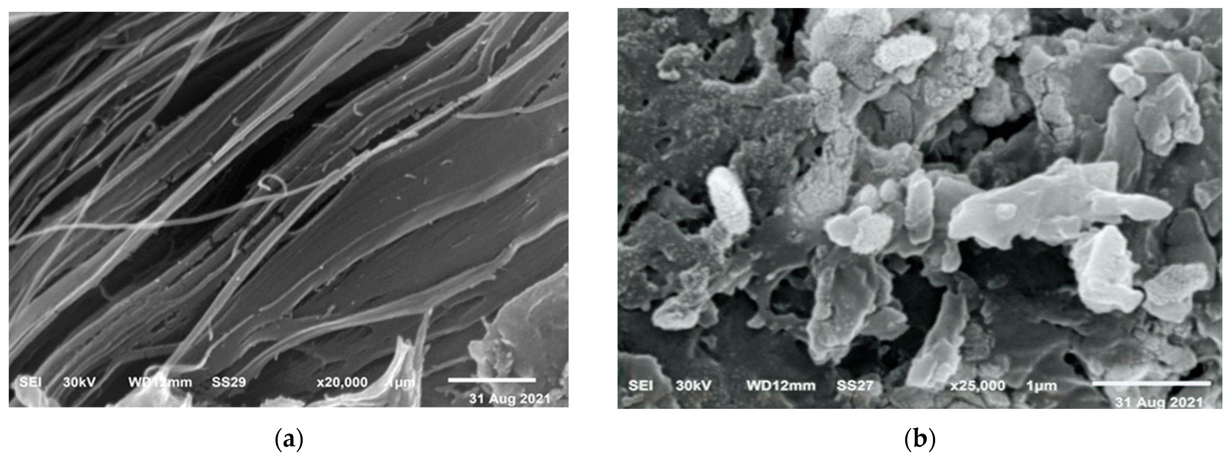
2.6.2. Scanning Electron Microscope (SEM)
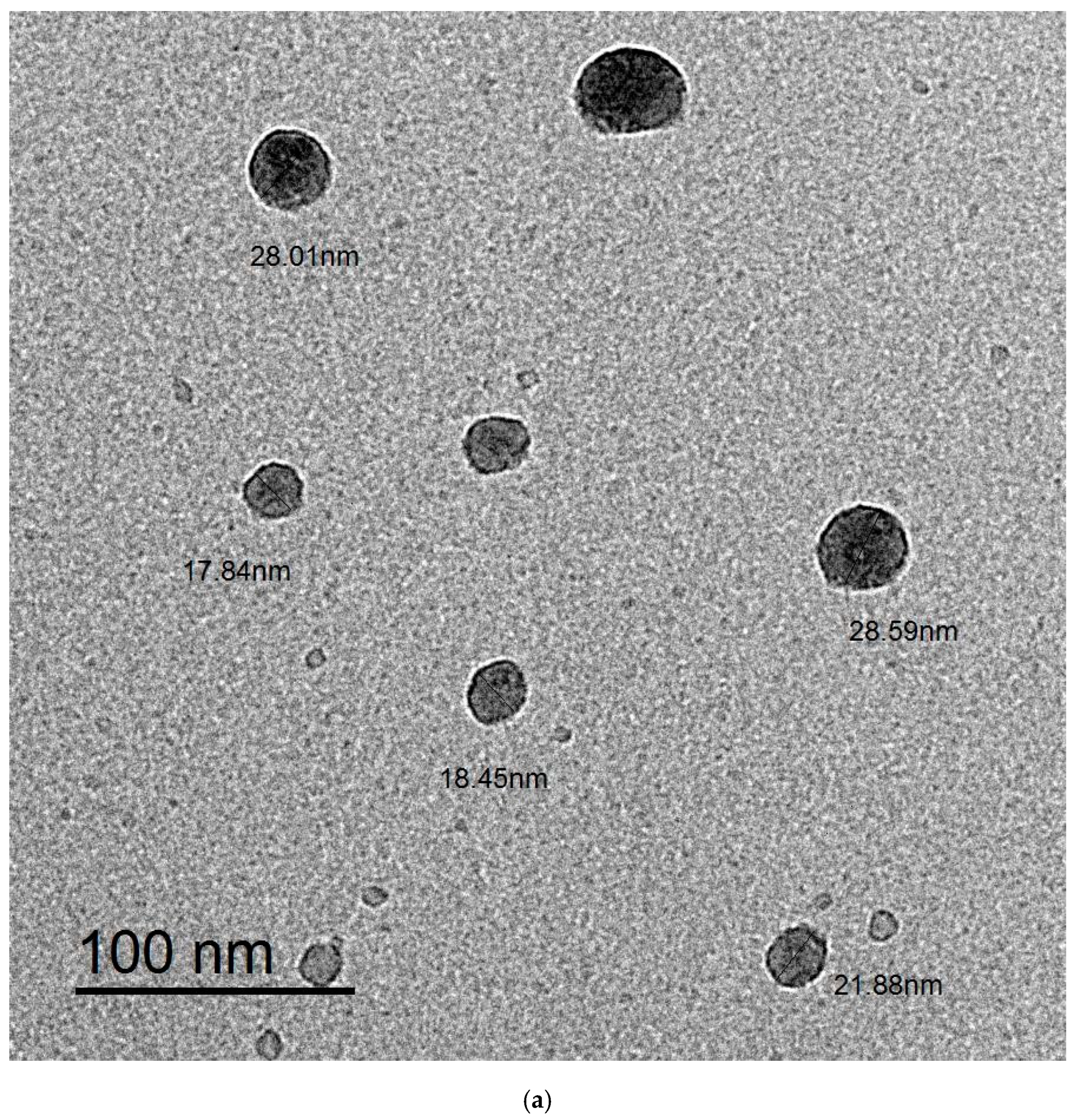
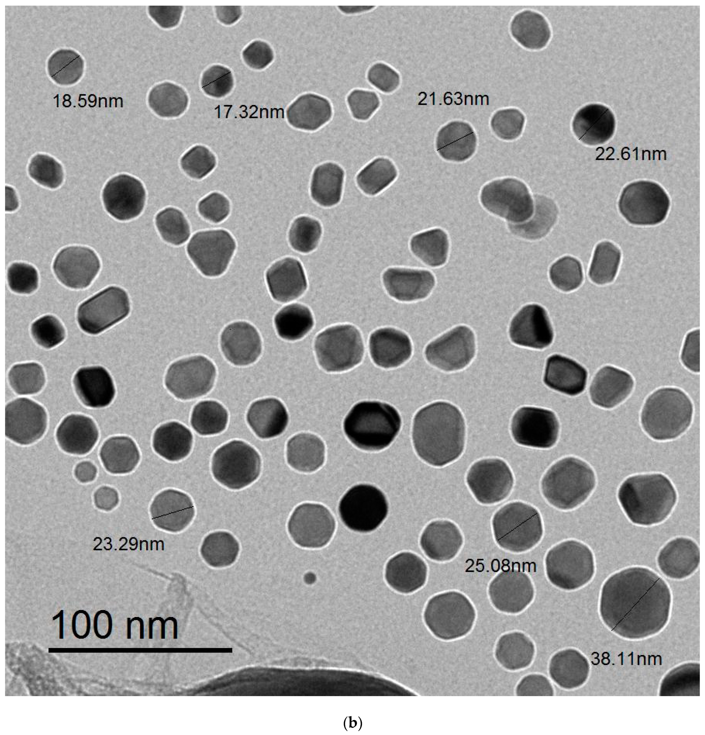
2.6.3. Transmission Electron Microscopy (TEM)
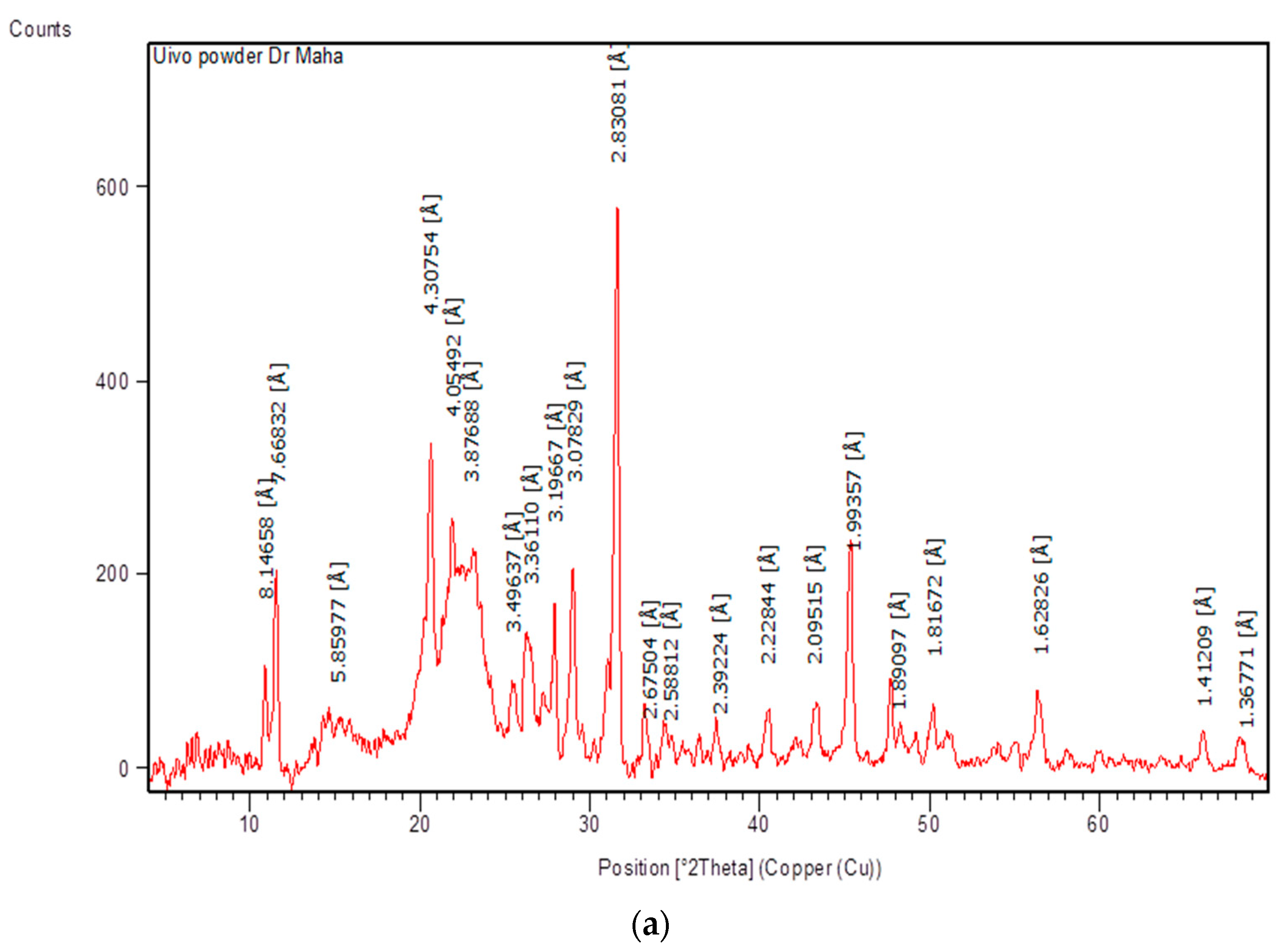
2.6.4. X-ray Powder Diffraction (XRD)
2.6.5. Energy-Dispersive Spectroscopy (EDS)
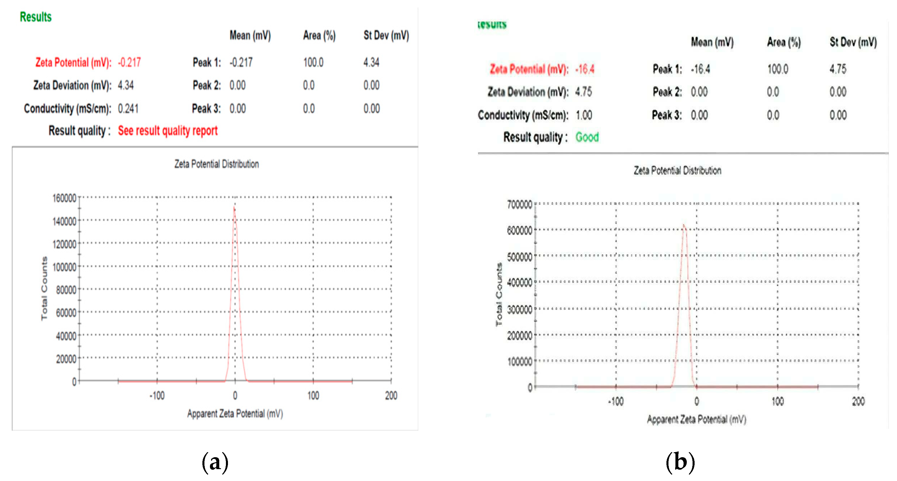
2.6.6. Zeta Potential Analysis
2.6.7. Differential Scanning Calorimetry (DSC)
2.7. Antibacterial Activities
2.8. Minimum Inhibitory Concentration (MIC)
2.9. Statistical Analysis
3. Results and Discussion
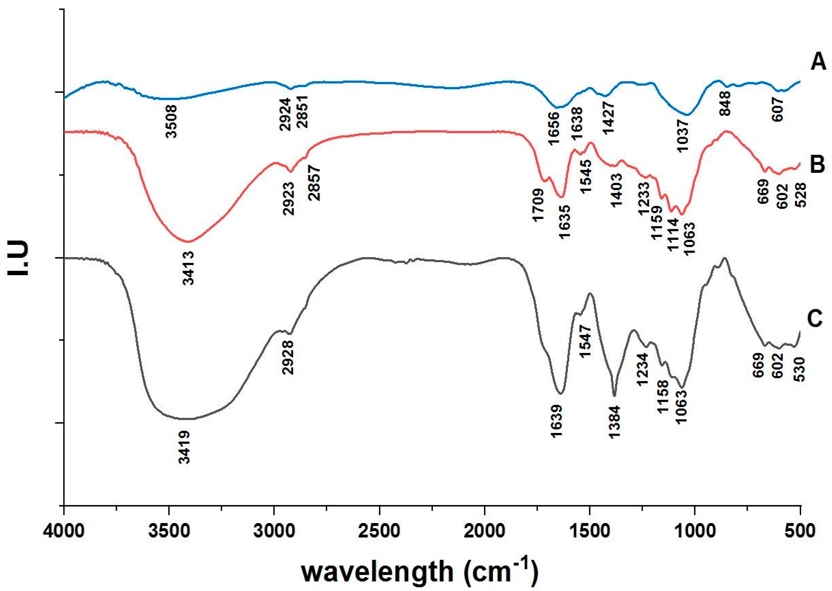
3.1. FT-IR Spectroscopy Analysis
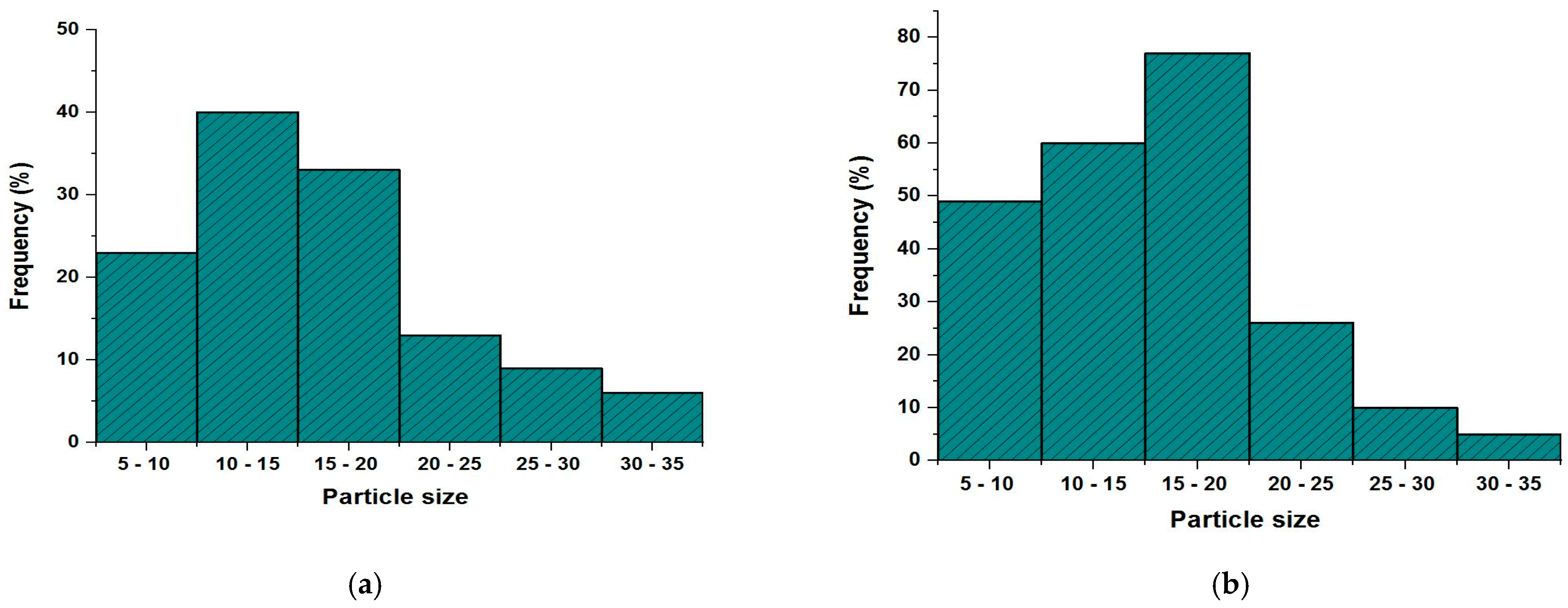
3.2. SEM and TEM Images
3.3. Energy-Dispersive X-ray Measurements
3.4. X-ray Diffraction
3.5. Zeta Potential Analysis
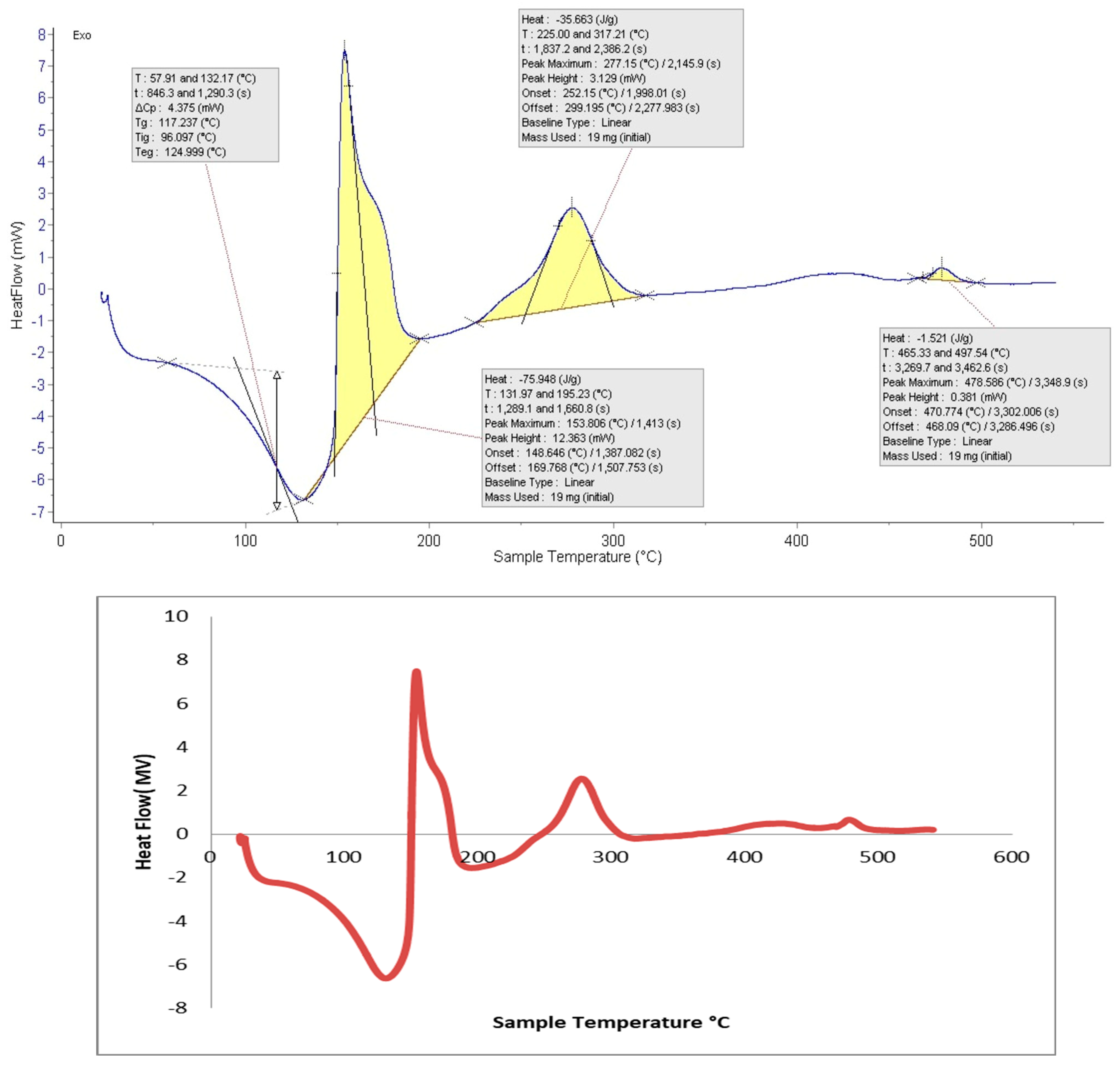
3.6. Differential Scanning Calorimetry (DSC)
3.7. Antimicrobial Activity
3.8. Antimicrobial Activity of Nanocellulose Blended with Fluoride
3.9. Minimum Inhibitory Concentration (MIC)
4. Conclusions
Author Contributions
Funding
Institutional Review Board Statement
Informed Consent Statement
Data Availability Statement
Conflicts of Interest
References
- Uerlich, M.F.; Baker, S.R.; Day, P.F.; Brown, L.; Vettore, M.V. Common Determinants of Dental Caries and Obesity in Children: A Multi-Ethnic Nested Birth Cohort Study in the United Kingdom. Int. J. Environ. Res. Public Health 2021, 18, 12561. [Google Scholar] [CrossRef]
- Seymour, B.; James, Z.; Shroff Karhade, D.; Barrow, J.; Pruneddu, A.; Anderson, N.K.; Definition of Global Health, T.F.F.T. A definition of global oral health: An expert consensus approach by the Consortium of Universities for Global Health’s Global Oral Health Interest Group. Glob. Health Action 2020, 13, 1814001. [Google Scholar] [CrossRef]
- Veiga, N.; Pereira, C.; Amaral, O. Prevalence and determinants of dental caries in Portuguese children. Procedia-Soc. Behav. Sci. 2015, 171, 995–1002. [Google Scholar] [CrossRef] [Green Version]
- Selwitz, R.H.; Ismail, A.I.; Pitts, N.B. Dental caries. Lancet 2007, 369, 51–59. [Google Scholar] [CrossRef]
- Featherstone, J.D. Dental caries: A dynamic disease process. Aust. Dent. J. 2008, 53, 286–291. [Google Scholar] [CrossRef]
- Rostinawati, T.; Aryani, H.; Iskandar, Y. Identification of bacteria causing dental caries through genetic testing and activity assay of toothpastes. J. Pharm. Sci. Res. 2018, 10, 511–513. [Google Scholar]
- Mitchell, T.J. The pathogenesis of streptococcal infections: From tooth decay to meningitis. Nat. Rev. Microbiol. 2003, 1, 219–230. [Google Scholar] [CrossRef]
- Yadav, K.; Prakash, S. Dental caries: A microbiological approach. J. Clin. Infect. Dis. Pract. 2017, 2, 1–15. [Google Scholar] [CrossRef]
- Hohwy, J.; Reinholdt, J.; Kilian, M. Population dynamics of Streptococcus mitis in its natural habitat. Infect. Immun. 2001, 69, 6055–6063. [Google Scholar] [CrossRef] [Green Version]
- Kulshrestha, A.; Gupta, P. Polymicrobial interaction in biofilm: Mechanistic insights. Pathog. Dis. 2022, 80, ftac010. [Google Scholar] [CrossRef]
- Loesche, W.J. Microbiology of Dental Decay and Periodontal Disease. In Medical Microbiology, 4th ed.; University of Texas Medical Branch at Galveston: Galveston, TX, USA, 1996; Chapter 99. Available online: https://www.ncbi.nlm.nih.gov/books/NBK8259/ (accessed on 10 October 2022).
- Desselberger, U. Emerging and re-emerging infectious diseases. J. Infect. 2000, 40, 3–15. [Google Scholar] [CrossRef] [PubMed]
- Abiodun Solanke, I.M.F.; Ajayi, D.M.; Arigbede, A.O. Nanotechnology and its application in dentistry. Ann. Med. Health Sci. Res. 2014, 4, 171–177. [Google Scholar] [CrossRef] [PubMed] [Green Version]
- De Kwaadsteniet, M.I.C.H.E.L.E.; Botes, M.; Cloete, T.E. Application of nanotechnology in antimicrobial coatings in the water industry. Nano 2011, 6, 395–407. [Google Scholar] [CrossRef]
- Bhutiya, P.L.; Misra, N.; Rasheed, M.A.; Hasan, S.Z. Silver Nanoparticles Deposited Algal Nanofibrous Cellulose Sheet for Antibacterial Activity. BioNanoScience 2020, 10, 23–33. [Google Scholar] [CrossRef]
- Moon, R.J.; Martini, A.; Nairn, J.; Simonsen, J.; Youngblood, J. Cellulose nanomaterials review: Structure, properties and nanocomposites. Chem. Soc. Rev. 2011, 40, 3941–3994. [Google Scholar] [CrossRef] [PubMed]
- AI-Jawhari, I.F.H. Nanocellulose for Sustainable Future Applications. In Handbook of Nanomaterials and Nanocomposites for Energy and Environmental Applications; Springer: Berlin/Heidelberg, Germany, 2020; pp. 1–12. [Google Scholar] [CrossRef]
- Naz, S.; Ali, J.S.; Zia, M. Nanocellulose isolation characterization and applications: A journey from non-remedial to biomedical claims. Bio-Des. Manuf. 2019, 2, 187–212. [Google Scholar] [CrossRef]
- Saleh, M.H.S.D.E.; Muhamad, M.D.I.I.; Mamat, S.N.H. Cellulose Nanofiber Isolation and Its Fabrication into Bio-Polymer-A Review. In Proceedings of the International Conference on Agricultural and Food Engineering for Life (Cafei2012), Putrajaya, Malaysia, 26–28 November 2012; Volume 26, p. 28. [Google Scholar]
- Iwamoto, S.; Kai, W.; Isogai, A.; Iwata, T. Elastic modulus of single cellulose microfibrils from tunicate measured by atomic force microscopy. Biomacromolecules 2009, 10, 2571–2576. [Google Scholar] [CrossRef]
- Klemm, D.; Kramer, F.; Moritz, S.; Lindström, T.; Ankerfors, M.; Gray, D.; Dorris, A. Nanocelluloses: A new family of nature-based materials. Angew. Chem. Int. Ed. 2011, 50, 5438–5466. [Google Scholar] [CrossRef]
- Beck, S.; Bouchard, J.; Berry, R. Controlling the reflection wavelength of iridescent solid films of nanocrystalline cellulose. Biomacromolecules 2011, 12, 167–172. [Google Scholar] [CrossRef]
- Lee, K.Y.; Aitomäki, Y.; Berglund, L.A.; Oksman, K.; Bismarck, A. On the use of nanocellulose as reinforcement in polymer matrix composites. Compos. Sci. Technol. 2014, 105, 15–27. [Google Scholar] [CrossRef] [Green Version]
- Jorfi, M.; Foster, E.J. Recent advances in nanocellulose for biomedical applications. J. Appl. Polym. Sci. 2015, 132. [Google Scholar] [CrossRef]
- Liu, A.; Walther, A.; Ikkala, O.; Belova, L.; Berglund, L.A. Clay nanopaper with tough cellulose nanofiber matrix for fire retardancy and gas barrier functions. Biomacromolecules 2011, 12, 633–641. [Google Scholar] [CrossRef]
- Lefatshe, K.; Muiva, C.M.; Kebaabetswe, L.P. Extraction of nanocellulose and in-situ casting of ZnO/cellulose nanocomposite with enhanced photocatalytic and antibacterial activity. Carbohydr. Polym. 2017, 164, 301–308. [Google Scholar] [CrossRef]
- Janpetch, N.; Saito, N.; Rujiravanit, R. Fabrication of bacterial cellulose-ZnO composite via solution plasma process for antibacterial applications. Carbohydr. Polym. 2016, 148, 335–344. [Google Scholar] [CrossRef]
- Dong, Y.Y.; Liu, S.; Liu, Y.J.; Meng, L.Y.; Ma, M.G. Ag@ Fe3O4@ cellulose nanocrystals nanocomposites: Microwave-assisted hydrothermal synthesis, antimicrobial properties, and good adsorption of dye solution. J. Mater. Sci. 2017, 52, 8219–8230. [Google Scholar] [CrossRef]
- Xiong, R.; Lu, C.; Wang, Y.; Zhou, Z.; Zhang, X. Nanofibrillated cellulose as the support and reductant for the facile synthesis of Fe3O4/Ag nanocomposites with catalytic and antibacterial activity. J. Mater. Chem. A 2013, 1, 14910–14918. [Google Scholar] [CrossRef]
- Dong, Y.Y.; Deng, F.; Zhao, J.J.; He, J.; Ma, M.G.; Xu, F.; Sun, R.C. Environmentally friendly ultrosound synthesis and antibacterial activity of cellulose/Ag/AgCl hybrids. Carbohydr. Polym. 2014, 99, 166–172. [Google Scholar] [CrossRef]
- Leverett, D.H. Fluorides and the changing prevalence of dental caries. Science 1982, 217, 26–30. [Google Scholar] [CrossRef] [Green Version]
- Featherstone, J.D. Prevention and reversal of dental caries: Role of low level fluoride. Community Dent. Oral Epidemiol. 1999, 27, 31–40. [Google Scholar] [CrossRef]
- Whelton, H.P.; Spencer, A.J.; Do, L.G.; Rugg-Gunn, A.J. Fluoride revolution and dental caries: Evolution of policies for global use. J. Dent. Res. 2019, 98, 837–846. [Google Scholar] [CrossRef]
- Palmer, J.D. Dental health in children—An improving picture? Br. Dent. J. 1980, 149, 48–50. [Google Scholar] [CrossRef]
- Pitts, N.B.; Zero, D.T.; Marsh, P.D.; Ekstrand, K.; Weintraub, J.A.; Ramos-Gomez, F.; Ismail, A. Dental caries. Nat. Rev. Dis. Prim. 2017, 3, 17030. [Google Scholar] [CrossRef] [Green Version]
- Arends, J.; Christoffersen, J. Nature and role of loosely bound fluoride in dental caries. J. Dent. Res. 1990, 69 (Suppl. S2), 601–605. [Google Scholar] [CrossRef]
- Cury, J.A.; Tenuta, L.M.A.; Ribeiro, C.C.C.; Paes Leme, A.F. The importance of fluoride dentifrices to the current dental caries prevalence in Brazil. Braz. Dent. J. 2004, 15, 167–174. [Google Scholar] [CrossRef] [Green Version]
- Elgamily, H.; Safy, R.; Makharita, R. Influence of Medicinal Plant Extracts on the Growth of Oral Pathogens Streptococcus Mutans and Lactobacillus Acidophilus: An In-Vitro Study. Open Access. Maced. J. Med. Sci. 2019, 7, 2328. [Google Scholar] [CrossRef] [Green Version]
- Chen, X.; Zou, L.Q.; Niu, J.; Liu, W.; Peng, S.F.; Liu, C.M. The stability, sustained release and cellular antioxidant activity of curcumin nanoliposomes. Molecules 2015, 20, 14293–14311. [Google Scholar] [CrossRef] [Green Version]
- Greve, C.; Preketes, N.K.; Costard, R.; Koeppe, B.; Fidder, H.; Nibbering, E.T.J.; Temps, F.; Mukamel, S.; Elsaesser, T. N–H stretching modes of adenosine monomer in solution studied by ultrafast nonlinear infrared spectroscopy and ab initio calculations. J. Phys. Chem. A 2012, 116, 7636–7644. [Google Scholar] [CrossRef] [Green Version]
- Marchewka, M.K. Infrared and Raman spectra of bis (melaminium) sulfate dihydrate. J. Chem. Res. 2003, 8, 518–521. [Google Scholar] [CrossRef]
- Stone, N.; Kendall, C.; Smith, J.; Crow, P.; Barr, H. Raman spectroscopy for identification of epithelial cancers. Faraday Discuss. 2004, 126, 141–157. [Google Scholar] [CrossRef]
- Choo, L.P.; Mansfield, J.R.; Pizzi, N.; Somorjai, R.L.; Jackson, M.; Halliday, W.C.; Mantsch, H.H. Infrared spectra of human central nervous system tissue: Diagnosis of alzheimer’s disease by multivariate analyses. Biospectroscopy 1995, 1, 141–148. [Google Scholar] [CrossRef]
- Wang, Y.; Zhou, Q.; Li, B.; Liu, B.; Wu, G.; Ibrahim, M.; Xie, G.; Li, H.; Sun, G. Differentiation in MALDI-TOF MS and FTIR spectra between two closely related species Acidovorax oryzae and Acidovorax citrulli. BMC Microbiol. 2012, 12, 182. [Google Scholar] [CrossRef] [PubMed] [Green Version]
- Stenstad, P.; Andresen, M.; Tanem, B.S.; Stenius, P. Chemical surface modifications of microfibrillated cellulose. Cellulose 2008, 15, 35–45. [Google Scholar] [CrossRef]
- Pramono, E.; Utomo, S.B.; Wulandari, V.; Clegg, F. FTIR studies on the effect of concentration of polyethylene glycol on polimerization of Shellac. J. Phys. Conf. Ser. 2016, 776, 012053. [Google Scholar]
- Brown, M.D.; Hart, C.; Gazi, E.; Gardner, P.; Lockyer, N.; Clarke, N. Influence of omega-6 PUFA arachidonic acid and bone marrow adipocytes on metastatic spread from prostate cancer. Br. J. Cancer 2010, 102, 403–413. [Google Scholar] [CrossRef]
- Wang, X.; Qi, Z.; Wang, S.; Liu, G.; Gao, H.; Tian, Y. The study of a single BGC823 cell using Fourier transform infrared microspectroscopic imaging. Spectrochim. Acta Part A Mol. Biomol. Spectrosc. 2011, 79, 1660–1662. [Google Scholar] [CrossRef]
- Verdonck, M.; Denayer, A.; Delvaux, B.; Garaud, S.; De Wind, R.; Desmedt, C.; Sotiriou, C.; Willard-Gallo, K.; Goormaghtigh, E. Characterization of human breast cancer tissues by infrared imaging. Analyst 2016, 141, 606–619. [Google Scholar] [CrossRef]
- Pandey, A.; Jariwala, K.N. Detection of adulteration in ghee from markets of Ahmedabad by FT-IR spectroscopy. J. Chem. Pharm. Res. 2015, 7, 10–14. [Google Scholar]
- Junhom, C.; Weerapreeyakul, N.; Tanthanuch, W.; Thumanu, K. FTIR microspectroscopy defines early drug resistant human hepatocellular carcinoma (HepG2) cells. Exp. Cell Res. 2016, 340, 71–80. [Google Scholar] [CrossRef]
- Rahman, M.S.; Rana, M.; Spitzhorn, L.-S.; Akhtar, N.; Hasan, M.Z.; Choudhury, N.; Fehm, T.; Czernuszka, J.T.; Adjaye, J.; Asaduzzaman, S.M. Fabrication of biocompatible porous scaffolds based on hydroxyapatite/collagen/chitosan composite for restoration of defected maxillofacial mandible bone. Prog. Biomater. 2019, 8, 137–154. [Google Scholar] [CrossRef] [Green Version]
- Dreissig, I.; Machill, S.; Salzer, R.; Krafft, C. Quantification of brain lipids by FTIR spectroscopy and partial least squares regression. Spectrochim. Acta Part A Mol. Biomol. Spectrosc. 2009, 71, 2069–2075. [Google Scholar] [CrossRef]
- Naseer, K.; Ali, S.; Qazi, J. ATR-FTIR spectroscopy as the future of diagnostics: A systematic review of the approach using bio-fluids. Appl. Spectrosc. Rev. 2021, 56, 85–97. [Google Scholar] [CrossRef]
- Fung, M.F.K.; Senterman, M.K.; Mikhael, N.Z.; Lacelle, S.; Wong, P.T. Pressure-tuning fourier transform infrared spectroscopic study of carcinogenesis in human endometrium. Biospectroscopy 1996, 2, 155–165. [Google Scholar] [CrossRef]
- Balan, V.; Mihai, C.T.; Cojocaru, F.D.; Uritu, C.M.; Dodi, G.; Botezat, D.; Gardikiotis, I. Vibrational spectroscopy fingerprinting in medicine: From molecular to clinical practice. Materials 2019, 12, 2884. [Google Scholar] [CrossRef] [Green Version]
- Müller, G.; Schöpper, C.; Vos, H.; Kharazipour, A.; Polle, A. FTIR-ATR spectroscopic analyses of changes in wood properties during particle-and fibreboard production of hard-and softwood trees. BioResources 2009, 4, 49–71. [Google Scholar] [CrossRef]
- Asare, S. Synthesis, Characterization and Molecular Dynamic Simulations of Aqueous Choline Chloride Deep Eutectic Solvents; South Dakota State University: Brookings, SD, USA, 2018. [Google Scholar]
- Suzuki, T.; Sato, T.; Zhang, J.; Kanao, M.; Higuchi, M.; Maki, H. Electrochemically switchable photoluminescence of an anionic dye in a cationic metallo-supramolecular polymer. J. Mater. Chem. C 2016, 4, 1594–1598. [Google Scholar] [CrossRef]
- Trivedi, M.; Branton, A.; Trivedi, D.; Shettigar, H.; Bairwa, K.; Jana, S. Fourier transform infrared and ultraviolet-visible spectroscopic characterization of bio;field treated salicylic acid and sparfloxacin. Nat. Prod. Chem. Res. 2015, 5, 1–6. [Google Scholar] [CrossRef]
- Jamil, B.; Habib, H.; Abbasi, S.A.; Ihsan, A.; Nasir, H.; Imran, M. Development of cefotaxime impregnated chitosan as nano-antibiotics: De novo strategy to combat biofilm forming multi-drug resistant pathogens. Front. Microbiol. 2016, 7, 330. [Google Scholar] [CrossRef] [Green Version]
- Baisya, S.S. Coordination Chemistry of Pteridine Ligands with Transition and d10 Metals. Ph.D. Dissertation, University of North Bengal, West Bengal, India, 2016. [Google Scholar]
- Xiang, Z.; Gao, W.; Chen, L.; Lan, W.; Zhu, J.Y.; Runge, T. A comparison of cellulose nanofibrils produced from Cladophora glomerata algae and bleached eucalyptus pulp. Cellulose 2016, 23, 493–503. [Google Scholar] [CrossRef]
- Peng, N.; Huang, D.; Gong, C.; Wang, Y.; Zhou, J.; Chang, C. Controlled arrangement of nanocellulose in polymeric matrix: From reinforcement to functionality. ACS Nano 2020, 14, 16169–16179. [Google Scholar] [CrossRef]
- Tan, Z.; Chen, S.; Peng, X.; Zhang, L.; Gao, C. Polyamide membranes with nanoscale Turing structures for water purification. Science 2018, 360, 518–521. [Google Scholar] [CrossRef] [Green Version]
- Hamouda, R.A.; Hussein, M.H.; Elhadary, A.M.A.; Abuelmagd, M.A. Extruded polysaccharide/protein matrix from Arthrospira platensis cultures mediated silver nanoparticles biosynthesis and capping. Appl. Nanosci. 2020, 10, 3839–3855. [Google Scholar] [CrossRef]
- Rajeshkumar, S.; Tharani, M.; Rajeswari, V.D.; Alharbi, N.S.; Kadaikunnan, S.; Khaled, J.M.; Govindarajan, M. Synthesis of greener silver nanoparticle-based chitosan nanocomposites and their potential antimicrobial activity against oral pathogens. Green Process. Synth. 2021, 10, 658–665. [Google Scholar] [CrossRef]
- Liu, G.Q.; Pan, X.J.; Li, J.; Li, C.; Ji, C.L. Facile preparation and characterization of anatase TiO2/nanocellulose composite for photocatalytic degradation of methyl orange. J. Saudi Chem. Soc. 2021, 25, 101383. [Google Scholar] [CrossRef]
- Ullah, S.; Ahmad, A.; Subhan, F.; Jan, A.; Raza, M.; Khan, A.U.; Rahman, A.-U.; Khan, U.A.; Tariq, M.; Yuan, Q. Tobramycin mediated silver nanospheres/graphene oxide composite for synergistic therapy of bacterial infection. J. Photochem. Photobiol. B Biol. 2018, 183, 342–348. [Google Scholar] [CrossRef] [PubMed]
- Dong, Y.Y.; Fu, L.H.; Liu, S.; Ma, M.G.; Wang, B. Silver-reinforced cellulose hybrids with enhanced antibacterial activity: Synthesis, characterization, and mechanism. RSC Adv. 2015, 5, 97359–97366. [Google Scholar] [CrossRef]
- Sasi, S.; Krishna, C.A.; Sugunan, S.K.; Chandran, A.; Nair, P.R.; Subramanian, K.R.V.; Mathew, S. Low cost, high efficiency flexible supercapacitor electrodes made from areca nut husk nanocellulose and silver nanoparticle embedded polyaniline. RSC Adv. 2021, 11, 29564–29575. [Google Scholar] [CrossRef]
- Nisizawa, K. Mode of action of cellulases. J. Ferment. Technol. 1973, 51, 267–304. [Google Scholar]
- Fan, L.; Zhang, H.; Gao, M.; Zhang, M.; Liu, P.; Liu, X. Cellulose nanocrystals/silver nanoparticles: In-situ preparation and application in PVA films. Holzforschung 2020, 74, 523–528. [Google Scholar] [CrossRef]
- Bai, L.; Liu, Y.; Ding, A.; Ren, N.; Li, G.; Liang, H. Fabrication and characterization of thin-film composite (TFC) nanofiltration membranes incorporated with cellulose nanocrystals (CNCs) for enhanced desalination performance and dye removal. Chem. Eng. J. 2019, 358, 1519–1528. [Google Scholar] [CrossRef]
- Shin, J.U.; Gwon, J.; Lee, S.Y.; Yoo, H.S. Silver-incorporated nanocellulose fibers for antibacterial hydrogels. ACS Omega 2018, 3, 16150–16157. [Google Scholar] [CrossRef]
- Abo-Elmagd, R.A.; Hussein, M.H.; Hamouda, R.A.; Shalan, A.E.; Abdelrazak, A. Statistical optimization of photo-induced biofabrication of silver nanoparticles using the cell extract of Oscillatoria limnetica: Insight on characterization and antioxidant potentiality. RSC Adv. 2020, 10, 44232–44246. [Google Scholar] [CrossRef] [PubMed]
- Hamouda, R.A.; Hussein, M.H.; Abo-Elmagd, R.A.; Bawazir, S.S. Synthesis and biological characterization of silver nanoparticles derived from the cyanobacterium Oscillatoria limnetica. Sci. Rep. 2019, 9, 13071. [Google Scholar] [CrossRef] [PubMed] [Green Version]
- Hamouda, R.A.; El Maksoud, A.I.A.; Wageed, M.; Alotaibi, A.S.; Elebeedy, D.; Khalil, H.; Hassan, A.; Abdella, A. Characterization and anticancer activity of biosynthesized Au/cellulose nanocomposite from Chlorella vulgaris. Polymers 2021, 13, 3340. [Google Scholar] [CrossRef]
- Prathapan, R.; Thapa, R.; Garnier, G.; Tabor, R.F. Modulating the zeta potential of cellulose nanocrystals using salts and surfactants. Colloids Surf. A Physicochem. Eng. Asp. 2016, 509, 11–18. [Google Scholar] [CrossRef]
- Asad, M.; Saba, N.; Asiri, A.M.; Jawaid, M.; Indarti, E.; Wanrosli, W.D. Preparation and characterization of nanocomposite films from oil palm pulp nanocellulose/poly (Vinyl alcohol) by casting method. Carbohydr. Polym. 2018, 191, 103–111. [Google Scholar] [CrossRef]
- de Oliveira, A.R.; Molina, E.F.; de Castro Mesquita, P.; Fonseca, J.L.C.; Rossanezi, G.; de Freitas Fernandes-Pedrosa, M.; de Oliveira, A.G.; da Silva-Júnior, A.A. Structural and thermal properties of spray-dried methotrexate-loaded biodegradable microparticles. J. Therm. Anal. Calorim. 2013, 112, 555–565. [Google Scholar] [CrossRef]
- Rantuch, P.; Chrebet, T.O.M.Á.Š. Thermal decomposition of cellulose insulation. Cellul. Chem. Technol. 2014, 48, 461–467. [Google Scholar]
- Zhu, P.; Ma, D. Double cold crystallization peaks of poly (ethylene terephthalate)—1. Samples isothermally crystallized at low temperature. Eur. Polym. J. 1997, 33, 1817–1818. [Google Scholar] [CrossRef]
- Mou, K.; Li, J.; Wang, Y.; Cha, R.; Jiang, X. 2, 3-Dialdehyde nanofibrillated cellulose as a potential material for the treatment of MRSA infection. J. Mater. Chem. B 2017, 5, 7876–7884. [Google Scholar] [CrossRef]
- Khulbe, K.C.; Matsuura, T. Membrane applications. In Nanotechnology in Membrane Processes; Springer: Cham, Switzerland, 2021; pp. 199–343. [Google Scholar] [CrossRef]
- Tyagi, P.; Mathew, R.; Opperman, C.H.; Jameel, H.; Gonzalez, R.W.; Lucia, L.A.; Hubbe, M.A.; Pal, L. High-strength antibacterial chitosan–cellulose nanocrystal composite tissue paper. Langmuir 2018, 35, 104–112. [Google Scholar] [CrossRef] [PubMed]
- Nguyen, H.-L.; Jo, Y.K.; Cha, M.; Cha, Y.J.; Yoon, D.K.; Sanandiya, N.D.; Prajatelistia, E.; Oh, D.X.; Hwang, D.S. Mussel-inspired anisotropic nanocellulose and silver nanoparticle composite with improved mechanical properties, electrical conductivity and antibacterial activity. Polymers 2016, 8, 102. [Google Scholar] [CrossRef] [Green Version]
- Marambio-Jones, C.; Hoek, E. A review of the antibacterial effects of silver nanomaterials and potential implications for human health and the environment. J. Nanoparticle Res. 2010, 12, 1531–1551. [Google Scholar] [CrossRef]
- Farooq, A.; Patoary, M.K.; Zhang, M.; Mussana, H.; Li, M.; Naeem, M.A.; Mushtaq, M.; Farooq, A.; Liu, L. Cellulose from sources to nanocellulose and an overview of synthesis and properties of nanocellulose/zinc oxide nanocomposite materials. Int. J. Biol. Macromol. 2020, 154, 1050–1073. [Google Scholar] [CrossRef]
- Martins, N.C.T.; Freire, C.; Pinto, R.; Fernandes, S.C.M.; Neto, C.; Silvestre, A.; Causio, J.; Baldi, G.; Sadocco, P.; Trindade, T. Electrostatic assembly of Ag nanoparticles onto nanofibrillated cellulose for antibacterial paper products. Cellulose 2012, 19, 1425–1436. [Google Scholar] [CrossRef]
- Percival, S.L.; Bowler, P.G.; Russell, D. Bacterial resistance to silver in wound care. J. Hosp. Infect. 2005, 60, 1–7. [Google Scholar] [CrossRef]
- El-Abd, N.M.; Hamouda, R.A.; Al-Shaikh, T.M.; Abdel-Hamid, M.S. Influence of biosynthesized silver nanoparticles using red alga Corallina elongata on broiler chicks’ performance. Green Process. Synth. 2022, 11, 238–253. [Google Scholar] [CrossRef]
- Hamouda, R.A.; Abd El-Mongy, M.; Eid, K.F. Comparative study between two red algae for biosynthesis silver nanoparticles capping by SDS: Insights of characterization and antibacterial activity. Microb. Pathog. 2019, 129, 224–2321. [Google Scholar] [CrossRef]
- Yin, I.X.; Zhao, I.S.; Mei, M.L.; Lo, E.C.M.; Tangm, J.; Li, Q.; So, L.Y.; Chu, C.H. Synthesis and Characterization of Fluoridated Silver Nanoparticles and Their Potential as a Non-Staining Anti-Caries Agent. Int. J. Nanomed. 2020, 15, 3207–3215. [Google Scholar] [CrossRef]
- dos Santos, V.E.; Filho, A.V.; Ribeiro Targino, A.G.; Pelagio Flores, M.A.; Galembeck, A.; Caldas, A.F. A New “Silver-Bullet” to Treat Caries in Children—Nano Silver Fluoride: A Randomised Clinical Trial. J. Dent. 2014, 42, 945–951. [Google Scholar] [CrossRef] [Green Version]
- Vijayakumar, M.; Sabari Lavanya, S.J.; Ponnudurai Arangannal, J.J.; AarthiJ, A.S. Nano Silver Fluoride-Overview. Eur. J. Mol. Clin. Med. 2020, 7, 6573–6580. [Google Scholar]
- Widakdo, J.; Wu, P.W.; Austria, H.F.M.; Hung, W.S.; Yu, P.J.; Wang, C.F.; Hu, C.C.; Lee, K.R.; Lai, J.Y. Dual functional GO-Ag incorporated nanocomposite pervaporation membrane with alcohol dehydration performance and enhanced antibacterial property. Mater. Today Chem. 2022, 24, 100985. [Google Scholar] [CrossRef]
- Widakdo, J.; Chen, T.M.; Lin, M.C.; Wu, J.H.; Lin, T.L.; Yu, P.J.; Hung, W.S.; Lee, K.R. Evaluation of the Antibacterial Activity of Eco-Friendly Hybrid Composites on the Base of Oyster Shell Powder Modified by Metal Ions and LLDPE. Polymers 2022, 14, 3001. [Google Scholar] [CrossRef] [PubMed]
- Selvaraj, P.; Neethu, E.; Rathika, P.; Jayaseeli, J.P.R.; Jermy, B.R.; AbdulAzeez, S.; Dhas, T.S. Antibacterial potentials of methanolic extract and silver nanoparticles from marine algae. Biocatal. Agric. Biotechnol. 2020, 28, 101719. [Google Scholar] [CrossRef]
- Aboelfetoh, E.F.; El-Shenody, R.A.; Ghobara, M.M. Eco-friendly synthesis of silver nanoparticles using green algae (Caulerpa serrulata): Reaction optimization, catalytic and antibacterial activities. Environ. Monit. Assess. 2017, 189, 349. [Google Scholar] [CrossRef]
- Kathiraven, T.; Sundaramanickam, A.; Shanmugam, N.; Balasubramanian, T. Green synthesis of silver nanoparticles using marine algae Caulerpa racemosa and their antibacterial activity against some human pathogens. Appl. Nanosci. 2015, 5, 499–504. [Google Scholar] [CrossRef] [Green Version]
- Borah, D.; Das, N.; Das, N.; Bhattacharjee, A.; Sarmah, P.; Ghosh, K.; Bhattacharjee, C.R. Alga-mediated facile green synthesis of silver nanoparticles: Photophysical, catalytic and antibacterial activity. Appl. Organomet. Chem. 2020, 34, e5597. [Google Scholar] [CrossRef]
- Venkatesan, J.; Kim, S.K.; Shim, M.S. Antimicrobial, antioxidant, and anticancer activities of biosynthesized silver nanoparticles using marine algae Ecklonia cava. Nanomaterials 2016, 6, 235. [Google Scholar] [CrossRef]
- Garza-Cervantes, J.A.; Mendiola-Garza, G.; de Melo, E.M.; Dugmore, T.I.; Matharu, A.S.; Morones-Ramirez, J.R. Antimicrobial activity of a silver-microfibrillated cellulose biocomposite against susceptible and resistant bacteria. Sci. Rep. 2020, 10, 7281. [Google Scholar] [CrossRef]
- Thiagamani, S.M.K.; Rajini, N.; Siengchin, S.; Rajulu, A.V.; Hariram, N.; Ayrilmis, N. Influence of silver nanoparticles on the mechanical, thermal and antimicrobial properties of cellulose-based hybrid nanocomposites. Compos. Part B Eng. 2019, 165, 516–525. [Google Scholar] [CrossRef]
- Adepu, S.; Khandelwal, M. Broad-spectrum antimicrobial activity of bacterial cellulose silver nanocomposites with sustained release. J. Mater. Sci. 2018, 53, 1596–1609. [Google Scholar] [CrossRef]
- Audtarat, S.; Hongsachart, P.; Dasri, T.; Chio-Srichan, S.; Soontaranon, S.; Wongsinlatam, W.; Sompech, S. Green synthesis of silver nanoparticles loaded into bacterial cellulose for antimicrobial application. Nanocomposites 2022, 2018, 34–46. [Google Scholar] [CrossRef]


| Wavenumber cm−1 | A | B | C | Active Groups | References |
|---|---|---|---|---|---|
| 3508 | D | ND | ND | O-H stretching | [39,40] |
| 3413 | ND | D | +6 | symmetric NH2 | [41] |
| 2924 | D | ND | +4 | C-H stretching | [42,43] |
| 2851 | D | +6 | ND | CH2 symmetric | [44] |
| 1718 | ND | D | ND | C=O | [45] |
| 109 | ND | D | ND | C=O stretching | [46] |
| 1656 | D | ND | ND | Amide I | [47] |
| 1638 | D | −3 | +1 | amide I | [43,48] |
| 1545 | ND | D | +3 | Peptide amide II | [49] |
| 1529 | ND | D | ND | amide II | [50] |
| 1427 | D | −24 | ND | CH3 | [51] |
| 1382 | ND | D | +2 | CH bending vibrations | [52] |
| 1233 | ND | D | +1 | PO2-asymmetric | [53] |
| 1197 | ND | D | ND | carbohydrates | [54] |
| 1159 | ND | D | +1 | (C-C/C-N stretching) | [55,56] |
| 1114 | ND | D | ND | O-H association band | [57] |
| 1037 | D | +36 | +36 | C=O stretch | [51] |
| 848 | D | ND | ND | C-H | [58] |
| 793 | D | ND | ND | C-C bond | [59] |
| 669 | ND | D | D | C-H bending | [60] |
| 607 | D | −5 | −5 | C≡C-H | [61] |
| 528 | ND | D | D | C-S stretching | [61,62] |
| S. No. | Peak Position 2θ | d-Spacing (Å) | hkl | Crystal Size L (nm) | Intensity % |
| 1 | 10.8 | 8.14658 | 200 | 17.2683 | 27.72 |
| 2 | 11.5 | 7.66832 | (210) | 8342 | 41.83 |
| 3 | 15.1 | 5.85977 | (220) | 48 | 17.57 |
| 4 | 20.6 | 4.30754 | (400) | 26 | 62.3 |
| 5 | 21.9 | 4.05492 | (330) | 8454 | 49.84 |
| 6 | 22.9 | 3.87688 | (331) | 8464 | 41.9 |
| 7 | 25.4 | 3.49637 | (422) | 3.8 | 23.61 |
| 8 | 26.5 | 3.3611 | (431) | 10.17 | 29.41 |
| 9 | 27.9 | 3.19667 | (432) | 38.79 | 36.88 |
| 10 | 29 | 3.07829 | (440) | 28.07 | 41.9 |
| 11 | 31.6 | 2.83081 | (610) | 27.83 | 100 |
| 12 | 33.5 | 2.67504 | (443) | 96.32 | 13 |
| 13 | 34.6 | 2.58812 | (622) | 96.62 | 12.72 |
| 14 | 37.6 | 2.39224 | (551) | 97.43 | 13.66 |
| 15 | 40.4 | 2.22844 | (559) | 74.24 | 2.22844 |
| 16 | 43.1 | 2.09515 | (733) | 117 | 2.09515 |
| 17 | 45.5 | 1.99357 | (831) | 9001 | 1.99357 |
| 18 | 48.1 | 1.89097 | (911) | 101 | 1.89097 |
| 19 | 50.2 | 1.81672 | (762) | 35.6 | 1.81672 |
| S. No. | Peak Position 2θ | d-Spacing (Å) | hkl | Crystal Size L (nm) | Intensity % |
| 1 | 11.5 | 7.68364 | (000) | 42.39 | 5.58 |
| 2 | 20.6 | 4.29569 | (100) | 35.72 | 10.51 |
| 3 | 23.2 | 3.82409 | (110) | 13.26 | 3.15 |
| 4 | 27.7 | 3.21829 | (111) | 28.96 | 45.55 |
| 5 | 28.9 | 3.08935 | (111) | 39.59 | 6.15 |
| 6 | 29.5 | 3.02172 | (111) | 72.69 | 0.95 |
| 7 | 30.9 | 2.88848 | (111) | 33.67 | 10.64 |
| 8 | 31.6 | 2.8291 | (111) | 73.84 | 32.05 |
| 9 | 32.1 | 2.78552 | (111) | 31.35 | 100 |
| 10 | 36.2 | 2.47933 | (200) | 49.32 | 2.42 |
| 11 | 37.2 | 2.41513 | (210) | 44.51 | 2.83 |
| 12 | 43.3 | 2.08628 | (211) | 25.22 | 1.3 |
| 13 | 45.4 | 1.99737 | (211) | 30.48 | 20.18 |
| 14 | 47.5 | 1.91105 | (211) | 19.21 | 1 |
| 15 | 49.0 | 1.85672 | (220) | 77.26 | 0.87 |
| 16 | 54.6 | 1.67844 | (310) | 27.93 | 14.53 |
| 17 | 55.8 | 1.64638 | (310) | 47.74 | 1.01 |
| 18 | 56.4 | 1.62887 | (310) | 29.93 | 4.54 |
| 19 | 57.3 | 1.60754 | (310) | 32.05 | 13.84 |
| 20 | 58.2 | 1.58487 | (311) | 60.36 | 0.6 |
| 21 | 66.0 | 1.41362 | (320) | 55.92 | 1.66 |
| 22 | 67.3 | 1.38953 | (321) | 41.65 | 5.48 |
| Strains | B | A | (C) with Cellulose Conc., % | Control | |||
|---|---|---|---|---|---|---|---|
| 1.6 | 0.8 | 0.4 | 0.2 | ||||
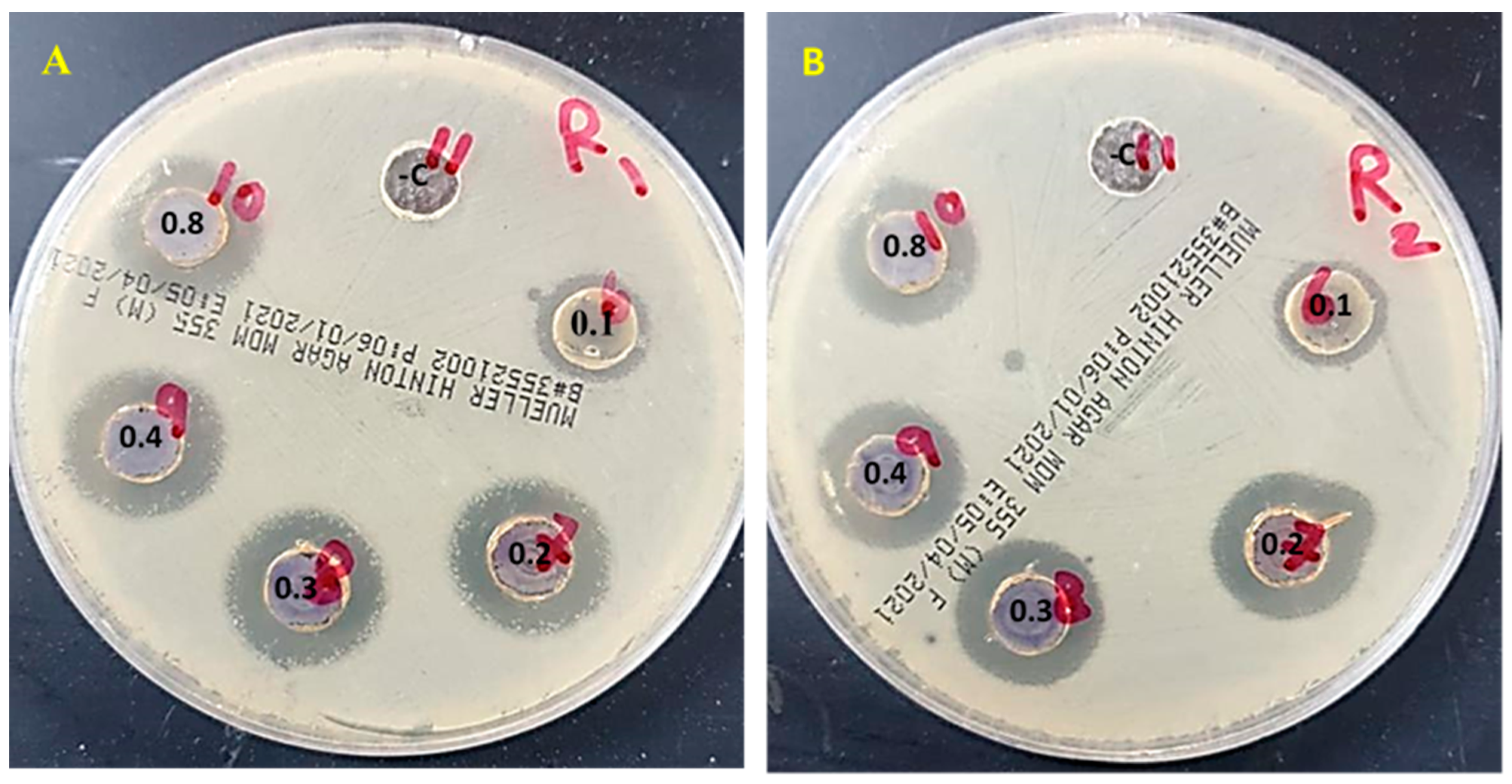
| S. mutans | 18 ± 0.2 c | 18 ± 0.1 c | 18 ± 0.2 c | 19 ± 0.1 c | 16 ± 0.00 b | 13 ± 0.2 a | 0 |
| L. acidophilus | 17 ± 0.13 c | 18 ± 0.1 c | 18 ± 0.05 c | 18 ± 0.05 c | 15 ± 0.1 b | 13 ± 0.1 a | 0 |
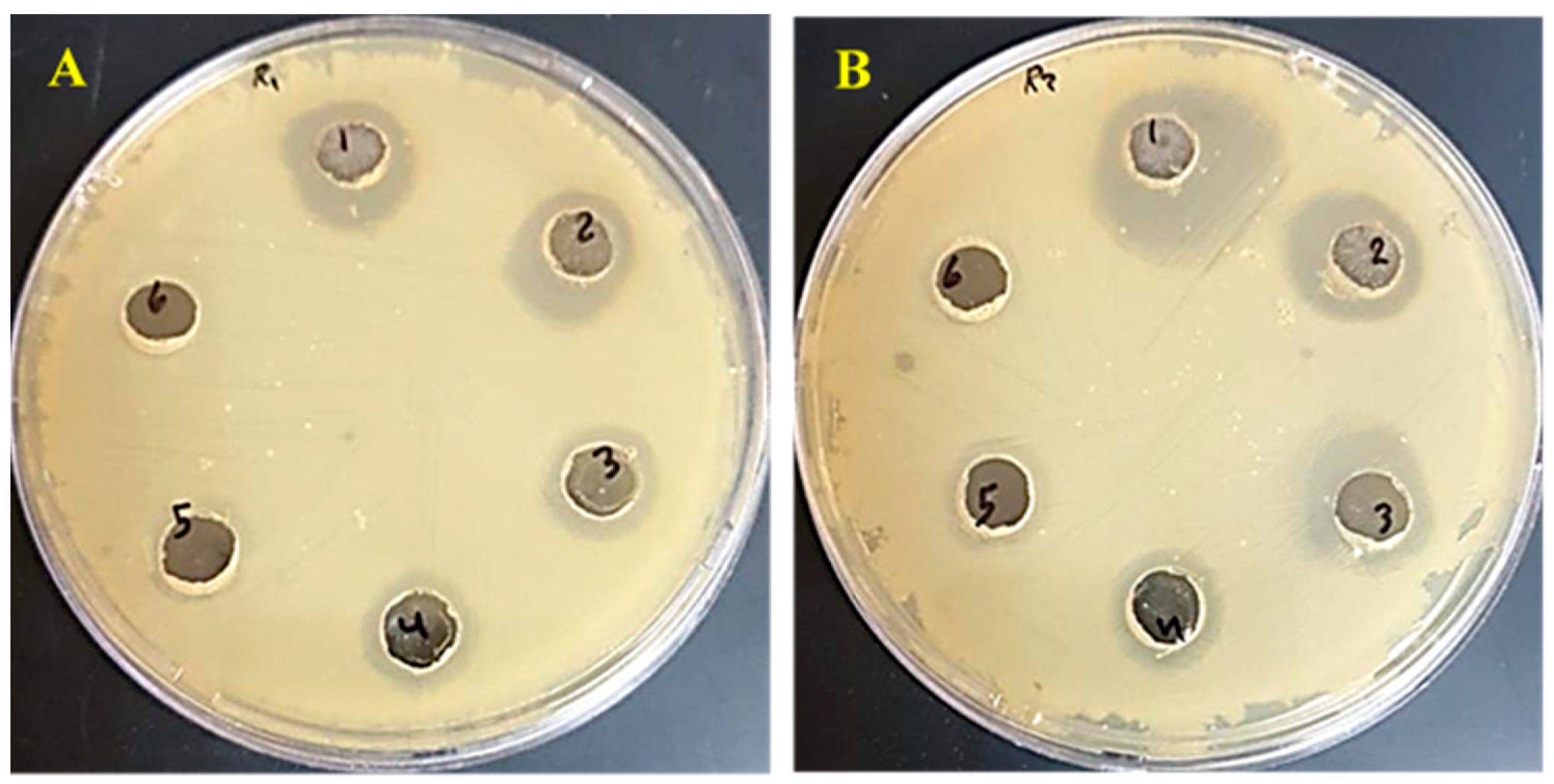
| Bacterial Strain | Nanocellulose | Ulva/AgNPs | Fluoride | Nanocellulose/Fluoride | Ulva/Ag/Cellulose Nanocomposites/Fluoride |
|---|---|---|---|---|---|
| S. mutans | 13 ± 0.2 a | 16 ± 0.00 b | 0 | 23 ± 2 c | 22 ± 1 c |
| L. acidophilus | 13 ± 0.2 a | 15 ± 0.1 b | 0 | 23 ± 1 c | 24 ± 2 c |
| Nanocomposites (g/mL) | 0.0018 | 0.0009 | 0.00045 | 0.000225 | 0.000112 | 0.000056 |
|---|---|---|---|---|---|---|
| S. mutans | 19 ± 0.1 | 18 ± 0.3 | 15 ± 0.1 | 14 ± 0.00 | 11 ± 0.00 | 0± |
| L. acidophilus | 19 ± 0.00 | 18 ± 0.1 | 15 ± 0.1 | 13 ± 0.00 | 12 ± 0.00 | 0± |
| Sources | Antibacterial Against | Reference | |
|---|---|---|---|
| AgNPs | Sargassum wightii | Micrococcus luteus, Serratia marcescens | [99] |
| Caulerpa serrulata | E. coli, Salmonella typhi | [100] | |
| Caulerpa racemosa | Staphylococcus aureus, Proteus mirabilis | [101] | |
| Chlorella ellipsoidea. | S. aureus, P. aeruginosa, K. pneumonia, E. coli | [102] | |
| Ecklonia cava | E. coli | [103] | |
| AgNPS/Cellulose | Orange peel waste | E. coli | [104] |
| Cotton pulp cellulose | E. coli, P. aeruginosa, S. aureus | [105] | |
| Bacterial cellulose | B. subtilis, S. aureus, E. coli | [106] | |
| Bacterial cellulose | Escherichia coli Staphylococcus aureus | [107] | |
| Ag/Ulva cellulose nanocomposites | S. mutans ATCC 25175 and L. acidophilus CH-2 | This study |
Disclaimer/Publisher’s Note: The statements, opinions and data contained in all publications are solely those of the individual author(s) and contributor(s) and not of MDPI and/or the editor(s). MDPI and/or the editor(s) disclaim responsibility for any injury to people or property resulting from any ideas, methods, instructions or products referred to in the content. |
© 2023 by the authors. Licensee MDPI, Basel, Switzerland. This article is an open access article distributed under the terms and conditions of the Creative Commons Attribution (CC BY) license (https://creativecommons.org/licenses/by/4.0/).
Share and Cite
Hamouda, R.A.; Qarabai, F.A.K.; Shahabuddin, F.S.; Al-Shaikh, T.M.; Makharita, R.R. Antibacterial Activity of Ulva/Nanocellulose and Ulva/Ag/Cellulose Nanocomposites and Both Blended with Fluoride against Bacteria Causing Dental Decay. Polymers 2023, 15, 1047. https://doi.org/10.3390/polym15041047
Hamouda RA, Qarabai FAK, Shahabuddin FS, Al-Shaikh TM, Makharita RR. Antibacterial Activity of Ulva/Nanocellulose and Ulva/Ag/Cellulose Nanocomposites and Both Blended with Fluoride against Bacteria Causing Dental Decay. Polymers. 2023; 15(4):1047. https://doi.org/10.3390/polym15041047
Chicago/Turabian StyleHamouda, Ragaa A., Fauzia A. K. Qarabai, Fathi S. Shahabuddin, Turki M. Al-Shaikh, and Rabab R. Makharita. 2023. "Antibacterial Activity of Ulva/Nanocellulose and Ulva/Ag/Cellulose Nanocomposites and Both Blended with Fluoride against Bacteria Causing Dental Decay" Polymers 15, no. 4: 1047. https://doi.org/10.3390/polym15041047