Article

Physical Chemical Investigation of Gamma-Irradiated Parchment for Preservation of Cultural Heritage

1
Horia Hulubei National Institute for Physics and Nuclear Engineering (IFIN-HH), Multipurpose Irradiation Facility (IRASM Department), 30 Reactorului Str., 077125 Magurele, Romania
2
National Research & Development Institute for Textile and Leather (INCDT-ICPI), 93 Ion Minulescu Str., 030508 Bucharest, Romania
3
Department of Analytical Chemistry and Physical Chemistry, Faculy of Chemistry, University of Bucharest, 4-12 Regina Elisabeta Bld., 030018 Bucharest, Romania
*
Author to whom correspondence should be addressed.
Polymers 2023, 15(4), 1034; https://doi.org/10.3390/polym15041034
Submission received: 10 January 2023 / Revised: 15 February 2023 / Accepted: 17 February 2023 / Published: 19 February 2023
(This article belongs to the Section Polymer Applications)

Abstract

:
The historical artefacts of parchment are prone to degradation if the storage conditions are improper due to the collagen structure having a limited stability under physical, chemical, and biological agent attacks. The parchment structure is difficult to characterize due to the variety of manufacturing traditions (eastern/western), intrinsic variability of skins (i.e., species, breeding variation, living conditions, effects of pathologies, etc.), biodeterioration, and aging, and the main concern in its analysis is its uniformity. The deterioration of parchment collagen produces a rather stiff or in some circumstances, a relaxed structure. Any intervention or treatment of unique, very precious cultural heritage artefacts must not negatively influence the properties of the component materials. Gamma irradiation is a relatively new technique of bioremediation. Data on the leather properties pre- and post-ionizing radiation bioremediation treatments are few in the literature. Fewer data are available on the historical leather and parchment physical chemical characteristics after ionizing gamma irradiation. This research had two main objectives: (i) the characterization of the parchment structure’s uniformity across the analyzed areas and its mechanical properties, i.e., tensile stress by mechanical tests and ATR-FTIR spectroscopy; and (ii) to establish parchment tolerance when exposed to ionizing gamma radiation as a pre-requisite for cultural heritage preservation irradiation treatment. It was found that the mechanical tests and ATR-FTIR spectroscopy may identify changes in the parchment’s irradiated structure and that the preservation of cultural heritage parchment artefacts may be performed at maximum 15 kGy gamma irradiation dose.

1. Introduction

Cultural heritage artefacts are prone to degradation due to physical, chemical, and biological factors. If physical and chemical degradation can be delayed by controlling the artefacts storage conditions, e.g., the temperature, relative humidity, and air purity, the biological attack, once installed, can only be stopped by a drastic intervention. Classical disinfection techniques based on using fumigation with various chemicals or solvents are ineffective against spores and eggs. New physical methods of conservation have been researched based on using lasers, plasma, or radiation treatments [1]. Among others, the ionizing radiation bioremediation treatment has the advantage of the certainty of the biocide effect, being a fast, mass, and safe treatment; no harmful chemicals and residues that may threaten the health of the museum personnel or general public are left in the materials [2]. Due to the novelty of the gamma irradiation treatment but also to the complexity and diversity of the constituents of cultural heritage items, the question that always remains to be answered is as follows: “Does the gamma radiation induces a supplementary degradation in the material?”. The subject is debated for numerous and diverse objects of wood [3], textile [4,5,6,7], paper [2,4,8,9,10,11,12,13], which are ornamented [14] or with polychromies [15,16,17,18]. It is also known that interventions for saving cultural heritage artefacts should be reversible. Nevertheless, the wood impregnation and consolidation using special resins and gamma radiation curing (Nucléart process), although irreversible, are used as cultural heritage artefacts salvation treatments [19]. In the literature, there are data on the behavior of the properties and mechanical parameters of leather under the effect of ionizing radiation, but there are fewer instances on historical examples, e.g., aged leather and parchment [20,21,22,23,24]. Parchment chemical structure is difficult to characterize due to its variability and aging conditions. The parchment is obtained from the skin of small animals such as sheep, goats, and calves through various treatments aimed at removing the hair by treatment with alkaline materials and drying in a tense state, and scraping and finishing by polishing and bleaching, in order to give the skin surface smoothness, suppleness, and opacity [25]. Although it is considered to be a primitive, coarse material, this is only in appearance, because the parchment has a sophisticated three-dimensional arrangement generated by its basic element, the collagen molecule, with a helical structure, which allows successive clockwise and counterclockwise windings and the formation of increasingly complex structures, such as collagen fibrils, collagen fibers, and three-dimensional collagen networks (e.g., derma, characterized by exceptional strength and durability). The millenary durability of the parchment is due, on one hand, to the simple chemical composition, but especially to this hierarchical structure of collagen, which is a complex structure that is difficult to evaluate as each hierarchical level requires a certain specific analysis technique, and a complete image of a parchment is obtained by correlating the results of all these techniques [26].
Skins display morphogenesis, which is formed from collagen molecules and diverse structures. The tanning of skins produces a crosslinked material while the parchment production generates a highly hygroscopic matrix due to the polar sites available for hydrogen bonding [27]. Microbiological infestation is common for parchment objects due to the presence of partially or completely gelatinized collagen fiber, and that is why safe storage implies dry conditions [27]. Chemical methods and freeze-drying were used to counter the mold growth on parchment. These methods may have adverse effects, producing changes in the material structure and in the case of the use of chemical fungicides, risks for one’s health and the environment may also appears. Gamma irradiation is researched as a new minimal invasive safe treatment technique, together with other physical techniques based on using plasma [28], lasers [29], e-beam, or X-rays [30,31,32]. The biocidal effect of gamma radiation is used for the sterilization of medical devices at industrial scale, which is effective for the whole volume of an artifact while plasma and e-beam techniques are used to treat surface attacks.
The main concern in the analysis of natural materials is their uniformity. One of the major problems when working with natural materials is the heterogeneity of the samples. In the case of skins, this is caused by the animal constitution and condition. Additionally, the parchment manufacturing process influences the structure of the final product. In order to exclude some of the mentioned influences, all the tested samples were collected from the middle section of one skin, close to the backbone of the animal, and the leather anatomical zone influence was proved by FTIR and mechanical tests. FTIR spectroscopy proved to be very useful in identifying the collagen structure and degree of conservation of various cultural heritage goods that were made of leather and parchment [33,34]. FTIR spectroscopy allows for the identification of changes at molecular and supramolecular levels by monitoring the band position and intensity. New bands in the FTIR spectrum, as well as the shift of band intensity, indicate the chemical or compositional modification of the samples, while the band position shifts indicate a conformation and/or crystallinity modification due to crosslinking, water adsorption/desorption, phase transitions, etc. [5,35]. The FTIR-ATR technique is commonly used in the study of collagenous structures allowing in-depth identification of alpha, beta, and random coil protein secondary structures [36,37]. Mechanical tests allow material characterization at a macroscale, and the combination of the FTIR analysis with mechanical test data may probe the correlation of molecular-scale modifications with the object’s macroproperties. This research had two main objectives: First, the characterization of the parchment’s structural uniformity across the analyzed areas and its mechanical properties, i.e., tensile stress. Secondly, to establish parchment tolerance when exposed to the ionizing gamma radiation as a pre-requisite for cultural heritage irradiation bioremediation treatment application. The motivation to treat the parchment at high gamma irradiation doses was simply to observe its tolerance to radiation. It was found that (i) mechanical tests and ATR-FTIR spectroscopy may identify changes in native parchment and gamma-irradiated structure; (ii) for cultural heritage parchment artefacts preservation the maximum allowed gamma treatment dose is 15 kGy.

2. Materials and Methods

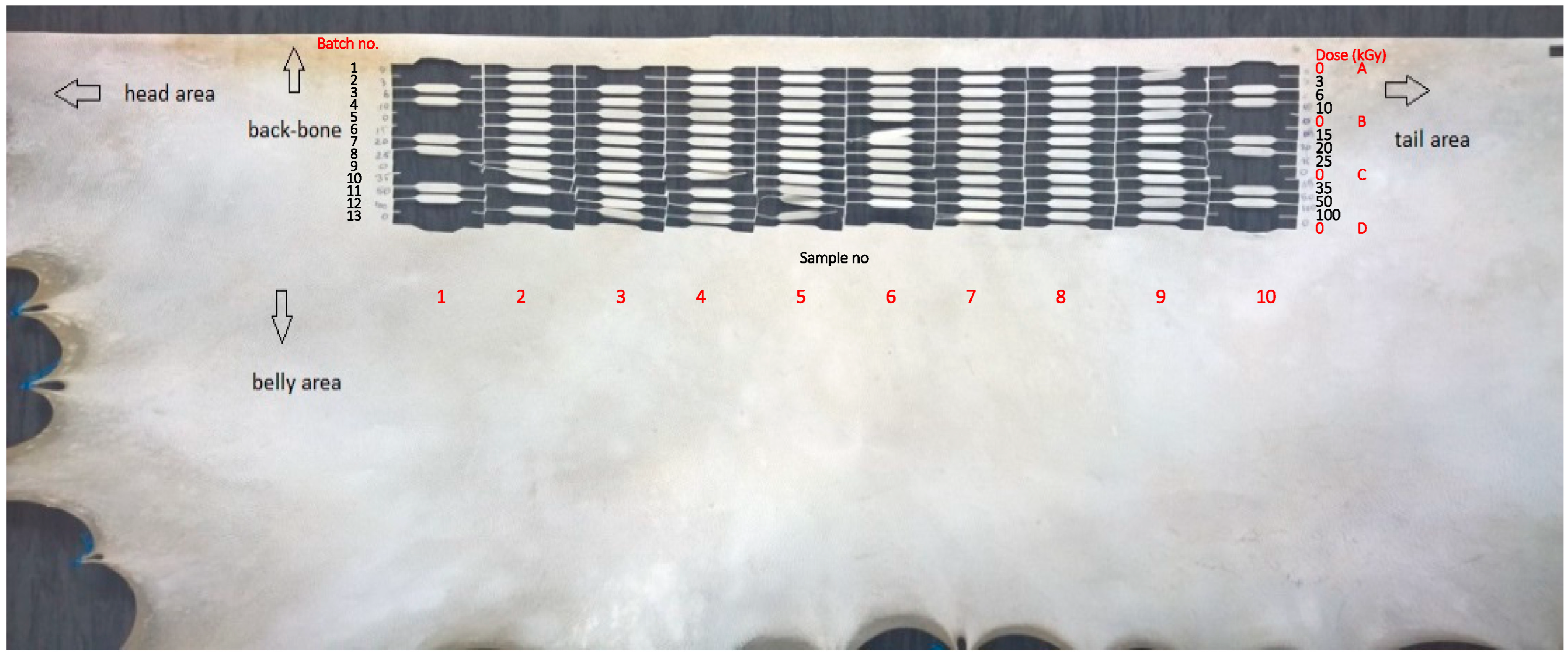
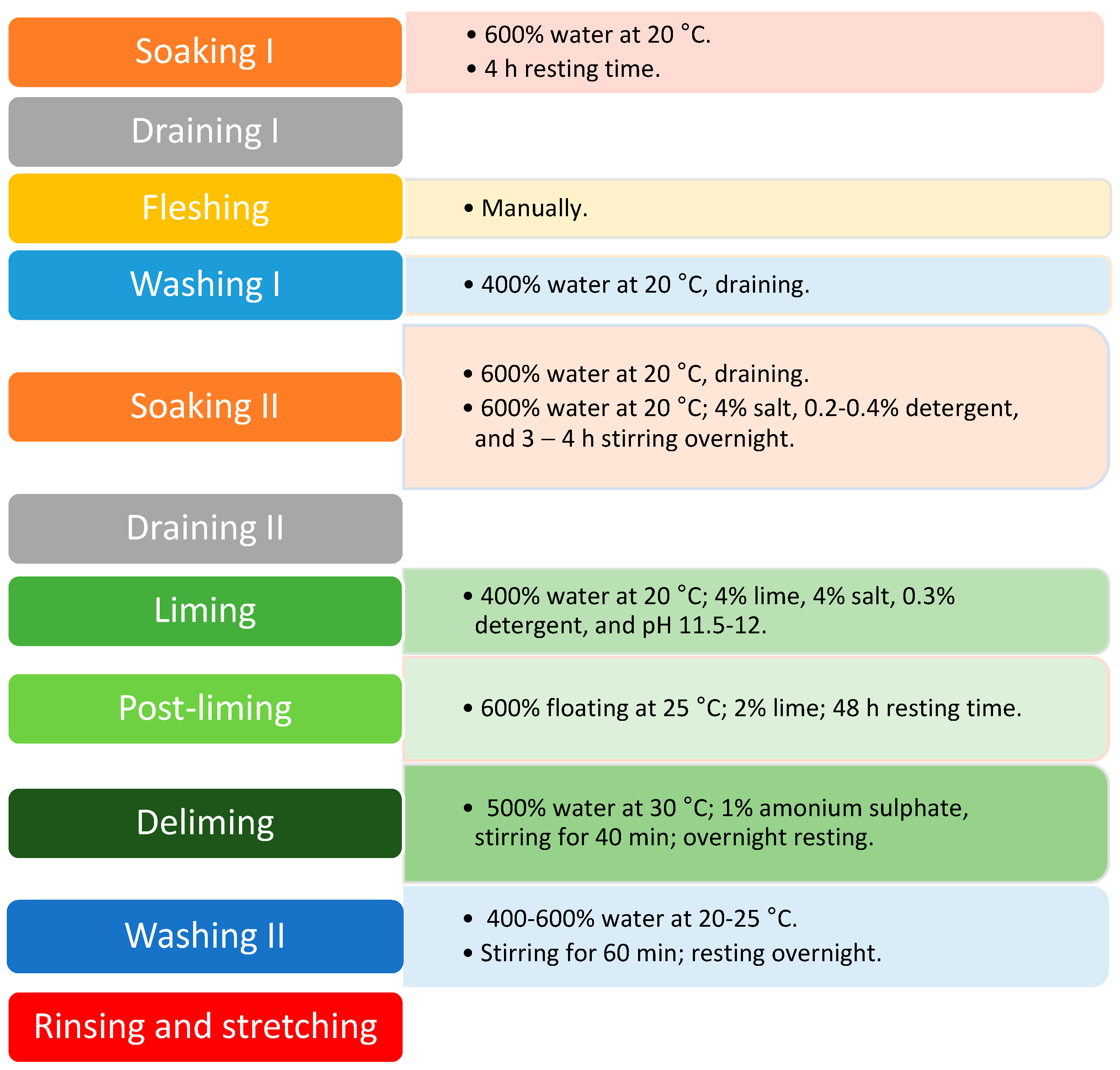
2.1. Parchment Preparation

The goat parchment was manufactured by INCDTP-ICPI, using traditional methods following the steps depicted in Scheme 1.
The processing operations of goat skins to obtain parchment are simple, only requiring washing, removal of hair and adhering residual flesh, and drying under tension. Washing is performed with Na2CO3 and house soap, and dehairing is performed with lime. Dehairing may take two days to one week based on the kind of skin and their curing state, environment temperature, and dehairing operation variables. In this case, the skin was dried for 3 days at normal temperature, between 22–28 °C, without ventilation in order to achieve a humidity of the material between 8–12%, depending on the relative humidity of the air. This humidity is specific to natural organic supports.

2.2. Gamma Irradiation of the Parchment

The samples were prepared for gamma irradiation and analysis at IFIN-HH, IRASM department, using a cutting tool according to ISO 37, type 4. The sampling was divided into 13 batches from the backbone to the belly area, as indicated in Figure 1; each batch contained 10 samples. The direction for collecting the samples was from head area to tail area. The direction of the samples was parallel to the backbone of the parchment skin. The samples were collected according to the parchment topography, i.e., starting from the backbone area downward to the belly area and from the head to the tail area. Figure 1 depicts the half goat skin parchment that was analyzed and the sample’s collection areas. Sample’s dimensions were measured using a Proma micrometer M026.
The samples were irradiated at IFIN-HH, IRASM department using a Cobalt-60 Gamma Cell 5000 research irradiator. There were 9 applied different irradiation doses: 3, 6, 10, 15, 20, 25, 35, 50, and 100 kGy, and 4 batches were kept as controls, at various distances from the backbone area to check parchment uniformity. The dose rate was approximately 6.51 kGy·h−1. The ethanol-chlorobenzene (ECB) dosimetry system was used to determine the applied dose and the dose uniformity. Between the irradiated batches were interleaved four non-irradiated batches (A, B, C, and D) in order to observe the uniformity and mechanical strength of the parchment. Non-irradiated batches were placed before 3 kGy and after 10, 25, and 100 kGy batches, as can be seen in Figure 1. Thus, their section intervals were obtained: interval no. 1 enclosing the batches: 0 kGy(A)–3 kGy–6 kGy–10 kGy–0 kGy(B); interval no. 2 enclosing the batches: 0 kGy(B)–15 kGy–20 kGy–25 kGy–0 kGy(C); and interval no. 3 comprising the batches: 0 kGy(C)–35 kGy–50 kGy–100 kGy–0 kGy(D).

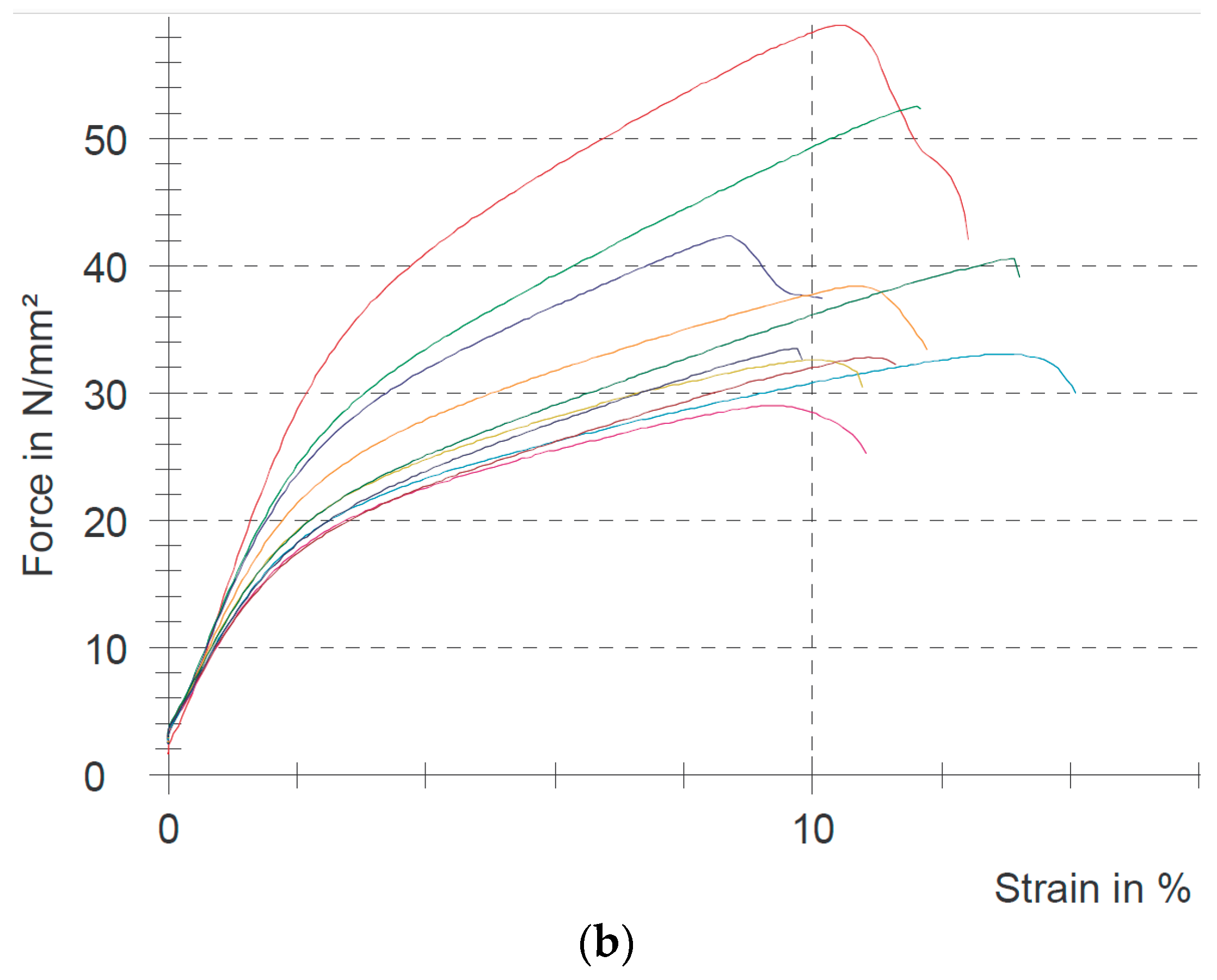
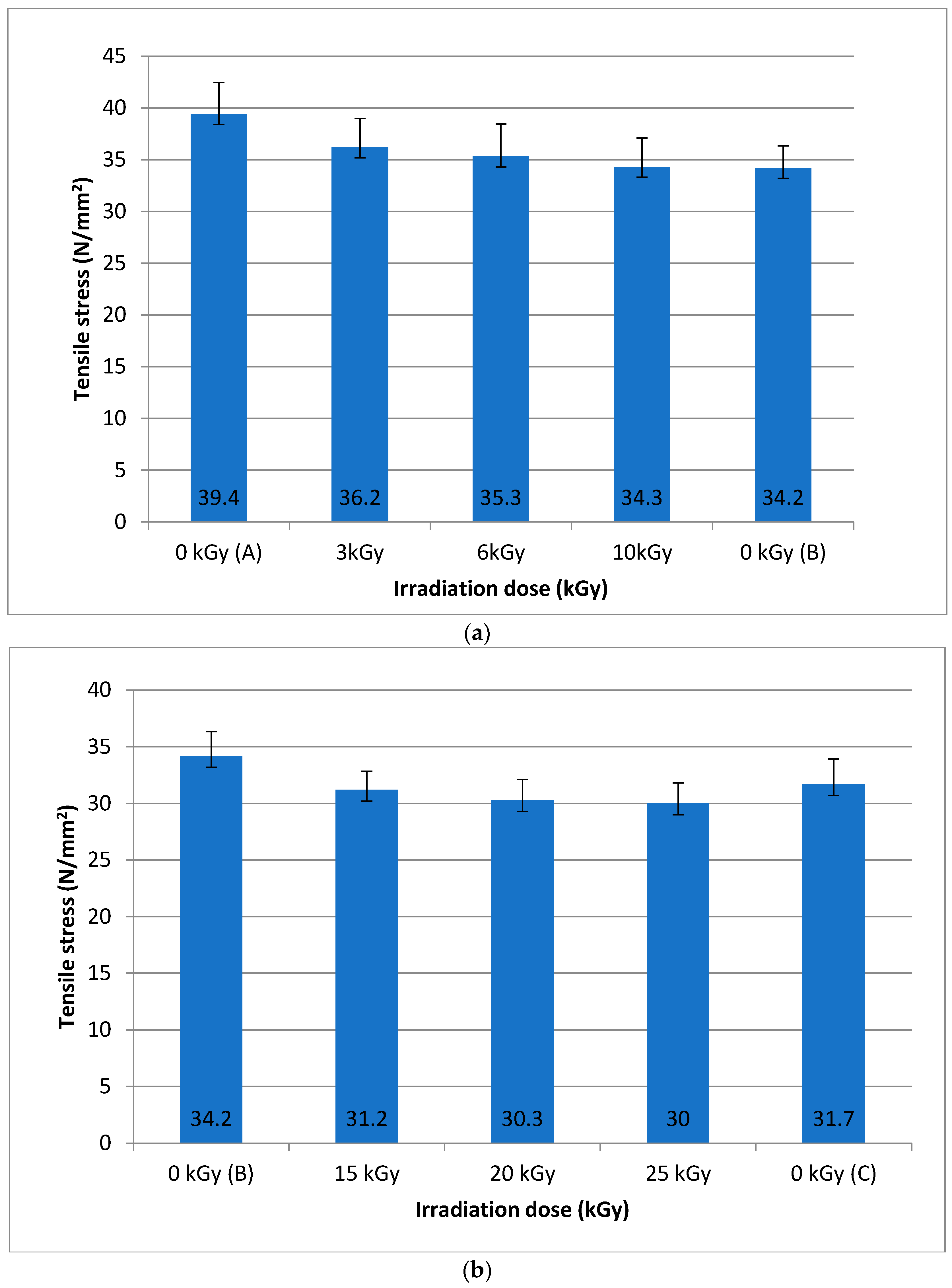
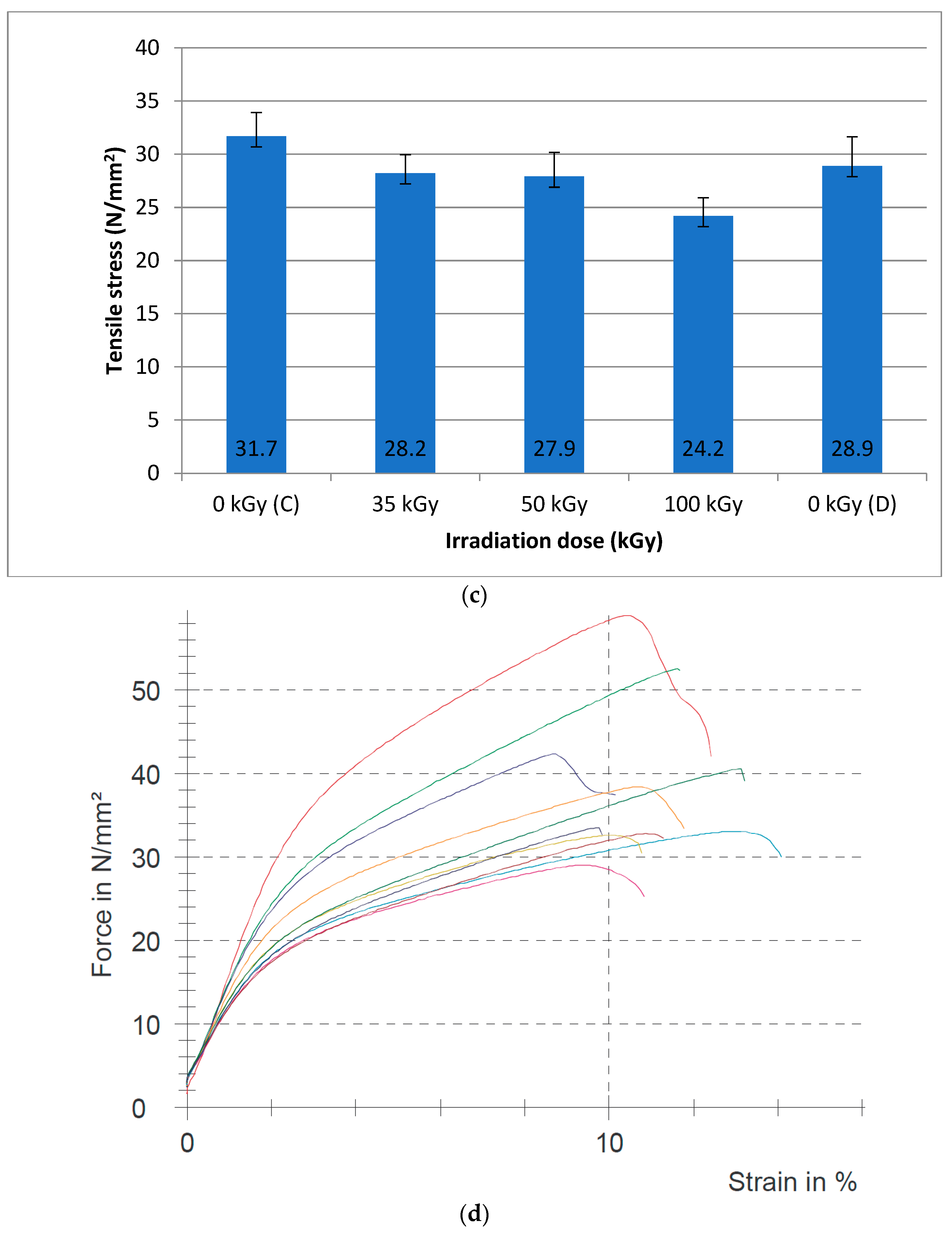
2.3. Mechanical Tests

The mechanical tests were conducted on a Zwick Roell testing machine with a 5 kN cell force.
The ISO 3376:2011 standard “Physical and mechanical tests–Determination of tensile strength and percentage extension” was not used for these tests due to the large dimension of the dumbbell, which results in a small number of testing samples and a large variation of tensile stress within the sample due to the heterogenic characteristics of natural materials. Instead, ISO 37 standard: “Rubber, vulcanized or thermoplastic-Determination of tensile stress–strain properties” was used. The dumbbell has the following characteristics: overall length, 35 mm; test length, 10 ± 0.5 mm; and width, 2 ± 0.1 mm. The testing speed was 200 mm·min−1 with a pre-load of 1 N. The reason for using such a small dumbbell is that it provided a large number of samples, which decreased the measuring errors induced by the heterogeneity of the natural material.

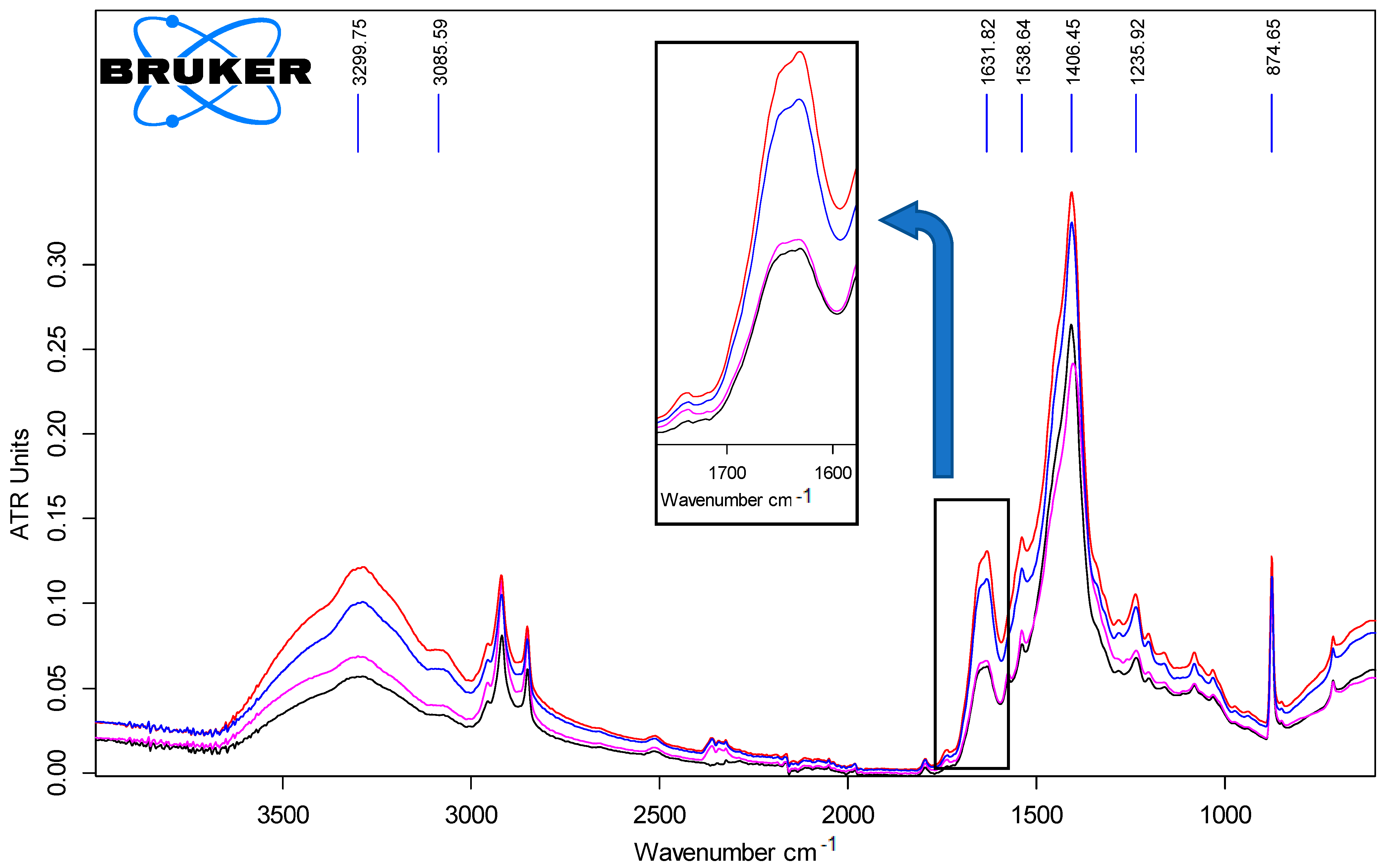
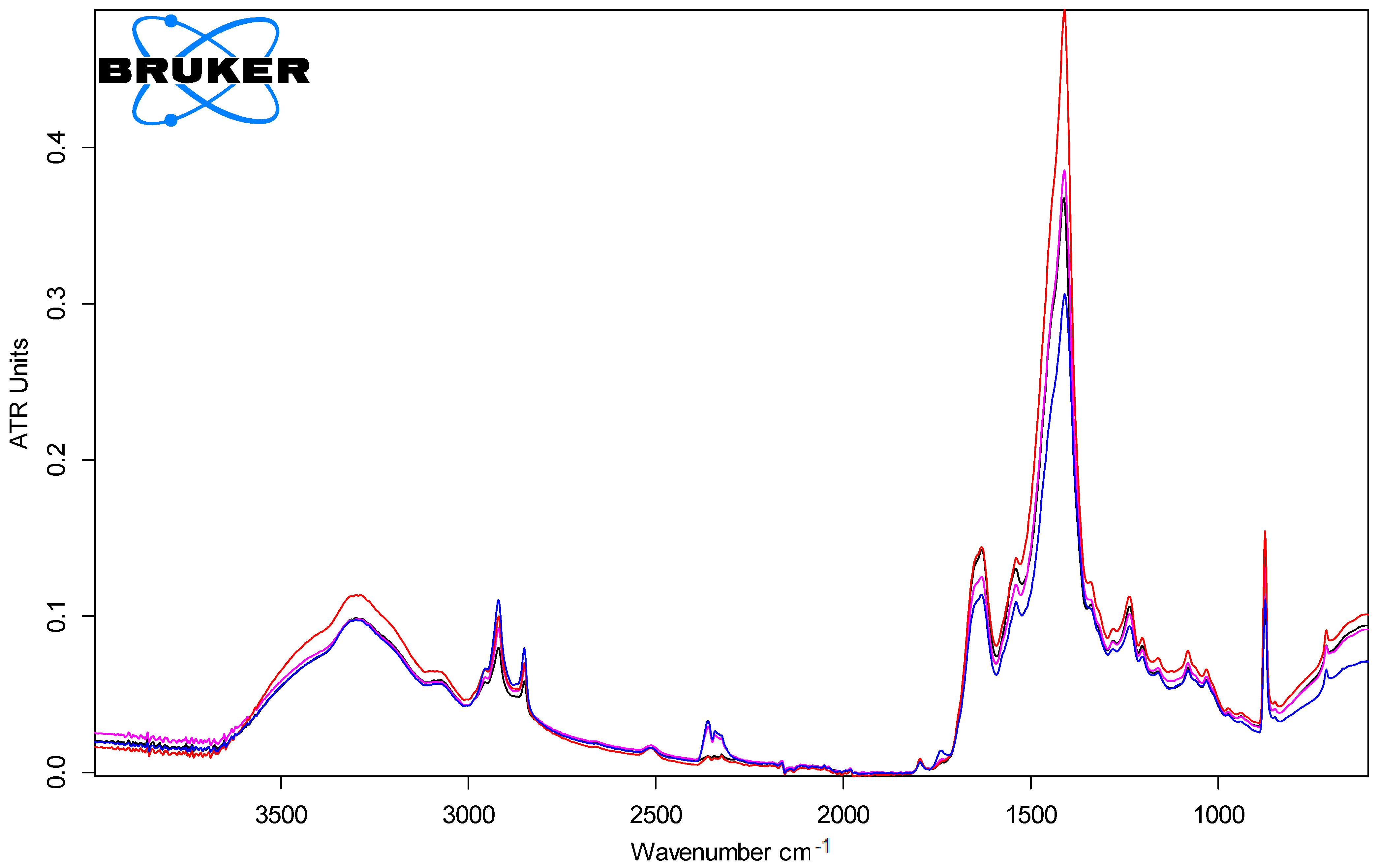
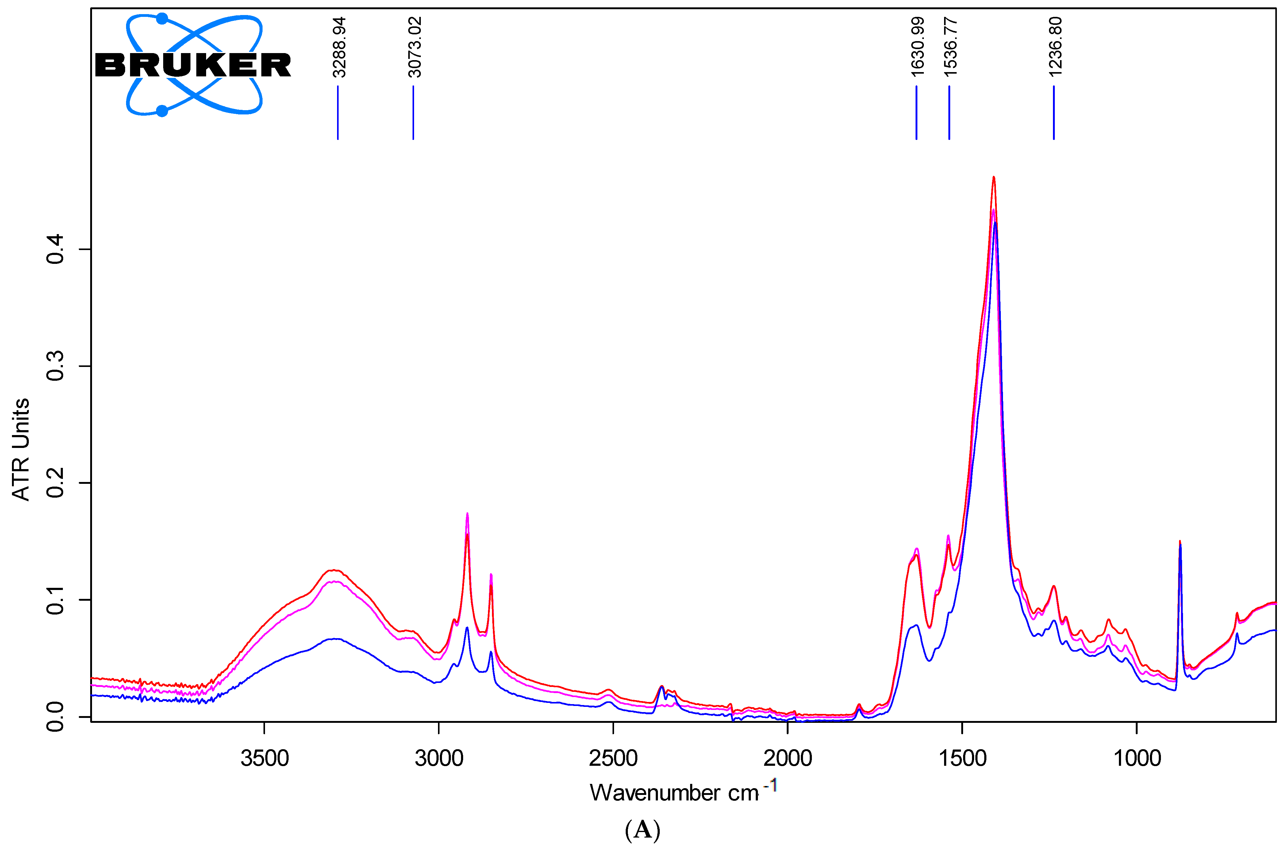
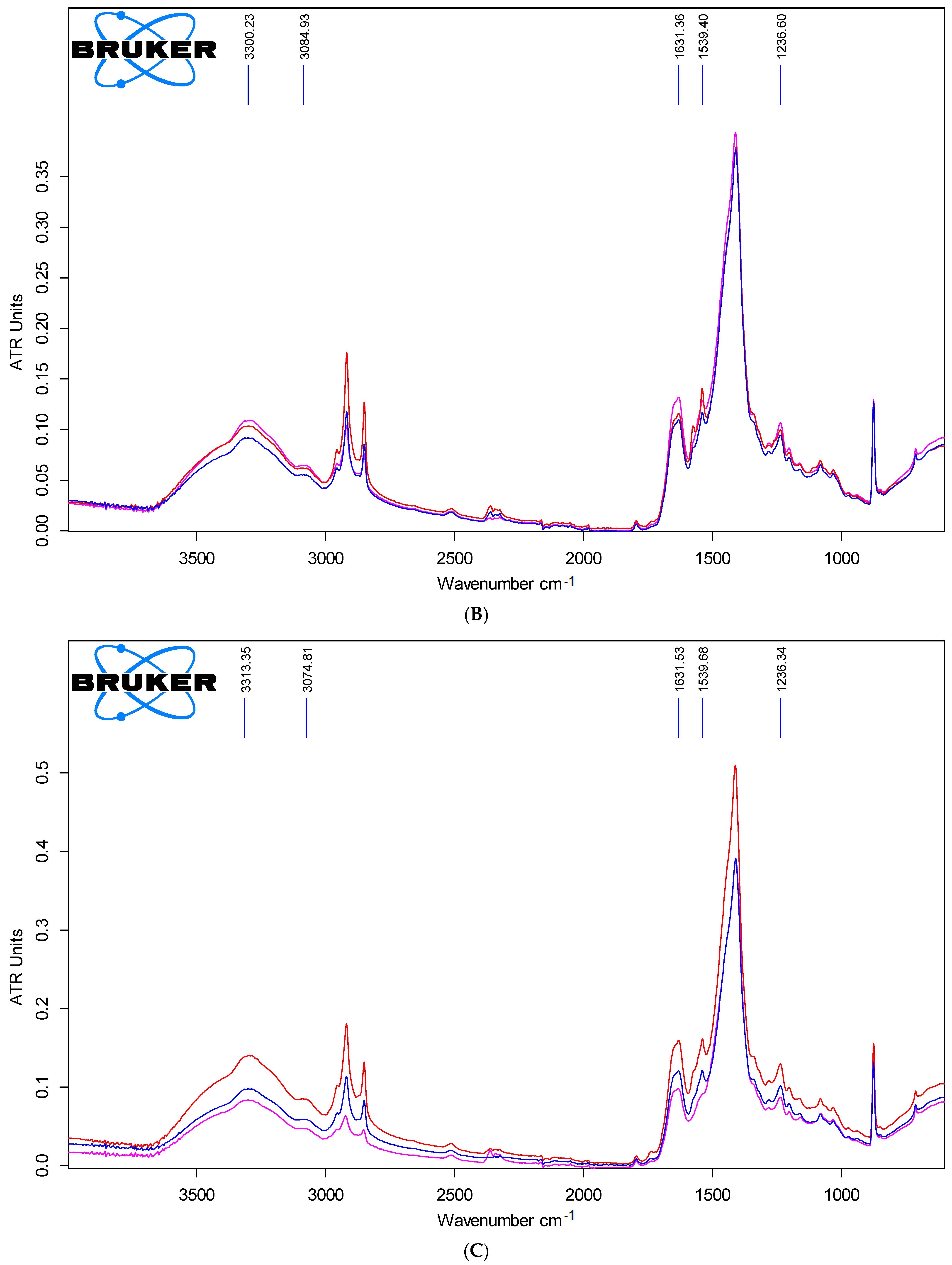
2.4. ATR-FTIR Spectroscopy Characterization

A second method of analysis was used, namely, FTIR by ATR technique, in order to observe the natural variability of the parchment structure and the possible changes at the molecular level after gamma irradiation, using a Bruker Vertex 70 machine, with a wavenumber range of 4000–400 cm−1, 4 cm−1 resolution, and diamond crystal ATR accessory. Ten measurements were conducted per sample and there were 256 scans per measurement. The results were mediated, thus a single spectrum resulted from the ten measurements. A total of eight control samples, four from the head area and four from the tail area, were collected from the extremes, head and tail, when furthering from the backbone to the belly. The samples were collected from the four interleaved non-irradiated batches corresponding to batch A, B, C, and D from head and tail areas to analyze anatomical position influence on the collagen parchment structure. In the case of the irradiated batches for each sample, there were also ten spectra measurements.

3. Results and Discussion

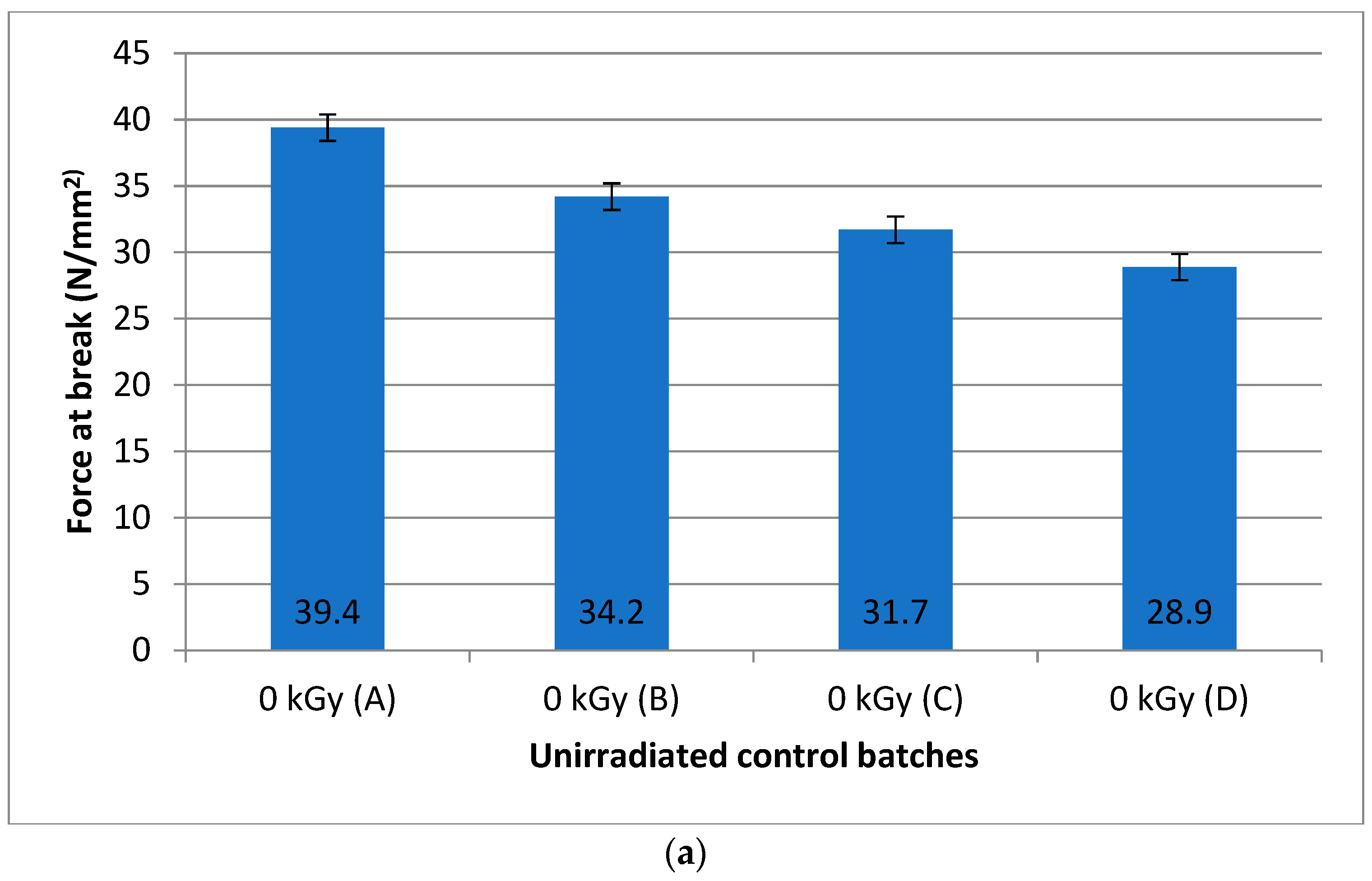
3.1. Physical Chemical Properties of Unirradiated Parchment

Figure 2 presents the tensile stress mean values obtained for the four non-irradiated batch samples A, B, C, and D, and for exemplification, the original stress–strain curves for unirradiated samples (batch A). All the experimental data are available upon request from the authors.
The control batch A is placed closest to the backbone while the control batch D is closest to the belly area. The highest average value for the tensile stress is 39.4 N/mm2, which is closest to the backbone (batch A), while the lowest value is 28.9 N/mm2 (batch D), which is farthest from the backbone. The natural decrease in the tensile stress that characterizes the parchment’s skin topography is obvious, while the standard error of the mean is low, comprised between 3.05 and 2.16 N/mm2. The results indicate that the natural mechanical resistance decreases along with the growth of the distance from the backbone to the belly area, from batch A to batch D.
The ATR-FTIR spectra of the control batches from the head area sampling are shown in Figure 3. It can be observed that there is a decrease in the intensity of the FTIR spectrum when furthering from the backbone area (A) to the belly area (D), which is in agreement with the measured decreased tensile stress. High mechanical resistance of the sample provides a good contact with the ATR crystal and a stronger intensity spectrum, while the loose parchment structure of the belly area produces a less intense vibration band spectrum. In the parchment FTIR spectra, the following main collagen bands are observed: amide A (3300 cm−1), amide B (3086 cm−1), amide I (1632 cm−1), amide II (1539 cm−1), and amide III (1233 cm−1) [34]. The multiple maxima of amide I of the 1650 cm−1 band indicate the various collagen protein conformations: alpha, beta, or random structures. The alpha helix structures are depicted by the left maximum of the amide I band while the random structures are described by the right maximum. An increase in the random structures peak maxima intensity in the belly area was observed, which is in accord with the mechanical tests that showed a decrease in the mechanical strength, i.e., tensile stress (see inset of Figure 3).
The ATR spectra from the tail area are shown in Figure 4 for the unirradiated control batches. The same behavior with the head samples is observed for the tail area samples, e.g., the decrease in the intensity of organized helix structures is depicted by the left maximum, and increase in random coil and unorganized structures is depicted by the right amide I band maximum for samples farthest of the backbone. In conclusion, there is no difference in behavior related to the decrease in the mechanical properties of the parchment from the backbone to the belly area for the tail and head areas.

3.2. Physical Chemical Properties of Gamma-Irradiated Parchment

The results of the mechanical tests obtained for the irradiated samples were analyzed by looking at the three intervals defined by the non-irradiated batches (control group, A, B, C, and D) as follows (see Figure 5a–c):
(a)
Interval no. 1: 0 kGy (A)–3 kGy–6 kGy–10 kGy–0 kGy (B);
(b)
Interval no. 2: 0 kGy (B)–15 kGy–20 kGy–25 kGy–0 kGy (C);
(c)
Interval no. 3: 0 kGy (C)–35 kGy–50 kGy–100 kGy–0 kGy (D).
The control group batches (A, B, C, and D) were created in order to analyze the behavior of the tensile stress of the samples before and after the irradiation treatment, taking into account the natural decrease in the tensile stress induced by the topography (anatomical zone) of the skin parchment.
The values for the irradiated samples at 3, 6, and 10 kGy, respectively, 36.2, 35.3, and 34.3 N/mm2 show a slight decrease from 3 to 6 and 10 kGy irradiation doses. Taking into consideration the calculated values of the standard error of the mean, respectively, 2.78, 3.13, and 2.79 N/mm2, one may conclude that the radiation has no damaging effect on the parchment because the observed decrease in the tensile stress is smaller than the natural decrease in the tensile stress in the direction from the backbone to belly area. Thus, the medium values for the irradiated samples are interposed between the non-irradiated batches, A and B samples, showing no influence of radiation treatment on the tensile stress.
For the interval no. 2, the tensile stress of 31.2 N/mm2 for 15 kGy is above the non-irradiated batch B value of 31.7 N/mm2 when including the values of the standard error of the mean 1.66 N/mm2, respectively, 2.22 N/mm2. This result concludes no damaging effect of the 15 kGy radiation treatment.
For the 20 and 25 kGy irradiation doses, the tensile stress values, respectively, 30.3 and 30 N/mm2 are below the non-irradiated second control batch B. Considering the values of the standard error of the mean 1.66, 1.83, and 1.81 N/mm2, we concluded that the gamma irradiation has a small damaging effect on the parchment, thus decreasing the tensile strength.
For the interval no. 3, a damaging effect is observed at 100 kGy, and the value of 24.2 N/mm2 is lower than the natural decrease in tensile stress for the non-irradiated batch D, 28.9 N/mm2. Additionally, for the irradiated samples at 35 and 50 kGy, the tensile stress is below the non-irradiated control batch D (28.9 N/mm2), i.e., 28.2 and 27.9 N/mm2, respectively.
For the irradiated samples, a slight decrease in the tensile stress can be considered a damaging effect, even if the standard error of the mean is taken into account, 1.75, respectively, 2.27 N/mm2.
Figure 6 demonstrates the ATR FTIR spectra of the gamma-irradiated parchment samples.
The irradiated samples have similar FTIR spectra, as compared to the controls, and the collagen structure is conserved but some changes in the position, shape, and band intensity can be observed. For the lowest irradiation doses of each interval, the intensities of the right and left maximum of the amide I band are comparable, while at higher doses, some increase in the random coil structure’s peak component is observed, i.e., the right maximum component intensity of the amide I band increases with the irradiation dose.
The ATR technique results correlates well with the mechanical tests results, showing at molecular level the small structural differences for the irradiated and non-irradiated collagen parchment samples.

4. Conclusions

In this paper, the natural variability of the collagen parchment structure was demonstrated by mechanical testing and the ATR-FTIR spectra analysis. The change in the amide I band intensity and shape is correlated with small changes in the collagen protein conformations and correlates with the mechanical behavior. Additionally, the influence of the gamma irradiation treatment on the parchment’s collagen was identified from small changes in the ratio between the alpha helix and random coil secondary protein structures that form the left and right maxima of the amide I vibration band. The variation of the tensile stress was considered when establishing the maximum allowed gamma irradiation dose.
In conclusion, the gamma radiation treatment for parchment disinfection is safe for doses up to 15 kGy, as observed from small modifications of the ATR spectrum and mechanical tests. Taking into account the necessity of using maximum doses of 15 kGy, it can be stated that radiation treatment is a viable alternative/process for cultural heritage decontamination and preservation. Further investigation may concern the natural and/or artificial ageing influence on the structure and stability of gamma-irradiated parchment for an even safer approach for the preservation of unique cultural heritage artefacts.

Author Contributions

Conceptualization, I.B.L. and I.S.; methodology, I.B.L., L.M., M.C. and I.S.; validation, I.B.L., M.C., and I.S.; formal analysis, I.B.L., M.C. and I.S.; investigation, I.B.L., M.C. and I.S.; resources, I.B.L. and I.S.; data curation, I.B.L. and I.S.; writing—original draft preparation, I.B.L.; writing—review and editing, I.S.; supervision, L.M.; project administration, I.S.; funding acquisition, I.S. All authors have read and agreed to the published version of the manuscript.

Funding

This research was funded by the Romanian Ministry of Research and Education, contract number 44 PCCDI/2018, C1.2.PFE_CDI.2021-587/contract no. 41PFE/30.12.2021, and the program Nucleus 10N/2019 “Multidisciplinary applications of gamma irradiation”, PN23210301 “Valorization and sustainable recovery of resources through ionizing radiations technologies for blue economy and circular economy”. The APC was funded by all the authors.

Institutional Review Board Statement

Not applicable.

Informed Consent Statement

Not applicable.

Data Availability Statement

Data are available upon request from the authors.

Acknowledgments

ATR-FTIR spectra acquisition by MSc Ovidiu Capraru and MSc Cosmin Herman is gratefully appreciated.

Conflicts of Interest

The authors declare no conflict of interest. The funders had no role in the design of the study; in the collection, analyses, or interpretation of data; in the writing of the manuscript, or in the decision to publish the results.

References

  1. Cortella, L.; Albino, C.; Tran, Q.K.; Froment, K. 50 years of French experience in using gamma rays as a tool for cultural heritage remedial conservation. Rad. Phys. Chem. 2020, 171, 108726. [Google Scholar] [CrossRef]
  2. Moise, I.V.; Virgolici, M.; Negut, C.D.; Manea, M.; Alexandru, M.; Trandafir, L.; Zorila, F.L.; Talasman, C.M.; Manea, D.; Nisipeanu, S.; et al. Establishing the irradiation dose for paper decontamination. Rad. Phys. Chem. 2012, 81, 1045–1050. [Google Scholar] [CrossRef]
  3. Choi, J.I.; Lim, S. Inactivation of fungal contaminants on Korean traditional cashbox by gamma irradiation. Rad. Phys. Chem. 2016, 118, 70–74. [Google Scholar] [CrossRef]
  4. Drábková, K.; Ďurovič, M.; Kučerová, I. Influence of gamma radiation on properties of paper and textile fibres during disinfection. Rad. Phys. Chem. 2018, 152, 75–80. [Google Scholar] [CrossRef]
  5. Geba, M.; Lisa, G.; Ursescu, C.M.; Olaru, A.; Spiridon, I.; Leon, A.L.; Stanculescu, I. Gamma irradiation of protein-based textiles for historical collections decontamination. J. Thermal Anal. Calorim. 2014, 118, 977–985. [Google Scholar] [CrossRef]
  6. Kavkler, K.; Pucić, I.; Zalar, P.; Demšar, A.; Mihaljević, B. Is it safe to irradiate historic silk textile against fungi? Rad. Phys. Chem. 2018, 150, 101–110. [Google Scholar] [CrossRef]
  7. Takacs, E.; Wojnárovits, L.; Borsa, J.; Földváry, C.; Hargittai, P.; Zöld, O. Effect of γ-irradiation on cotton-cellulose. Rad. Phys. Chem. 1999, 55, 663–666. [Google Scholar] [CrossRef]
  8. Area, M.C.; Calvo, A.M.; Felissia, F.E.; Docters, A.; Miranda, M.V. Influence of dose and dose rate on the physical properties of commercial papers commonly used in libraries and archives. Rad. Phys. Chem. 2014, 96, 217–222. [Google Scholar] [CrossRef]
  9. Bicchieri, M.; Monti, M.; Piantanida, G.; Sodo, A. Effects of gamma irradiation on deteriorated paper. Rad. Phys. Chem. 2016, 125, 21–26. [Google Scholar] [CrossRef]
  10. Choi, J.I.; Chung, Y.J.; Lee, K.S.; Lee, J.W. Effect of radiation on disinfection and mechanical properties of Korean traditional paper, Hanji. Rad. Phys. Chem. 2012, 81, 1051–1054. [Google Scholar] [CrossRef]
  11. Kantoğlu, Ö.; Ergun, E.; Ozmen, D.; Halkman, H.B. A biological survey on the Ottoman Archive papers and determination of the D10 value. Rad. Phys. Chem. 2018, 144, 204–210. [Google Scholar] [CrossRef]
  12. Kodama, Y.; Rodrigues, O., Jr.; Garcia, R.H.L.; de Souza Santos, P.; Vasquez, P.A. Study of free radicals in gamma irradiated cellulose of cultural heritage materials using Electron Paramagnetic Resonance. Rad. Phys. Chem. 2016, 124, 169–173. [Google Scholar] [CrossRef]
  13. Trandafir, L.; Zorila, F.L.; Alexandru, M.; Ene, M.; Constantin, M.; Alistar, A.; Cutrubinis, M.; Iordache, O.; Stanculescu, R.I. Radioresistance of biodegradation fungi and its importance in establishing the decontamination dose. In Proceedings of the 5th International Conference on Advanced Materials and Systems (ICAMS), Bucharest, Romania, 23–25 October 2014; pp. 561–566. [Google Scholar]
  14. Marušić, K.; Pucić, I.; Desnica, V. Ornaments in radiation treatment of cultural heritage: Color and UV–Vis spectral changes in irradiated nacres. Rad. Phys. Chem. 2016, 124, 62–67. [Google Scholar] [CrossRef]
  15. Manea, M.M.; Moise, I.V.; Virgolici, M.; Negut, C.D.; Barbu, O.H.; Cutrubinis, M.; Fugaru, V.; Stanculescu, I.R.; Ponta, C.C. Spectroscopic evaluation of painted layer structural changes induced by gamma radiation in experimental models. Rad. Phys. Chem. 2012, 81, 160–167. [Google Scholar] [CrossRef]
  16. Manea, M.M.; Negut, C.D.; Stanculescu, I.R.; Ponta, C.C. Irradiation effects on canvas oil painting: Spectroscopic observations. Rad. Phys. Chem. 2012, 81, 1595–1599. [Google Scholar] [CrossRef]
  17. Negut, C.D.; Bercu, V.; Duliu, O.G. Defects induced by gamma irradiation in historical pigments. J. Cultural Heritage 2012, 13, 397–403. [Google Scholar] [CrossRef]
  18. Yoon, M.; Kim, D.W.; Choi, J.I.; Chung, Y.J.; Kang, D.I.; Kim, G.H.; Son, K.T.; Park, H.J.; Lee, J.W. Effect of gamma irradiation on Korean traditional multicolored paintwork. Rad. Phys. Chem. 2015, 115, 112–118. [Google Scholar] [CrossRef]
  19. International Atomic Energy Agency. Uses of Ionizing Radiation for Tangible Cultural Heritage Conservation; IAEA Radiation Technology series no. 6; IAEA: Vienna, Austria, 2017; Available online: https://www.iaea.org/publications/10937/uses-of-ionizing-radiation-for-tangible-cultural-heritage-conservation (accessed on 15 February 2023).
  20. Lungu, I.B.; Moise, V.I.; Cutrubinis, M.; Stanculescu, I.R. Study on mechanical proprieties of gamma irradiated leather and parchment. In Proceedings of the 5th International Conference on Advanced Materials and Systems, Bucharest, Romania, 23–25 October 2014; pp. 527–532. Available online: https://icams.ro/icamsresurse/2014/full_papers/5_Cultural_Heritage/05.pdf (accessed on 15 February 2023).
  21. Nunes, I.; Mesquita, N.; Verde, S.C.; Trigo, M.J.; Ferreira, A.; Carolino, M.M.; Portugal, A.; Botelho, M.L. Gamma radiation effects on physical properties of parchment documents: Assessment of Dmax. Rad. Physics Chem. 2012, 81, 1943–1946. [Google Scholar] [CrossRef]
  22. Csepregi, Á.; Szikszai, Z.; Targowski, P.; Sylwestrzak, M.; Müller, K.; Huszánk, R.; Angyal, A.; Donczo, B.; Kertesz, Z.; Szarka, M.; et al. Possible modifications of parchment during ion beam analysis. Herit. Sci. 2022, 10, 1–13. [Google Scholar] [CrossRef]
  23. Marušić, K.; Mlinarić, N.M.; Mihaljević, B. Radiation treatment of cultural heritage objects made of leather treated with common preservatives. Rad. Phys. Chem. 2022, 197, 110126. [Google Scholar] [CrossRef]
  24. Vadrucci, M.; De Bellis, G.; Mazzuca, C.; Mercuri, F.; Borgognoni, F.; Schifano, E.; Uccelletti, D.; Cicero, C. Effects of the ionizing radiation disinfection treatment on historical leather. Front. Mater. 2020, 7, 21. [Google Scholar] [CrossRef] [Green Version]
  25. Miu, L.; Badea, E. Parchment Manufacturing. In Parchment...a Story. The Unseen Face of Documents on Parchment Issued by the Royal Chancellery during the Time of Stefan cel Mare; Excelenta prin Cultura: Bucuresti, Romania, 2015; pp. 30–35. [Google Scholar]
  26. Poulsen, D.V.; Badea, E. (Eds.) Guidelines—Parchment Assessment Report, International Seminar and Workshop; Conservation and Restoration of Parchments; Civico Stampa: Torino, Italy, 2008. [Google Scholar]
  27. Kite, M.; Thomson, R. (Eds.) Conservation of Leather and Related Materials; Routledge: Oxfordshire, UK, 2006; pp. 202–224. [Google Scholar]
  28. Tiňo, R.; Vizárová, K.; Krčma, F. Plasma surface cleaning of cultural heritage objects. In Nanotechnologies and Nanomaterials for Diagnostic, Conservation and Restoration of Cultural Heritage; Elsevier: Amsterdam, The Netherlands, 2019; pp. 239–275. [Google Scholar]
  29. Kennedy, C.J.; Vest, M.; Cooper, M.; Wess, T.J. Laser cleaning of parchment: Structural, thermal and biochemical studies into the effect of wavelength and fluence. Appl. Surf. Sci. 2004, 227, 151–163. [Google Scholar] [CrossRef]
  30. Vadrucci, M.; Cicero, C.; Borgognoni, F.; Ceres, G.; Perini, N.; Migliore, L.; Mercuri, F.; Orazi, N.; Paoloni, S.; Rubechini, A. Parchment disinfection treatment by ionizing radiation. In Proceedings of the 2018 Metrology for Archaeology and Cultural Heritage (MetroArchaeo), Cassino, Italy, 22–24 October 2018; pp. 367–372. [Google Scholar] [CrossRef]
  31. Vadrucci, M.; Cicero, C.; Parisse, P.; Casalis, L.; De Bellis, G. Surface evaluation of the effect of X-rays irradiation on parchment artefacts through AFM and SEM. Appl. Surf. Sci. 2020, 513, 145881. [Google Scholar] [CrossRef]
  32. Vadrucci, M. A Machine for Ionizing Radiation Treatment of Bio-Deteriogens Infesting Artistic Objects. Quantum Beam Sci. 2022, 6, 33. [Google Scholar] [CrossRef]
  33. Vadrucci, M.; Cicero, C.; Mazucca, C.; Mercuri, F.; Missori, M.; Orazi, N.; Severini, L.; Zammit, U. Effect of X-ray and artificial aging on parchment. Eur. Phys. J. Plus 2021, 136, 1–16. [Google Scholar] [CrossRef]
  34. Carsote, C.; Şendrea, C.; Micu, M.C.; Adams, A.; Badea, E. Micro-DSC, FTIR-ATR and NMR MOUSE study of the dose-dependent effects of gamma irradiation on vegetable-tanned leather: The influence of leather thermal stability. Rad. Phys. Chem. 2021, 189, 109712. [Google Scholar] [CrossRef]
  35. Chirila, L.; Popescu, A.; Stanculescu, I.R.; Cutrubinis, M.; Cerempei, A.; Sandu, I. Gamma irradiation effects on natural dyeing performances of wool fabrics. Rev. Chim. 2016, 67, 2628–2633. [Google Scholar]
  36. Olariu, L.; Dumitriu, B.G.; Gaidau, C.; Stanca, M.; Tanase, L.M.; Ene, M.D.; Stanculescu, I.R.; Tablet, C. Bioactive Low Molecular Weight Keratin Hydrolysates for Improving Skin Wound Healing. Polymers 2022, 14, 1125. [Google Scholar] [CrossRef]
  37. Gaidau, C.; Stanculescu, I.R.; Stanca, M.; Cutrubinis, M.; Trandafir, L.; Alexandru, M.; Alexe, C.A. Gamma irradiation a green alternative for hides and leather conservation. Rad. Phys. Chem. 2021, 182, 109369. [Google Scholar] [CrossRef]
Scheme 1. Framework procedure for skin processing to obtain parchment.
Polymers 15 01034 sch001
Figure 1. Half skin parchment sample after mechanical tests samples withdraw, with indication of sampling areas (head, backbone, belly, and tail), batches number (left column numbers), sample number (horizontal line numbers), and irradiation doses (right column numbers; in the case of control samples, the batches A, B, C, and D are highlighted also).
Polymers 15 01034 g001
Figure 2. (a) Tensile stress mean values for unirradiated (0 kGy) samples (control batches) from head to tail area (A, B, C and D); (b) an example of the stress–strain curves for the A batch samples.
Polymers 15 01034 g002aPolymers 15 01034 g002b
Figure 3. ATR spectra for non-irradiated (control) batches from head area (A to D batches); red, blue, and magenta curves depict samples near the backbone, and black curve depicts the sample farthest from backbone; inset with the amide I band depicting modification of the helix and random coil components, e.g., left and right maxima of the amide I band.
Polymers 15 01034 g003
Figure 4. The ATR spectra for non-irradiated control batches from tail area (A–D); red, blue, and magenta curves depict samples near the backbone, and black curve depicts the sample farthest from the backbone.
Polymers 15 01034 g004
Figure 5. Tensile stress mean values of gamma-irradiated samples: (a) interval 1 results from control batch A to B; (b) interval 2 results from control batch B to C; and (c) interval 3 results from batch C to D. (d) An example of the stress–strain curves for the 3 kGy irradiated batch samples.
Polymers 15 01034 g005aPolymers 15 01034 g005b
Figure 6. The ATR-FTIR spectra for parchment irradiated samples; (A) interval no. 1: 3–6–10 kGy; (B) interval no. 2: 15–20–25 kGy; and (C) interval no. 3: 35–50–100 kGy. Amide I band depicts modifications of the helix and random coil components, for lowest doses (blue lines) and, respectively, higher doses (red and magenta lines); increase of the right maxima of the band responsible for random structures is observed.
Polymers 15 01034 g006aPolymers 15 01034 g006b
Disclaimer/Publisher’s Note: The statements, opinions and data contained in all publications are solely those of the individual author(s) and contributor(s) and not of MDPI and/or the editor(s). MDPI and/or the editor(s) disclaim responsibility for any injury to people or property resulting from any ideas, methods, instructions or products referred to in the content.

