
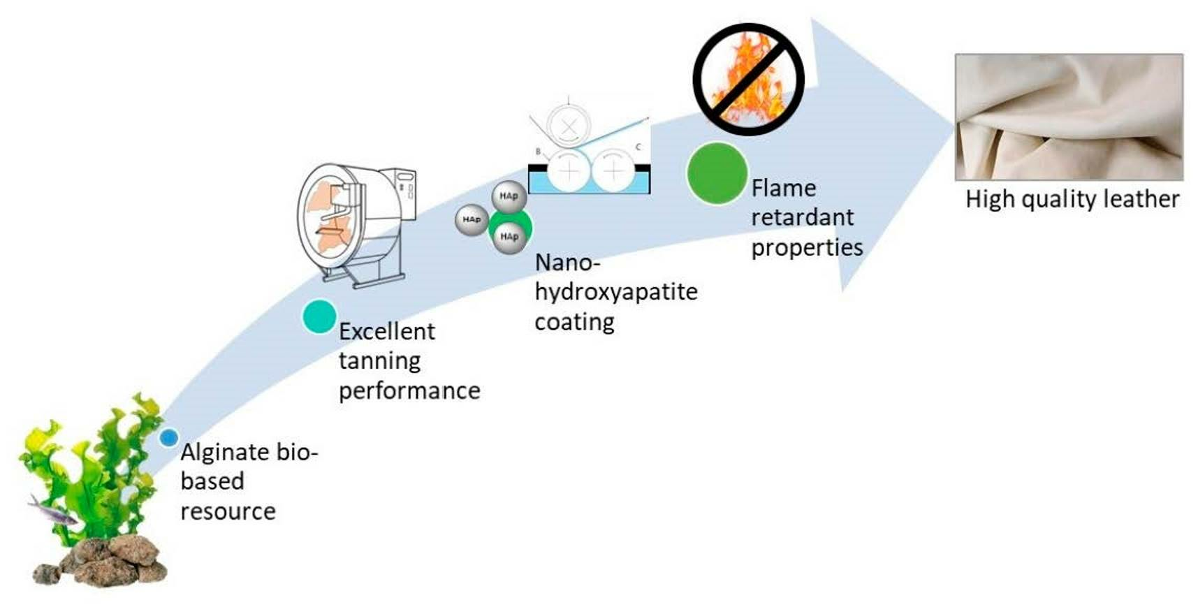
Cleaner Leather Tanning and Post-Tanning Processes Using Oxidized Alginate as Biodegradable Tanning Agent and Nano-Hydroxyapatite as Potential Flame Retardant
 ,
,  , ,
, ,
Abstract
:
1. Introduction
1.1. Sustainability Issues in the Tanning Industry
1.2. Oxidized Sodium Alginate as an Alternative Chrome-Free Tanning Material
1.3. Nano-Hydroxyapatite as Less-Toxic Additive for Improving the Flame Retardancy of Leather
2. Materials and Methods
2.1. Sodium Alginate Oxidation
2.2. Nano-Hydroxyapatite Synthesis
2.3. Analyses Methods and Techniques
3. Results and Discussion
3.1. Laboratory-Scale Tanning Test Using OSA and Nano-HAp: Study of Their Interaction with Collagen Using Micro-DSC, NMR-MOUSE, ATR-FTIR and SEM-EDS
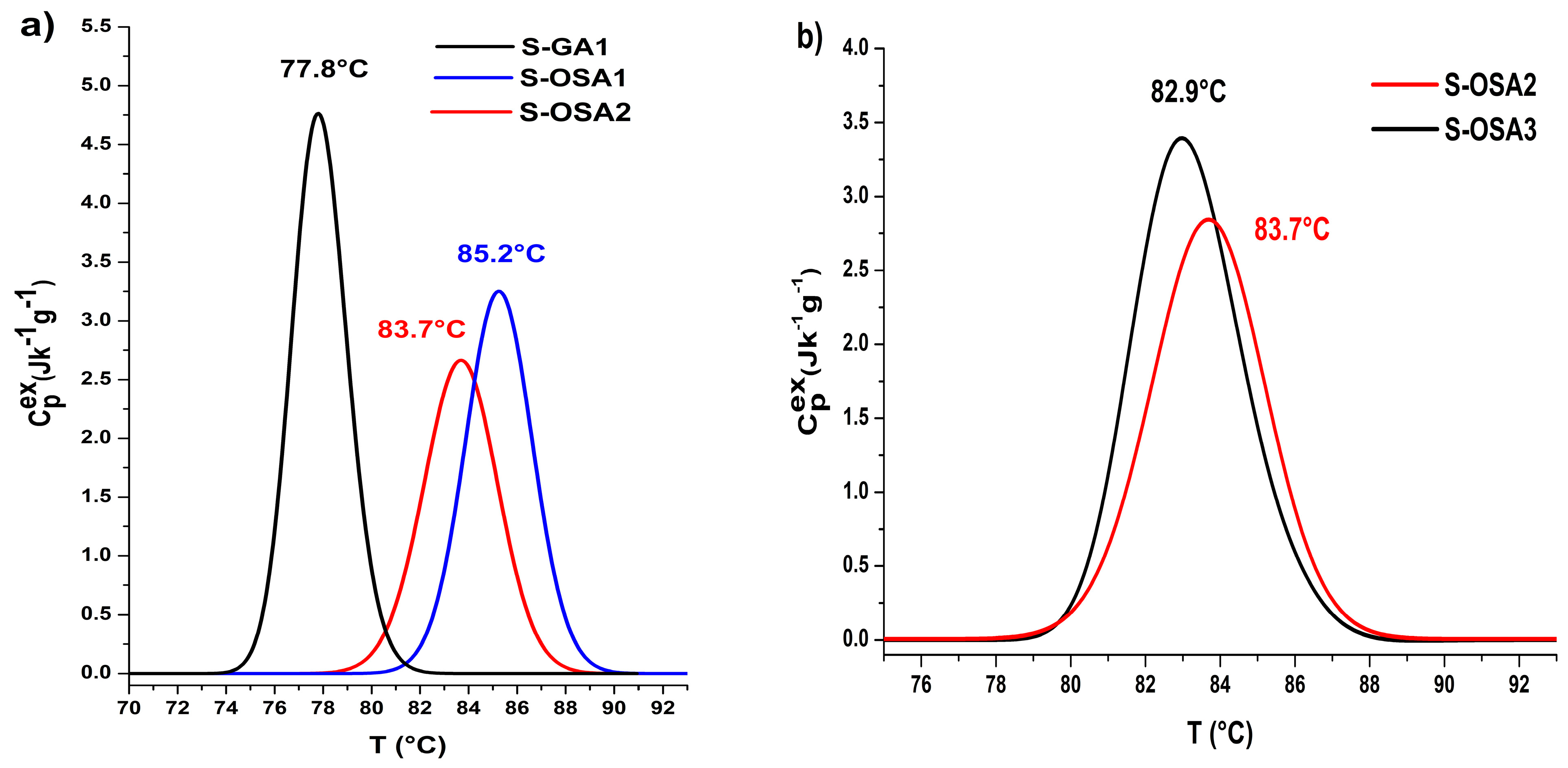
3.1.1. Hydrothermal Stability of Collagen-OSA Chemical Matrix by Micro-DSC
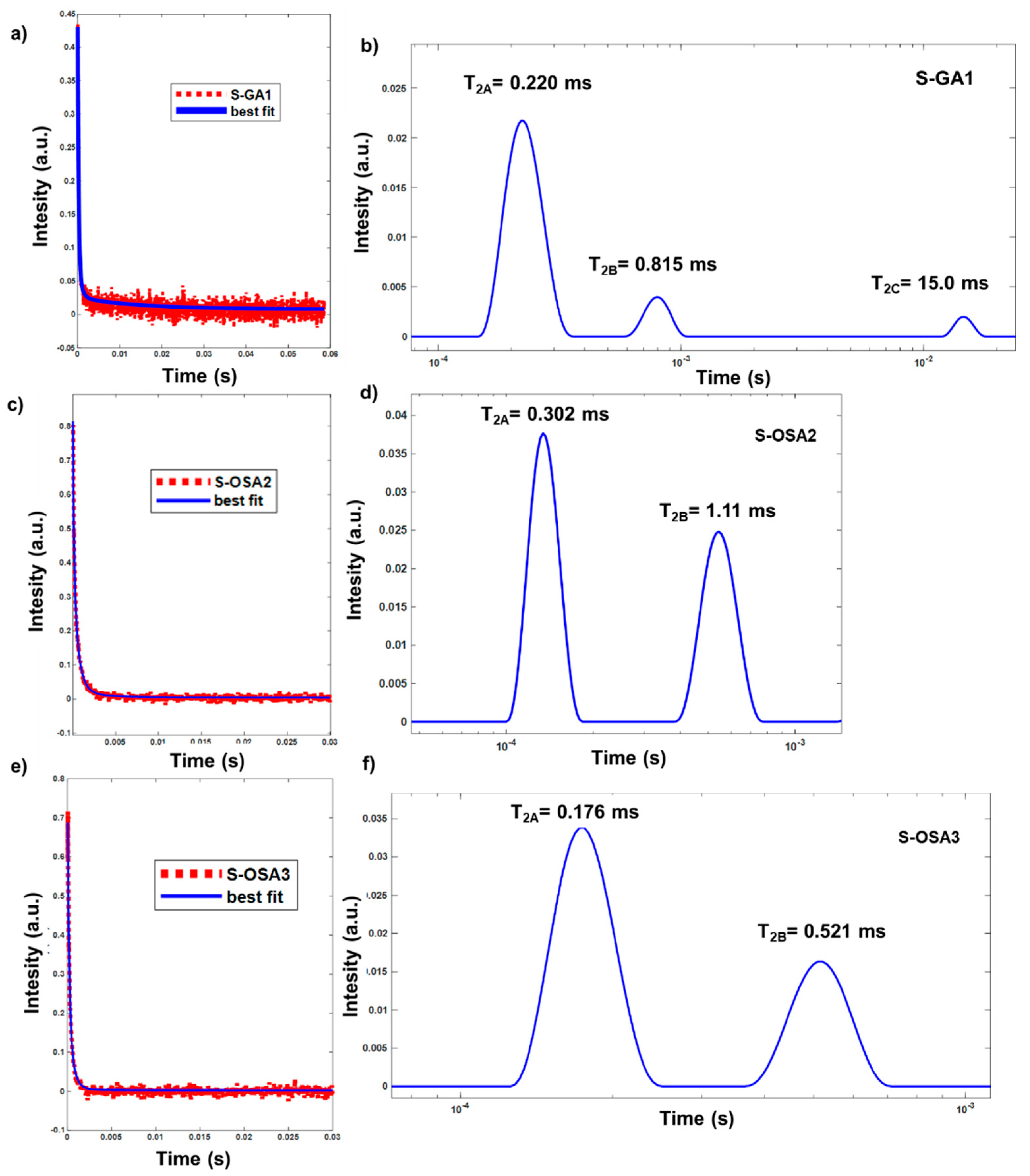
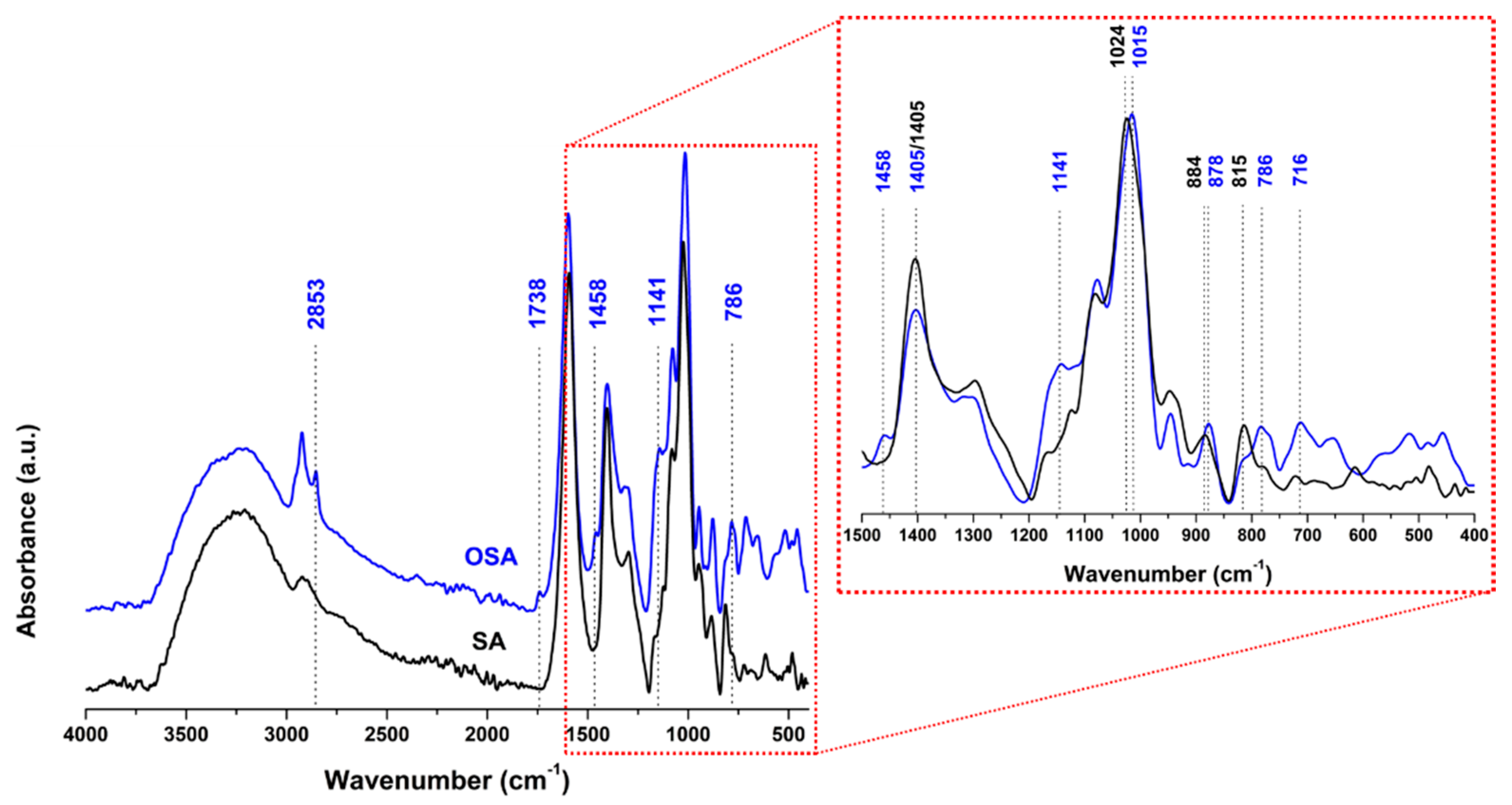
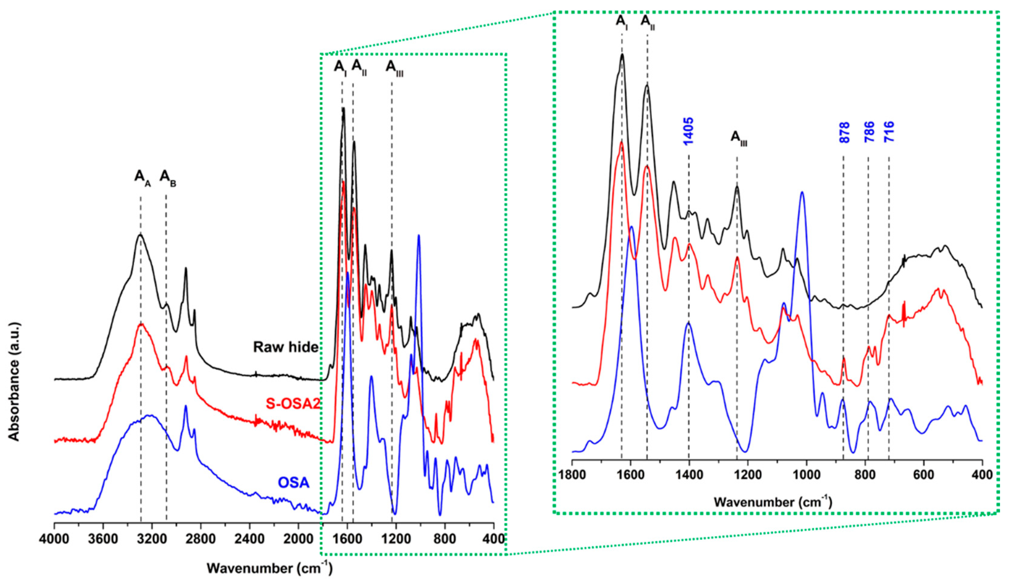
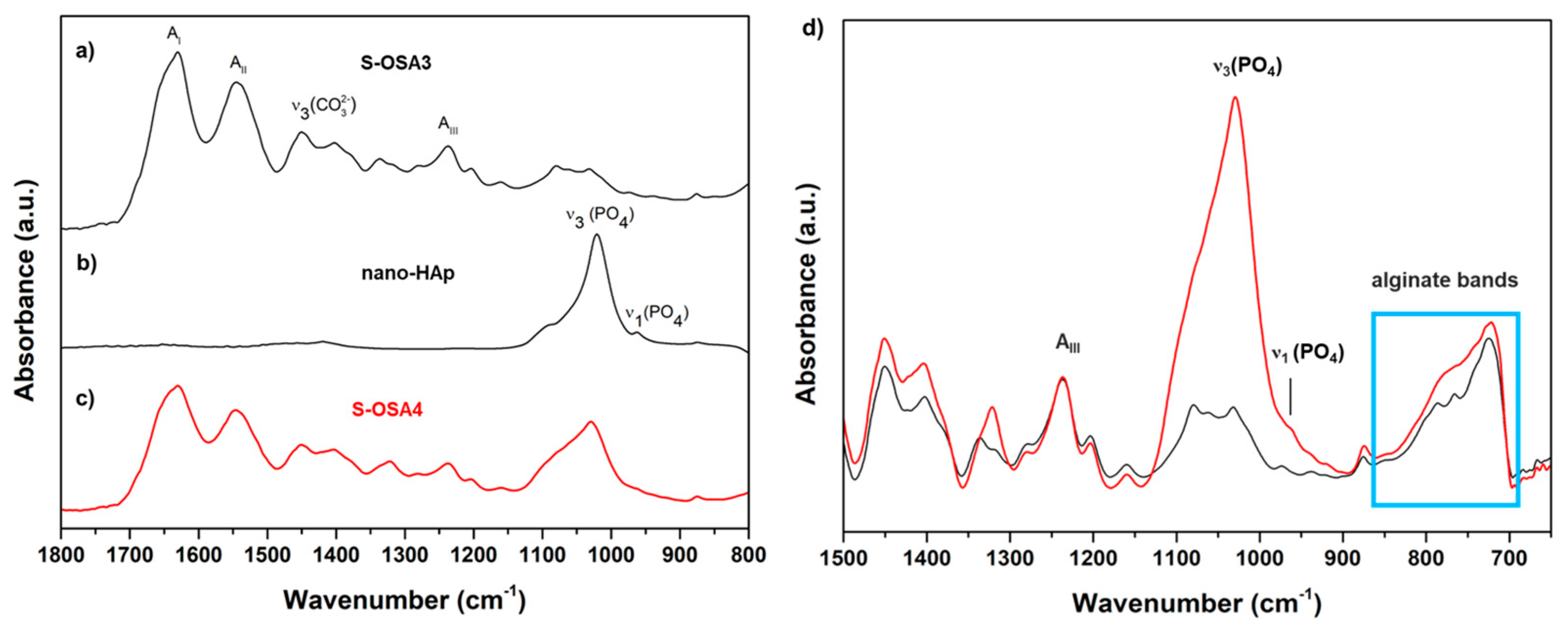
3.1.2. 1H Unilateral NMR and FTIR-ATR Analysis of Collagen-OSA Chemical Matrix in Leather
3.2. Laboratory Scale Tanning Test with OSA as Tanning Agent and Nano-HAp Wet Treatment
3.3. From Laboratory to Industrial Scale: A Scale-Up Framework for a Tanning Process Using OSA and Nano-HAp
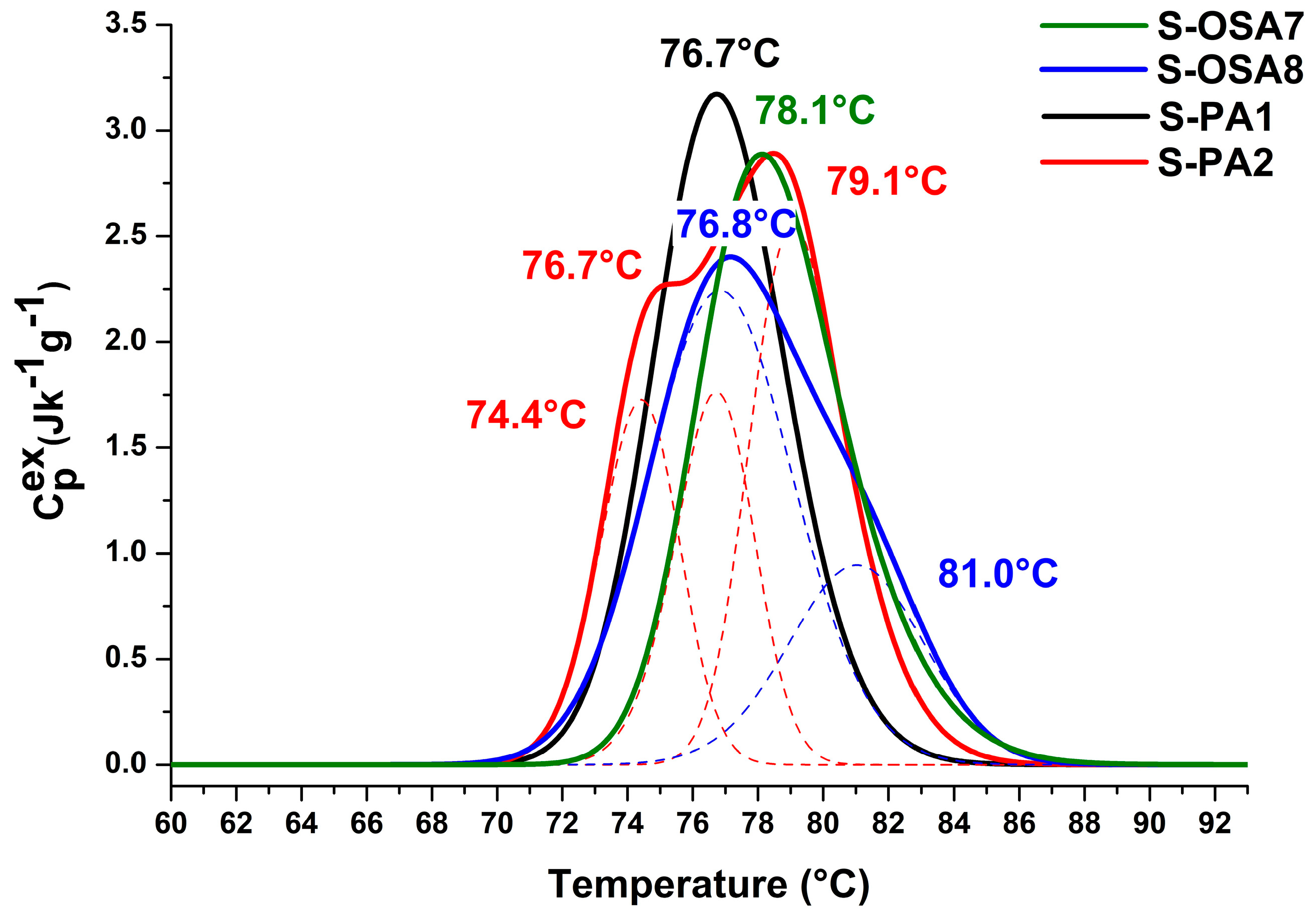
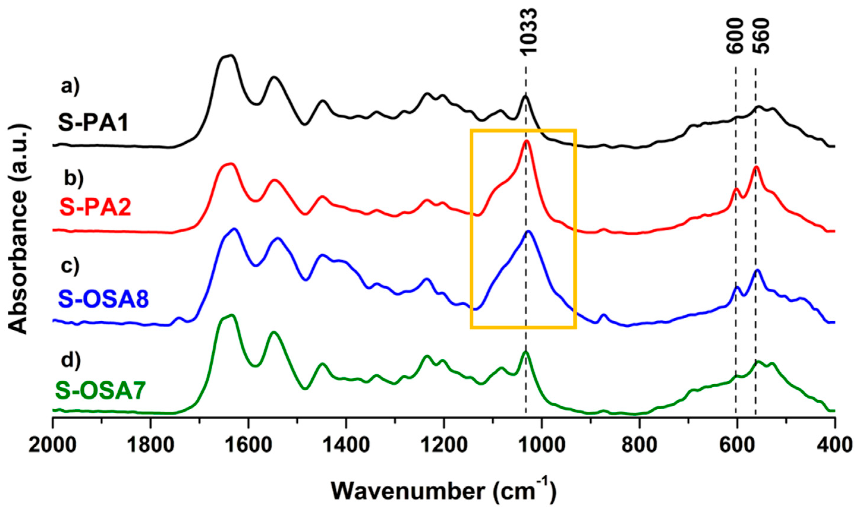
3.3.1. Thermal Stability and Chemical Characterization
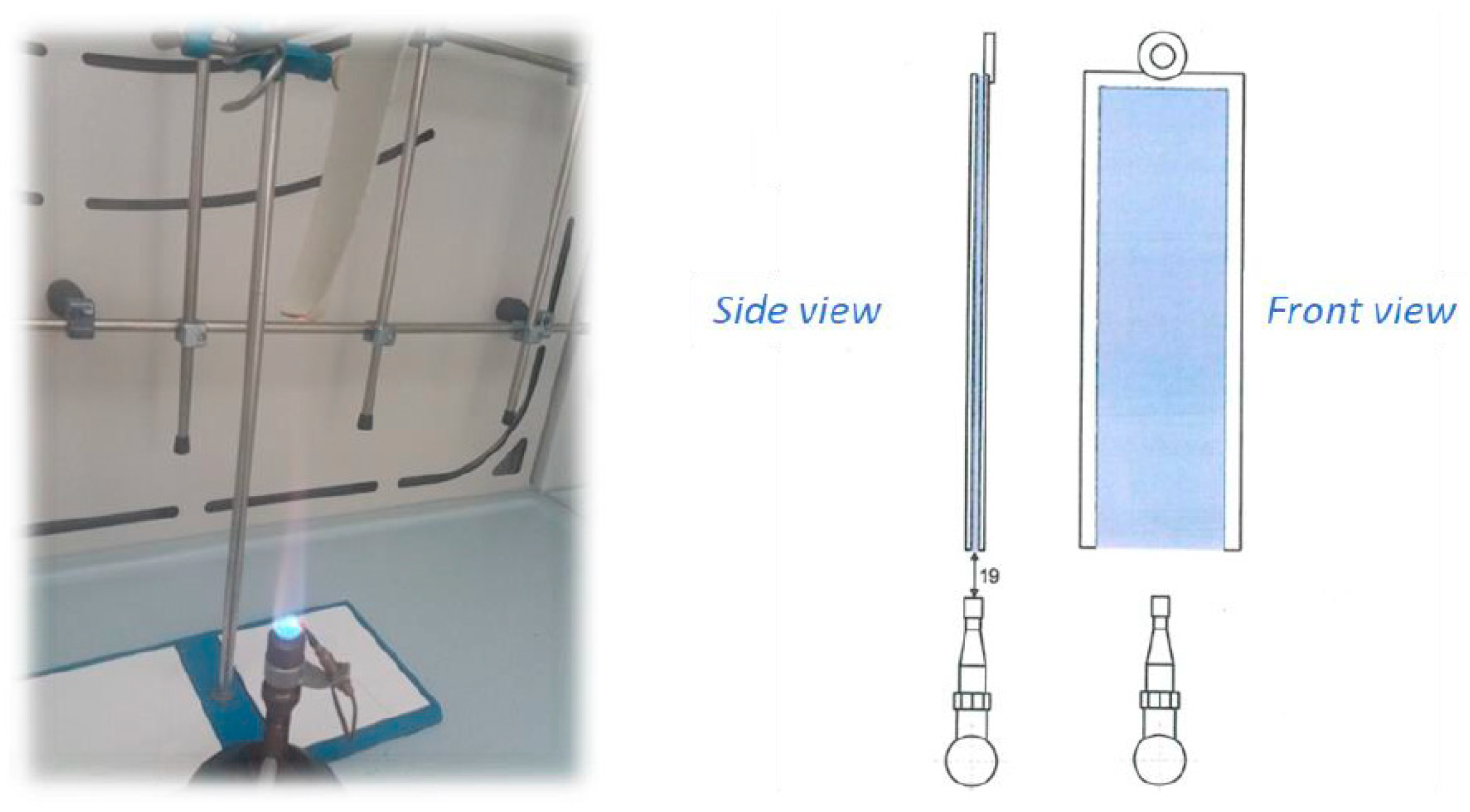
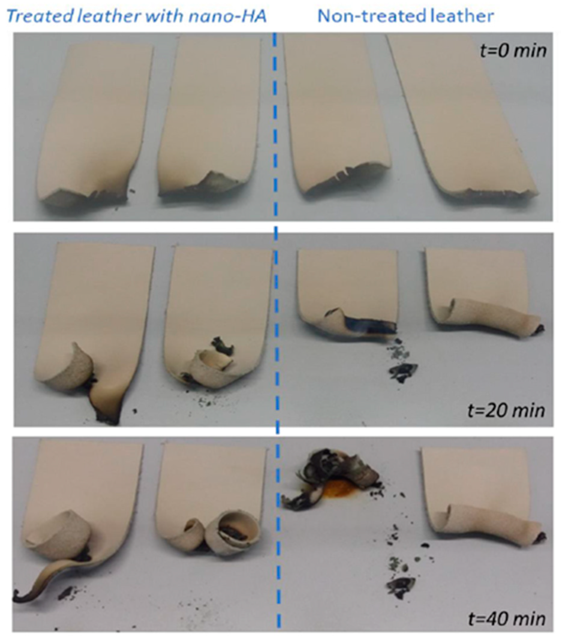
3.3.2. Fire Resistance Characterization
3.3.3. Physical Mechanical Characterization
- -
- In terms of tensile strength, tearing, and cracking resistance, S-OSA7 and S-OSA8 demonstrated behavior resembling that of commercial leathers.
- -
- OSA-tanned leathers displayed superior deformation and elastic–plastic behavior.
- -
- Nano-HAp treatment increased cracking resistance while slightly reducing tearing resistance.

| Test Name | Technical Characteristic | UM | S-OSA7 | S-OSA8 | S-PA1 | S-PA2 | Standard Method |
|---|---|---|---|---|---|---|---|
| Thickness | Thickness | mm | 1.7 | 1.8 | 2.1 | 1.9 | SR EN ISO 2589:2016 [92] |
| Tensile strength and percent elongation | Elongation at cracking | % | 54.5 | 56.5 | 47.5 | 55.4 | SR EN ISO 3376:2020 [93] |
| Elongation at break | % | 63.6 | 54.5 | 71.3 | 55.1 | ||
| Tensile strength | N/mm2 | 18.7 | 12.3 | 20.3 | 12.0 | ||
| Tear strength | N/mm2 | 12.9 | 13.7 | 13.3 | 13.8 | ||
| Tear strength in extension | Tear resistance | N | 53.2 | 45.0 | 57.5 | 42.7 | SR EN ISO 3377-1:2012 [94] |
| Tear resistance on two edges | Tear resistance | N | 123.2 | 77.7 | 147.3 | 77.7 | SR EN ISO 3377-2:2016 [95] |
| Softness | Ring opening Ø 20 mm | mm | 2.6 | 2.3 | 3.0 | 1.9 | SR EN ISO 17235:2016 [96] |
| Ø 25 mm | mm | 4.3 | 3.1 | 3.9 | 2.3 | ||
| Ø 35 mm | mm | 5.9 | 4.4 | 5.4 | 3.5 |
4. Conclusions
Supplementary Materials
Author Contributions
Funding
Institutional Review Board Statement
Data Availability Statement
Acknowledgments
Conflicts of Interest
References
- Ma’arfi, F.; Khan, M.Y.; Husain, A.; Khanam, A.; Hasan, Z. Contamination of Water Resources with Potentially Toxic Elements and Human Health Risk Assessment: Part 1. In Contamination of Water; Elsevier: Amsterdam, The Netherlands, 2021; pp. 123–141. ISBN 978-0-12-824058-8. [Google Scholar] [CrossRef]
- Mishra, S.; Bharagava, R.N. Toxic and Genotoxic Effects of Hexavalent Chromium in Environment and Its Bioremediation Strategies. J. Environ. Sci. Health Part C 2016, 34, 1–32. [Google Scholar] [CrossRef] [PubMed]
- Deng, Y.; Wang, M.; Tian, T.; Lin, S.; Xu, P.; Zhou, L.; Dai, C.; Hao, Q.; Wu, Y.; Zhai, Z.; et al. The Effect of Hexavalent Chromium on the Incidence and Mortality of Human Cancers: A Meta-Analysis Based on Published Epidemiological Cohort Studies. Front. Oncol. 2019, 9, 24. [Google Scholar] [CrossRef] [PubMed]
- Zeiner, M.; Rezić, I.; Steffan, I. Determination of Total Chromium in Tanned Leather Samples Used in Car Industry. Coll. Antropol. 2011, 30, 89–92. Available online: https://hrcak.srce.hr/file/97033 (accessed on 1 December 2023).
- Pouliot, B.P.; Mass, J.; Kaplan, L. Using XRF for the Identification of Chrome Tanning in Leather. Miami, Florida, 16 May 2015; p. 120. Available online: https://www.culturalheritage.org/docs/default-source/publications/annualmeeting/2015-posters/2015am_poster_92.pdf?sfvrsn=127da700_7 (accessed on 1 December 2023).
- Velusamy, M.; Chakali, B.; Ganesan, S.; Tinwala, F.; Shanmugham Venkatachalam, S. Investigation on Pyrolysis and Incineration of Chrome-Tanned Solid Waste from Tanneries for Effective Treatment and Disposal: An Experimental Study. Environ. Sci. Pollut. Res. 2020, 27, 29778–29790. [Google Scholar] [CrossRef] [PubMed]
- Ammenn, J.; Huebsch, C.; Schilling, E.; Dannheim, B. Chemistry of Syntans and Their Influence on Leather Quality. J. Am. Leather Chem. Assoc. 2015, 110, 349–354. Available online: https://www.researchgate.net/publication/289629872_Chemistry_of_syntans_and_their_influence_on_leather_quality (accessed on 1 December 2023).
- Yi, Y.; Jiang, Z.; Yang, S.; Ding, W.; Wang, Y.; Shi, B. Formaldehyde Formation during the Preparation of Dialdehyde Carboxymethyl Cellulose Tanning Agent. Carbohydr. Polym. 2020, 239, 116217. [Google Scholar] [CrossRef]
- Wu, Y.; Yuan, L.; Sheng, N.; Gu, Z.; Feng, W.; Yin, H.; Morsi, Y.; Mo, X. A Soft Tissue Adhesive Based on Aldehyde-Sodium Alginate and Amino-Carboxymethyl Chitosan Preparation through the Schiff Reaction. Front. Mater. Sci. 2017, 11, 215–222. [Google Scholar] [CrossRef]
- An, H.; Yu, H.; Wei, Y.; Liu, F.; Ye, J. Disrupted Metabolic Pathways and Potential Human Diseases Induced by Bisphenol S. Environ. Toxicol. Pharmacol. 2021, 88, 103751. [Google Scholar] [CrossRef]
- Fathima, N.N.; Kumar, T.P.; Kumar, D.R.; Rao, J.R.; Nair, B.U. Wet White Leather Processing: A New Combination Tanning System. J. Am. Leather Chem. Assoc. 2006, 101, 58–65. Available online: https://www.researchgate.net/publication/288775821_Wet_white_leather_processing_A_new_combination_tanning_system (accessed on 1 December 2023).
- Ding, W.; Yi, Y.; Wang, Y.; Zhou, J.; Shi, B. Preparation of a Highly Effective Organic Tanning Agent with Wide Molecular Weight Distribution from Bio-Renewable Sodium Alginate. ChemistrySelect 2018, 3, 12330–12335. [Google Scholar] [CrossRef]
- Balakrishnan, B.; Lesieur, S.; Labarre, D.; Jayakrishnan, A. Periodate Oxidation of Sodium Alginate in Water and in Ethanol–Water Mixture: A Comparative Study. Carbohydr. Res. 2005, 340, 1425–1429. [Google Scholar] [CrossRef] [PubMed]
- Jejurikar, A.; Seow, X.T.; Lawrie, G.; Martin, D.; Jayakrishnan, A.; Grøndahl, L. Degradable Alginate Hydrogels Crosslinked by the Macromolecular Crosslinker Alginate Dialdehyde. J. Mater. Chem. 2012, 22, 9751. [Google Scholar] [CrossRef]
- Aroguz, A.Z.; Baysal, K.; Adiguzel, Z.; Baysal, B.M. Alginate/Polyoxyethylene and Alginate/Gelatin Hydrogels: Preparation, Characterization, and Application in Tissue Engineering. Appl. Biochem. Biotechnol. 2014, 173, 433–448. [Google Scholar] [CrossRef] [PubMed]
- Rayatpisheh, S.; Poon, Y.F.; Cao, Y.; Feng, J.; Chan, V.; Chan-Park, M.B. Aligned 3D Human Aortic Smooth Muscle Tissue via Layer by Layer Technique inside Microchannels with Novel Combination of Collagen and Oxidized Alginate Hydrogel. J. Biomed. Mater. Res. A 2011, 98A, 235–244. [Google Scholar] [CrossRef]
- Ravichandran, V.; Jayakrishnan, A. Synthesis and Evaluation of Anti-Fungal Activities of Sodium Alginate-Amphotericin B Conjugates. Int. J. Biol. Macromol. 2018, 108, 1101–1109. [Google Scholar] [CrossRef] [PubMed]
- Lee, K.Y.; Mooney, D.J. Alginate: Properties and Biomedical Applications. Prog. Polym. Sci. 2012, 37, 106–126. [Google Scholar] [CrossRef] [PubMed]
- Sarker, B.; Papageorgiou, D.G.; Silva, R.; Zehnder, T.; Gul-E-Noor, F.; Bertmer, M.; Kaschta, J.; Chrissafis, K.; Detsch, R.; Boccaccini, A.R. Fabrication of Alginate–Gelatin Crosslinked Hydrogel Microcapsules and Evaluation of the Microstructure and Physico-Chemical Properties. J. Mater. Chem. B 2014, 2, 1470. [Google Scholar] [CrossRef]
- Reakasame, S.; Boccaccini, A.R. Oxidized Alginate-Based Hydrogels for Tissue Engineering Applications: A Review. Biomacromolecules 2018, 19, 3–21. [Google Scholar] [CrossRef]
- Wang, J.; Fu, W.; Zhang, D.; Yu, X.; Li, J.; Wan, C. Evaluation of Novel Alginate Dialdehyde Cross-Linked Chitosan/Calcium Polyphosphate Composite Scaffolds for Meniscus Tissue Engineering. Carbohydr. Polym. 2010, 79, 705–710. [Google Scholar] [CrossRef]
- Park, H.; Lee, K.Y. Cartilage Regeneration Using Biodegradable Oxidized Alginate/Hyaluronate Hydrogels: Cartilage Regeneration Using Biodegradable Hydrogels. J. Biomed. Mater. Res. A 2014, 102, 4519–4525. [Google Scholar] [CrossRef]
- Maiti, S.; Singha, K.; Ray, S.; Dey, P.; Sa, B. Adipic Acid Dihydrazide Treated Partially Oxidized Alginate Beads for Sustained Oral Delivery of Flurbiprofen. Pharm. Dev. Technol. 2009, 14, 461–470. [Google Scholar] [CrossRef]
- Ding, F.; Wu, S.; Wang, S.; Xiong, Y.; Li, Y.; Li, B.; Deng, H.; Du, Y.; Xiao, L.; Shi, X. A Dynamic and Self-Crosslinked Polysaccharide Hydrogel with Autonomous Self-Healing Ability. Soft Matter 2015, 11, 3971–3976. [Google Scholar] [CrossRef] [PubMed]
- Bouhadir, K.H.; Lee, K.Y.; Alsberg, E.; Damm, K.L.; Anderson, K.W.; Mooney, D.J. Degradation of Partially Oxidized Alginate and Its Potential Application for Tissue Engineering. Biotechnol. Prog. 2001, 17, 945–950. [Google Scholar] [CrossRef]
- Liao, H.; Zhang, H.; Chen, W. Differential Physical, Rheological, and Biological Properties of Rapid in Situ Gelable Hydrogels Composed of Oxidized Alginate and Gelatin Derived from Marine or Porcine Sources. J. Mater. Sci. Mater. Med. 2009, 20, 1263–1271. [Google Scholar] [CrossRef]
- Mu, B.; Lu, C.; Liu, P. Disintegration-Controllable Stimuli-Responsive Polyelectrolyte Multilayer Microcapsules via Covalent Layer-by-Layer Assembly. Colloids Surf. B Biointerfaces 2011, 82, 385–390. [Google Scholar] [CrossRef]
- Wright, B.; De Bank, P.A.; Luetchford, K.A.; Acosta, F.R.; Connon, C.J. Oxidized Alginate Hydrogels as Niche Environments for Corneal Epithelial Cells: Oxidised Alginate Hydrogels as Niche Environments. J. Biomed. Mater. Res. A 2014, 102, 3393–3400. [Google Scholar] [CrossRef]
- Registration Dossier—ECHA. Available online: https://echa.europa.eu/it/registration-dossier/-/registered-dossier/5745 (accessed on 15 November 2023).
- Potassium Periodate|7790-21-8 Supplier and Manufacturer—BuyersGuideChem. Available online: https://www.buyersguidechem.com/chemical_supplier/Potassium_periodate (accessed on 7 December 2022).
- Lucia, A.; Herwijnen, H.W.G.; Oberlerchner, J.T.; Rosenau, T.; Beaumont, M. Resource-Saving Production of Dialdehyde Cellulose: Optimization of the Process at High Pulp Consistency. ChemSusChem 2019, 12, 4679–4684. [Google Scholar] [CrossRef] [PubMed]
- Kisukuri, C.M.; Bednarz, R.J.; Kampf, C.; Arndt, S.; Waldvogel, S.R. Robust and Self-Cleaning Electrochemical Production of Periodate. ChemSusChem 2022, 15, e202200874. [Google Scholar] [CrossRef] [PubMed]
- Selvaraju, S.; Ramalingam, S.; Rao, J.R. Preparation and Application of Biodegradable Nanocomposite for Cleaner Leather Processing. J. Clean. Prod. 2017, 158, 225–232. [Google Scholar] [CrossRef]
- Kale, M.B.; Luo, Z.; Zhang, X.; Dhamodharan, D.; Divakaran, N.; Mubarak, S.; Wu, L.; Xu, Y. Waterborne Polyurethane/Graphene Oxide-Silica Nanocomposites with Improved Mechanical and Thermal Properties for Leather Coatings Using Screen Printing. Polymer 2019, 170, 43–53. [Google Scholar] [CrossRef]
- Ma, J.; Lv, X.; Gao, D.; Li, Y.; Lv, B.; Zhang, J. Nanocomposite-Based Green Tanning Process of Suede Leather to Enhance Chromium Uptake. J. Clean. Prod. 2014, 72, 120–126. [Google Scholar] [CrossRef]
- Gao, D.; Ma, J.; Lv, B.; Zhang, J. Special Review: Collagen Modification Using Nanotechnologies: A Review. J. Am. Leather Chem. Assoc. 2013, 108, 392–400. Available online: https://www.researchgate.net/publication/286303232_Special_review_Collagen_modification_using_nanotechnologies_A_review (accessed on 1 December 2023).
- Wegst, U.G.K.; Bai, H.; Saiz, E.; Tomsia, A.P.; Ritchie, R.O. Bioinspired Structural Materials. Nat. Mater. 2015, 14, 23–36. [Google Scholar] [CrossRef] [PubMed]
- Chen, F.; Huang, P.; Zhu, Y.-J.; Wu, J.; Zhang, C.-L.; Cui, D.-X. The Photoluminescence, Drug Delivery and Imaging Properties of Multifunctional Eu3+/Gd3+ Dual-Doped Hydroxyapatite Nanorods. Biomaterials 2011, 32, 9031–9039. [Google Scholar] [CrossRef]
- Chen, F.; Zhu, Y.-J. Multifunctional Calcium Phosphate Nanostructured Materials and Biomedical Applications. Curr. Nanosci. 2014, 10, 465–485. [Google Scholar] [CrossRef]
- Liu, H.; Peng, H.; Wu, Y.; Zhang, C.; Cai, Y.; Xu, G.; Li, Q.; Chen, X.; Ji, J.; Zhang, Y.; et al. The Promotion of Bone Regeneration by Nanofibrous Hydroxyapatite/Chitosan Scaffolds by Effects on Integrin-BMP/Smad Signaling Pathway in BMSCs. Biomaterials 2013, 34, 4404–4417. [Google Scholar] [CrossRef]
- Karalkeviciene, R.; Raudonyte-Svirbutaviciene, E.; Zarkov, A.; Yang, J.-C.; Popov, A.I.; Kareiva, A. Solvothermal Synthesis of Calcium Hydroxyapatite via Hydrolysis of Alpha-Tricalcium Phosphate in the Presence of Different Organic Additives. Crystals 2023, 13, 265. [Google Scholar] [CrossRef]
- Farkas, N.-I.; Marincaș, L.; Barabás, R.; Bizo, L.; Ilea, A.; Turdean, G.L.; Toșa, M.; Cadar, O.; Barbu-Tudoran, L. Preparation and Characterization of Doxycycline-Loaded Electrospun PLA/HAP Nanofibers as a Drug Delivery System. Materials 2022, 15, 2105. [Google Scholar] [CrossRef]
- Liu, X.L.; Hu, Z.W.; Zhang, S.; Gu, X.Y.; Ma, W.J. Effect of Hydroxyapatite on Fire Resistance and Smoke Suppression of Polyurethane Fire-Retardant Coating. Xiandai Huagong/Modern Chem. Ind. 2015, 35, 88–91. Available online: https://www.researchgate.net/publication/283130315_Effect_of_hydroxyapatite_on_fire_resistance_and_smoke_suppression_of_polyurethane_fire-retardant_coating (accessed on 1 December 2023).
- Elbasuney, S.; Maraden, A. Novel Thermoset Nanocomposite Intumescent Coating Based on Hydroxyapatite Nanoplates for Fireproofing of Steel Structures. J. Inorg. Organomet. Polym. Mater. 2020, 30, 820–830. [Google Scholar] [CrossRef]
- Dong, Q.-X.; Chen, Q.-J.; Yang, W.; Zheng, Y.-L.; Liu, X.; Li, Y.-L.; Yang, M.-B. Thermal Properties and Flame Retardancy of Polycarbonate/Hydroxyapatite Nanocomposite. J. Appl. Polym. Sci. 2008, 109, 659–663. [Google Scholar] [CrossRef]
- Vahabi, H.; Shabanian, M.; Aryanasab, F.; Mangin, R.; Laoutid, F.; Saeb, M.R. Inclusion of Modified Lignocellulose and Nano-Hydroxyapatite in Development of New Bio-Based Adjuvant Flame Retardant for Poly(Lactic Acid). Thermochim. Acta 2018, 666, 51–59. [Google Scholar] [CrossRef]
- Dholakiya, B.Z. Use of Non-Traditional Fillers to Reduce Flammability of Polyester Resin Composites. Polimeri 2009, 30, 10–17. Available online: https://www.researchgate.net/publication/250928194_Use_of_non-traditional_fillers_to_reduce_flammability_of_polyester_resin_composites (accessed on 1 December 2023).
- Ingrao, C.; Vesce, E.; Evola, R.S.; Rebba, E.; Arcidiacono, C.; Martra, G.; Beltramo, R. Chemistry behind Leather: Life Cycle Assessment of Nano-Hydroxyapatite Preparation on the Lab-Scale for Fireproofing Applications. J. Clean. Prod. 2021, 279, 123837. [Google Scholar] [CrossRef]
- Usenko, C.; Abel, E.; Hopkins, A.; Martinez, G.; Tijerina, J.; Kudela, M.; Norris, N.; Joudeh, L.; Bruce, E. Evaluation of Common Use Brominated Flame Retardant (BFR) Toxicity Using a Zebrafish Embryo Model. Toxics 2016, 4, 21. [Google Scholar] [CrossRef]
- Carsote, C.; Badea, E. Micro Differential Scanning Calorimetry and Micro Hot Table Method for Quantifying Deterioration of Historical Leather. Herit. Sci. 2019, 7, 48. [Google Scholar] [CrossRef]
- Carsote, C.; Şendrea, C.; Micu, M.-C.; Adams, A.; Badea, E. Micro-DSC, FTIR-ATR and NMR MOUSE Study of the Dose-Dependent Effects of Gamma Irradiation on Vegetable-Tanned Leather: The Influence of Leather Thermal Stability. Radiat. Phys. Chem. 2021, 189, 109712. [Google Scholar] [CrossRef]
- Sendrea, C.; Carsote, C.; Radu, M.; Badea, E.; Miu, L. The Effect of Gamma Irradiation on Shrinkage Activity of Collagen in Vegetable Tanned Leather. Rev Chim 2017, 68, 1535–1538. [Google Scholar] [CrossRef]
- Blümich, B.; Perlo, J.; Casanova, F. Mobile Single-Sided NMR. Prog. Nucl. Magn. Reson. Spectrosc. 2008, 52, 197–269. [Google Scholar] [CrossRef]
- Sakhno, Y.; Ivanchenko, P.; Iafisco, M.; Tampieri, A.; Martra, G. A Step toward Control of the Surface Structure of Biomimetic Hydroxyapatite Nanoparticles: Effect of Carboxylates on the {010} P-Rich/Ca-Rich Facets Ratio. J. Phys. Chem. C 2015, 119, 5928–5937. [Google Scholar] [CrossRef]
- Ding, W.; Zhou, J.; Zeng, Y.; Wang, Y.; Shi, B. Preparation of Oxidized Sodium Alginate with Different Molecular Weights and Its Application for Crosslinking Collagen Fiber. Carbohydr. Polym. 2017, 157, 1650–1656. [Google Scholar] [CrossRef] [PubMed]
- Aina, V.; Lusvardi, G.; Annaz, B.; Gibson, I.R.; Imrie, F.E.; Malavasi, G.; Menabue, L.; Cerrato, G.; Martra, G. Magnesium- and Strontium-Co-Substituted Hydroxyapatite: The Effects of Doped-Ions on the Structure and Chemico-Physical Properties. J. Mater. Sci. Mater. Med. 2012, 23, 2867–2879. [Google Scholar] [CrossRef] [PubMed]
- Silva, V.M.T.M.; Quadros, P.A.; Laranjeira, P.E.M.S.C.; Dias, M.M.; Lopes, J.C.B. A Novel Continuous Industrial Process for Producing Hydroxyapatite Nanoparticles. J. Dispers. Sci. Technol. 2008, 29, 542–547. [Google Scholar] [CrossRef]
- Motskin, M.; Wright, D.M.; Muller, K.; Kyle, N.; Gard, T.G.; Porter, A.E.; Skepper, J.N. Hydroxyapatite Nano and Microparticles: Correlation of Particle Properties with Cytotoxicity and Biostability. Biomaterials 2009, 30, 3307–3317. [Google Scholar] [CrossRef] [PubMed]
- Covington, A.D.; Wise, W.R. (Eds.) Collagen and Skin Structure. In Tanning Chemistry: The Science of Leather; The Royal Society of Chemistry: London, UK, 2019; ISBN 978-1-78801-204-1. [Google Scholar] [CrossRef]
- Della Gatta, G.; Richardson, M.J.; Sarge, S.M.; Stølen, S. Standards, Calibration, and Guidelines in Microcalorimetry. Part 2. Calibration Standards for Differential Scanning Calorimetry* (IUPAC Technical Report). Pure Appl. Chem. 2006, 78, 1455–1476. [Google Scholar] [CrossRef]
- Proietti, N.; Di Tullio, V.; Carsote, C.; Badea, E. 13C Solid-State NMR Complemented by ATR-FTIR and Micro-DSC to Study Modern Collagen-Based Material and Historical Leather. Magn. Reson. Chem. 2020, 58, 840–859. [Google Scholar] [CrossRef]
- Covington, A.D.; Wise, W.R. Tanning Chemistry the Science of Leather; The Royal Society of Chemistry: Cambridge, UK, 2019; ISBN 98-1-8801-204-1. [Google Scholar]
- Li, X.; Wang, Y.N.; Li, J.; Shi, B. Effect of Sodium Chloride on Structure of Collagen Fiber Network in Pickling and Tanning. J. Am. Leather Chem. Assoc. 2016, 111, 230–237. [Google Scholar]
- Duan, L.; Li, J.; Li, C.; Li, G. Effects of NaCl on the Rheological Behavior of Collagen Solution. Korea-Aust. Rheol. J. 2013, 25, 137–144. [Google Scholar] [CrossRef]
- Yu, G.; Niu, C.; Liu, J.; Wu, J.; Jin, Z.; Wang, Y.; Zhao, K. Preparation and Properties of Self-Cross-Linking Hydrogels Based on Chitosan Derivatives and Oxidized Sodium Alginate. ACS Omega 2023, 8, 19752–19766. [Google Scholar] [CrossRef]
- Covington, A.D.; Wise, W.R. (Eds.) Theory of Tanning: The Concept of Link–Lock. In Tanning Chemistry: The Science of Leather; The Royal Society of Chemistry: London, UK, 2019; ISBN 978-1-78801-204-1. [Google Scholar]
- Masic, A.; Badea, E.; Ceccarelli, R.; Della Gatta, G.; Coluccia, S. Studio comparativo DSC e SEM/ESEM di pergamene antiche e invecchiate artificialmente. In Proceedings of the Lo stato dell’arte2- Conservazione, Confronto e Restauro di Esperienze, Il Prato, Genova, Italy, 29 September 2004; pp. 52–61. [Google Scholar]
- Quaratesi, I.; Bruno, I.; Pauciulo, A.; Bartiromo, A.R.; Badea, E.; Carşote, C.; Neri, P.; Talotta, C.; Gliubizzi, R.; Di Tullio, V.; et al. Side-Chain Poly[2]Pseudorotaxanes Containing β-Cyclodextrin for More Sustainable Tanning Process. Polym. Test. 2023, 129, 108268. [Google Scholar] [CrossRef]
- Carşote, C.; Badea, E.; Miu, L.; Gatta, G.D. Study of the Effect of Tannins and Animal Species on the Thermal Stability of Vegetable Leather by Differential Scanning Calorimetry. J. Therm. Anal. Calorim. 2016, 124, 1255–1266. [Google Scholar] [CrossRef]
- Cucos, A.; Gaidau, C.; Badea, E.; Miu, L. Influence of Glycerin on Denaturation Temperature of Chrome- and Vegetable-Tanned Leather. Rev. Roum. Chim. 2015, 60, 1093. Available online: https://www.researchgate.net/profile/Elena-Badea/publication/305641297_Influence_of_glycerin_on_denaturation_temperature_of_chrome-_and_vegetable-tanned_leather/links/57f6513f08ae91deaa5ebf2d/Influence-of-glycerin-on-denaturation-temperature-of-chrome-and-vegetable-tanned-leather.pdf (accessed on 1 December 2023).
- Chen, W.; Chen, Z.; Long, Z.; Shan, Z. Development of Aldehyde and Similar-to-Aldehyde Tanning Agents. Text. Res. J. 2022, 92, 3387–3397. [Google Scholar] [CrossRef]
- Sendrea, C.; Badea, E.; Miu, L.; Ignat, M.; Iovu, H. Unilateral NMR for Damage Assessment of Vegetable—Tanned Leather. Correlation with Hydrotermal Properties. In Proceedings of the ICAMS 2014 5th International Conference on Advanced Materials and Systems, Bucharest, Romania, 23–25 October 2014. [Google Scholar] [CrossRef]
- Van Stiphout, T.A.P.; Pel, L.; Galvosas, P.; Prabakar, S.; Holmes, G. NMR Transverse Relaxation Analysis of Leather Looseness; Eindhoven University of Thecnology: Eindhoven, The Netherlands, 2015; p. 50. Available online: https://pure.tue.nl/ws/files/57723539/Stiphout_2015.pdf (accessed on 1 December 2023).
- Sendrea, C.; Micu, M.-C.; Hadimbu, E.; Paunescu, S.M.; Caniola, I.M.; Ignat, M.; Miu, L.; Badea, E. Micro DSC and NMR MOUSE Studies of Collagen–Vegetable Tannin Interaction Mechanism during Leather Making. In Proceedings of the 8th International Conference on Advanced Materials and Systems, Online Event, 1–3 October 2020; INCDTP—Leather and Footwear Research Institute (ICPI): Bucharest, Romania, 2020; pp. 561–566. [Google Scholar] [CrossRef]
- Wang, H.; Chen, X.; Wen, Y.; Li, D.; Sun, X.; Liu, Z.; Yan, H.; Lin, Q. A Study on the Correlation between the Oxidation Degree of Oxidized Sodium Alginate on Its Degradability and Gelation. Polymers 2022, 14, 1679. [Google Scholar] [CrossRef]
- Ghanbari, M.; Salavati-Niasari, M.; Mohandes, F. Thermosensitive Alginate–Gelatin–Nitrogen-Doped Carbon Dots Scaffolds as Potential Injectable Hydrogels for Cartilage Tissue Engineering Applications. RSC Adv. 2021, 11, 18423–18431. [Google Scholar] [CrossRef] [PubMed]
- Li, L.-Y.; Zhao, Y.-Q.; He, Y.; Chi, C.-F.; Wang, B. Physicochemical and Antioxidant Properties of Acid- and Pepsin-Soluble Collagens from the Scales of Miiuy Croaker (Miichthys miiuy). Mar. Drugs 2018, 16, 394. [Google Scholar] [CrossRef] [PubMed]
- Sellimi, S.; Younes, I.; Ayed, H.B.; Maalej, H.; Montero, V.; Rinaudo, M.; Dahia, M.; Mechichi, T.; Hajji, M.; Nasri, M. Structural, Physicochemical and Antioxidant Properties of Sodium Alginate Isolated from a Tunisian Brown Seaweed. Int. J. Biol. Macromol. 2015, 72, 1358–1367. [Google Scholar] [CrossRef]
- Shekh, M.I.; Zhu, G.; Xiong, W.; Wu, W.; Stadler, F.J.; Patel, D.; Zhu, C. Dynamically Bonded, Tough, and Conductive MXene@oxidized Sodium Alginate: Chitosan Based Multi-Networked Elastomeric Hydrogels for Physical Motion Detection. Int. J. Biol. Macromol. 2023, 224, 604–620. [Google Scholar] [CrossRef] [PubMed]
- Amirrah, I.N.; Lokanathan, Y.; Zulkiflee, I.; Wee, M.F.M.R.; Motta, A.; Fauzi, M.B. A Comprehensive Review on Collagen Type I Development of Biomaterials for Tissue Engineering: From Biosynthesis to Bioscaffold. Biomedicines 2022, 10, 2307. [Google Scholar] [CrossRef]
- Cutini, M.; Corno, M.; Costa, D.; Ugliengo, P. How Does Collagen Adsorb on Hydroxyapatite? Insights From Ab Initio Simulations on a Polyproline Type II Model. J. Phys. Chem. C 2019, 123, 7540–7550. [Google Scholar] [CrossRef]
- Miles, C.A.; Avery, N.C. Thermal Stabilization of Collagen in Skin and Decalcified Bone. Phys. Biol. 2011, 8, 026002. [Google Scholar] [CrossRef]
- Miles, C.A.; Ghelashvili, M. Polymer-in-a-Box Mechanism for the Thermal Stabilization of Collagen Molecules in Fibers. Biophys. J. 1999, 76, 3243–3252. [Google Scholar] [CrossRef] [PubMed]
- Şendrea, C.; Carsote, C.; Badea, E.; Adams, A.; Niculescu, M.; Iovu, H. Non-Invasive Characterization of Collagen Based Materials by NMR-Mouse and ATR-FTIR. Sci. Bull.-Univ. Politeh. Buchar. 2016, 78, 27–38. Available online: https://www.scientificbulletin.upb.ro/rev_docs_arhiva/fulle96_692257.pdf (accessed on 1 December 2023).
- Hassani, A.; Avci, Ç.B.; Kerdar, S.N.; Amini, H.; Amini, M.; Ahmadi, M.; Sakai, S.; Bagca, B.G.; Ozates, N.P.; Rahbarghazi, R.; et al. Interaction of Alginate with Nano-Hydroxyapatite-Collagen Using Strontium Provides Suitable Osteogenic Platform. J. Nanobiotechnology 2022, 20, 310. [Google Scholar] [CrossRef] [PubMed]
- Ficai, A.; Andronescu, E.; Ghitulica, C.; Voicu, G.; Trandafir, V.; Mânzu, D.; Ficai, M.; Pall, S. Colagen/Hydroxyapatite Interactions in Composite Biomaterials. Mater. Plast. 2009, 46, 11–15. Available online: https://www.researchgate.net/publication/242223232_Colagen_Hydroxyapatite_Interactions_in_Composite_Biomaterials (accessed on 1 December 2023).
- Chandía, N.P.; Matsuhiro, B.; Mejías, E.; Moenne, A. Alginic Acids in Lessonia Vadosa: Partial Hydrolysis and Elicitor Properties of the Polymannuronic Acid Fraction. J. Appl. Phycol. 2004, 16, 127–133. [Google Scholar] [CrossRef]
- Kourkoumelis, N.; Lani, A.; Tzaphlidou, M. Infrared Spectroscopic Assessment of the Inflammation-Mediated Osteoporosis (IMO) Model Applied to Rabbit Bone. J. Biol. Phys. 2012, 38, 623–635. [Google Scholar] [CrossRef]
- Lambri, M.L.; Giordano, E.D.; Bozzano, P.B.; Bonifacich, F.G.; Pérez-Landazábal, J.I.; Zelada, G.I.; Gargicevich, D.; Recarte, V.; Lambri, O.A. Thermal Degradation of Type I Collagen from Bones. J. Renew. Mater. 2016, 4, 251–257. [Google Scholar] [CrossRef]
- Joshi, M.; Butola, B.S. Application Technologies for Coating, Lamination and Finishing of Technical Textiles. In Advances in the Dyeing and Finishing of Technical Textiles; Elsevier: Amsterdam, The Netherlands, 2013; pp. 355–411. ISBN 978-0-85709-433-9. [Google Scholar] [CrossRef]
- Zhang, T.; Cai, W.; Chu, F.; Zhou, F.; Liang, S.; Ma, C.; Hu, Y. Hydroxyapatite/Polyurea Nanocomposite: Preparation and Multiple Performance Enhancements. Compos. Part Appl. Sci. Manuf. 2020, 128, 105681. [Google Scholar] [CrossRef]
- ISO 2589:2016; IULTCS/IUP 4 Leather, Physical and Mechanical Tests, Determination of Thickness. ISO: Geneva, Switzerland, 2016. Available online: https://www.iso.org/standard/68859.html (accessed on 1 December 2023).
- ISO 3376:2020; IULTCS/IUP 6 Leather, Physical and Mechanical Tests, Determination of Tensile Strength and Percentage Elongation. ISO: Geneva, Switzerland, 2020. Available online: https://www.iso.org/standard/75173.html (accessed on 1 December 2023).
- SR EN ISO 3377-1:2012; Piei Finite. Încercări Fizice şi Mecanice. Determinarea Forţei de Sfâşiere. Partea 1: Sfâşierea pe o Singură Margine. ISO: Geneva, Switzerland, 2012. Available online: https://magazin.asro.ro/ro/standard/197178 (accessed on 1 December 2023).
- ISO 3377-2:2016; IULTCS/IUP 8 Leather, Physical and Mechanical Tests, Determination of Tear Load. ISO: Geneva, Switzerland, 2016. Available online: https://www.iso.org/standard/68861.html (accessed on 1 December 2023).
- ISO 17235:2015; Leather-Physical and Mechanical Tests-Determination of Softness. ISO: Geneva, Switzerland, 2015. Available online: https://www.en-standard.eu/une-en-iso-17235-2016-leather-physical-and-mechanical-tests-determination-of-softness-iso-17235-2015/ (accessed on 1 December 2023).


| Sample Symbol | Tanning Agent | NaCl (%) | Ts (°C) | Tmax (°C) | Tonset (°C) | ΔH (J/g) | ΔT1/2 (°C) |
|---|---|---|---|---|---|---|---|
| S-GA1 | commercial glutaraldehyde (GA) | 12 | 79 | 77.8 | 75.4 | 13.6 | 2.6 |
| S-OSA1 | OSA (SA:KIO4 molar ratio 1:0.8) | 12 | 88 | 85.2 | 82.3 | 12.8 | 3.2 |
| S-OSA2 | OSA (SA:KIO4 molar ratio 1:0.8) | 6 | 86 | 83.7 | 80.3 | 11.3 | 3.4 |
| S-OSA3 | OSA (SA:KIO4 molar ratio 1:0.2) | 6 | 84 | 82.9 | 80.1 | 10.2 | 3.5 |
| Sample Name | Tanning Agent | T1 (ms) | WA (%) | T2A (ms) | WB (%) | T2B (ms) | WC (%) | T2C (ms) |
|---|---|---|---|---|---|---|---|---|
| S-GA1 | commercial glutaraldehyde (GA) | 30 | 85 | 0.220 | 12 | 0.815 | 3 | 15.0 |
| S-OSA2 | OSA (SA:KIO4 molar ratio 1:0.8) | 34 | 86 | 0.302 | 14 | 1.11 | - | - |
| S-OSA3 | OSA (SA:KIO4 molar ratio 1:0.2) | 31 | 64 | 0.176 | 32 | 0.521 | - | - |
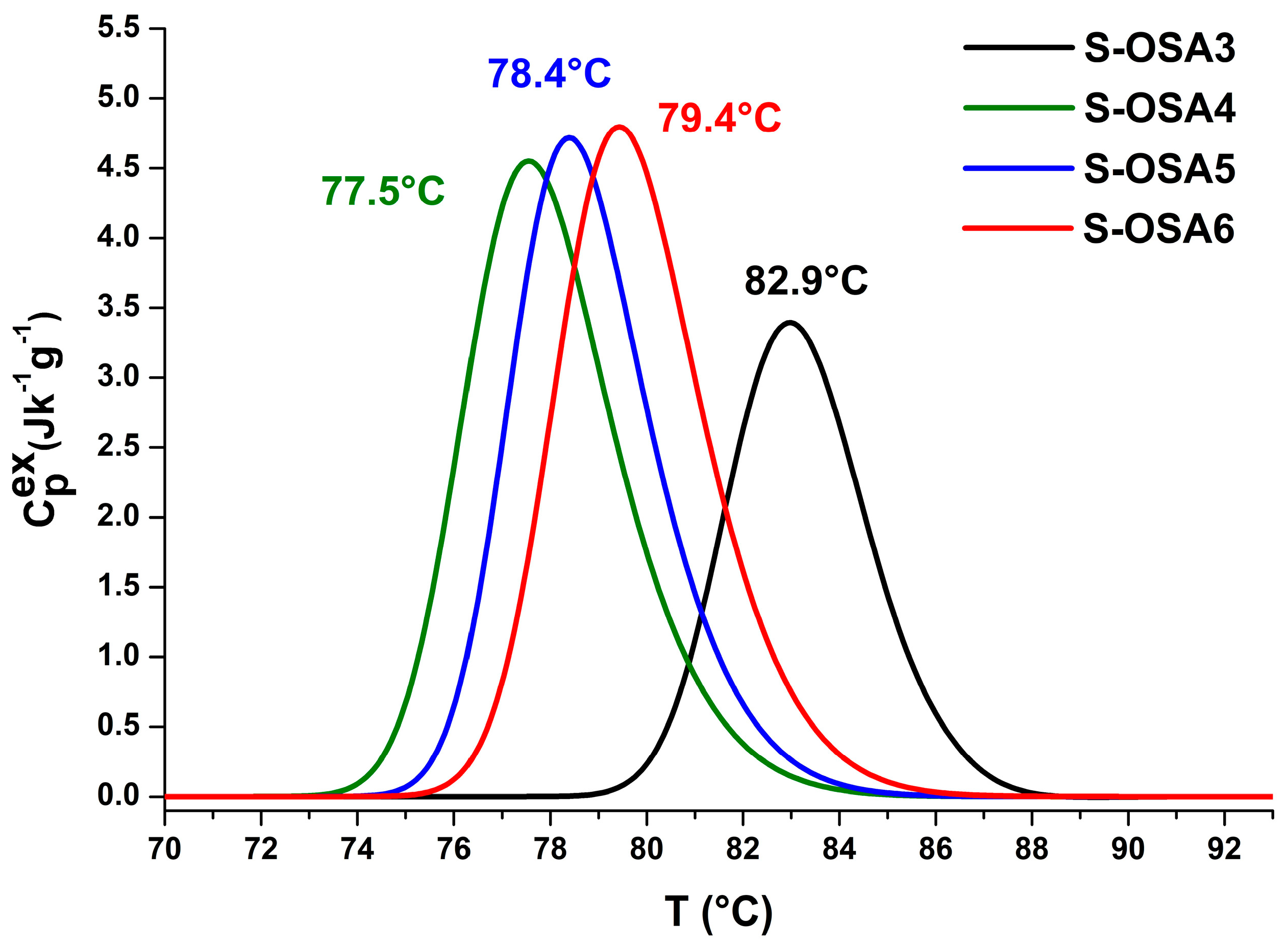
| Sample Name | nHAp (%) | Tonset (°C) | Tmax (°C) | ΔH (J/g) | ΔT1/2 (°C) |
|---|---|---|---|---|---|
| S-OSA3 | 0 | 80.1 | 82.9 | 12.8 | 3.5 |
| S-OSA4 | 1.0 | 74.6 | 77.5 | 18.0 | 3.6 |
| S-OSA5 | 1.5 | 75.6 | 78.4 | 17.4 | 3.3 |
| S-OSA6 | 3.0 | 76.2 | 79.4 | 18.5 | 3.5 |
| Sample Symbol | Tanning Agent/Nano-Hap | Timax (°C) | Tonset (°C) | ΣΔHi (J·g−1) | % ΔHi | ΔT1/2 (°C) |
|---|---|---|---|---|---|---|
| S-OSA7 | OSA (SA:KIO4 molar ratio of 1:0.2) | T1 = 78.1 | 73.7 | 16.1 | ΔH1 = 100 | 5.1 |
| S-OSA8 | OSA (SA:KIO4 molar ratio of 1:0.2) + nano-Hap (1%) | T1 = 81.0 T2 = 76.8 | 72.1 | 17.5 | ΔH1 = 28 ΔH2 = 72 | 7.1 |
| S-PA1 | commercial poly-aldehyde (PA) | T1 = 76.7 | 72.7 | 18.1 | ΔH2 = 100 | 4.7 |
| S-PA2 | commercial poly-aldehyde (PA) + nano-Hap (1%) | T1 = 79.1 T2 = 76.7 | 71.5 | 20.6 | ΔH1 = 26.0 ΔH2 = 74.0 | 7.2 |
| Sample | Tanning Agent/Nano-HAp | T1 (ms) | WA | T2A (ms) | WB | T2B (ms) |
|---|---|---|---|---|---|---|
| S-OSA7 | OSA (SA:KIO4 molar ratio 1:0.2) | 34 | 86 | 0.302 | 14 | 1.11 |
| S-OSA8 | OSA (SA:KIO4 molar ratio 1:0.2) + 1% nHAp | 25.4 | 92 | 0.244 | 8 | 1.23 |
| S-PA1 | commercial poly-aldehyde (PA) | 29 | 83 | 0.205 | 17 | 0.756 |
| S-PA2 | commercial poly-aldehyde (PA) + 1% nHAp | 26 | 89 | 0.236 | 9 | 0.854 |
Disclaimer/Publisher’s Note: The statements, opinions and data contained in all publications are solely those of the individual author(s) and contributor(s) and not of MDPI and/or the editor(s). MDPI and/or the editor(s) disclaim responsibility for any injury to people or property resulting from any ideas, methods, instructions or products referred to in the content. |
© 2023 by the authors. Licensee MDPI, Basel, Switzerland. This article is an open access article distributed under the terms and conditions of the Creative Commons Attribution (CC BY) license (https://creativecommons.org/licenses/by/4.0/).
Share and Cite
Quaratesi, I.; Micu, M.C.; Rebba, E.; Carsote, C.; Proietti, N.; Di Tullio, V.; Porcaro, R.; Badea, E. Cleaner Leather Tanning and Post-Tanning Processes Using Oxidized Alginate as Biodegradable Tanning Agent and Nano-Hydroxyapatite as Potential Flame Retardant. Polymers 2023, 15, 4676. https://doi.org/10.3390/polym15244676
Quaratesi I, Micu MC, Rebba E, Carsote C, Proietti N, Di Tullio V, Porcaro R, Badea E. Cleaner Leather Tanning and Post-Tanning Processes Using Oxidized Alginate as Biodegradable Tanning Agent and Nano-Hydroxyapatite as Potential Flame Retardant. Polymers. 2023; 15(24):4676. https://doi.org/10.3390/polym15244676
Chicago/Turabian StyleQuaratesi, Ilaria, Maria Cristina Micu, Erica Rebba, Cristina Carsote, Noemi Proietti, Valeria Di Tullio, Rita Porcaro, and Elena Badea. 2023. "Cleaner Leather Tanning and Post-Tanning Processes Using Oxidized Alginate as Biodegradable Tanning Agent and Nano-Hydroxyapatite as Potential Flame Retardant" Polymers 15, no. 24: 4676. https://doi.org/10.3390/polym15244676