
Crosslinked Chitosan Films Supplemented with Randia sp. Fruit Extract
 , ,
, ,  and
and
Abstract
:1. Introduction
2. Materials and Methods
2.1. Reagents and Solvents
2.2. Randia Extract
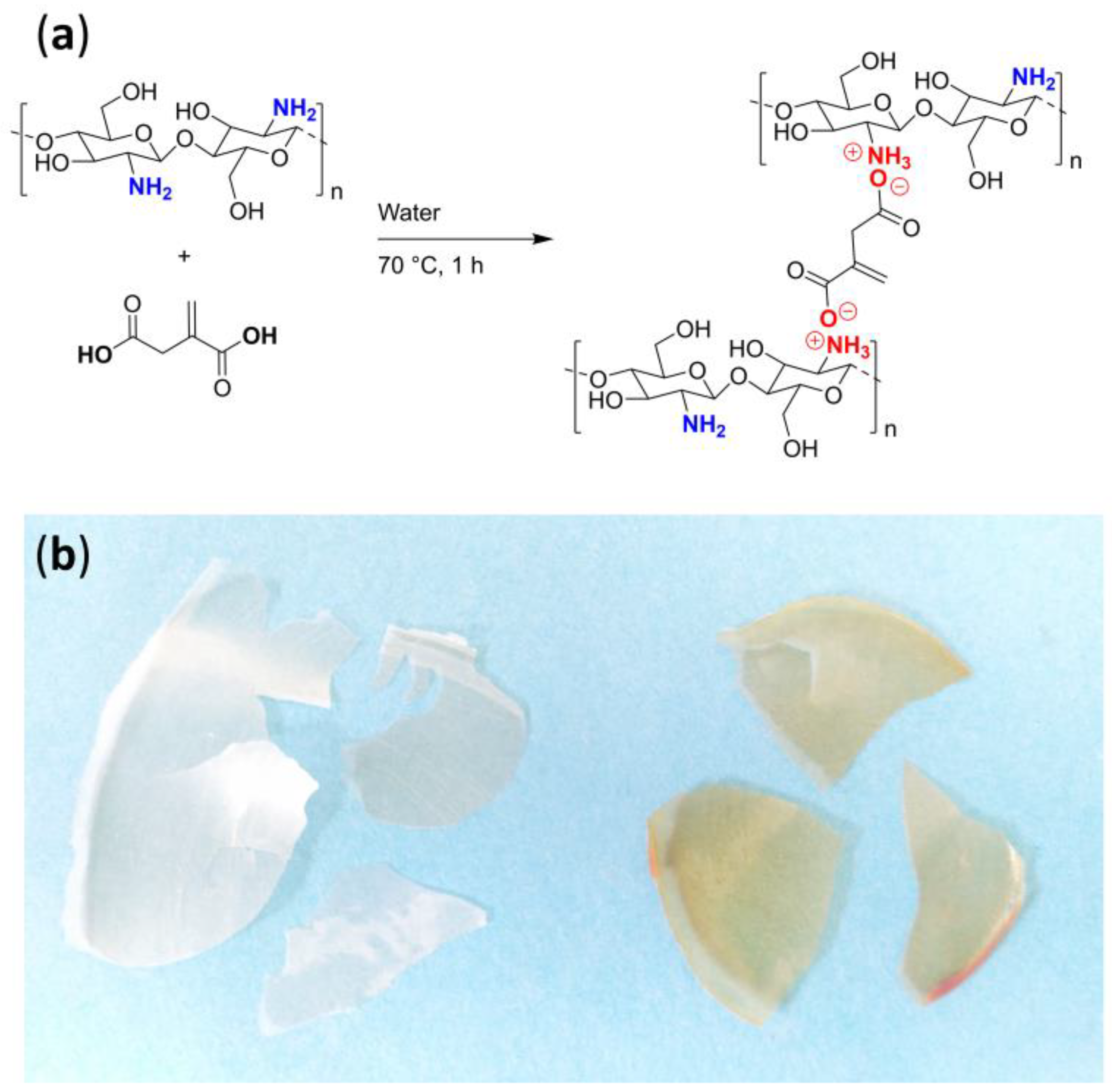
2.3. Synthesis of Chitosan/Itaconic Acid Film
2.4. Synthesis of the Chitosan/Itaconic Acid/Randia Film
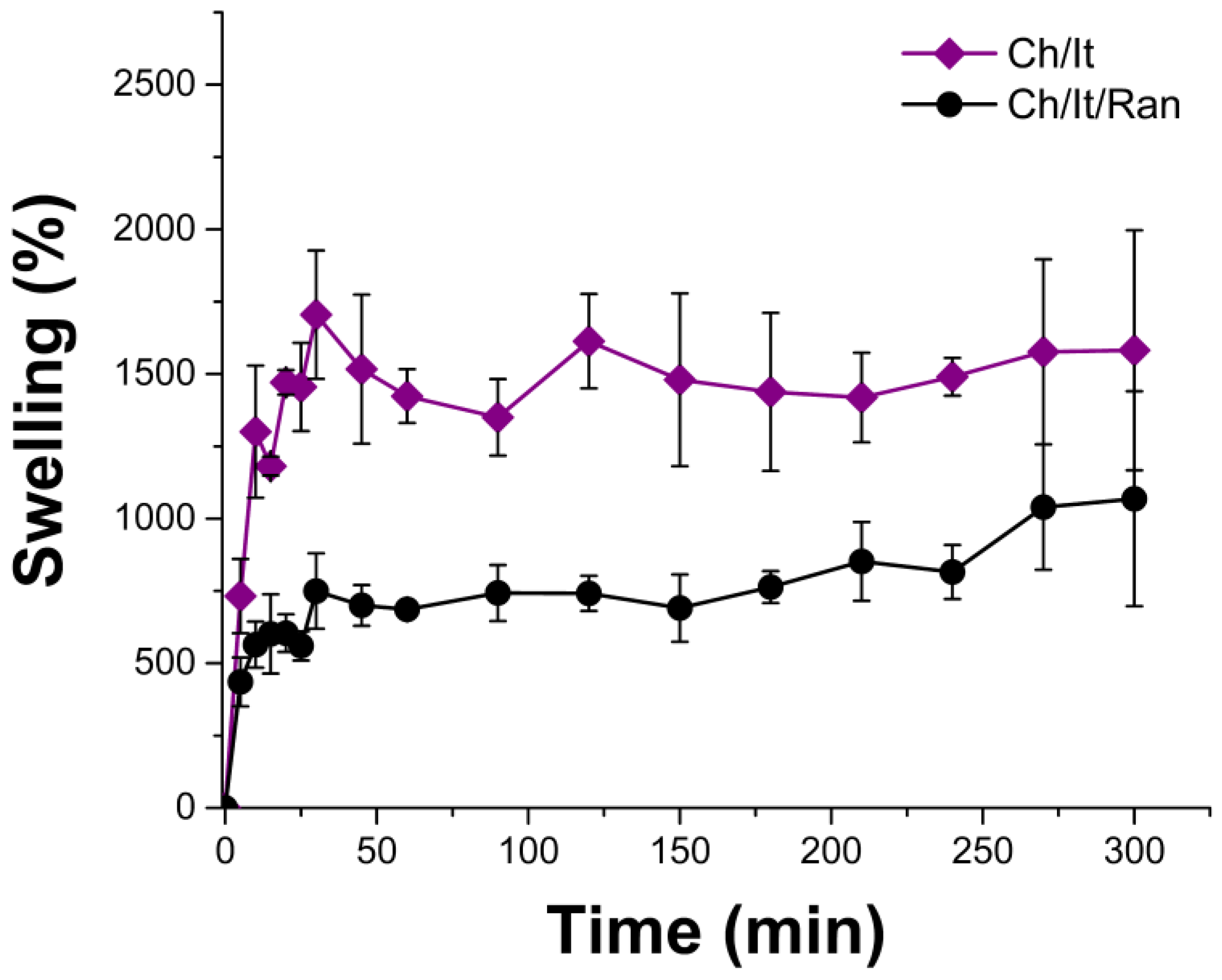
2.5. Limit Swelling
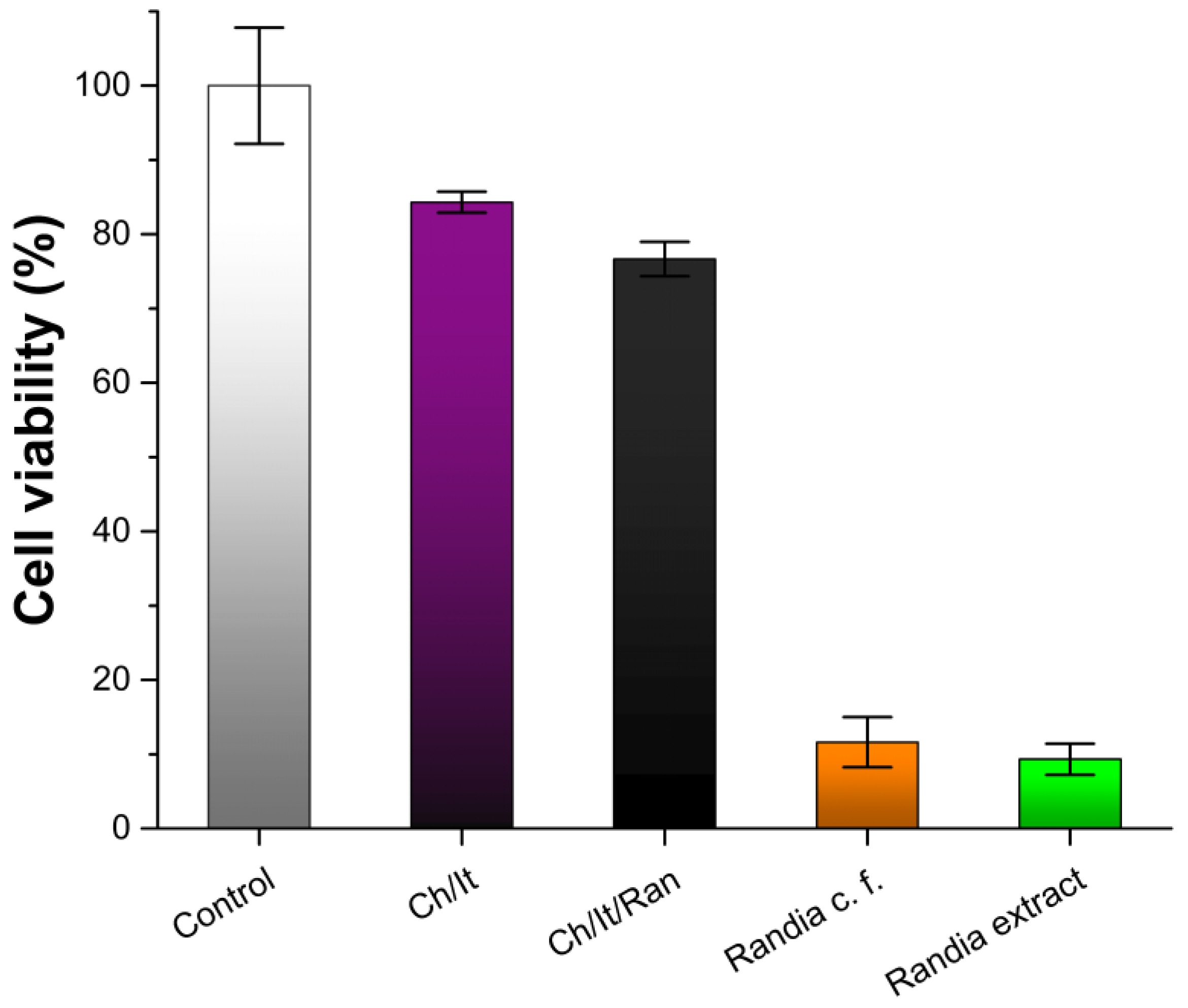
2.6. Cell Viability
2.7. Instrumental
2.7.1. UV-Vis Spectrophotometry
2.7.2. Infrared Spectroscopy
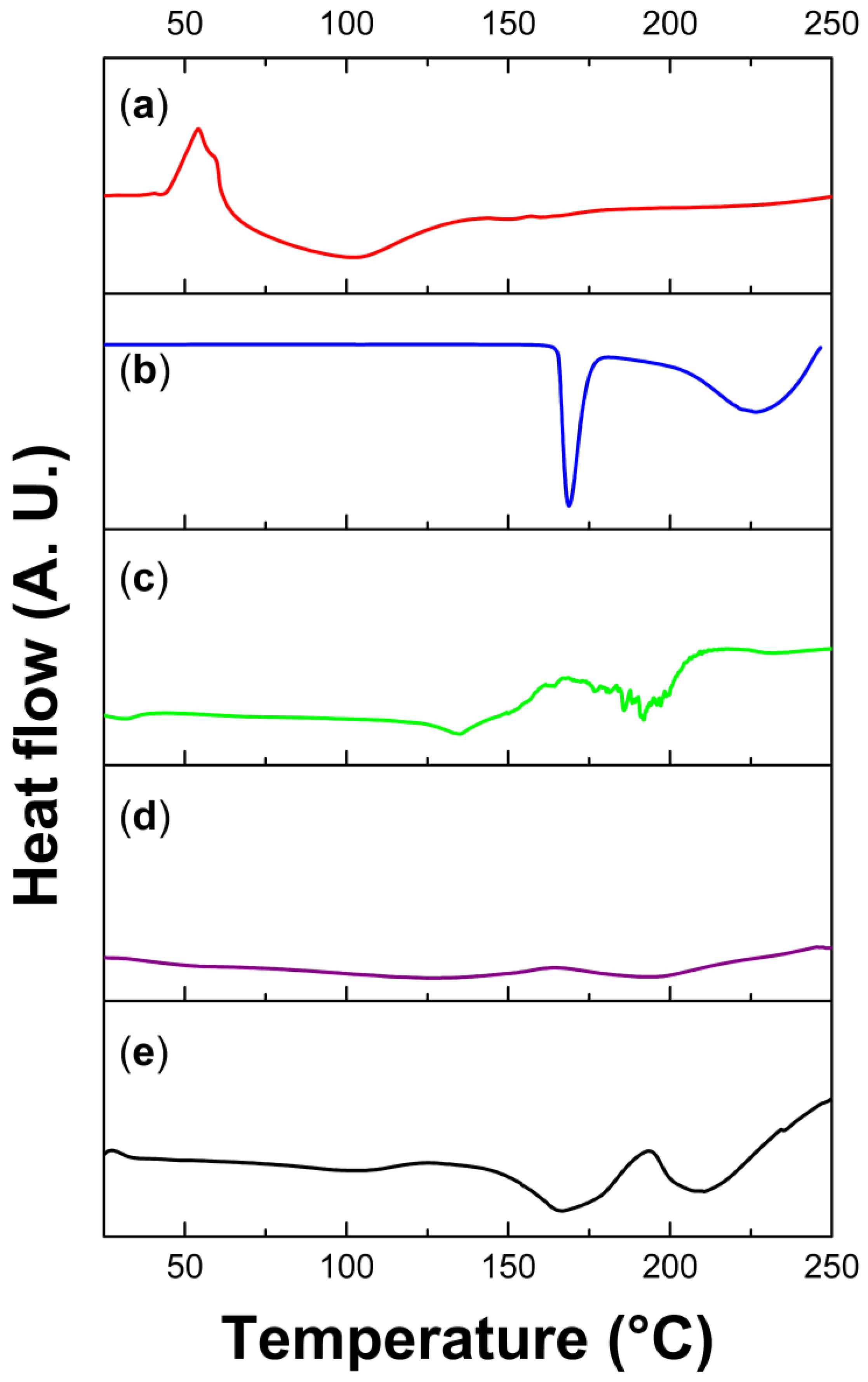
2.7.3. Differential Scanning Calorimetry (DSC)
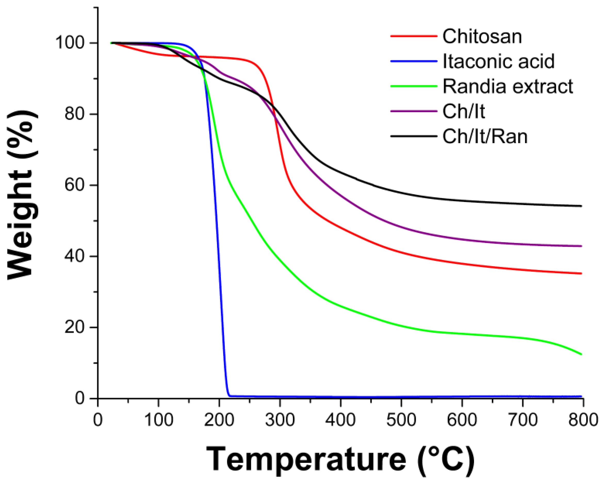
2.7.4. Thermogravimetric Analysis (TGA)
2.7.5. Contact Angle
2.7.6. Direct Analysis in Real-Time Mass Spectrometry (DART-MS)
3. Results and Analysis
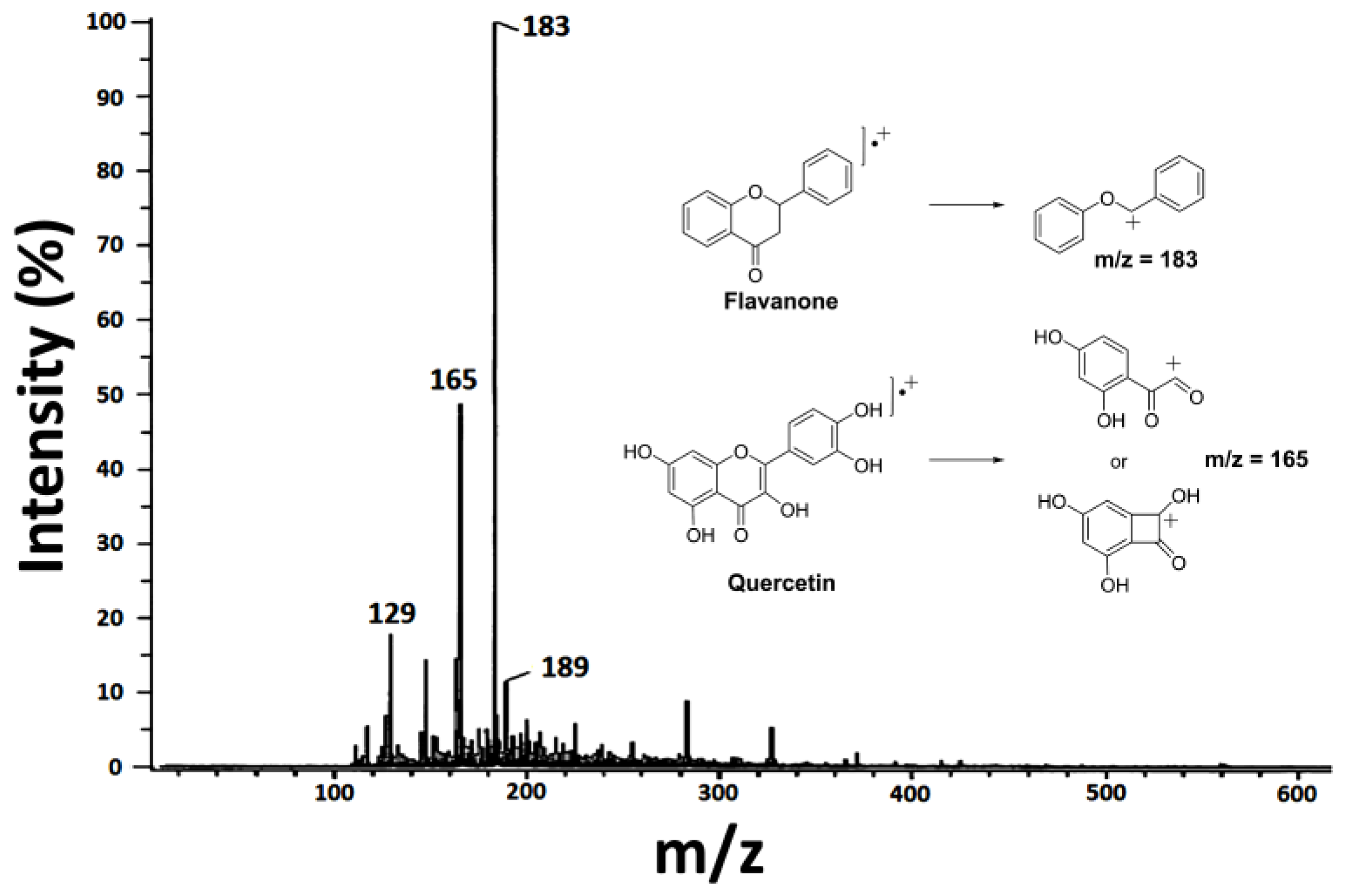
3.1. Mass Spectrometry of Randia Extract
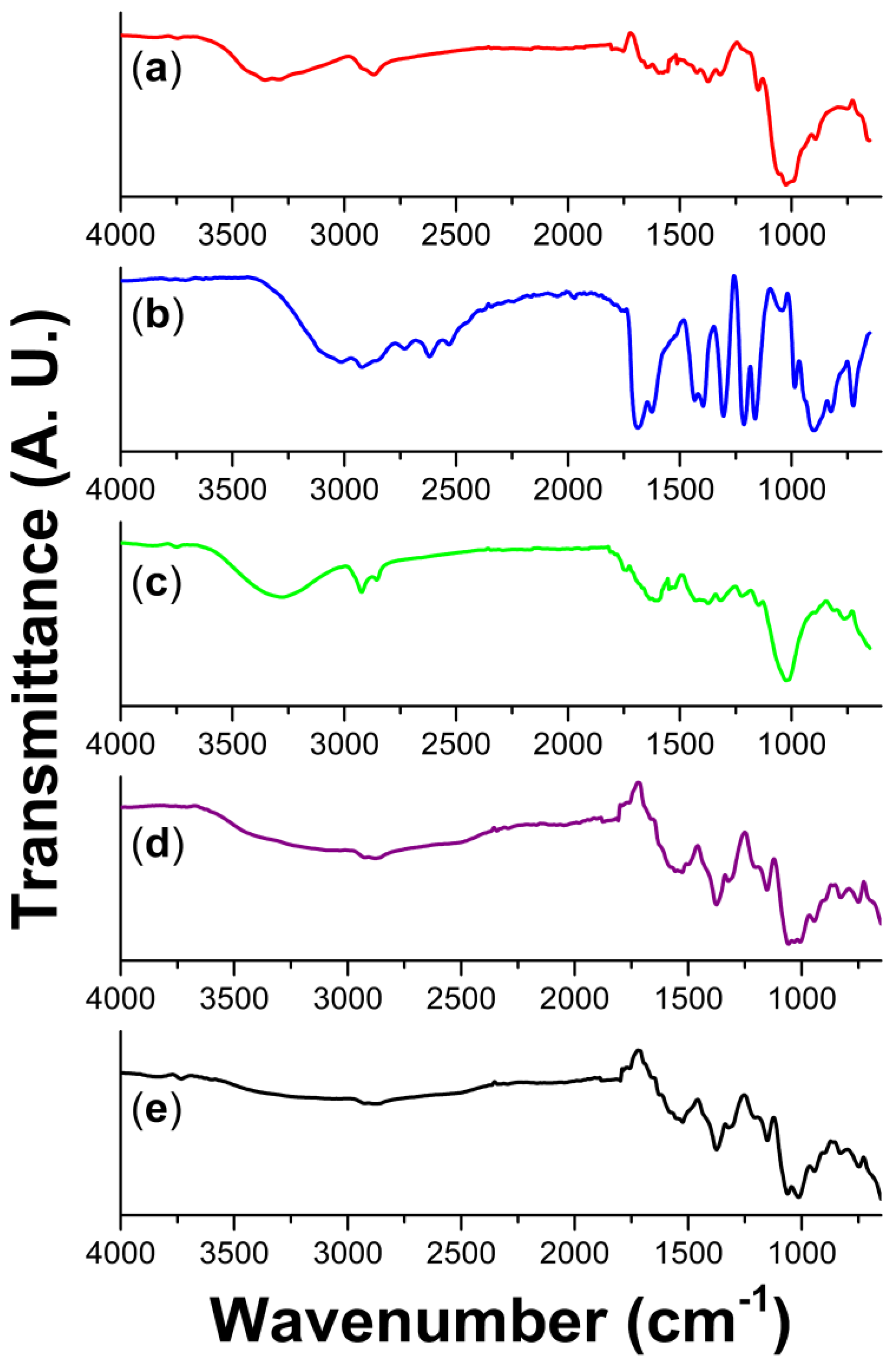
3.2. Spectroscopic Analysis
3.3. Mechanism of Crosslinking
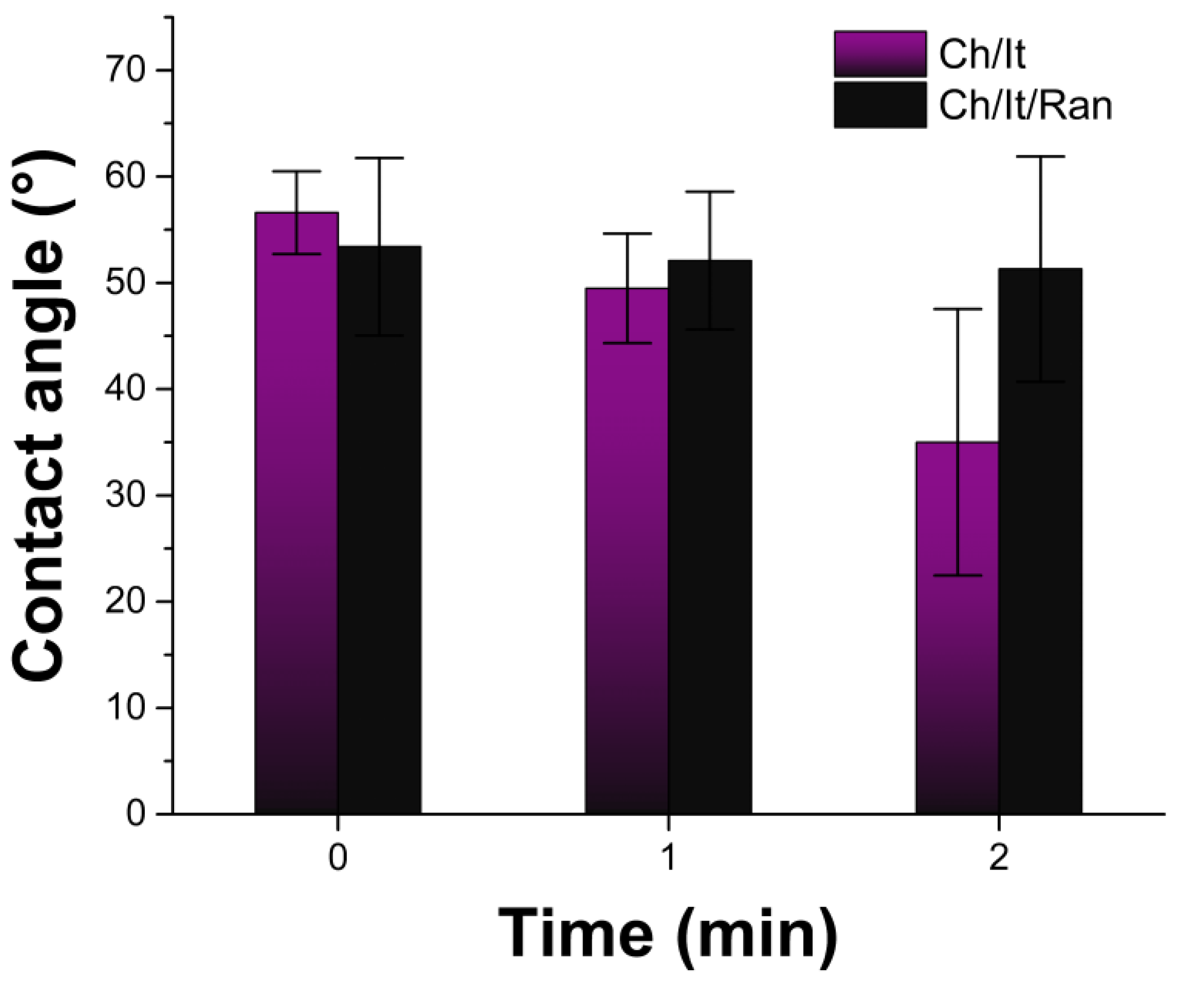
3.4. Hydrophilicity: Contact Angle and Swelling
3.5. Thermal Analysis
3.6. Cell Viability
4. Conclusions
Author Contributions
Funding
Institutional Review Board Statement
Data Availability Statement
Acknowledgments
Conflicts of Interest
References
- Birajdar, M.S.; Joo, H.; Koh, W.-G.; Park, H. Natural bio-based monomers for biomedical applications: A review. Biomater. Res. 2021, 25, 8. [Google Scholar] [CrossRef] [PubMed]
- Huang, S.J. Polymer waste management–biodegradation, incineration, and recycling. J. Macromol. Sci. Part A 1995, 32, 593–597. [Google Scholar] [CrossRef]
- Tudor, V.C.; Mocuta, D.N.; Teodorescu, R.F.; Smedescu, D.I. The issue of plastic and microplastic pollution in soil. Mater. Plast. 2019, 56, 484–487. [Google Scholar] [CrossRef]
- Elliott, J.E.; Elliott, K.H. Tracking marine pollution. Science 2013, 340, 556–558. [Google Scholar] [CrossRef]
- Kharrazi, S.M.; Younesi, H.; Abedini-Torghabeh, J. Microbial biodegradation of waste materials for nutrients enrichment and heavy metals removal: An integrated composting-vermicomposting process. Int. Biodeterior. Biodegrad. 2014, 92, 41–48. [Google Scholar] [CrossRef]
- Ferrero, F.; Periolatto, M. Antimicrobial finish of textiles by chitosan UV-curing. J. Nanosci. Nanotechnol. 2012, 12, 4803–4810. [Google Scholar] [CrossRef]
- Peers, S.; Montembault, A.; Ladavière, C. Chitosan hydrogels for sustained drug delivery. J. Control. Release 2020, 326, 150–163. [Google Scholar] [CrossRef] [PubMed]
- Milosavljević, N.B.; Ristić, M.Đ.; Perić-Grujić, A.A.; Filipović, J.M.; Štrbac, S.B.; Rakočević, Z.L.; Kalagasidis Krušić, M.T. Hydrogel based on chitosan, itaconic acid and methacrylic acid as adsorbent of Cd2+ ions from aqueous solution. Chem. Eng. J. 2010, 165, 554–562. [Google Scholar] [CrossRef]
- Yang, Y.; Chen, G.; Murray, P.; Zhang, H. Porous chitosan by crosslinking with tricarboxylic acid and tuneable release. SN Appl. Sci. 2020, 2, 435. [Google Scholar] [CrossRef] [Green Version]
- Noel, S.P.; Courtney, H.; Bumgardner, J.D.; Haggard, W.O. Chitosan films: A potential local drug delivery system for antibiotics. Clin. Orthop. Relat. Res. 2008, 466, 1377–1382. [Google Scholar] [CrossRef] [PubMed] [Green Version]
- Flores-Rojas, G.G.; López-Saucedo, F.; Vera-Graziano, R.; Magaña, H.; Mendizábal, E.; Bucio, E. Silver Nanoparticles Loaded on Polyethylene Terephthalate Films Grafted with Chitosan. Polymers 2023, 15, 125. [Google Scholar] [CrossRef] [PubMed]
- Ortega, A.; Sánchez, A.; Burillo, G. Binary graft of poly(N-vinylcaprolactam) and poly(acrylic acid) onto chitosan hydrogels using ionizing radiation for the retention and controlled release of therapeutic compounds. Polymers 2021, 13, 2641. [Google Scholar] [CrossRef]
- Lukman Hekiem, N.L.; Md Ralib, A.A.; Mohd Hatta, M.A.; Ahmad, F.B.; Nordin, A.N.; Ab Rahim, R.; Za’bah, N.F. Effect of chitosan dissolved in different acetic acid concentration towards VOC sensing performance of quartz crystal microbalance overlay with chitosan. Mater. Lett. 2021, 291, 129524. [Google Scholar] [CrossRef]
- Zhang, H.; Kong, M.; Jiang, Q.; Hu, K.; Ouyang, M.; Zhong, F.; Qin, M.; Zhuang, L.; Wang, G. Chitosan membranes from acetic acid and imidazolium ionic liquids: Effect of imidazolium structure on membrane properties. J. Mol. Liq. 2021, 340, 117209. [Google Scholar] [CrossRef]
- Yang, S.; Liu, L.; Chen, H.; Wei, Y.; Dai, L.; Liu, J.; Yuan, F.; Mao, L.; Li, Z.; Chen, F.; et al. Impact of different crosslinking agents on functional properties of curcumin-loaded gliadin-chitosan composite nanoparticles. Food Hydrocoll. 2021, 112, 106258. [Google Scholar] [CrossRef]
- Wang, J.; Zhuang, S. Chitosan-based materials: Preparation, modification and application. J. Clean. Prod. 2022, 355, 131825. [Google Scholar] [CrossRef]
- Kyzas, G.Z.; Siafaka, P.I.; Lambropoulou, D.A.; Lazaridis, N.K.; Bikiaris, D.N. Poly(itaconic acid)-grafted chitosan adsorbents with different cross-linking for Pb(II) and Cd(II) uptake. Langmuir 2014, 30, 120–131. [Google Scholar] [CrossRef] [PubMed]
- Ko, E.; Kim, H. Preparation of chitosan aerogel crosslinked in chemical and ionical ways by non-acid condition for wound dressing. Int. J. Biol. Macromol. 2020, 164, 2177–2185. [Google Scholar] [CrossRef]
- Sirviö, J.A.; Kantola, A.M.; Komulainen, S.; Filonenko, S. Aqueous modification of chitosan with itaconic acid to produce strong oxygen barrier film. Biomacromolecules 2021, 22, 2119–2128. [Google Scholar] [CrossRef]
- WFO Randia Capitata DC. Available online: http://www.worldfloraonline.org/taxon/wfo-0000294365 (accessed on 27 January 2023).
- Gallardo-Casas, C.; Guevara-Balcázar, G.; Morales-Ramos, E.; Tadeo-Jiménez, Y.; Gutiérrez-Flores, O.; Jiménez-Sánchez, N.; Valadez-Omaña, M.; Valenzuela-Vargas, M.; Castillo-Hernández, M. Ethnobotanic study of Randia aculeata (Rubiaceae) in Jamapa, Veracruz, Mexico, and its anti-snake venom effects on mouse tissue. J. Venom. Anim. Toxins Incl. Trop. Dis. 2012, 18, 287–294. [Google Scholar] [CrossRef] [Green Version]
- Cuevas-Juárez, E.; Yuriar-Arredondo, K.Y.; Pío-León, J.F.; Montes-Avila, J.; López-Angulo, G.; Páz Díaz-Camacho, S.; Delgado-Vargas, F. Antioxidant and α-glucosidase inhibitory properties of soluble melanins from the fruits of Vitex mollis Kunth, Randia echinocarpa Sessé et Mociño and Crescentia alata Kunth. J. Funct. Foods 2014, 9, 78–88. [Google Scholar] [CrossRef]
- Montes-Avila, J.; Ojeda-Ayala, M.; López-Angulo, G.; Pío-León, J.F.; Díaz-Camacho, S.P.; Ochoa-Terán, A.; Delgado-Vargas, F. Physicochemical properties and biological activities of melanins from the black-edible fruits Vitex mollis and Randia echinocarpa. J. Food Meas. Charact. 2018, 12, 1972–1980. [Google Scholar] [CrossRef]
- Kumar, D.; Mudgade, S.C.; Bhat, Z.A.; Bhujbal, S.S.; Rub, R. Anti allergic and anti-inflammatory effects of the fruits of Randia dumetorum Lamk. Orient. Pharm. Exp. Med. 2011, 11, 161–167. [Google Scholar] [CrossRef]
- Kandimalla, R.; Kalita, S.; Saikia, B.; Choudhury, B.; Singh, Y.P.; Kalita, K.; Dash, S.; Kotoky, J. Antioxidant and hepatoprotective potentiality of Randia dumetorum Lam. Leaf and bark via inhibition of oxidative stress and inflammatory cytokines. Front. Pharmacol. 2016, 7, 205. [Google Scholar] [CrossRef] [PubMed] [Green Version]
- Martínez-Barbosa, M.E.; Moreno-Corral, R.A. Washable, reusable and disposable medical textiles. In Medical Textiles from Natural Resources; Elsevier: Amsterdam, The Netherlands, 2022; pp. 717–765. [Google Scholar]
- Jiménez Ortega, L.A.; Barrientos Ramírez, L.; Tena Meza, M.P. Evaluación Fisicoquímica y fitoquímica de frutos de sapuche (Randia laevigata Standl.). e-CUCBA 2020, 13, 30–39. [Google Scholar] [CrossRef]
- Santos-Cervantes, M.E.; Ibarra-Zazueta, M.E.; Loarca-Piña, G.; Paredes-López, O.; Delgado-Vargas, F. Antioxidant and antimutagenic activities of Randia echinocarpa fruit. Plant Foods Hum. Nutr. 2007, 62, 71–77. [Google Scholar] [CrossRef]
- Bezerra da Cruz-Silva, S.C.; Matias, R.; Bono, J.A.M.; Santos, K.S.; Ludwig, J. Antifungal potential of extracts and fractions of Randia nitida leaves on soybean pathogens and their phytochemistry. Rev. Caatinga 2016, 29, 594–602. [Google Scholar] [CrossRef] [Green Version]
- Scigelova, M.; Hornshaw, M.; Giannakopulos, A.; Makarov, A. Fourier transform mass spectrometry. Mol. Cell. Proteom. 2011, 10, M111.009431. [Google Scholar] [CrossRef] [Green Version]
- Fernandes Queiroz, M.; Melo, K.; Sabry, D.; Sassaki, G.; Rocha, H. Does the use of chitosan contribute to oxalate kidney stone formation? Mar. Drugs 2014, 13, 141–158. [Google Scholar] [CrossRef] [Green Version]
- Cozzolino, D. Near infrared spectroscopy in natural products analysis. Planta Med. 2009, 75, 746–756. [Google Scholar] [CrossRef] [Green Version]
- Silverstein, R.M.; Webster, F.X.; Kiemle, D.J. Spectrometric Identification of Organic Compounds, 7th ed.; John Wiley & Sons: Hoboken, NJ, USA, 2005; ISBN 0471393622. [Google Scholar]
- Baranović, G.; Šegota, S. Infrared spectroscopy of flavones and flavonols. Reexamination of the hydroxyl and carbonyl vibrations in relation to the interactions of flavonoids with membrane lipids. Spectrochim. Acta Part A Mol. Biomol. Spectrosc. 2018, 192, 473–486. [Google Scholar] [CrossRef]
- Kim, L.-S.; Hong, S.-J.; Son, M.-K.; Lee, Y.-H. Polymeric and compositional properties of novel extracellular microbial polyglucosamine biopolymer from new strain of citrobacter sp. BL-4. Biotechnol. Lett. 2006, 28, 241–245. [Google Scholar] [CrossRef] [PubMed]
- Farahani, B.V.; Ghasemzadeh, H.; Afraz, S. Thermodynamic studies of insulin loading into a glucose responsive hydrogel based on chitosan-polyacrylamide-polyethylene glycol. J. Chin. Chem. Soc. 2016, 63, 438–444. [Google Scholar] [CrossRef]
- Qi, P.; Chen, H.-L.; Nguyen, H.T.H.; Lin, C.-C.; Miller, S.A. Synthesis of biorenewable and water-degradable polylactam esters from itaconic acid. Green Chem. 2016, 18, 4170–4175. [Google Scholar] [CrossRef]
- Milosavljević, N.B.; Kljajević, L.M.; Popović, I.G.; Filipović, J.M.; Kalagasidis Krušić, M.T. Chitosan, itaconic acid and poly(vinyl alcohol) hybrid polymer networks of high degree of swelling and good mechanical strength. Polym. Int. 2010, 59, 686–694. [Google Scholar] [CrossRef]
- Pérez-Calderón, J.; Santos, M.V.; Zaritzky, N. Synthesis, characterization and application of cross-linked chitosan/oxalic acid hydrogels to improve azo dye (Reactive Red 195) adsorption. React. Funct. Polym. 2020, 155, 104699. [Google Scholar] [CrossRef]
- Gabriele, F.; Donnadio, A.; Casciola, M.; Germani, R.; Spreti, N. Ionic and covalent crosslinking in chitosan-succinic acid membranes: Effect on physicochemical properties. Carbohydr. Polym. 2021, 251, 117106. [Google Scholar] [CrossRef] [PubMed]
- Lee, L.-H. Roles of molecular interactions in adhesion, adsorption, contact angle and wettability. J. Adhes. Sci. Technol. 1993, 7, 583–634. [Google Scholar] [CrossRef]
- Malkin, A.; Ilyin, S.; Roumyantseva, T.; Kulichikhin, V. Rheological evidence of gel formation in dilute poly(acrylonitrile) solutions. Macromolecules 2013, 46, 257–266. [Google Scholar] [CrossRef]
- Kulichikhin, V.G.; Ilyin, S.O.; Mironova, M.V.; Berkovich, A.K.; Nifant’ev, I.E.; Malkin, A.Y. From polyacrylonitrile, its solutions, and filaments to carbon fibers: I. Phase state and rheology of basic polymers and their solutions. Adv. Polym. Technol. 2018, 37, 1076–1084. [Google Scholar] [CrossRef]
- Rizwan, M.; Yahya, R.; Hassan, A.; Yar, M.; Azzahari, A.D.; Selvanathan, V.; Sonsudin, F.; Abouloula, C.N. pH sensitive hydrogels in drug delivery: Brief history, properties, swelling, and release mechanism, material selection and applications. Polymers 2017, 9, 137. [Google Scholar] [CrossRef] [Green Version]
- Blanco, Y.S.; Topel, Ö.; Bajnóczi, É.G.; Werner, J.; Björneholm, O.; Persson, I. Chemical equilibria of aqueous ammonium–carboxylate systems in aqueous bulk, close to and at the water–air interface. Phys. Chem. Chem. Phys. 2019, 21, 12434–12445. [Google Scholar] [CrossRef] [PubMed] [Green Version]
- Shan, S.; Herschlag, D. The change in hydrogen bond strength accompanying charge rearrangement: Implications for enzymatic catalysis. Proc. Natl. Acad. Sci. USA 1996, 93, 14474–14479. [Google Scholar] [CrossRef] [Green Version]
- Cunha da Cruz, J.; Machado de Castro, A.; Camporese Sérvulo, E.F. World market and biotechnological production of itaconic acid. 3 Biotech. 2018, 8, 138. [Google Scholar] [CrossRef] [PubMed]
- Damiani, E.; Solorio, J.A.; Doyle, A.P.; Wallace, H.M. How reliable are in vitro IC50 values? Values vary with cytotoxicity assays in human glioblastoma cells. Toxicol. Lett. 2019, 302, 28–34. [Google Scholar] [CrossRef] [Green Version]
- Leoni, G.; Neumann, P.-A.; Sumagin, R.; Denning, T.L.; Nusrat, A. Wound repair: Role of immune–epithelial interactions. Mucosal Immunol. 2015, 8, 959–968. [Google Scholar] [CrossRef] [Green Version]
- Coraux, C.; Hajj, R.; Lesimple, P.; Puchelle, E. In vivo models of human airway epithelium repair and regeneration. Eur. Respir. Rev. 2005, 14, 131–136. [Google Scholar] [CrossRef]


| Sample | Wavenumber (cm−1) | Type |
|---|---|---|
| Chitosan | 3354, 3296 (wide band) | O-H st and N-H st |
| 2868 | C-H st | |
| 1575 | N-H bd | |
| 1373 | C-H (methyl) bd | |
| 1145, 1025 | C-O st as and sym | |
| Itaconic acid | 3000 (wide band) | O-H st |
| 3013, 2917 | C-H st | |
| 1682 | C=O st | |
| 1623 | C=C | |
| 1395 | C-H bd | |
| 1304, 1213 | C-O st | |
| Randia extract | 3291 | O-H st |
| 2920 | C-H st | |
| 1699 | C=O st | |
| 1594 | C=C | |
| 1403 | C-N | |
| 1021 | C-O st | |
| Ch/It | 3030 (wide band) | O-H st |
| 2885 | C-H st | |
| 1528 | C=C | |
| 1375 | C-H methyl bd and O-C=O as st | |
| 1066, 1022 | C-O st | |
| Ch/It/Ran | 3000 (wide band) | O-H st |
| 2878 | C-H st | |
| 1524 | C=C | |
| 1375 | C-H methyl bd and O-C=O as st | |
| 1151, 1061, 1013 | C-O st, as and sym |
| Sample | DSC | TGA | |||
|---|---|---|---|---|---|
| Transition (°C) | Thermal Transition | 10 wt% Loss (°C) | Decomposition Temperature Td (°C) | % Char Yield (800 °C) | |
| Chitosan | 101 | Endo | 275 | 297 | 35.4 |
| Itaconic acid | 168 | Endo | 176 | 203 | 0.6 |
| Randia extract | 135, | Endo | 176 | 192, 258 | 12.5 |
| 191 | |||||
| Ch/It | 128 | Endo | 224 | 196, 302 | 42.1 |
| Ch/It/Ran | 170, | Endo | 200.3 | 136, 185,311 | 54.2 |
| 210 | |||||
| Chitosan (%wt.) | Itaconic Acid (%wt.) | Randia Extract (%wt.) | |
|---|---|---|---|
| Ch/It | 70 | 30 | - |
| Ch/It/Ran | 55 | 23 | 22 |
Disclaimer/Publisher’s Note: The statements, opinions and data contained in all publications are solely those of the individual author(s) and contributor(s) and not of MDPI and/or the editor(s). MDPI and/or the editor(s) disclaim responsibility for any injury to people or property resulting from any ideas, methods, instructions or products referred to in the content. |
© 2023 by the authors. Licensee MDPI, Basel, Switzerland. This article is an open access article distributed under the terms and conditions of the Creative Commons Attribution (CC BY) license (https://creativecommons.org/licenses/by/4.0/).
Share and Cite
López-Saucedo, F.; Buendía-González, L.; Magaña, H.; Flores-Rojas, G.G.; Bucio, E. Crosslinked Chitosan Films Supplemented with Randia sp. Fruit Extract. Polymers 2023, 15, 2724. https://doi.org/10.3390/polym15122724
López-Saucedo F, Buendía-González L, Magaña H, Flores-Rojas GG, Bucio E. Crosslinked Chitosan Films Supplemented with Randia sp. Fruit Extract. Polymers. 2023; 15(12):2724. https://doi.org/10.3390/polym15122724
Chicago/Turabian StyleLópez-Saucedo, Felipe, Leticia Buendía-González, Héctor Magaña, Guadalupe Gabriel Flores-Rojas, and Emilio Bucio. 2023. "Crosslinked Chitosan Films Supplemented with Randia sp. Fruit Extract" Polymers 15, no. 12: 2724. https://doi.org/10.3390/polym15122724