

Synthesis of PMMA Microspheres with Tunable Diameters: Evaluation as a Template in the Synthesis of Tin Oxide Coatings
 , , and
Abstract
:1. Introduction
2. Materials and Methods
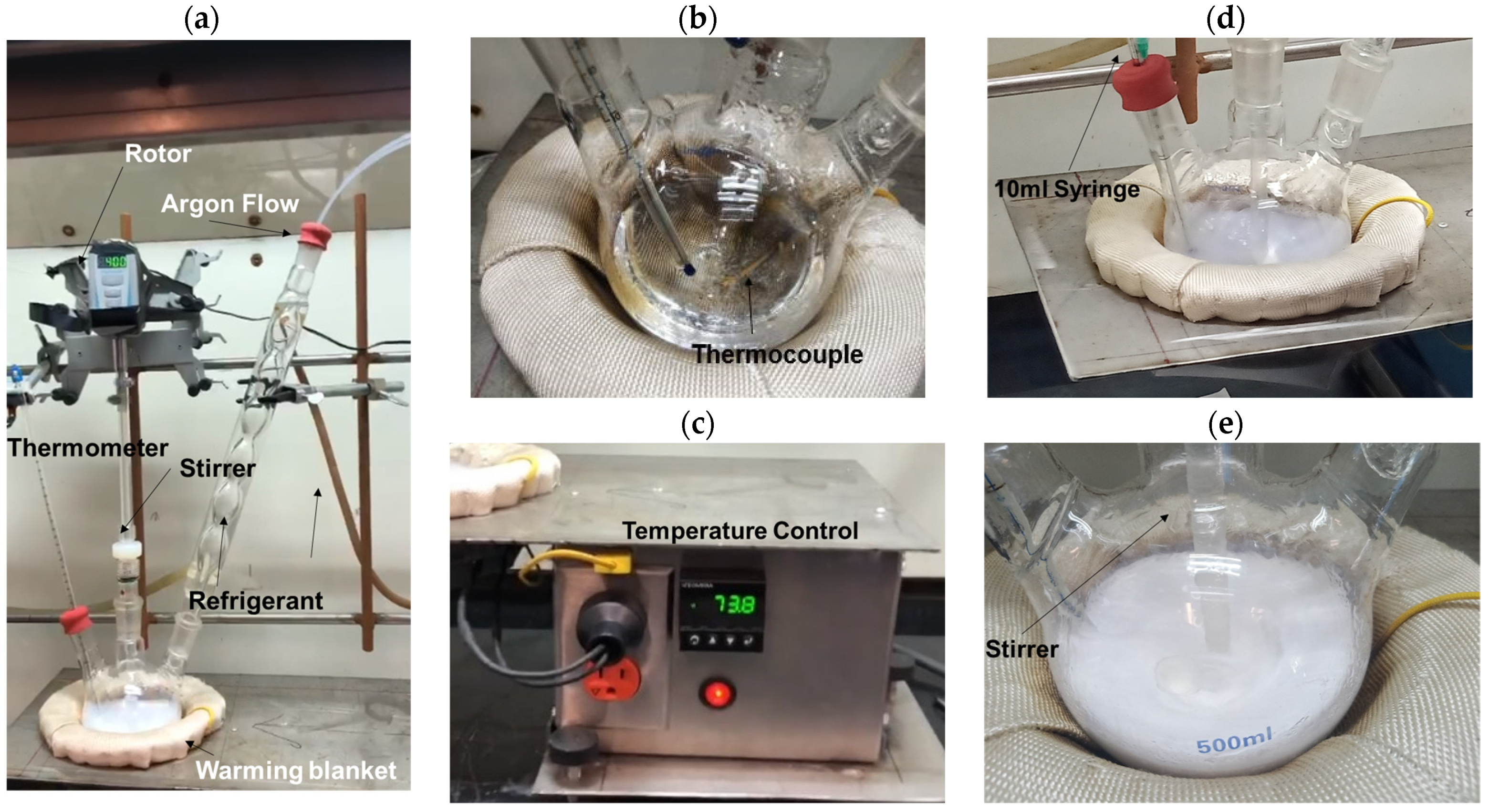
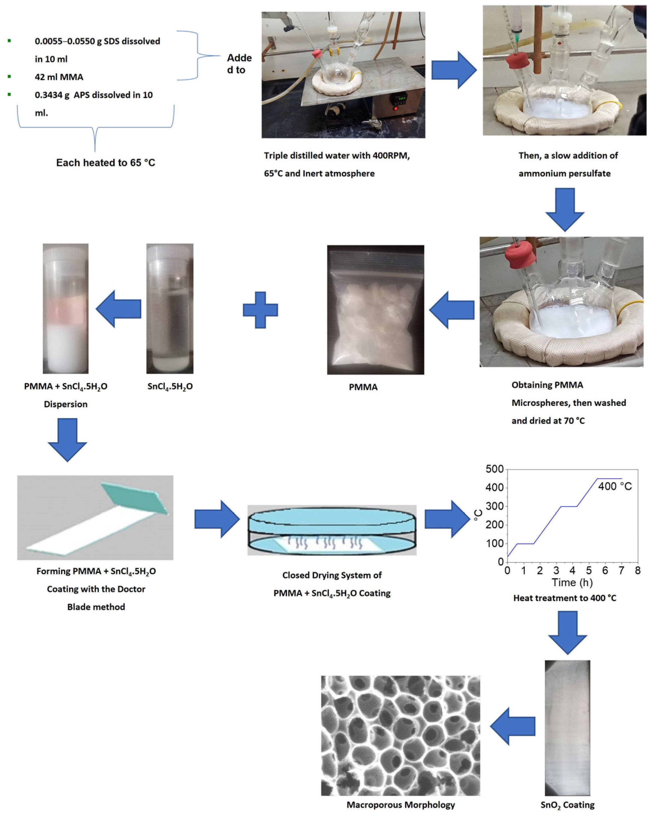
2.1. Synthesis of PMMA
2.2. Synthesis of Porous Tin Oxide Coatings
2.3. Characterization
3. Results and Discussion
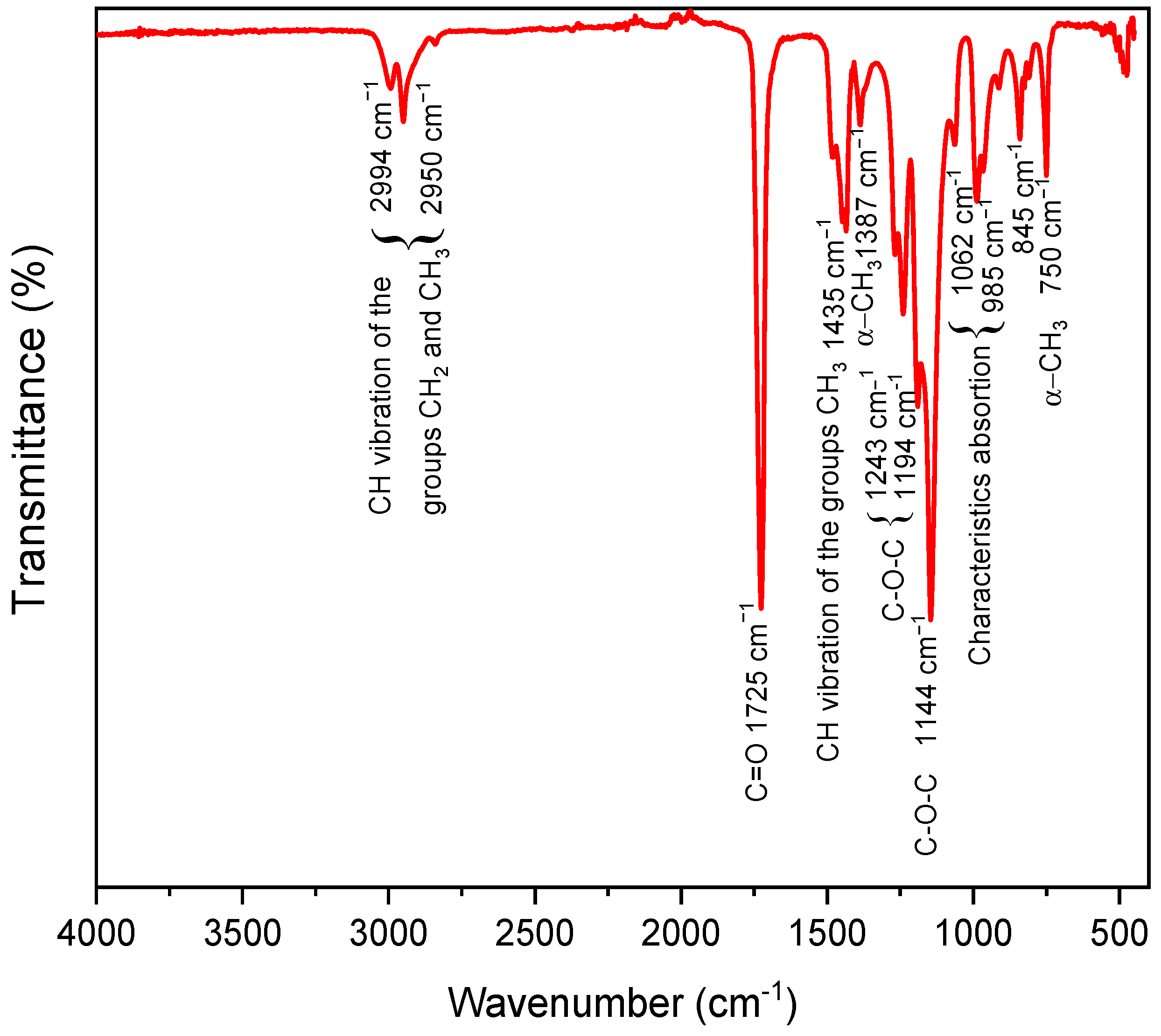
3.1. FTIR Analysis
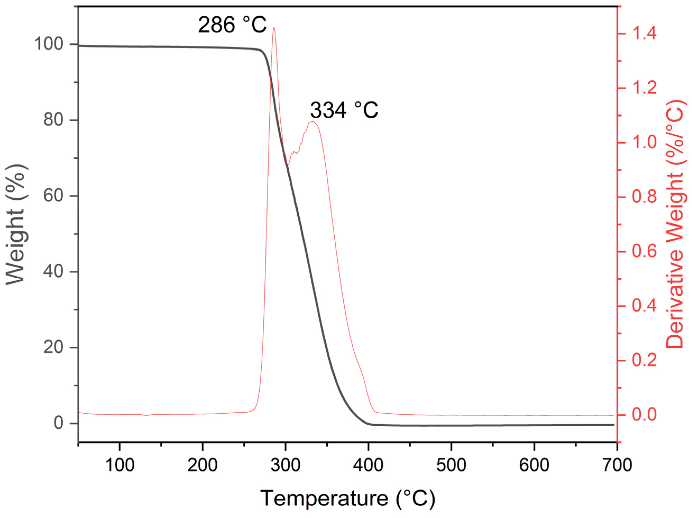
3.2. TGA Analysis
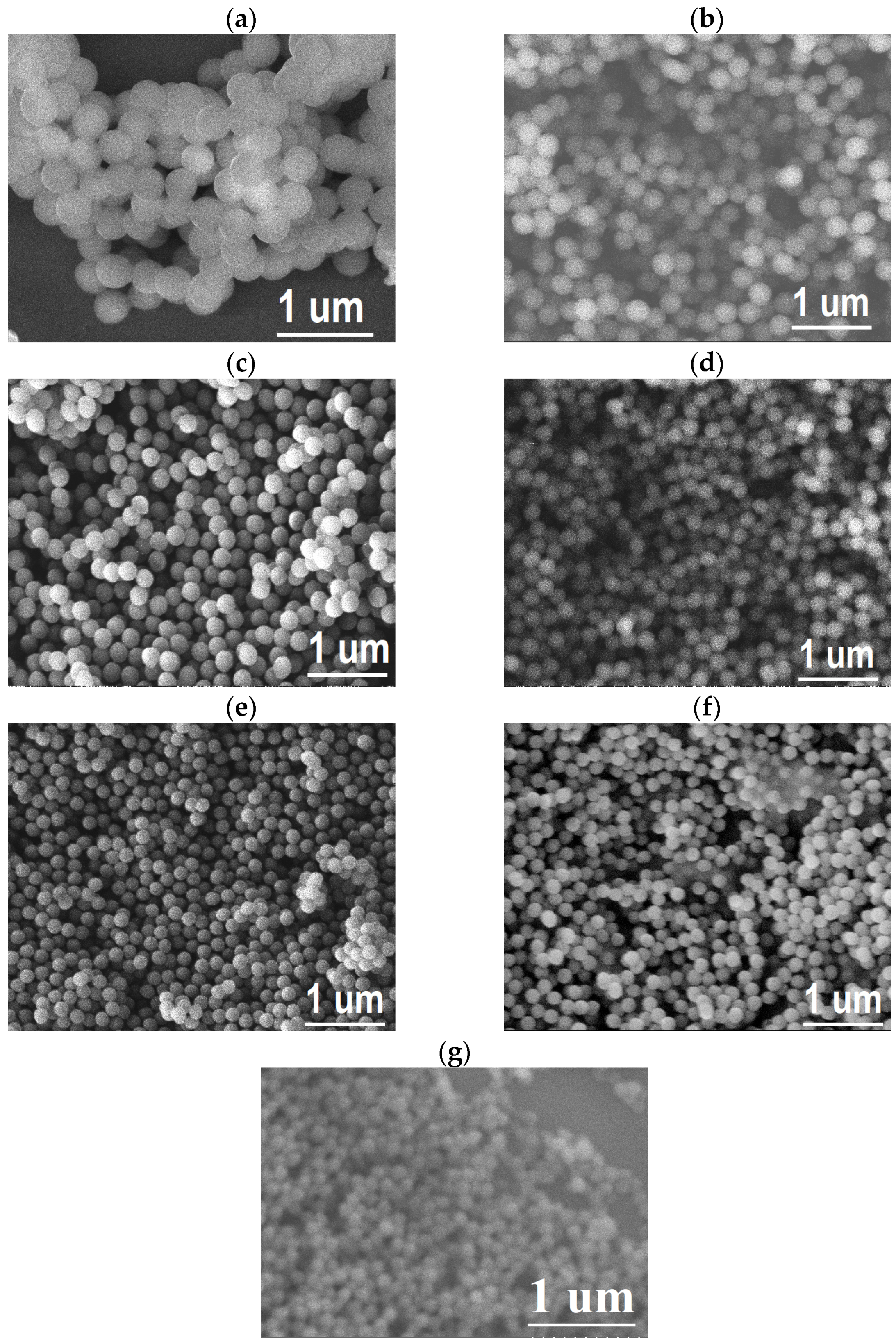
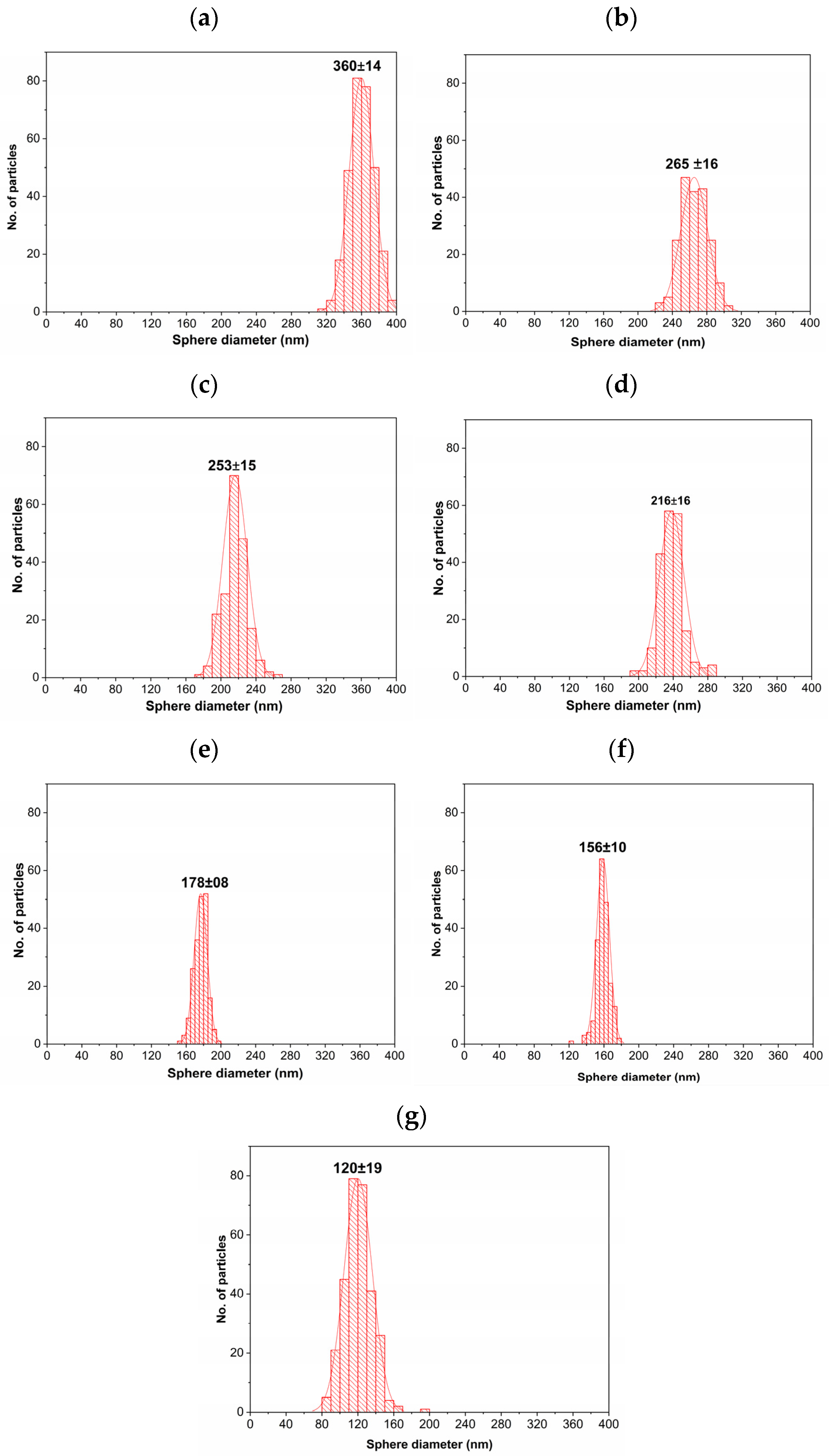
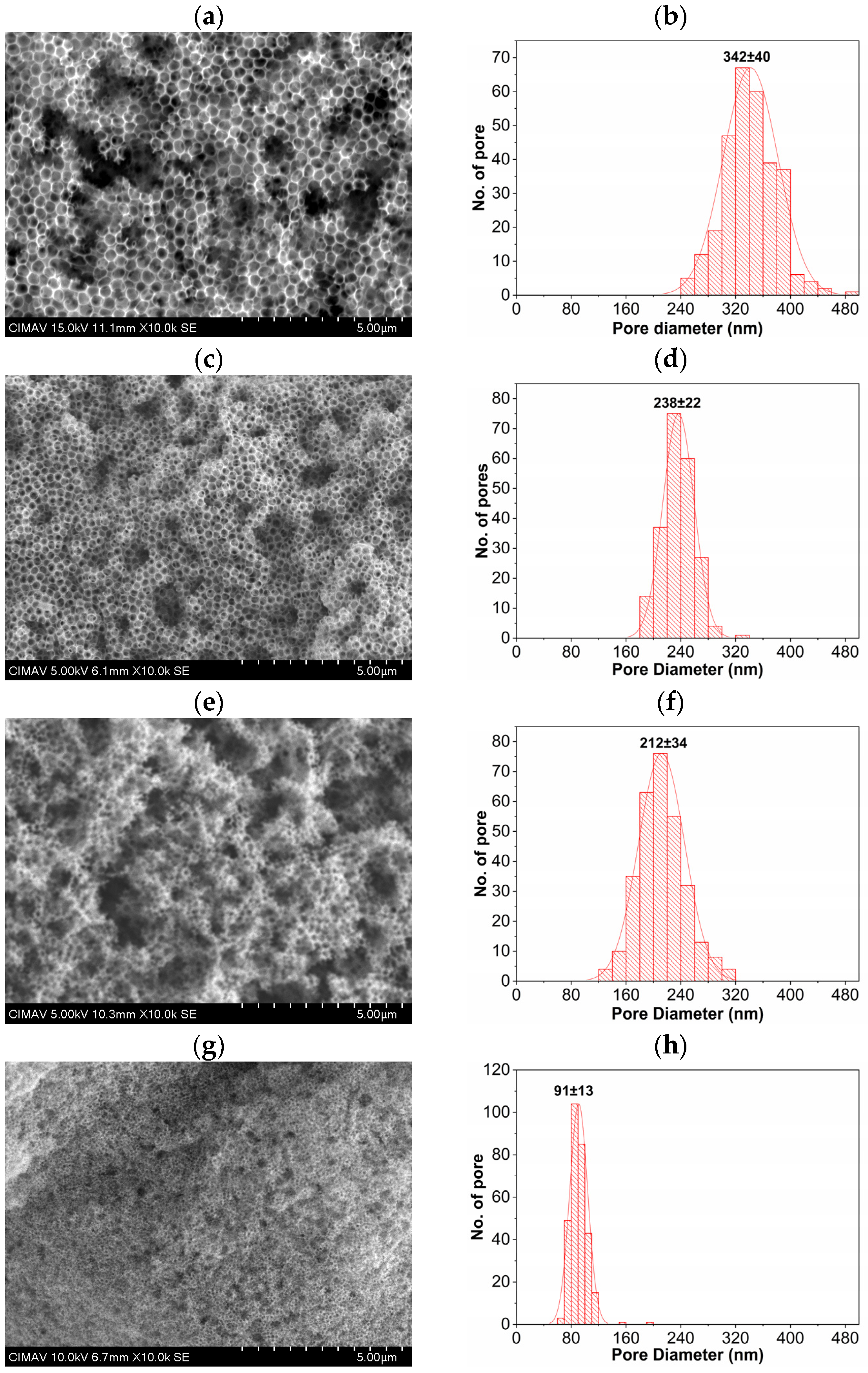
3.3. Morphology by SEM
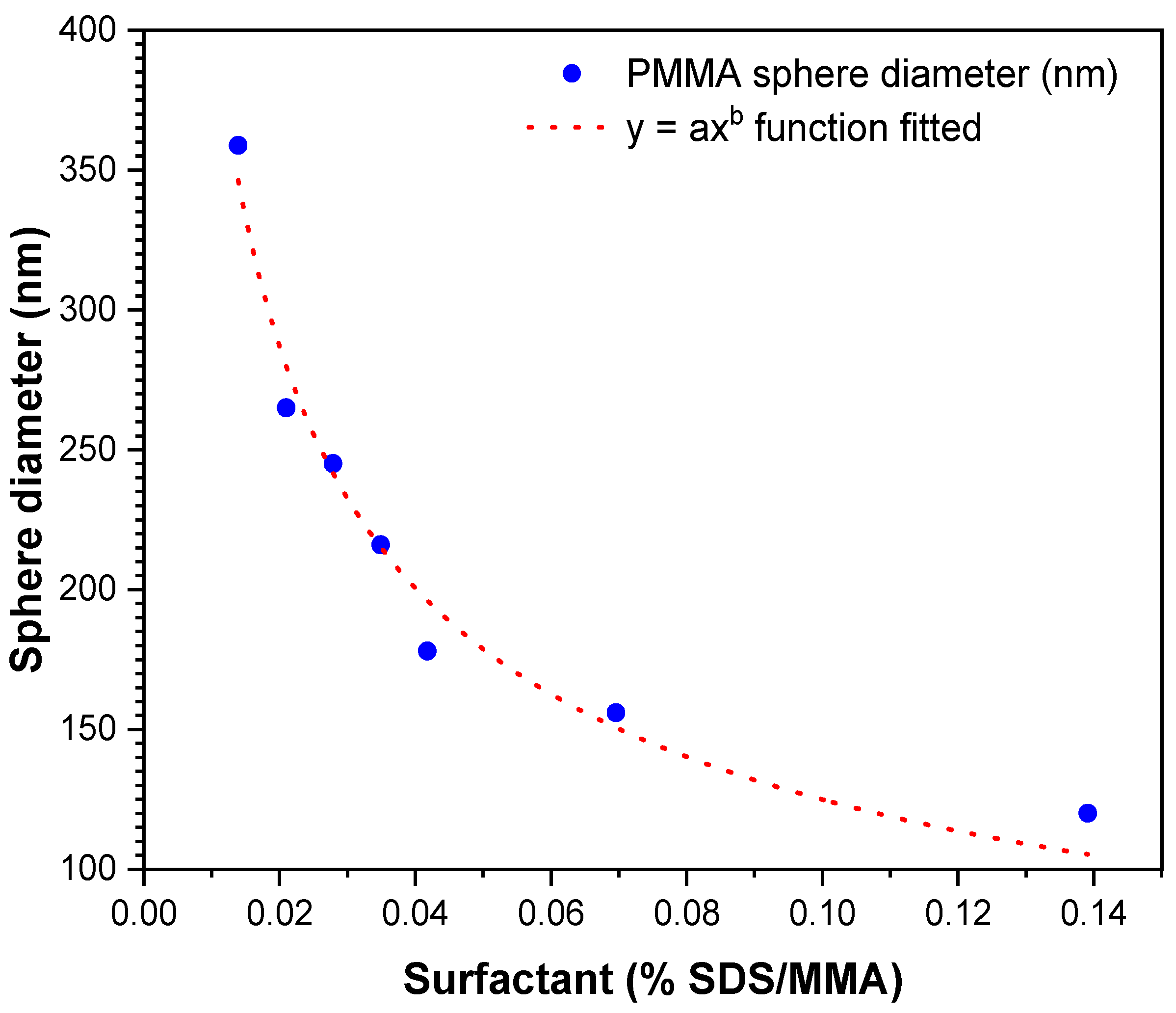
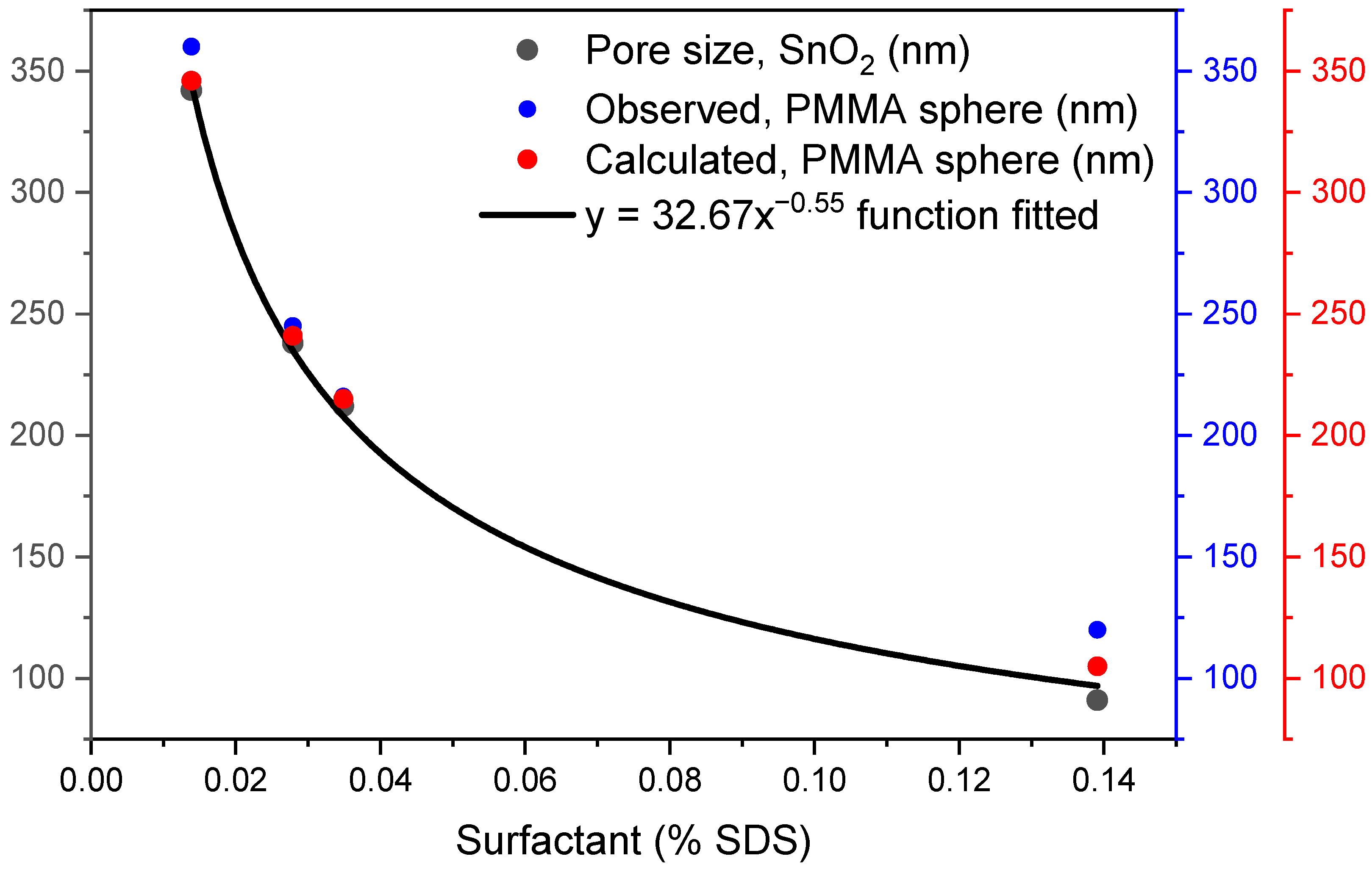
3.4. Fit a Function
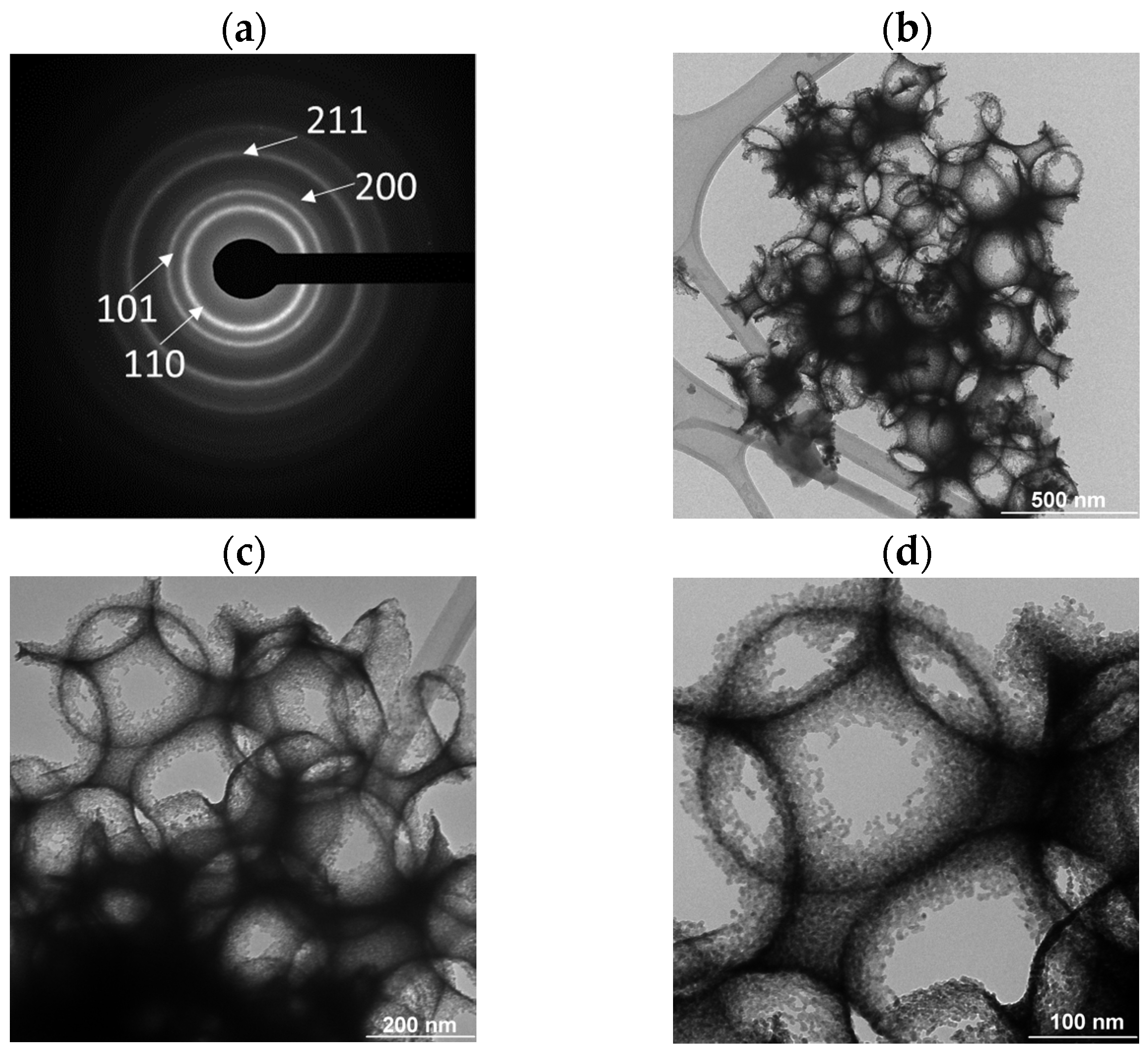
3.5. Synthesis of SnO2 Coatings
4. Conclusions
Author Contributions
Funding
Institutional Review Board Statement
Informed Consent Statement
Data Availability Statement
Acknowledgments
Conflicts of Interest
References
- Yuan, M.; Huang, D.; Zhao, Y. Development of Synthesis and Application of High Molecular Weight Poly(Methyl Methacrylate). Polymers 2022, 14, 2632. [Google Scholar] [CrossRef] [PubMed]
- Duan, G.; Zhang, C.; Li, A.; Yang, X.; Lu, L.; Wang, X. Preparation and Characterization of Mesoporous Zirconia Made by Using a Poly (Methyl Methacrylate) Template. Nanoscale Res. Lett. 2008, 3, 118. [Google Scholar] [CrossRef] [PubMed]
- Hyodo, T.; Sasahara, K.; Shimizu, Y.; Egashira, M. Preparation of Macroporous SnO2 Films Using PMMA Microspheres and Their Sensing Properties to NOx and H2. Sens. Actuators B Chem. 2005, 106, 580–590. [Google Scholar] [CrossRef]
- Kamitani, K.; Hyodo, T.; Shimizu, Y.; Egashira, M. Fabrication of Porous Alumina Ceramics Having Cell Windows with Controlled Size by PMMA Template Method. J. Mater. Sci. 2010, 45, 3602–3609. [Google Scholar] [CrossRef]
- Khan, H.; Samanta, S.; Seth, M.; Jana, S. Fabrication and Photoelectrochemical Activity of Hierarchically Porous TiO2–ZnO Heterojunction Film. J. Mater. Sci. 2020, 55, 11907–11918. [Google Scholar] [CrossRef]
- Sordello, F.; Minero, C. Photocatalytic Hydrogen Production on Pt-Loaded TiO2 Inverse Opals. Appl. Catal. B Environ. 2015, 163, 452–458. [Google Scholar] [CrossRef]
- Masuda, Y. Recent Advances in SnO2 Nanostructure Based Gas Sensors. Sens. Actuators B Chem. 2022, 364, 131876. [Google Scholar] [CrossRef]
- Tan, Y.; Zhang, J. Highly Sensitive Ethanol Gas Sensors Based on Co-Doped SnO2 Nanobelts and Pure SnO2 Nanobelts. Phys. E Low-Dimens. Syst. Nanostructures 2023, 147, 115604. [Google Scholar] [CrossRef]
- Rameshkumar, C.; Ananth, D.; Divyalakshmi, V.; Balakrishnan, M.; Senthilkumar, G.; Subalakshmi, R. An Investigation of SnO2 Nanofilm for Solar Cell Application by Spin Coating Technique. AIP Conf. Proc. 2021, 2341, 040024. [Google Scholar] [CrossRef]
- Alfaro Cruz, M.R.; Saldaña-Ramírez, A.; Juárez-Ramírez, I.; Torres-Martínez, L.M. Development of SnO2–ZnO Thin Films as a Photocatalyst for Obtaining Alternative Fuels through Photocatalytic Reactions. Solid State Sci. 2023, 137, 107112. [Google Scholar] [CrossRef]
- Jo, M.-H.; Koo, B.-R.; Ahn, H.-J. Accelerating F-Doping in Transparent Conducting F-Doped SnO2 Films for Electrochromic Energy Storage Devices. Ceram. Int. 2020, 46, 25066–25072. [Google Scholar] [CrossRef]
- Wu, S.; Zhao, Q.; Miao, D.; Dong, Y. Synthesis and Characterization of Sb-Doped SnO2-(CeO2-TiO2) Composite Thin Films Deposited on Glass Substrates for Antistatic Electricity and UV-Shielding. J. Rare Earths 2010, 28, 189–193. [Google Scholar] [CrossRef]
- Bathula, B.; Gurugubelli, T.R.; Yoo, J.; Yoo, K. Recent Progress in the Use of SnO2 Quantum Dots: From Synthesis to Photocatalytic Applications. Catalysts 2023, 13, 765. [Google Scholar] [CrossRef]
- Zhang, K.; Tan, X.; Xiang, P.; Li, B.; Li, J.; Ren, Y.; Zhu, Y.; Liu, Y.; Yan, W.; Chen, X.; et al. Modifying the Photoelectric Performance of SnO2 via D-Arginine Monohydrochloride for High-Performance Perovskite Solar Cells. J. Alloys Compd. 2023, 946, 169361. [Google Scholar] [CrossRef]
- Goseki, R.; Ishizone, T. Poly(Methyl Methacrylate) (PMMA). In Encyclopedia of Polymeric Nanomaterials; Kobayashi, S., Müllen, K., Eds.; Springer: Berlin/Heidelberg, Germany, 2021; pp. 1–11. ISBN 978-3-642-36199-9. [Google Scholar]
- Forte, M.A.; Silva, R.M.; Tavares, C.J.; Silva, R.F. e Is Poly(Methyl Methacrylate) (PMMA) a Suitable Substrate for ALD?: A Review. Polymers 2021, 13, 1346. [Google Scholar] [CrossRef]
- Gao, J.; Jiang, F.; Zhai, G. Ultra-High Molecular Weight Alpha-Amino Poly(Methyl Methacrylate) with High T g through Emulsion Polymerization by Using Transition Metal Cation-Tertiary Amine Pairs as a Mono-Centered Initiator. Macromol. React. Eng. 2016, 10, 269–279. [Google Scholar] [CrossRef]
- Arora, P.; Jain, R.; Mathur, K.; Sharma, A.; Gupta, A. Synthesis of Polymethyl Methacrylate (PMMA) by Batch Emulsion Polymerization. Afr. J. Pure Appl. Chem. 2010, 4, 152–157. [Google Scholar]
- Hamlaoui, F.Z.; Naar, N. Improvement of the Structural and Electrical Properties of PMMA/PANI-MA Blends Synthesized by Interfacial in Situ Polymerization in a Continuous Organic Phase. Polym. Bull. 2022, 79, 37–63. [Google Scholar] [CrossRef]
- Chatterjee, A.; Mishra, S. Novel Synthesis with an Atomized Microemulsion Technique and Characterization of Nano-Calcium Carbonate (CaCO3)/Poly(Methyl Methacrylate) Core-Shell Nanoparticles. Particuology 2013, 11, 760–767. [Google Scholar] [CrossRef]
- Yang, Y.; He, J.; Zhang, Y.; Hong, Y.; Wang, X. Understanding the Interface Structures of Water-Based and Solvent-Based Poly(Methyl Methacrylate) Coatings at the Molecular Level. Appl. Surf. Sci. 2022, 579, 152239. [Google Scholar] [CrossRef]
- Yang, M.; Wang, L.; Xia, Y. Ammonium Persulphate Induced Synthesis of Polymethyl Methacrylate Grafted Sodium Alginate Composite Films with High Strength for Food Packaging. Int. J. Biol. Macromol. 2019, 124, 1238–1245. [Google Scholar] [CrossRef] [PubMed]
- Li, X.; Huang, Y.; Dan, Y. Synthesis of Sub-100 Nm PMMA Nanoparticles Initiated by Ammonium Persulfate/Ascorbic Acid in Acetone-Water Mixture. Colloid Polym. Sci. 2020, 298, 225–232. [Google Scholar] [CrossRef]
- Parra, C.; Albano, C.; González, G. Effect of Surfactant Type on the Synthesis of PMMA Using Redox Initiation and High Frequency Ultrasound. Polym. Eng. Sci. 2008, 48, 2066–2073. [Google Scholar] [CrossRef]
- Baissac, L.; Buron, C.C.; Hallez, L.; Berçot, P.; Hihn, J.-Y.; Chantegrel, L.; Gosse, G. Synthesis of Sub-Micronic and Nanometric PMMA Particles via Emulsion Polymerization Assisted by Ultrasound: Process Flow Sheet and Characterization. Ultrason. Sonochem. 2018, 40, 183–192. [Google Scholar] [CrossRef]
- Mahmoudian, M.; Torbati, S.; AliMirzayi, N.; Nozad, E.; Kochameshki, M.G.; Shokri, A. Preparation and Investigation of Poly(Methylmethacrylate) Nano-Capsules Containing Haloxyfop-R-Methyl and Their Release Behavior. J. Environ. Sci. Health -Part B Pestic. Food Contam. Agric. Wastes 2020, 55, 301–309. [Google Scholar] [CrossRef]
- Vargas-Salazar, C.Y.; Ovando-Medina, V.M.; Ledezma-Rodríguez, R.; Peralta, R.D.; Martínez-Gutiérrez, H. Ultrasound-Assisted Polymerization of Methyl Methacrylate Using the Reactive Surfactant Hitenol BC10 in a Semicontinuous Heterophase Process. Iran. Polym. J. Engl. Ed. 2015, 24, 41–50. [Google Scholar] [CrossRef]
- Kamras, B.L.; Mirzanasiri, N.; Korir, D.K.; Mandal, S.; Hariharakumar, S.L.; Petros, R.A.; Marpu, S.B.; Simmons, D.P.; Omary, M.A. Formula-Driven, Size-Tunable Synthesis of PMMA Nanoparticles by Varying Surfactant Concentration. Materials 2020, 13, 1834. [Google Scholar] [CrossRef]
- Bao, J.; Zhang, A. Poly(methyl methacrylate) nanoparticles prepared through microwave emulsion polymerization. J. Appl. Polym. Sci. 2004, 93, 2815–2820. Available online: https://onlinelibrary.wiley.com/doi/abs/10.1002/app.20758 (accessed on 10 March 2023). [CrossRef]
- Xie, C.; Leng, K.; Sheng, J.; Wang, X.; Li, Q.; Song, L.; Liu, L.; Sun, H.; Huang, X.; Wang, Z.; et al. Preparation of Poly(Methyl Methacrylate) Microspheres via Photopolymerization Initiated by LED Light Source. Colloid Polym. Sci. 2020, 298, 1285–1291. [Google Scholar] [CrossRef]
- Yoshida, S.; Kikuchi, S.; Kanehashi, S.; Okamoto, K.; Ogino, K. Microfluidic Fabrication of Morphology-Controlled Polymeric Microspheres of Blends of Poly(4-Butyltriphenylamine) and Poly(Methyl Methacrylate). Materials 2018, 11, 582. [Google Scholar] [CrossRef]
- Gao, Y.; Zhang, J.; Liang, J.; Yuan, D.; Zhao, W. Research Progress of Poly(Methyl Methacrylate) Microspheres: Preparation, Functionalization and Application. Eur. Polym. J. 2022, 175, 111379. [Google Scholar] [CrossRef]
- Ortiz-Landeros, J.; Pfeiffer, H. Métodos De Síntesis De Microesferas Poliméricas Y Su Uso En El Proceso De Síntesis De Materiales Cerámicos Macroporosos. Tip Rev. Espec. En Cienc. Quím.-Biológicas 2010, 13, 113–120. [Google Scholar]
- Chern, C.S. Emulsion Polymerization Mechanisms and Kinetics. Prog. Polym. Sci. 2006, 31, 443–486. [Google Scholar] [CrossRef]
- Czajka, A.; Lovell, P.A.; Armes, S.P. Time-Resolved Small-Angle X-Ray Scattering Studies during the Aqueous Emulsion Polymerization of Methyl Methacrylate. Macromolecules 2022, 55, 10188–10196. [Google Scholar] [CrossRef]
- Mendoza-Castellanos, J.L.; Morales-Mendoza, J.E.; Paraguay-Delgado, F. Synthesis and Characterization of Macroporous Tin Oxide Coatings. J. Mater. Res. Technol. 2022, 19, 4092–4102. [Google Scholar] [CrossRef]
- Abdelrazek, E.M.; Hezma, A.M.; El-khodary, A.; Elzayat, A.M. Spectroscopic Studies and Thermal Properties of PCL/PMMA Biopolymer Blend. Egypt. J. Basic Appl. Sci. 2016, 3, 10–15. [Google Scholar] [CrossRef]
- Sayyah, S.M.; Khaliel, A.B.; El-Shafiey, Z.A.; Barsoum, B.N. Infrared Studies on Polymethyl Methacrylate Doped with a Sulphur-Containing Ligand and Its Cobalt (II) Complex During Gamma Radiolysis. Int. J. Polym. Mater. Polym. Biomater. 2005, 54, 445–466. [Google Scholar] [CrossRef]
- Wypych-Puszkarz, A.; Cetinkaya, O.; Yan, J.; Udovytska, R.; Jung, J.; Jenczyk, J.; Nowaczyk, G.; Jurga, S.; Ulański, J.; Matyjaszewski, K.; et al. Molecular Dynamics and Structure of Poly(Methyl Methacrylate) Chains Grafted from Barium Titanate Nanoparticles. Molecules 2022, 27, 6372. [Google Scholar] [CrossRef]
- Ali, U.; Karim, K.J.B.A.; Buang, N.A. A Review of the Properties and Applications of Poly (Methyl Methacrylate) (PMMA). Polym. Rev. 2015, 55, 678–705. [Google Scholar] [CrossRef]
- Novaković, K.; Katsikas, L.; Popović, I.G. The Thermal Degradation of Poly(Iso-Butyl Methacrylate) and Poly(Sec-Butyl Methacrylate). J. Serbian Chem. Soc. 2000, 65, 867–875. [Google Scholar] [CrossRef]
- Rojas, A.F.; Aranzazu, L.M.; Gaviria, G.H.; Carrero, J.I. Degradación Térmica de Polimetíl Metacrilato En Múltiples Extrusiones. Ingeniería y Competitividad 2014, 16, 131–142. Available online: http://www.scielo.org.co/scielo.php?script=sci_arttext&pid=S0123-30332014000200012&lng=es&tlng= (accessed on 27 February 2023). [CrossRef]
- Guo, Z.; Liu, J.; Li, Y.; Lin, H.; Wang, H.; Tam, K.C.; Liu, G. Effects of Dispersion Techniques on the Emulsion Polymerization of Methyl Methacrylate. Colloid Polym. Sci. 2021, 299, 1147–1159. [Google Scholar] [CrossRef]
- Shang, L.; Nienhaus, K.; Nienhaus, G.U. Engineered nanoparticles interacting with cells: Size matters. J. Nanobiotechnol. 2014, 12, 5. Available online: https://jnanobiotechnology.biomedcentral.com/articles/10.1186/1477-3155-12-5 (accessed on 7 March 2023). [CrossRef] [PubMed]
- Zhang, H.; Dong, F.; Fang, S.; Ye, C.; Wang, M.; Cheng, H.; Han, Z.; Zhai, S. Fabrication of Macroporous Titanium Dioxide Film Using PMMA Microspheres as Template. J. Colloid Interface Sci. 2012, 386, 73–79. [Google Scholar] [CrossRef] [PubMed]
- Sadakane, M.; Takahashi, C.; Kato, N.; Ogihara, H.; Nodasaka, Y.; Doi, Y.; Hinatsu, Y.; Ueda, W. Three-Dimensionally Ordered Macroporous (3DOM) Materials of Spinel-Type Mixed Iron Oxides. Synthesis, Structural Characterization, and Formation Mechanism of Inverse Opals with a Skeleton Structure. Bull. Chem. Soc. Jpn. 2007, 80, 677–685. [Google Scholar] [CrossRef]
- Waterhouse, G.I.N.; Metson, J.B.; Idriss, H.; Sun-Waterhouse, D. Physical and Optical Properties of Inverse Opal CeO2 Photonic Crystals. Chem. Mater. 2008, 20, 1183–1190. [Google Scholar] [CrossRef]
- Bosco, J.P.; Sasaki, K.; Sadakane, M.; Ueda, W.; Chen, J.G. Synthesis and Characterization of Three-Dimensionally Ordered Macroporous (3DOM) Tungsten Carbide: Application to Direct Methanol Fuel Cells. Chem. Mater. 2010, 22, 966–973. [Google Scholar] [CrossRef]
- Li, D.; Zhao, Z.; Kong, X.; Tian, M.; Wang, Y.; Cheng, Y.; Cao, T.; Zhou, J.; Fan, X.; Gou, L. Solution-Processable Three-Dimensionally Macroporous KTiOPO4/SiO2 Inverse Opal Powders with Enhanced Second Harmonic Emission. J. Alloys Compd. 2018, 746, 256–261. [Google Scholar] [CrossRef]
- Qin, J.; Cui, Z.; Yang, X.; Zhu, S.; Li, Z.; Liang, Y. Synthesis of Three-Dimensionally Ordered Macroporous LaFeO3 with Enhanced Methanol Gas Sensing Properties. Sens. Actuators B Chem. 2015, 209, 706–713. [Google Scholar] [CrossRef]


| Surfactant | Surfactant (%) | Sphere Diameter (nm) | Synthesis Method | Reference |
|---|---|---|---|---|
| SDS | 0.008–0.2 | 97–23 | Microwave heating | [28] |
| SDS | 0.09–0.017 | 60–20 | Magnetic stirring and ultrasound | [43] |
| SDS | 0.0139–0.1391 | 360–120 | Emulsion polymerization | This work |
| Parameter | Value |
|---|---|
| Equation | y = axb |
| a | 37.99 ± 5.75 |
| b | −0.5168 ± 0.0406 |
| R2 | 0.9746 |
| SDS (%) | PMMA Sphere Diameter (nm) | |
|---|---|---|
| Measured SEM Analyses | Calculated | |
| 0.0139 | 360 ± 14 | 346.31 |
| 0.0210 | 265 ± 16 | 279.80 |
| 0.0279 | 245± 15 | 241.59 |
| 0.0349 | 216 ± 14 | 215.19 |
| 0.0418 | 178 ± 08 | 196.03 |
| 0.0696 | 156 ± 10 | 150.62 |
| 0.1391 | 120 ± 19 | 105.30 |
Disclaimer/Publisher’s Note: The statements, opinions and data contained in all publications are solely those of the individual author(s) and contributor(s) and not of MDPI and/or the editor(s). MDPI and/or the editor(s) disclaim responsibility for any injury to people or property resulting from any ideas, methods, instructions or products referred to in the content. |
© 2023 by the authors. Licensee MDPI, Basel, Switzerland. This article is an open access article distributed under the terms and conditions of the Creative Commons Attribution (CC BY) license (https://creativecommons.org/licenses/by/4.0/).
Share and Cite
Mendoza-Castellanos, J.L.; Pantoja-Espinoza, J.C.; Rodríguez-Pacheco, L.C.; Paraguay-Delgado, F. Synthesis of PMMA Microspheres with Tunable Diameters: Evaluation as a Template in the Synthesis of Tin Oxide Coatings. Polymers 2023, 15, 2419. https://doi.org/10.3390/polym15112419
Mendoza-Castellanos JL, Pantoja-Espinoza JC, Rodríguez-Pacheco LC, Paraguay-Delgado F. Synthesis of PMMA Microspheres with Tunable Diameters: Evaluation as a Template in the Synthesis of Tin Oxide Coatings. Polymers. 2023; 15(11):2419. https://doi.org/10.3390/polym15112419
Chicago/Turabian StyleMendoza-Castellanos, José L., Juan C. Pantoja-Espinoza, Luis C. Rodríguez-Pacheco, and Francisco Paraguay-Delgado. 2023. "Synthesis of PMMA Microspheres with Tunable Diameters: Evaluation as a Template in the Synthesis of Tin Oxide Coatings" Polymers 15, no. 11: 2419. https://doi.org/10.3390/polym15112419