

Biocompatible Polymer-Grafted TiO2 Nanoparticle Sonosensitizers Prepared Using Phosphonic Acid-Functionalized RAFT Agent

Abstract
:1. Introduction
2. Results and Discussion
3. Conclusions
4. Materials and Methods
4.1. Materials
4.2. Apparatus
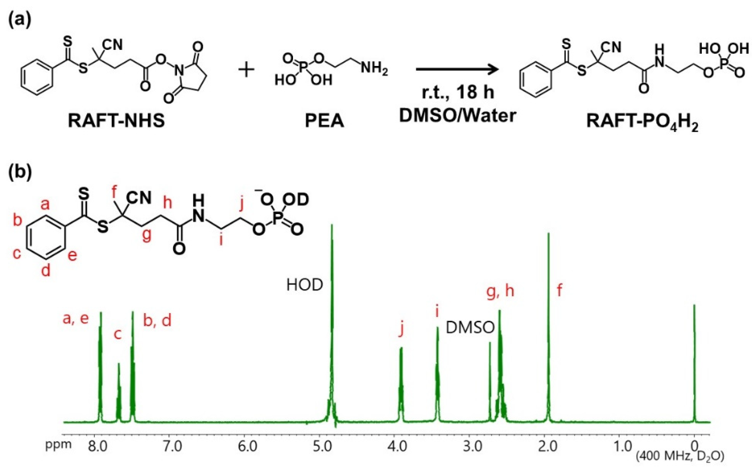
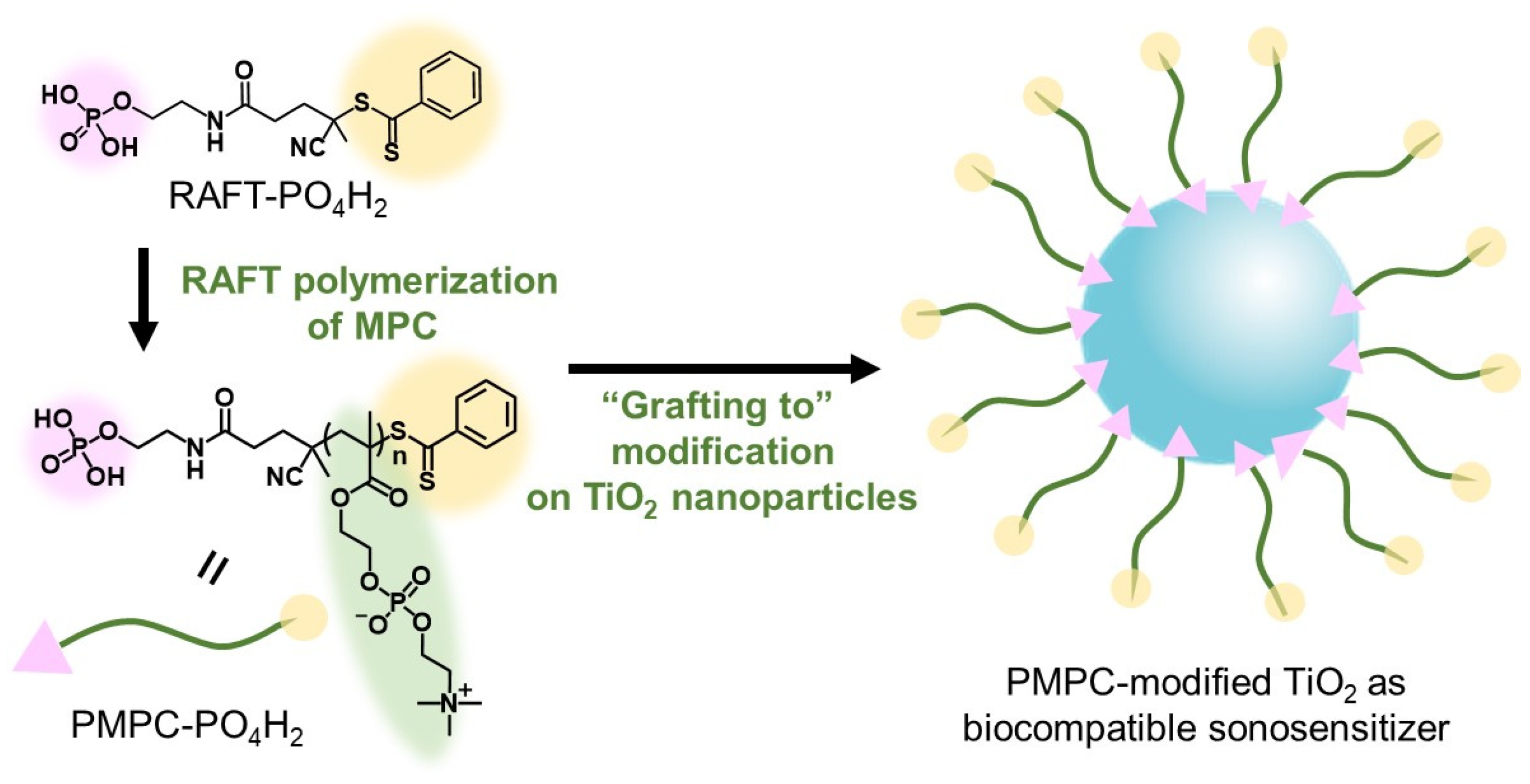
4.3. Synthesis of RAFT- PO4H2
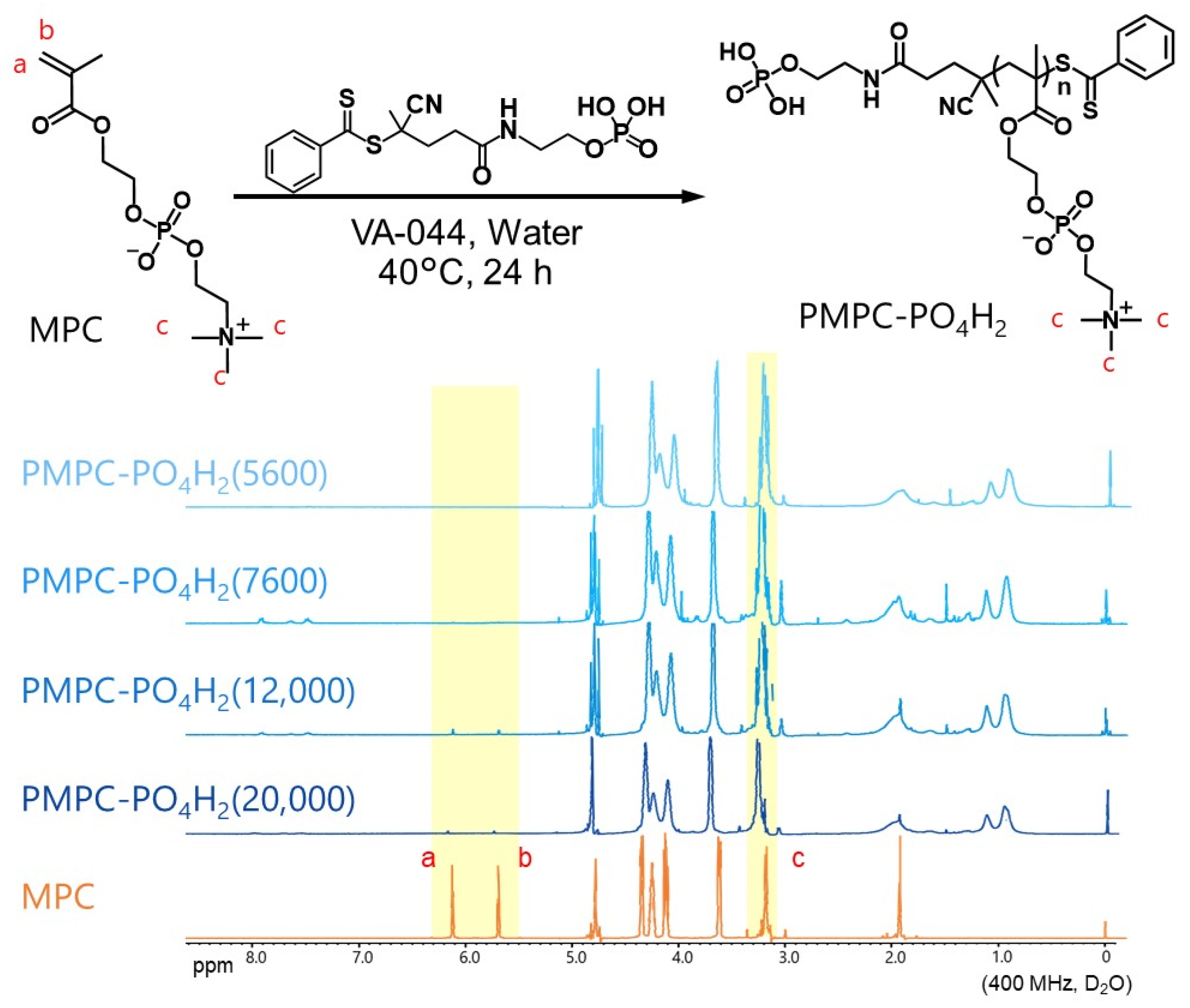
4.4. Synthesis of PMPC by RAFT Polymerization
4.5. PMPC-Modified TiO2 Nanoparticles
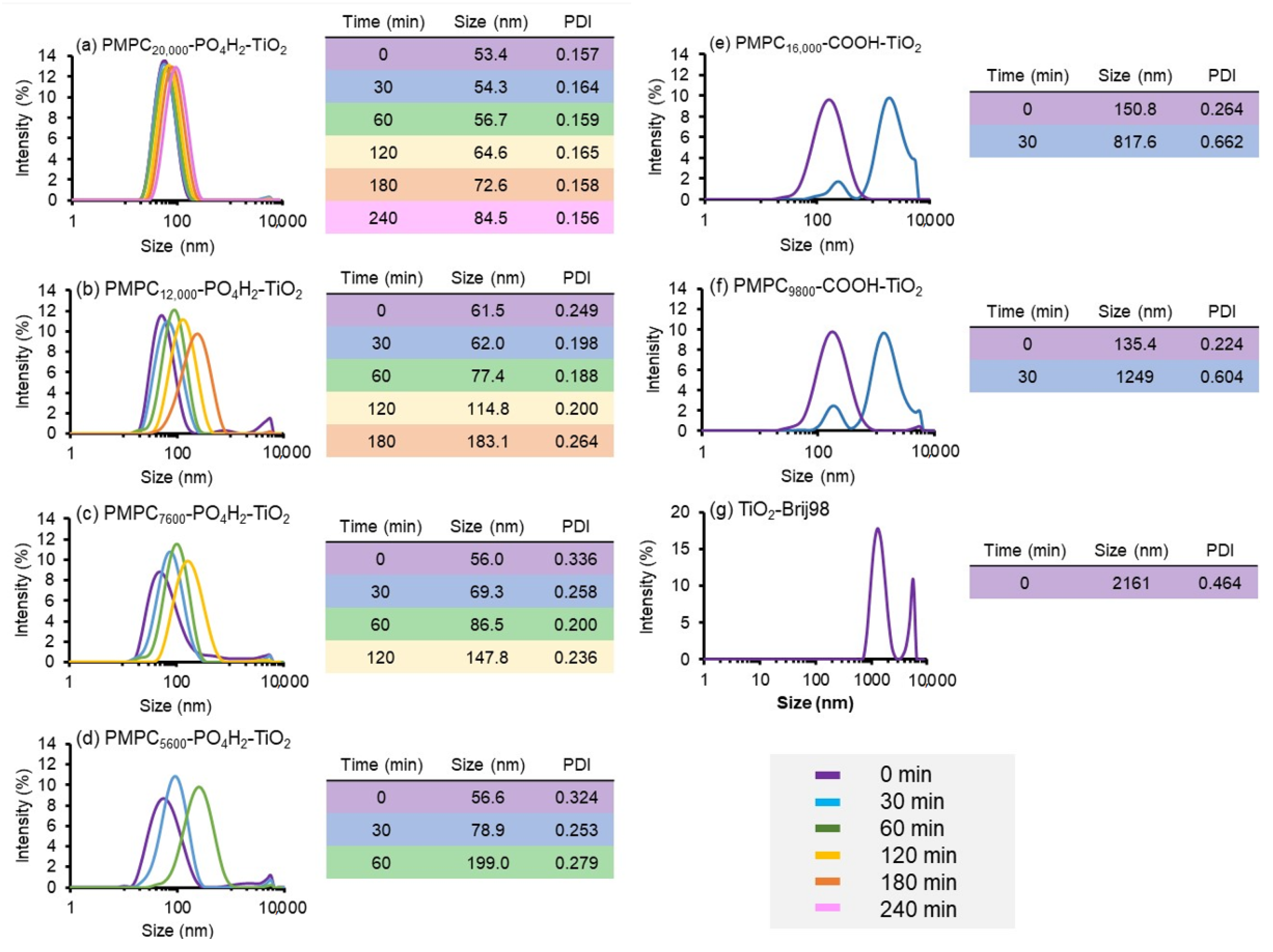
4.6. Colloidal Stability of PMPC-Modified TiO2 Nanoparticles in PBS
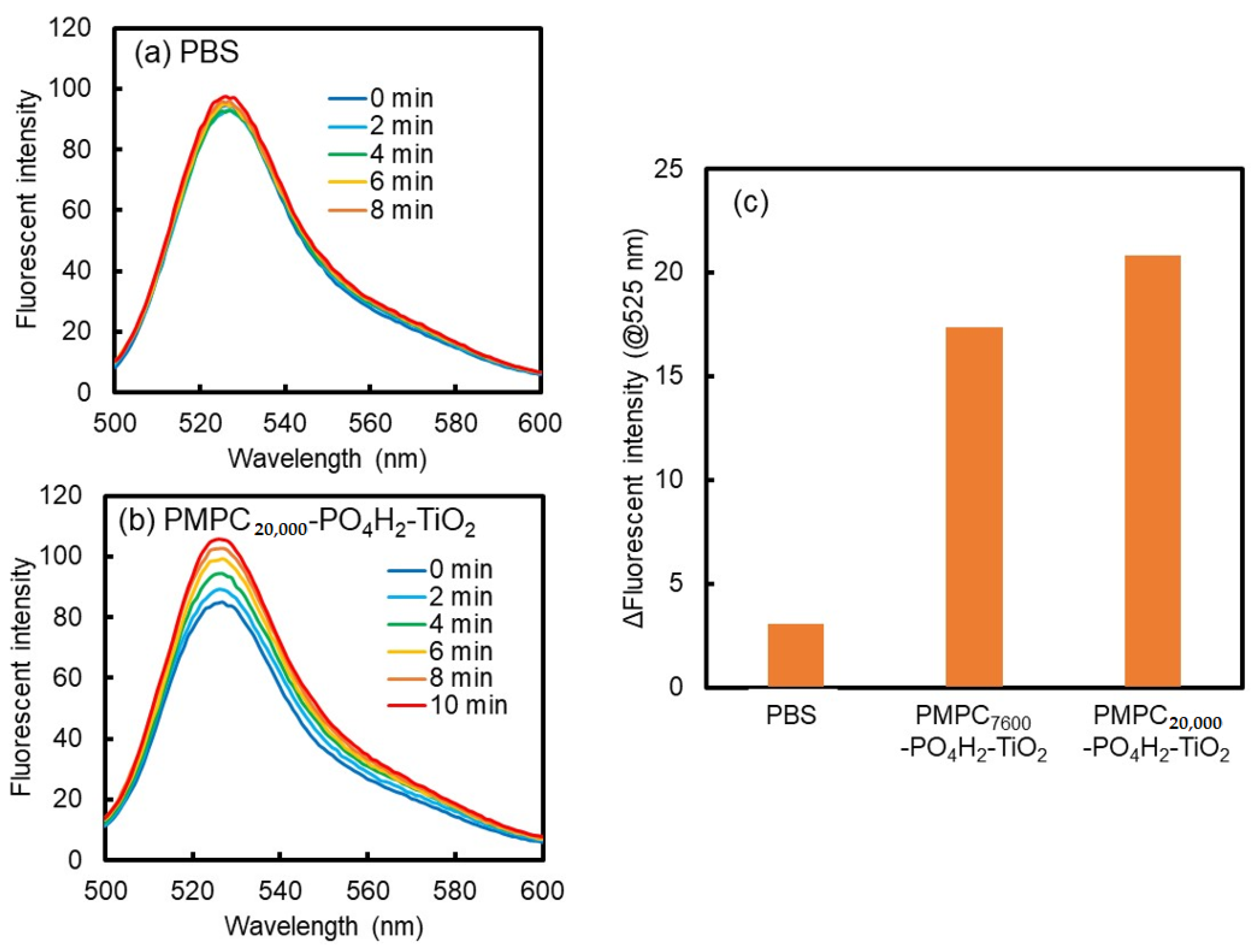
4.7. Sonosensitizing Effect of PMPC-Modified TiO2 Nanoparticles
Supplementary Materials
Author Contributions
Funding
Institutional Review Board Statement
Data Availability Statement
Acknowledgments
Conflicts of Interest
References
- Qian, X.; Zheng, Y.; Chen, Y. Micro/Nanoparticle-Augmented Sonodynamic Therapy (SDT): Breaking the Depth Shallow of Photoactivation. Adv. Mater. 2016, 28, 8097–8129. [Google Scholar] [CrossRef]
- Son, S.; Kim, J.H.; Wang, X.; Zhang, C.; Yoon, S.A.; Shin, J.; Sharma, A.; Lee, M.H.; Cheng, L.; Wu, J.; et al. Multifunctional sonosensitizers in sonodynamic cancer therapy. Chem. Soc. Rev. 2020, 49, 3244–3261. [Google Scholar] [CrossRef] [PubMed]
- Chen, H.; Zhou, X.; Gao, Y.; Zheng, B.; Tang, F.; Huang, J. Recent progress in development of new sonosensitizers for sonodynamic cancer therapy. Drug Discov. Today 2014, 19, 502–509. [Google Scholar] [CrossRef] [PubMed]
- Tachibana, K.; Feril, L.B., Jr.; Ikeda-Dantsuji, Y. Sonodynamic therapy. Ultrasonics 2008, 48, 253–259. [Google Scholar] [CrossRef] [PubMed]
- Zeng, J.; Gu, C.; Geng, X.; Lin, K.; Xie, Y.; Chen, X. Combined photothermal and sonodynamic therapy using a 2D black phosphorus nanosheets loaded coating for efficient bacterial inhibition and bone-implant integration. Biomaterials 2023, 297, 122122. [Google Scholar] [CrossRef]
- Zhao, Y.; Huang, T.; Wang, S.; Yao, S.; Hu, Q.; Wan, X.; Guo, N.; Zhang, Y.; Li, L. Manganese oxide-modified bismuth oxychloride piezoelectric nanoplatform with multiple enzyme-like activities for cancer sonodynamic therapy. J. Colloid Interface Sci. 2023, 640, 839–850. [Google Scholar] [CrossRef]
- An, J.; He, X.; Ma, H.; Li, Y.; Li, Y.; Zhang, X.; Shuai, Q.; Wang, Y.; Liu, W.; Li, W.; et al. Radionuclide labeled nanocarrier for imaging guided combined radionuclide, sonodynamic, and photothermal therapy of pancreatic tumours. J. Colloid Interface Sci. 2023, 642, 789–799. [Google Scholar] [CrossRef]
- Wei, D.; Yu, Y.; Zhang, X.; Wang, Y.; Chen, H.; Zhao, Y.; Wang, F.; Rong, G.; Wang, W.; Kang, X.; et al. Breaking the Intracellular Redox Balance with Diselenium Nanoparticles for Maximizing Chemotherapy Efficacy on Patient-Derived Xenograft Models. ACS Nano 2020, 14, 16984–16996. [Google Scholar] [CrossRef]
- Han, X.; Huang, J.; Jing, X.; Yang, D.; Lin, H.; Wang, Z.; Li, P.; Chen, Y. Oxygen-Deficient Black Titania for Synergistic/Enhanced Sonodynamic and Photoinduced Cancer Therapy at Near Infrared-II Biowindow. ACS Nano 2018, 12, 4545–4555. [Google Scholar] [CrossRef]
- Gil You, D.; Deepagan, V.G.; Um, W.; Jeon, S.; Son, S.; Chang, H.; Yoon, H.I.; Cho, Y.W.; Swierczewska, M.; Lee, S.; et al. ROS-generating TiO2 nanoparticles for non-invasive sonodynamic therapy of cancer. Sci. Rep. 2016, 6, 23200. [Google Scholar] [CrossRef]
- Huang, P.; Qian, X.; Chen, Y.; Yu, L.; Lin, H.; Wang, L.; Zhu, Y.; Shi, J. Metalloporphyrin-Encapsulated Biodegradable Nanosystems for Highly Efficient Magnetic Resonance Imaging-Guided Sonodynamic Cancer Therapy. J. Am. Chem. Soc. 2017, 139, 1275–1284. [Google Scholar] [CrossRef]
- Zhang, Y.; Yong, L.; Luo, Y.; Ding, X.; Xu, D.; Gao, X.; Yan, S.; Wang, Q.; Luo, J.; Pu, D.; et al. Enhancement of HIFU ablation by sonosensitizer-loading liquid fluorocarbon nanoparticles with pre-targeting in a mouse model. Sci. Rep. 2019, 9, 6982. [Google Scholar] [CrossRef]
- Wang, Z.; Yu, N.; Zhang, J.; Ren, Q.; Li, M.; Chen, Z. Nanoscale Hf-hematoporphyrin frameworks for synergetic sonodynamic/radiation therapy of deep-seated tumors. J. Colloid Interface Sci. 2022, 626, 803–814. [Google Scholar] [CrossRef]
- Lu, Z.; Bai, S.; Jiang, Y.; Wu, S.; Xu, D.; Chen, Y.; Lan, Y.; An, Y.; Mao, J.; Liu, X.; et al. Porphyrin-Based Covalent Organic Framework for Imaging-Guided Cancer Combinatorial Immuno-Sonodynamic Therapy. Adv. Funct. Mat. 2022, 32, 2207749. [Google Scholar] [CrossRef]
- He, D.; Wang, W.; Feng, N.; Zhang, Z.; Zhou, D.; Zhang, J.; Luo, H.; Li, Y.; Chen, X.; Wu, J. Defect-Modified nano-BaTiO3 as a Sonosensitizer for Rapid and High-Efficiency Sonodynamic Sterilization. ACS Appl. Mater. Int. 2023, 15, 15140–15151. [Google Scholar] [CrossRef]
- Zhong, X.; Wang, X.; Cheng, L.; Tang, Y.; Zhan, G.; Gong, F.; Zhang, R.; Hu, J.; Liu, Z.; Yang, X. GSH-Depleted PtCu3 nanocages for chemodynamic- enhanced sonodynamic cancer therapy. Adv. Funct. Mat. 2020, 30, 1907954. [Google Scholar] [CrossRef]
- Cheng, S.; Chen, L.; Gong, F.; Yang, X.; Han, Z.; Wang, Y.; Ge, J.; Gao, X.; Li, Y.; Zhong, X.; et al. PtCu Nanosonosensitizers with Inflammatory Microenvironment Regulation for Enhanced Sonodynamic Bacterial Elimination and Tissue Repair. Adv. Funct. Mater. 2023, in press. [Google Scholar] [CrossRef]
- Iyer, A.K.; Khaled, G.; Fang, J.; Maeda, H. Exploiting the enhanced permeability and retention effect for tumor targeting. Drug Discov. Today 2006, 11, 812–818. [Google Scholar] [CrossRef]
- Matsumura, Y.; Maeda, H. A new concept for macromolecular therapeutics in cancer chemotherapy: Mechanism of tumoritropic accumulation of proteins and the antitumor agent smancs. Cancer Res. 1986, 46, 6387–6392. [Google Scholar]
- Keller, A.A.; Wang, H.; Zhou, D.; Lenihan, H.S.; Cherr, G.; Cardinale, B.J.; Miller, R.; Ji, Z. Stability and Aggregation of Metal Oxide Nanoparticles in Natural Aqueous Matrices. Environ. Sci. Technol. 2010, 44, 1962–1967. [Google Scholar] [CrossRef]
- Yamaguchi, S.; Kobayashi, H.; Narita, T.; Kanehira, K.; Sonezaki, S.; Kubota, Y.; Terasaka, S.; Iwasaki, Y. Novel Photodynamic Therapy Using Water-dispersed TiO2-Polyethylene Glycol Compound: Evaluation of Antitumor Effect on Glioma Cells and Spheroids In Vitro. Photochem. Photobiol. 2010, 86, 964–971. [Google Scholar] [CrossRef] [PubMed]
- Shen, S.; Guo, X.; Wu, L.; Wang, M.; Wang, X.; Kong, F.; Shen, H.; Xie, M.; Ge, Y.; Jin, Y. Dual-core@shell-structured Fe3O4-NaYF 4@TiO2 nanocomposites as a magnetic targeting drug carrier for bioimaging and combined chemo-sonodynamic therapy. J. Mat. Chem. B 2014, 2, 5775–5784. [Google Scholar] [CrossRef] [PubMed]
- Harada, A.; Ono, M.; Yuba, E.; Kono, K. Titanium dioxide nanoparticle-entrapped polyion complex micelles generate singlet oxygen in the cells by ultrasound irradiation for sonodynamic therapy. Biomater. Sci. 2012, 1, 65–73. [Google Scholar] [CrossRef] [PubMed]
- Yamamoto, S.; Ono, M.; Yuba, E.; Harada, A. In Vitro Sonodynamic Therapeutic Effect of Polyion Complex Micelles Incorporating Titanium Dioxide Nanoparticles. Nanomaterials 2017, 7, 268. [Google Scholar] [CrossRef]
- Otsu, T. Iniferter concept and living radical polymerization. J. Polym. Sci. Part A: Polym. Chem. 2000, 38, 2121–2136. [Google Scholar] [CrossRef]
- Matyjaszewski, K.; Tsarevsky, N.V. Nanostructured functional materials prepared by atom transfer radical polymerization. Nat. Chem. 2009, 1, 276–288. [Google Scholar] [CrossRef]
- Braunecker, W.A.; Matyjaszewski, K. Controlled/living radical polymerization: Features, developments, and perspectives. Prog. Polym. Sci. 2007, 32, 93–146. [Google Scholar] [CrossRef]
- Hawker, C.J.; Bosman, A.W.; Harth, E. New Polymer Synthesis by Nitroxide Mediated Living Radical Polymerizations. Chem. Rev. 2001, 101, 3661–3688. [Google Scholar] [CrossRef]
- Kamigaito, M.; Ando, T.; Sawamoto, M. Metal-Catalyzed Living Radical Polymerization. Chem. Rev. 2001, 101, 3689–3745. [Google Scholar] [CrossRef]
- Terashima, T. Functional spaces in star and single-chain polymers via living radical polymerization. Polym. J. 2014, 46, 664–673. [Google Scholar] [CrossRef]
- Lutz, J.-F.; Ouchi, M.; Liu, D.R.; Sawamoto, M. Sequence-Controlled Polymers. Science 2013, 341, 1238149. [Google Scholar] [CrossRef]
- Satoh, K. Controlled/living polymerization of renewable vinyl monomers into bio-based polymers. Polym. J. 2015, 47, 527–536. [Google Scholar] [CrossRef]
- Yamago, S. Precision Polymer Synthesis by Degenerative Transfer Controlled/Living Radical Polymerization Using Organotellurium, Organostibine, and Organobismuthine Chain-Transfer Agents. Chem. Rev. 2009, 109, 5051–5068. [Google Scholar] [CrossRef]
- Goto, A.; Fukuda, T. Kinetics of living radical polymerization. Prog. Polym. Sci. 2004, 29, 329–385. [Google Scholar] [CrossRef]
- Goto, A.; Zushi, H.; Hirai, N.; Wakada, T.; Tsujii, Y.; Fukuda, T. Living radical polymerizations with germanium, tin, and phosphorus catalysts—Reversible chain transfer catalyzed polymerizations (RTCPs). J. Am. Chem. Soc. 2007, 129, 13347–13354. [Google Scholar] [CrossRef]
- Goto, A.; Suzuki, T.; Ohfuji, H.; Tanishima, M.; Fukuda, T.; Tsujii, Y.; Kaji, H. Reversible Complexation Mediated Living Radical Polymerization (RCMP) Using Organic Catalysts. Macromolecules 2011, 44, 8709–8715. [Google Scholar] [CrossRef]
- Rosen, B.M.; Percec, V. Single-Electron Transfer and Single-Electron Transfer Degenerative Chain Transfer Living Radical Polymerization. Chem. Rev. 2009, 109, 5069–5119. [Google Scholar] [CrossRef]
- Moad, G.; Rizzardo, E.; Thang, S. Living radical polymerization by the raft process—A second update Aust. J. Chem. 2009, 62, 1402–1472. [Google Scholar]
- Corrigan, N.; Jung, K.; Moad, G.; Hawker, C.J.; Matyjaszewski, K.; Boyer, C. Reversible-deactivation radical polymerization (Controlled/living radical polymerization): From discovery to materials design and applications. Prog. Polym. Sci. 2020, 111, 101311. [Google Scholar] [CrossRef]
- David, G.; Boyer, C.; Tonnar, J.; Ameduri, B.; Lacroix-Desmazes, P.; Boutevin, B. Use of Iodocompounds in Radical Polymerization. Chem. Rev. 2006, 106, 3936–3962. [Google Scholar] [CrossRef]
- Kitayama, Y.; Okubo, M. Emulsifier-free, organotellurium-mediated living radical emulsion polymerization (emulsion TERP) of styrene: Poly(dimethylaminoethyl methacrylate) macro-TERP agent. Polym. Chem. 2013, 5, 2784–2792. [Google Scholar] [CrossRef]
- Kitayama, Y.; Chaiyasat, A.; Minami, H.; Okubo, M. Emulsifier-Free, Organotellurium-Mediated Living Radical Emulsion Polymerization of Styrene: Polymerization Loci. Macromolecules 2010, 43, 7465–7471. [Google Scholar] [CrossRef]
- Zetterlund, P.B.; Kagawa, Y.; Okubo, M. Controlled/Living Radical Polymerization in Dispersed Systems. Chem. Rev. 2008, 108, 3747–3794. [Google Scholar] [CrossRef] [PubMed]
- Zetterlund, P.B.; Thickett, S.C.; Perrier, S.; Bourgeat-Lami, E.; Lansalot, M. Controlled/Living Radical Polymerization in Dispersed Systems: An Update. Chem. Rev. 2015, 115, 9745–9800. [Google Scholar] [CrossRef] [PubMed]
- Wang, B.; Li, B.; Zhao, B.; Li, C. Amphiphilic Janus gold nanoparticles via combining “solid-state grafting-to” and “grafting-from” methods. J. Am. Chem. Soc. 2008, 130, 11594–11595. [Google Scholar] [CrossRef]
- Wang, Y.-X.; Li, Y.; Qiao, S.-H.; Kang, J.; Shen, Z.-L.; Zhang, N.-N.; An, Z.; Wang, X.; Liu, K. Polymers via Reversible Addition–Fragmentation Chain Transfer Polymerization with High Thiol End-Group Fidelity for Effective Grafting-To Gold Nanoparticles. J. Phys. Chem. Lett. 2021, 12, 4713–4721. [Google Scholar] [CrossRef]
- Biggs, C.; Walker, M.; Gibson, M. “Grafting to” of RAFTed responsive polymers to glass substrates by thiol-ene and critical comparison to thiol-gold coupling. Biomacromolecules 2016, 17, 2626–2633. [Google Scholar] [CrossRef]
- Li, D.; He, Q.; Li, J. Smart core/shell nanocomposites: Intelligent polymers modified gold nanoparticles. Adv. Colloid Interface Sci. 2009, 149, 28–38. [Google Scholar] [CrossRef]
- Kitayama, Y.; Takeuchi, T. Localized Surface Plasmon Resonance Nanosensing of C-Reactive Protein with Poly(2-methacryloyloxyethyl phosphorylcholine)-Grafted Gold Nanoparticles Prepared by Surface-Initiated Atom Transfer Radical Polymerization. Anal. Chem. 2014, 86, 5587–5594. [Google Scholar] [CrossRef]
- Kitayama, Y.; Takeuchi, T. Synthesis of CO2/N2-triggered reversible stability-controllable poly(2-(diethylamino)ethyl methacrylate)-grafted-AuNPs by surface-initiated atom transfer radical polymerization. Langmuir 2014, 30, 12684–12689. [Google Scholar] [CrossRef]
- Hojjati, B.; Charpentier, P. Synthesis and kinetics of graft polymerization of methyl methacrylate from the RAFT coordinated surface of nano-TiO2. J. Polym. Sci. A Polym. Chem. 2008, 46, 3926–3937. [Google Scholar] [CrossRef]
- Wang, W.; Cao, H.; Zhu, G.; Wang, P. A facile strategy to modify TiO2 nanoparticles via surface-initiated atrp of styrene. J. Polym. Sci. A Polym. Chem. 2010, 48, 1782–1790. [Google Scholar] [CrossRef]
- Hojjati, B.; Sui, R.; Charpentier, P. Synthesis of TiO2/PAA nanocomposite by RAFT polymerization. Polymer 2007, 48, 5850–5858. [Google Scholar] [CrossRef]
- Guo, S.; Zhang, Q.; Wang, D.; Wang, L.; Lin, F.; Wilson, P.; Haddleton, D. Bioinspired coating of TiO2 nanoparticles with antimicrobial polymers by Cu(0)-LRP: Grafting to vs. grafting from. Polym. Chem. 2017, 8, 6570–6580. [Google Scholar] [CrossRef]
- Gao, W.; Dickinson, L.; Grozinger, C.; Morin, F.; Reven, L. Order-disorder transitions in self-assembled monolayers: A 13C solid-state NMR study. Langmuir 1997, 13, 115–118. [Google Scholar] [CrossRef]
- Pawsey, S.; Yach, K.; Halla, J.; Reven, L. Self-Assembled Monolayers of Alkanoic Acids: A Solid-State NMR Study. Langmuir 2000, 16, 3294–3303. [Google Scholar] [CrossRef]
- Zhao, D.; Chen, C.; Wang, Y.; Ji, H.; Ma, W.; Zang, L.; Zhao, J. Surface Modification of TiO2 by Phosphate: Effect on Photocatalytic Activity and Mechanism Implication. J. Phys. Chem. C 2008, 112, 5993–6001. [Google Scholar] [CrossRef]
- Yu, J.C.; Ho, W.; Yu, J.; Hark, S.K.; Iu, K. Effects of Trifluoroacetic Acid Modification on the Surface Microstructures and Photocatalytic Activity of Mesoporous TiO2 Thin Films. Langmuir 2003, 19, 3889–3896. [Google Scholar] [CrossRef]
- Ishihara, K.; Ziats, N.P.; Tierney, B.P.; Nakabayashi, N.; Anderson, J.M. Protein adsorption from human plasma is reduced on phospholipid polymers. J. Biomed. Mater. Res. 1991, 25, 1397–1407. [Google Scholar] [CrossRef]
- Ishihara, K.; Nomura, H.; Mihara, T.; Kurita, K.; Iwasaki, Y.; Nakabayashi, N. Why do phospholipid polymers reduce protein adsorption? J. Biomed. Mater. Res. 1998, 39, 323–330. [Google Scholar] [CrossRef]
- Pawsey, S.; Yach, K.; Reven, L. Self-Assembly of Carboxyalkylphosphonic Acids on Metal Oxide Powders. Langmuir 2002, 18, 5205–5212. [Google Scholar] [CrossRef]
- Gao, W.; Dickinson, L.; Grozinger, C.; Morin, F.G.; Reven, L. Self-Assembled Monolayers of Alkylphosphonic Acids on Metal Oxides. Langmuir 1996, 12, 6429–6435. [Google Scholar] [CrossRef]


| Run | RAFT Agent | [MPC]0/[RAFT Agent]0/ | Conv. (%) | Mn | Mn,th | Mw/Mn |
|---|---|---|---|---|---|---|
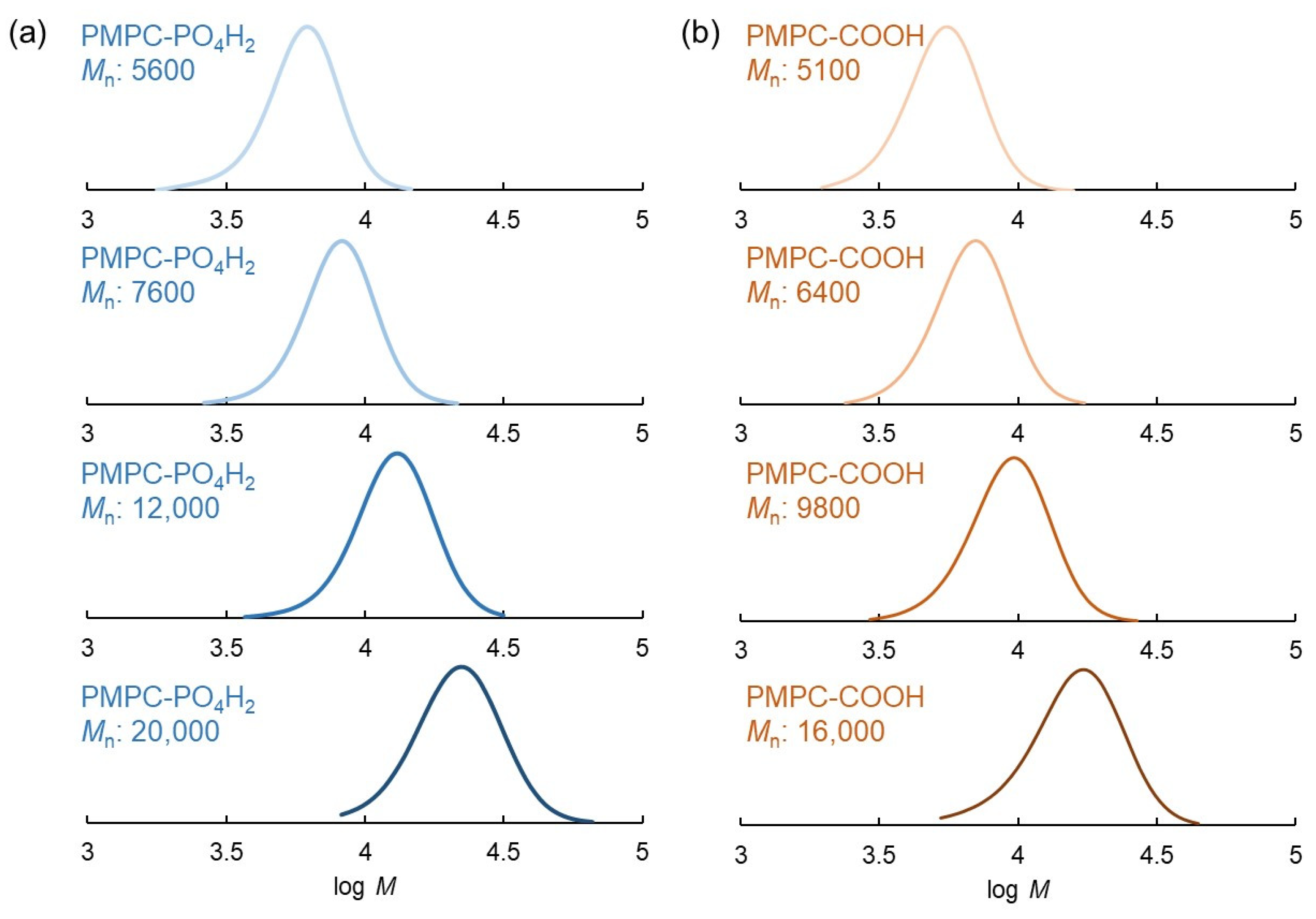
| 1 b | RAFT-PO4H2 | 10/1 | >99 | 5600 | 3400 | 1.10 |
| 2 b | RAFT-PO4H2 | 20/1 | >99 | 7600 | 6300 | 1.11 |
| 3 b | RAFT-PO4H2 | 40/1 | >99 | 12,000 | 12,200 | 1.12 |
| 4 b | RAFT-PO4H2 | 80/1 | >99 | 20,000 | 24,000 | 1.15 |
| 5 c | RAFT-COOH | 10/1 | >99 | 5100 | 3200 | 1.11 |
| 6 c | RAFT-COOH | 20/1 | >99 | 6400 | 6200 | 1.11 |
| 7 c | RAFT-COOH | 40/1 | >99 | 9800 | 12,100 | 1.16 |
| 8 c | RAFT-COOH | 80/1 | >99 | 16,000 | 24,000 | 1.21 |
| Sample | Concentration (mg/mL) | Size (nm) | PDI |
|---|---|---|---|
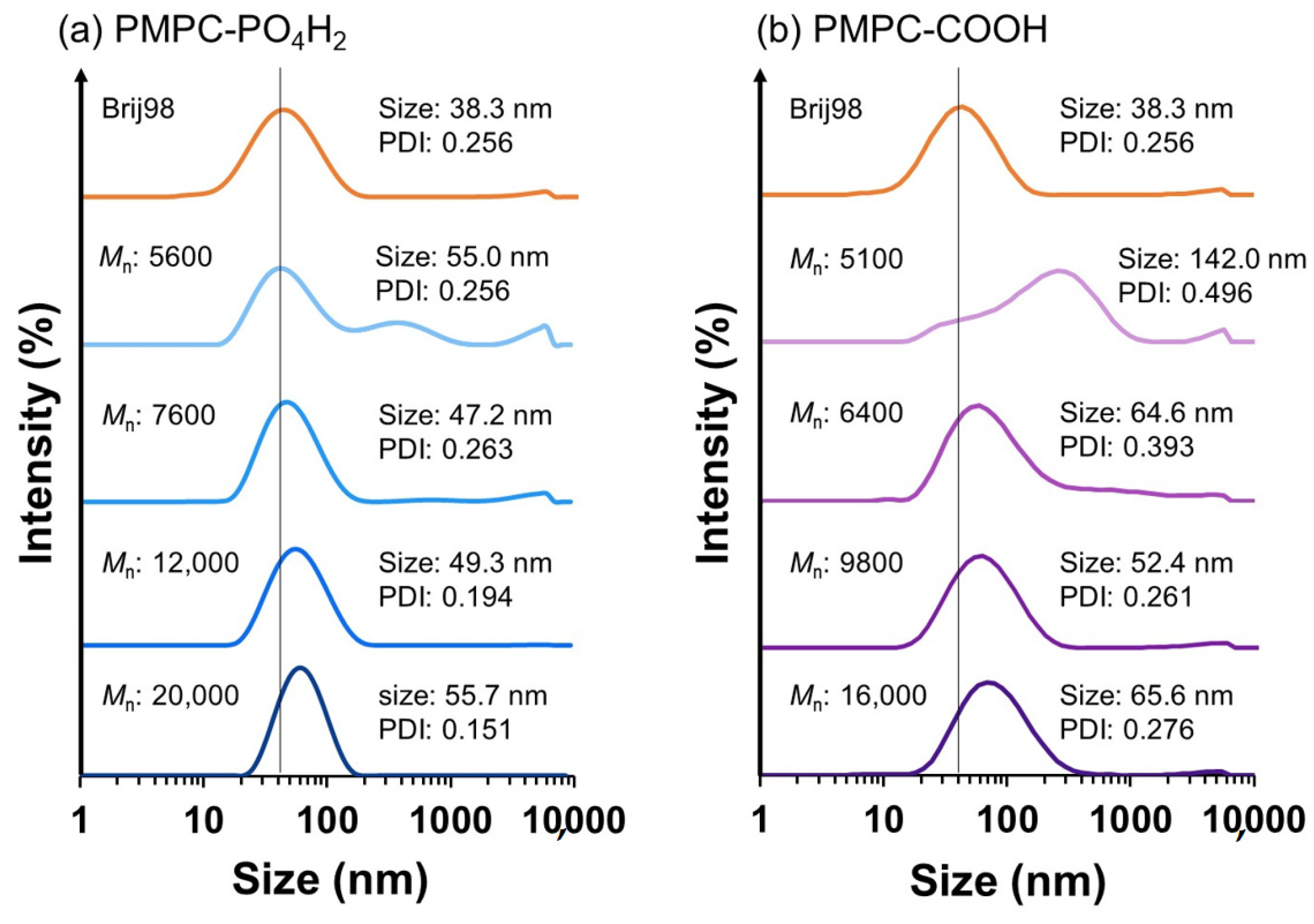
| Original | - | 38.3 | 0.256 |
| PMPC-PO4H2 a | 2.5 | 41.6 | 0.181 |
| 5.0 | 49.1 | 0.264 | |
| 10 | 47.2 | 0.263 | |
| 20 | 49.3 | 0.271 |
Disclaimer/Publisher’s Note: The statements, opinions and data contained in all publications are solely those of the individual author(s) and contributor(s) and not of MDPI and/or the editor(s). MDPI and/or the editor(s) disclaim responsibility for any injury to people or property resulting from any ideas, methods, instructions or products referred to in the content. |
© 2023 by the authors. Licensee MDPI, Basel, Switzerland. This article is an open access article distributed under the terms and conditions of the Creative Commons Attribution (CC BY) license (https://creativecommons.org/licenses/by/4.0/).
Share and Cite
Kitayama, Y.; Katayama, A.; Shao, Z.; Harada, A. Biocompatible Polymer-Grafted TiO2 Nanoparticle Sonosensitizers Prepared Using Phosphonic Acid-Functionalized RAFT Agent. Polymers 2023, 15, 2426. https://doi.org/10.3390/polym15112426
Kitayama Y, Katayama A, Shao Z, Harada A. Biocompatible Polymer-Grafted TiO2 Nanoparticle Sonosensitizers Prepared Using Phosphonic Acid-Functionalized RAFT Agent. Polymers. 2023; 15(11):2426. https://doi.org/10.3390/polym15112426
Chicago/Turabian StyleKitayama, Yukiya, Aoi Katayama, Zhicheng Shao, and Atsushi Harada. 2023. "Biocompatible Polymer-Grafted TiO2 Nanoparticle Sonosensitizers Prepared Using Phosphonic Acid-Functionalized RAFT Agent" Polymers 15, no. 11: 2426. https://doi.org/10.3390/polym15112426