Article

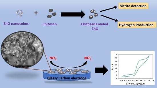
Zinc Nanocomposite Supported Chitosan for Nitrite Sensing and Hydrogen Evolution Applications

1
Department of Chemistry, College of Science, Princess Nourah bint Abdulrahman University, P.O. Box 84428, Riyadh 11671, Saudi Arabia
2
Chemistry Department, Faculty of Science, Cairo University, Giza 12613, Egypt
3
Physics Department, Faculty of Science, King Abdulaziz University, Jeddah 21589, Saudi Arabia
4
Department of Chemistry, Faculty of Applied Science, Umm Al-Qura University, Makkah 24230, Saudi Arabia
*
Authors to whom correspondence should be addressed.
Polymers 2023, 15(10), 2357; https://doi.org/10.3390/polym15102357
Submission received: 24 April 2023 / Revised: 12 May 2023 / Accepted: 15 May 2023 / Published: 18 May 2023

Abstract

:
Nanoparticles of ZnO-Chitosan (Zn-Chit) composite were prepared using precipitation methods. Several analytical techniques, such as scanning electron microscope (SEM), transmitted electron microscope (TEM), powder X-ray diffraction (XRD), infrared spectroscopy (IR), and thermal analysis, were used to characterize the prepared composite. The activity of the modified composite was investigated for nitrite sensing and hydrogen production applications using various electrochemical techniques. A comparative study was performed for pristine ZnO and ZnO loaded on chitosan. The modified Zn-Chit has a linear range of detection 1–150 µM and a limit of detection (LOD) = 0.402 µM (response time ~3 s). The activity of the modified electrode was investigated in a real sample (milk). Furthermore, the anti-interference capability of the surface was utilized in the presence of several inorganic salts and organic additives. Additionally, Zn-Chit composite was employed as an efficient catalyst for hydrogen production in an acidic medium. Thus, the electrode showed long-term stability toward fuel production and enhanced energy security. The electrode reached a current density of 50 mA cm−2 at an overpotential equal to −0.31 and −0.2 V (vs. RHE) for GC/ZnO and GC/Zn-Chit, respectively. Electrode durability was studied for long-time constant potential chronoamperometry for 5 h. The electrodes lost 8% and 9% of the initial current for GC/ZnO and GC/Zn-Chit, respectively.

Graphical Abstract

1. Introduction

Nitrite is a crucial element for food additives, concrete admixtures, and the cycling of soil [1,2]. The World Health Organization (WHO) has classified nitrite as a highly hazardous substance because of its potential to threaten human health (such as through carcinogenesis) and cause water and soil contamination over time [3]. Furthermore, nitroaromatic hydrocarbons, nitramines, and nitrates, which comprise the most used high-energy explosives, are mostly degraded by nitrite, which is one of the key components of improvised explosives [4]. The urgent requirement for on-site detection of trace nitrite is driven by the need to ensure sustainable environmental development and safeguard public health and security [5,6,7,8]. Many attempts have been undertaken to measure nitrite below one micromolar, such as by high-performance liquid chromatography, surface-enhanced Raman spectroscopy, fluorescence spectroscopy, and electrochemical detection [9,10].
Although most of these techniques perform well in lab settings, electrochemical methods are very sensitive and selective for detecting drugs and contaminated water [11,12,13,14]. Various benefits include affordability, reaction speed, simplicity, and reproducibility [15,16]. Using nanomaterials has led to the development of different electrochemical systems, including those incorporating nanostructured metal oxides [17]. Zinc oxide (ZnO) is a substance easily used in medical and biological applications, such as in wearable technology. ZnO is one of the greatest promising semiconductor materials for electrochemical sensor fabrications because of its high capacity to react with oxygen [18]. Several Zn-based electrodes were employed for the electrochemical detection of nitrite, such as flower-ZnO [19], Zn-Schiff base [20], ZnS [20], and ZnTiO3 [21].
Chitosan, a derivative of chitin, is commonly utilized in different applications. Recent developments in fermentation technology have enabled the production of novel chitosan with physiochemical properties distinct from waste materials, thereby offering a viable alternative to conventional sources such as crab shells. Chitosan is commonly used for immobilization due to its favorable environmental characteristics, high absorption capacity, remarkable layer-forming capabilities, superior permeability, heightened thermal stability, robust mechanical strength, biocompatibility, and convenient accessibility [22]. Chitosan exhibits distinctive structural and functional characteristics, such as non-toxicity, hydrophilicity, superior adhesion, biocompatibility, environmental sustainability, antibacterial and antimicrobial properties, and non-carcinogenicity, which render it highly versatile and applicable across diverse domains. This substance holds considerable importance in various fields, such as biomedicine, sensor technology, cosmetics, biochemistry, biotechnology, pharmaceuticals, food additives, preservative materials, water purification, antiseptic dyes, and agricultural applications [23,24,25,26,27].
Hydrogen gas (H2) plays a crucial role in facilitating the shift towards a sustainable energy system, owing to its ability to serve as a high-energy density for renewable energy [28,29,30]. Although water electrolysis presents an attractive option for green hydrogen production utilizing renewable energy sources, its global contribution to hydrogen production remains limited. This is primarily due to the requirement for costly electrocatalysts to offset the Ohmic losses associated with the kinetic overpotential of the system [31,32,33]. The literature reports zinc-based electrodes as an efficient surface for hydrogen production, such as ZnMn2O4, Zn-Ni-P, and Zn-AgIn5S8 [34,35,36].
In the present work, a composite of Zn-Chitosan is prepared as a dual functional catalyst for electrochemical detection of nitrite and hydrogen production enhancement. The prepared catalyst was characterized using several electrochemical techniques. Different kinetics parameters are estimated to evaluate the process efficiency. Additionally, real nitrite samples were used to construct calibration curves. A comparative study was performed for pristine ZnO and Zn-Chitosan composites for hydrogen production in an acidic medium.

2. Materials and Methods

2.1. Preparation of ZnO Nanoparticles

Using a hydrothermal process, hexagonal nanocubes of zinc oxide were created. In a typical experiment, 1.2 g of Zn(NO3)2·6H2O were dissolved in double-distilled water, followed by the slow addition of 0.1 M of KOH. This resulted in a mixture of 100 mL, which was then agitated for 20 min while being dispersed in an ultrasonic water bath for 30 min. The mixture was then placed into a Teflon-lined stainless-steel autoclave, which was then dried in an oven before undergoing a 12-h hydrothermal treatment at 180 °C. The item was filtered, cleaned with ethanol and water, and then dried in an oven at 60 °C.

2.2. Preparation of Zn-Chit Composite

Zn-Chit composite was prepared by a mixture of chitosan solution mixed with ZnO nanoparticles: 0.5 g of chitosan (Sigma Aldrich, St. Louis, MO, USA, 50,000–190,000 Da (based on viscosity), and 75–85% deacetylated) was added to 40 mL absolute ethanol in a 250 mL beaker, and then the temperature was raised gently with stirring. 0.5 g of ZnO nanoparticles were added to the mixture. The solution temperature was cooled to room temperature. The addition of the chitosan to the chitosan solution led to crosslinking and the encapsulation of ZnO nanoparticles. Chitosan in acidic media becomes a polyelectrolyte because of the protonation of the –NH2 groups. The following equilibrium reaction describes the state of ionization. Therefore, about 1 mL of acetic acid (10%) was added to the mixture and stirred until the solution turned viscous. After ten minutes, the mixture underwent filtration and was rinsed with distilled water. The product was dried in an oven set at 80 °C for 3 h.

2.3. Fabrication of Electrode

A glassy carbon electrode with a diameter of 3 mm (0.0707 cm2) was used as the working electrode. It was first polished with gentle emery paper and cleaned with ethanol and double-distilled water. After that, the cast solution was created by dispersing 20 mg of the catalyst powder (ZnO or Zn-Chit) in 0.5 mL of ethanol and 0.5 mL of 5 wt% Nafion using an ultrasonic bath for 1 h. The modified electrode was made in the following manner: GC/ZnO and GC/Zn-Chit are accomplished by a drop cast of 20 µL of catalyst solution onto electrode’s surface, allowing it to dry for 4 h at 50 °C. The Autolab PGSTAT128N was used to perform all electrochemical experiments. The electrochemistry program NOVA (Version 2.1, Metrohm Autolab, Utrecht, The Netherlands) fits the impedance spectrum. Ag/AgCl/KCl (sat.) and Pt wire were employed as reference and counter electrodes.
GC/ZnO and GC/Zn-Chit were also used as working electrodes. All potential values in this work were compared to a reference electrode made of Ag/AgCl/sat.KCl for nitrite electrochemical sensor application. Furthermore, the potential was referenced to a reversible hydrogen electrode (RHE) for hydrogen evolution reaction (HER) applications. The electrochemical impedance spectroscopy measurements were adjusted to a constant AC voltage value using an AC voltage amplitude of 10 mV and a frequency range of 1 × 10⁴ Hz to 0.1 Hz.
The potential was referenced to the reversible hydrogen electrode (RHE) regarding the following equations [37]:
ERHE = EAg/AgCl + E° Ag/AgCl + 0.059 pH
Electrochemical experiments were carried out in a 0.5 M supporting electrolyte solution with H2SO4. The potential was standardized to a hydrogen electrode that is reversible as follows:
ERHE = EAg/AgCl + 0.197       (0.5 M H2SO4 ~ pH = 0) & (E° Ag/AgCl = 0.197 V)

3. Result & Discussion

3.1. Catalyst Characterization

The structure of modified Zn-Chit materials was determined using X-ray diffraction, as illustrated in Figure 1. The study reports the observation of seven maxima for ZnO, with respective Miller indices of {100}, {002}, {101}, {102}, {110}, {103}, and {112}, at 2θ = 31, 34, 36, 47, 56, 62, and 69, as documented in reference [38]. Furthermore, the observed peak at a 2θ value of approximately 20 degrees can be attributed to the crystallographic plane with Miller indices of {110} for chitosan, as reported in previous studies [39]. Moreover, the homogeneous and consistent dispersion of ZnO onto chitosan substrate promotes the adsorption of nitrite onto the electrode interface. Transmission electron microscopy (TEM) was employed as the conventional technique for determining the dimensions of ZnO nanoparticles. The mean particle size of zinc oxide was estimated to be around 70 nm. (Figure 1b).
The scanning electron microscope (SEM) was utilized to characterize the surface morphology of Zn-Chit, as illustrated in Figure 1c,d. The scanning electron microscopy (SEM) images of ZnO nanoparticles incorporated within chitosan sheets revealed the presence of hexagonal nanocubes.
Energy-dispersive X-ray spectroscopy (EDX), a technique for elemental analysis, was employed to identify the existence of elements, including Zn, C, N, and O (see Figure 1e). The atomic ratios of Zn and O suggest the existence of ZnO and the lack of adulteration from other constituents in the specimens. Additionally, the presence of the carbon and nitrogen on surface attributed to the chemical structure of chitosan.
Zn-Chit samples were characterized using FT-IR, revealing a range of absorption bands that facilitate identifying specific functional groups. Figure 2a illustrates the infrared spectra of the Zn-Chit sample. The stretching vibrations observed at wave number 3460 cm−1 were attributed to O–H and N–H bond-stretching while the stretching of C–H was identified at 2941 cm−1. The absorption bands observed at 1633, 1573, 1436, and 1374 cm−1 correspond to the C=O stretching of the amide band, accompanied by the bending of N–H, C–H, and O–H, respectively. The spectral analysis revealed that the band at 1162 cm−1 corresponded to the anti-symmetric stretching of the (C–O–C) bridge.
Additionally, the skeletal vibrations involving C–O stretching were predicted to occur at 1084 and 1023 cm−1 [40,41]. The spectrum of synthesized ZnO nanoparticles reveals a fundamental vibration mode at 3425 cm−1, identified as O–H stretching and deformation. This mode is associated with the adsorption of water on the metal surface. The tetrahedral coordination of Zn is accountable for its absorption at 873 cm−1. The vibrational stretching of ZnO nanoparticles is indicated by the observed bands within the range of 732 to 609 cm−1.
A thermal analytical technique, namely TGA, was utilized to estimate the thermal durability and chemical decomposition. As depicted in Figure 2b, the thermogravimetric analysis curve of Zn-Chit is presented. The composite material exhibited four distinct thermal transitions at 82, 277.2, 388.4, and 454.2 °C. The thermal transition observed at approximately 85 °C is commonly attributed to the water removal. Additionally, it has been reported that the thermal decomposition of chitosan occurs through a two-stage process [42]. It is anticipated that the degradation of chitosan will result in thermal decomposition peaks at 277.2 and 388.4 °C. The fourth thermal transition within the temperature range of 454.2 °C is associated with ZnO.

3.2. Zn-Chit Composite for Nitrite Detection

The present study investigated the efficacy of various modified GC electrodes, specifically GC/Chitosan, GC/ZnO, and GC/Zn-Chit, in detecting nitrites through the cyclic voltammetry (CV) technique. The experiment was utilized in a solution of 0.05 M phosphate buffer (pH = 7) in the presence of 50 µM of nitrite, with a sweep rate of 10 mV s−1. One oxidation peak was observed in the potential range 0.8~1.0 V (vs. Ag/AgCl). For instance, the estimated Eps were observed to be 1.01, 0.94, 1.02, and 0.99 V for GC, GC/Chitosan, GC/ZnO, and GC/Zn-Chit, respectively. The forward peak indicates the transformation of N O 2 into N O 3 . The electrochemical detection of nitrite is deemed irreversible due to an undefined backward peak and the lack of reduction peaks. Figure 3 shows the activity of the Zn-Chit surface toward nitrite detection compared to pristine ZnO counterparts. However, the well distribution of ZnO on chitosan sheets enhanced the detection process by increasing the compatibility between the ZnO and the glassy carbon electrode.
Furthermore, chitosan facilitates the adsorption of ions from bulk to surface. However, several chitosan composites were reported in the literature as efficient surfaces for nitrite detection in an aqueous medium, such as graphene/polypyrrole/chitosan, Au-chitosan, and Au@RGO-Chitosan [43,44,45]. Therefore, adding the chitosan to ZnO nanoparticles enhanced the anodic oxidation current of the nitrite from 58 to 92 µA. Moreover, the activity of bare glassy carbon and chitosan was studied for nitrite detection. The adsorption step is essential for the electrochemical process [46]. Furthermore, chitosan showed lower activity toward nitrite detection than ZnO due to the absence of active centers for the charge transfer process.
Since the electrode surface’s sensitivity toward nitrite is considered as an essential parameter for the study, the calibration curve was studied for different surfaces in the presence of least amount of nitrite. The limit of detection is a method of analysis that can calculate the smallest amount of analyte detected in a sample [47]. Contrarily, the limit of quantization is the lowest drug concentration that can be quantitatively detected with the claimed accuracy and precision [48]. Various values (from 1 µM to 150 µM) were used to produce the calibration curve for nitrite ion detection and sensitivity determination. The chronoamperometric method was used to determine the limit of detection and linear dynamic range by gradually introducing nitrite into drinking water. The electrochemical behavior of the GC/ZnO- and GC/Zn-Chitosan-modified electrodes was investigated by observing their constant potential chronoamperograms. The experiments were conducted in PBS with a pH of 7 and a concentration of 0.05 M at a constant potential of 0.9 V (vs. Ag/AgCl), as illustrated in Figure 4a,b. Figure 4c,d illustrates the relationship between the nitrite concentrations and oxidation current within a concentration range of 1–150 µM nitrite. Furthermore, the slope of the calibration curve was used to assess the limits of quantization and detection.
Equations (3) and (4) represent the linear ranges for the modified electrodes GC/ZnO and GC/Zn-Chit, as follows:
i(µA) = 0.74 CZnO (µM) + 3.7
i(µA) = 1.27 CZn-Chit (µM) + 4.16
Additionally, using the equations LOD = 3 s/m and LOQ = 10 s/m, respectively, the detection and quantization limits are computed [15], where m is the slope of the calibration curve and s is the standard deviation. The LOD and LOQ for electrodes were determined as 0.689 µM and 2.3 µM for GC/ZnO, 0.402 µM, and 1.34 µM for GC/Zn-Chit, respectively.
The modified GC/ZnO and GC/Zn-Chit electrodes’ linear dynamic range and limit of detection for determining nitrite were compared to those of other results reported in the published literature, and the results are shown in Table 1.
For a complete understanding of nitrite sensing, kinetic parameters were estimated for nitrite oxidation over the modified electrodes. Figure 5a,b show Cvs. of the modified GC/ZnO and GC/Zn-Chit in a solution of 0.05 M PBS and 50 µM of nitrite at a wide range of scan rate (0.005 to 0.2 V s−1 (vs. Ag/AgCl)).
The diffusion coefficient (D) can be estimated using the Randles–Sevcik equation for irreversible process as follows [56,57,58,59,60,61,62]:
Ip = 2.99 × 10⁵ n A Co [(1 − α) no D ʋ] 0.5
where, i is the nitrite’s oxidation current, n is the number of electrons (n = 1), A is the electrode’s surface area, D is the analyte diffusion coefficient, Co is the analyte concentration, and ν is the scan rate.
The diffusion coefficient was estimated using Randles–Sevick by constructing the linear relation between the nitrite oxidation current versus the square root of the scan rate (see Figure 5c). The provided diffusion coefficients are 6.3 × 1 0 5 and 2.4 × 1 0 4 cm2 s−1 for GC/ZnO and GC/Zn-Chit electrodes, respectively. A higher diffusion coefficient value for a chitosan-based surface regards the chitosan’s higher ability to adsorb the nitrite.
Figure 5d represents a linear relation between peak potential versus the logarithm of scan rate for different modified surfaces. Thus, reversibility can be confirmed by the shift of the Ep positively as the scan rate increases [63]. The shift in the peak potential’s position was observed by increasing the scan rate values according to the Laviron equation for irreversible reactions [63,64,65,66,67]:
E pa ( V )   =   E °     R T α n F   ln   R T k s α n F   +   R T α n F   ln   ν
Epa is the peak potential, R is the universal gas constant, E° is the formal potential, T is the temperature, n is the number of electrons, v is the scanning rate, and F is the Faraday constant.
The transfer coefficient (α) is the kinetic parameter that infers the reaction’s propensity for moving in the oxidation/reduction direction. When (α) is smaller than 0.5, the direction of oxidation is preferred. Considering the Laviron relation, the transfer coefficients were calculated using the linear relationship between Log (v) and Epa as 0.32 and 0.61 for modified GC/ZnO and GC/Zn-Chit, respectively. However, the symmetry factor, charge transfer coefficient (α), indicates that the oxidation of nitrite upon GC/Zn-Chit is more favorable compared to its GC/ZnO counterparts. The interaction between electrode and nitrite was established by linear relation between Ep vs. Log(i), and Ip vs. scan rate. (see Figure 5e,f). Whereas, the linear relation reflects the adsorption of nitrite on electrode surfaces [68,69]. Additionally, reaction is considered to be controlled by mixed adsorption/desorption process.
Figure 6a shows the chronoamperograms of GC/Zn-Chit for different nitrite concentrations at constant potential of 900 mV (vs. Ag/AgCl). however, the constant potential chronoamperometery technique was employed to estimate the diffusion coefficients for nitrite toward the modified electrode.
The Cottrell equation [70,71,72] was used to determine the average diffusion coefficient of nitrite molecules at a GC/Zn-Chit-modified electrode at a potential of 0.85 V (vs. Ag/AgCl) in a solution of 0.05 M PBS and several nitrite concentrations, as follows:
i = nFACoD0.5(π) −0.5 (t)−0.5
where, i is the current, n is the number of oxidizing electrons (n = 1), F is the Faraday constant and A is the electrode surface area, and t is the operating time. As represteneted in Figure 6b, diffusion coefficient estimated using the slope of the Cottrell relation. Whereas, the provided diffusion values for nitrite equaled 6.1 × 10⁻⁴, 3.07 × 10⁻⁴, 4.7 × 10⁻⁴, 6.2 × 10⁻⁴, and 5.4 × 10⁻⁴ cm2 mol−1 for 10, 20, 30, 40, and 50 µM, respectively.
The activity of both electrodes was utilized by EIS. Figure 7 represents Nyquist plot for the GC/ZnO and GC/Zn-Chit surfaces at a constant potential of 0.85 V (vs. Ag/AgCl). However, EIS data was fitted using NOVA software. The inset figure represents the fitting circuits of the corresponding Nyquist results. However, circuit number one is attributed to the GC/ZnO electrode that included solution resistance (Rs) connected in series with a cell of charge transfer resistance (Rc) connected to a constant phase element (CPE). On the other hand, Nyquist data for GC/Zn-Chit fitted by circuit number two includes solution resistance (Rs) connected to two cells of resistance and a constant phase element. Whereas the presence of two cells is attributed to two layers of surface behavior. However, the Nyquist plot’s diameter reflects the electrode’s activity. The lower diameter represents the higher charge transfer process and respectable activity toward nitrite sensing. The charge transfer resistances of 2480.8 and 1192.3 Ω were evaluated for GC/ZnO and GC/Zn-Chit electrodes, respectively. Table 2 contains the reported fitting data for the modified electrodes.
Real samples were employed to determine the electrodes’ activity in real-time applications. Thus, three types of samples were prepared for electrochemical analysis in milk. In the pre-treatment process of milk, a mixture of 15 g milk and 2 mL semi-saturated ammonium sulfate solution was subjected to a temperature of 65 °C for 15 min to induce protein precipitation. Subsequently, the acquired supernatant was diluted to a volume of 45 mL using a 0.1 M K2S2O8/PBS (pH 6.0) solution and administered [73]. Figure 8a shows the DPV of the different spiked nitrite concentrations in milk samples at a wide range of nitrite concentrations (10 to 500 µM). The calibration curve was established and attributed to the milk sample, as illustrated in Figure 8b. Linear dynamic regions were detected at 10–500 µM with corresponding limit of detections equal to 8.82 µM. In order to perform electrochemical detection for the real samples, the standard addition technique was employed. This involved the addition of various concentrations of nitrite (20, 80, 150, 230, and 300 μM) to the treated extracts from milk. Table 3 shows the real sample detection recovery.
To examine the anti-interference capabilities of the sensor, a GC/Zn-Chit sensor was utilized to measure a variety of potentially interfering substances, including inorganic ions such as CuSO4, MgCl2, CaCl2, NaCl, and NH4Cl, as well as organic substances such as ascorbic acid, uric acid, glucose, and dopamine. Figure 9 depicts the chronoamperogram of the modified electrode GC/Zn-Chit. Firstly, nitrite solution was spiked to the solution, whereas the electrode response was observed as an increase in current. Then, the interfering solutions were added to the solution with only a slight change in the measured current. Finally, the reactivity of the electrode was investigated by spiking different concentrations of nitrite in the solution. Thus, the observed current increased again, reflecting the electrode surface’s regeneration and the electrode’s high anti-interference ability.

3.3. Zn-Chit Composite for Hydrogen Production

Hydrogen evolution reactions on GC/ZnO- and GC/Zn-Chit-modified surfaces were investigated. Figure 10a represents the LSV of the modified electrodes in a solution of 0.5 M H2SO4 at a scan rate of 10 mV s−1. However, the presence of chitosan and ZnO composite enhanced the efficiency of the hydrogen production process. Whereas, the mechanism of hydrogen evolution mainly consists of adsorption steps, as follows [74,75]:
Volmer   step :   H + + e H a d s
Tafel   step :   2 H a d s H 2
Heyrovsky   step :   H + + H a d s + e H 2
Regarding the previously mentioned equations, the first stage of the HER process requires the adsorption of hydrogen ions (Volmer step) on the electrode surface. Next, two hydrogen ions that have been adsorbed on the surface are either recombined (Tafel step) or a direct bond is formed between an adsorbed hydrogen atom and a hydrated proton in the medium (Heyrovsky step).
Accordingly, chitosan was reported to enhance the catalyst’s ability to generate more hydrogen. For instance, a combination between metal oxides was found to shift the overpotential of hydrogen production toward a more positive value that indicates the reaction became more thermodynamically favored, such as CdS-Chitosan, CoNPs-Chitosan, and Pt-Chitosan-TiO2 [76,77,78]. By comparing pristine and chitosan-modified electrodes, both electrodes reached a current density of 50 mA cm−2 at over potential of −0.31 and −0.2 V (vs. RHE) for GC/ZnO and GC/Zn-Chit, respectively. On the other hand, a Tafel slopes curve was established for different surfaces to determine each electrode’s thermodynamic favorability (see Figure 10b). Consequently, the Tafel slopes provided for GC/ZnO and GC/Zn-Chit electrodes are 103 and 126 mV dec−1, respectively. Figure 10c shows the long-term stability of HER upon modified GC/ZnO and GC/Zn-Chit electrodes for 5 h. The chronoamperometry was performed in a solution of 0.5 M H2SO4 at a potential of −0.3 V (vs. RHE). High electrode stability was observed, and the outcome current density decreased by 8% and 9% for GC/ZnO and GC/Zn-Chit electrodes, respectively. Electrochemical impedance spectroscopy (EIS) was employed to characterize HER upon modified GC/ZnO and GC/Zn-Chit electrodes. As represented in Figure 10d, Nyquist plot of modified GC/ZnO and GC/Zn-Chit electrodes in a solution of 0.5 M H2SO4 at constant potential equaled −0.3 V (vs. RHE). The EIS data were fitted by NOVA software. The inset of Figure 10d illustrates the fitting circuit for the modified electrode. The equivalent circuit for HER consisted of solution resistance (Rs) connected in parallel with two similar resistance circuits and a constant phase element (CPE). The presence of a constant phase element corresponds to the surface roughness of the electrode. The following are essentially identical parameters for the constant phase element and capacitance [79]:
Z = 1 / Y o ( j ω ) α
where, constant phase element and capacitance are equaled (α = 1). However, the charge transfer resistances for GC/ZnO and GC/Zn-Chit are 216 and 110 Ω, respectively. The fitted EIS data is reported in Table 4 for GC/ZnO and GC/Zn-Chit.

4. Conclusions

The chitosan-based composite was successfully prepared for nitrite sensing and hydrogen production applications. Comparative studies between pristine and chitosan-based ZnO showed a synergistic effect between chitosan and ZnO nanoparticles. The modified GC/Zn-Chit electrodes showed efficient activity toward nitrite detection with a linear detection range of 1 to 150 µM and a detection limit of 0.402 µM. Therefore, a synergistic effect between ZnO nanoparticles and chitosan membrane could be described by the outstanding activity of modified GC/Zn-Chit toward nitrite detection. The electrode showed anti-interference ability in the presence of several metal ions and organic additives. The ability of nitrite detection was studied for milk as real samples. Furthermore, the modified surfaces found efficient activity toward hydrogen production. The electrode reached 50 mA cm−2 at overpotentials of −0.31 and −0.2 V (vs. RHE) for GC/ZnO and GC/Zn-Chit, respectively.

Author Contributions

Formal analysis, M.A.H., S.S.M., N.S.A.-K., F.S.A., R.A.P., S.S.N. and H.A.A.; Funding acquisition, N.S.A.-K., F.S.A., R.A.P., M.A.H., S.S.N., S.S.M. and H.A.A.; Methodology, M.A.H., S.S.M., H.A.A. and R.A.P.; Data curation, M.A.H., S.S.N. and S.S.M.; Conceptualization, S.S.M. and M.A.H.; Validation, S.S.M. and M.A.H.; Project administration, N.S.A.-K., F.S.A., M.A.H., S.S.M. and S.S.N.; Resources and Software, S.S.M. and M.A.H.; Writing—original draft, S.S.M. and M.A.H.; Writing—review and editing, N.S.A.-K., F.S.A., R.A.P., S.S.M., H.A.A. and M.A.H. All authors have read and agreed to the published version of the manuscript.

Funding

This research was funded by “Princess Nourah bint Abdulrahman University Researchers Supporting Project number (PNURSP2023R85), Princess Nourah Bint Abdulrahman university, Riyadh, Saudi Arabia”.

Institutional Review Board Statement

Not applicable.

Informed Consent Statement

Not applicable.

Data Availability Statement

The data used for research described in this manuscript are available upon request from corresponding authors: shymaasamir80@cu.edu.eg; shymaa@sci.cu.edu.eg (S.S.M). maadel@cu.edu.eg; maahefnawy@gmail.com (M.A.H).

Acknowledgments

The authors extend their sincere appreciation to “Princess Nourah bint Abdulrahman University Researchers Supporting Project number (PNURSP2023R85), Princess Nourah Bint Abdulrahman university, Riyadh, Saudi Arabia”, and “Cairo University, Egypt”.

Conflicts of Interest

The authors state that the publishing of this work does not include any conflict of interest.

References

  1. EFSA Panel on Food Additives and Nutrient Sources added to Food (ANS); Mortensen, A.; Aguilar, F.; Crebelli, R.; Di Domenico, A.; Dusemund, B.; Frutos, M.J.; Galtier, P.; Gott, D.; Gundert-Remy, U.; et al. Gundert-Remy, Re-evaluation of potassium nitrite (E 249) and sodium nitrite (E 250) as food additives. EFSA J. 2017, 15, e04786. [Google Scholar]
  2. Das, J.K.; Pradhan, B. Study on influence of nitrite and phosphate based inhibiting admixtures on chloride interaction, rebar corrosion, and microstructure of concrete subjected to different chloride exposures. J. Build. Eng. 2022, 50, 104192. [Google Scholar] [CrossRef]
  3. Zhang, T.; Liu, Y.; Li, J.; Ren, W.; Dou, X. High-performance fluorescent and colorimetric dual-mode nitrite sensor boosted by a versatile coumarin probe equipped with diazotization-coupling reaction-sites. Sens. Actuators B Chem. 2023, 379, 133261. [Google Scholar] [CrossRef]
  4. Zhang, T.; Hu, X.; Zu, B.; Dou, X. A March to Shape Optical Artificial Olfactory System toward Ultrasensitive Detection of Improvised Explosives. Adv. Photon-Res. 2022, 3, 2200006. [Google Scholar] [CrossRef]
  5. Yang, R.; Lin, Y.; Yang, J.; He, L.; Tian, Y.; Hou, X.; Zheng, C. Headspace Solid-Phase Microextraction Following Chemical Vapor Generation for Ultrasensitive, Matrix Effect-Free Detection of Nitrite by Microplasma Optical Emission Spectrometry. Anal. Chem. 2021, 93, 6972–6979. [Google Scholar] [CrossRef]
  6. Han, Y.; Zhang, R.; Dong, C.; Cheng, F.; Guo, Y. Sensitive electrochemical sensor for nitrite ions based on rose-like AuNPs/MoS2/graphene composite. Biosens. Bioelectron. 2019, 142, 111529. [Google Scholar] [CrossRef]
  7. Zhang, J.; Yang, J.; Chen, J.; Zhu, Y.; Hu, K.; Ma, Q.; Zuo, Y. A novel propylene glycol alginate gel based colorimetric tube for rapid detection of nitrite in pickled vegetables. Food Chem. 2022, 373, 131678. [Google Scholar] [CrossRef]
  8. Hao, Y.; Yang, Z.; Dong, W.; Liu, Y.; Song, S.; Hu, Q.; Shuang, S.; Dong, C.; Gong, X. Intelligently design primary aromatic amines derived carbon dots for optical dual-mode and smartphone imaging detection of nitrite based on specific diazo coupling. J. Hazard. Mater. 2022, 430, 128393. [Google Scholar] [CrossRef]
  9. Chen, J.; Pang, S.; He, L.; Nugen, S.R. Highly sensitive and selective detection of nitrite ions using Fe3O4@SiO2/Au magnetic nanoparticles by surface-enhanced Raman spectroscopy. Biosens. Bioelectron. 2016, 85, 726–733. [Google Scholar] [CrossRef]
  10. Büldt, A.; Karst, U. Determination of Nitrite in Waters by Microplate Fluorescence Spectroscopy and HPLC with Fluorescence Detection. Anal. Chem. 1999, 71, 3003–3007. [Google Scholar] [CrossRef]
  11. Laghlimi, C.; Moutcine, A.; Elamrani, M.; Chtaini, A.; Isaad, J.; Belkhanchi, H.; Ziat, Y. Investigation on square wave and cyclic voltammetry approaches of the Pb2+, Cd2+, Co2+ and Hg2+ in tap water of Beni Mellal city (Morocco), Desalin. Water Treat. 2022, 280, 251–261. [Google Scholar] [CrossRef]
  12. Mohanraj, J.; Durgalakshmi, D.; Rakkesh, R.A.; Balakumar, S.; Rajendran, S.; Karimi-Maleh, H. Facile synthesis of paper based graphene electrodes for point of care devices: A double stranded DNA (dsDNA) biosensor. J. Colloid Interface Sci. 2020, 566, 463–472. [Google Scholar] [CrossRef] [PubMed]
  13. Laghlimi, C.; Moutcine, A.; Chtaini, A.; Isaad, J.; Soufi, A.; Ziat, Y.; Amhamdi, H.; Belkhanchi, H. Recent advances in electrochemical sensors and biosensors for monitoring drugs and metabolites in pharmaceutical and biological samples. ADMET DMPK 2023. [Google Scholar] [CrossRef]
  14. Ifguis, O.; Moutcine, A.; Laghlimi, C.; Ziat, Y.; Bouhdadi, R.; Chtaini, A.; Moubarik, A.; Mbarki, M. Biopolymer-Modified Carbon Paste Electrode for the Electrochemical Detection of Pb(II) in Water. J. Anal. Methods Chem. 2022, 2022, 5348246. [Google Scholar] [CrossRef] [PubMed]
  15. Hefnawy, M.A.; Medany, S.S.; Fadlallah, S.A.; El-Sherif, R.M.; Hassan, S.S. Novel Self-assembly Pd(II)-Schiff Base Complex Modified Glassy Carbon Electrode for Electrochemical Detection of Paracetamol. Electrocatalysis 2022, 13, 598–610. [Google Scholar] [CrossRef]
  16. Hefnawy, M.A.; Fadlallah, S.A.; El-Sherif, R.M.; Medany, S.S. Competition between enzymatic and non-enzymatic electrochemical determination of cholesterol. J. Electroanal. Chem. 2023, 930, 117169. [Google Scholar] [CrossRef]
  17. Salih, E.; Mekawy, M.; Hassan, R.Y.A.; El-Sherbiny, I.M. Synthesis, characterization and electrochemical-sensor applications of zinc oxide/graphene oxide nanocomposite. J. Nanostruct. Chem. 2016, 6, 137–144. [Google Scholar] [CrossRef]
  18. Marie, M.; Mandal, S.; Manasreh, O. An Electrochemical Glucose Sensor Based on Zinc Oxide Nanorods. Sensors 2015, 15, 18714–18723. [Google Scholar] [CrossRef]
  19. Marlinda, A.R.; Pandikumar, A.; Yusoff, N.; Huang, N.M.; Lim, H.N. Electrochemical sensing of nitrite using a glassy carbon electrode modified with reduced functionalized graphene oxide decorated with flower-like zinc oxide. Microchim. Acta 2015, 182, 1113–1122. [Google Scholar] [CrossRef]
  20. Akbari, Z.; Montazerozohori, M.; Bruno, G.; Moulaee, K.; Neri, G. Development of a novel electrochemical nitrite sensor based on Zn-Schiff base complexes. Appl. Organomet. Chem. 2022, 36, e6610. [Google Scholar] [CrossRef]
  21. Ehsan, M.A.; Khaledi, H.; Pandikumar, A.; Rameshkumar, P.; Huang, N.M.; Arifin, Z.; Mazhar, M. Nitrite ion sensing properties of ZnTiO3–TiO2 composite thin films deposited from a zinc–titanium molecular complex. New J. Chem. 2015, 39, 7442–7452. [Google Scholar] [CrossRef]
  22. Annu; Raja, A.N. Recent development in chitosan-based electrochemical sensors and its sensing application. Int. J. Biol. Macromol. 2020, 164, 4231–4244. [Google Scholar] [CrossRef] [PubMed]
  23. Rinaudo, M. Chitin and chitosan: Properties and applications. Prog. Polym. Sci. 2006, 31, 603–632. [Google Scholar] [CrossRef]
  24. Kas, H.S. Chitosan: Properties, preparations and application to microparticulate systems. J. Microencapsul. 1997, 14, 689–711. [Google Scholar] [CrossRef] [PubMed]
  25. de Alvarenga, E.S. Characterization and properties of chitosan. Biotechnol. Biopolym. 2011, 91, 48–53. [Google Scholar]
  26. Aranaz, I.; Alcántara, A.R.; Civera, M.C.; Arias, C.; Elorza, B.; Caballero, A.H.; Acosta, N. Chitosan: An Overview of Its Properties and Applications. Polymers 2021, 13, 3256. [Google Scholar] [CrossRef]
  27. Li, Q.; Dunn, E.T.; Grandmaison, E.W.; Goosen, M.F.A. Applications and Properties of Chitosan. J. Bioact. Compat. Polym. 1992, 7, 370–397. [Google Scholar] [CrossRef]
  28. Lamb, J.J.; Austbo, B. Hydrogen, Biomass, Bioenergy; Elsevier: Amsterdam, The Netherlands, 2020. [Google Scholar]
  29. Fang, Z.; Smith, R.L.; Qi, X. Production of Hydrogen from Renewable Resources; Springer: Berlin/Heidelberg, Germany, 2015. [Google Scholar]
  30. Santhanam, K.S.V.; Press, R.J.; Miri, M.J.; Bailey, A.V.; Takacs, G.A. Introduction to Hydrogen Technology; John Wiley & Sons: Hoboken, NJ, USA, 2017. [Google Scholar]
  31. Sherif, S.A.; Goswami, D.Y.; Stefanakos, E.K.L.; Steinfeld, A. Handbook of Hydrogen Energy; CRC Press: Boca Raton, FL, USA, 2014. [Google Scholar]
  32. Vayssieres, L. On Solar Hydrogen and Nanotechnology; John Wiley & Sons: Hoboken, NJ, USA, 2010. [Google Scholar]
  33. Lee, S.; Speight, J.G.; Loyalka, S.K. Handbook of Alternative Fuel Technologies; CRC Press: Boca Raton, FL, USA, 2014. [Google Scholar]
  34. Yang, Y.; Mao, B.; Gong, G.; Li, D.; Liu, Y.; Cao, W.; Xing, L.; Zeng, J.; Shi, W.; Yuan, S. In-situ growth of Zn–AgIn5S8 quantum dots on g-C3N4 towards 0D/2D heterostructured photocatalysts with enhanced hydrogen production. Int. J. Hydrog. Energy 2019, 44, 15882–15891. [Google Scholar] [CrossRef]
  35. Bessekhouad, Y.; Trari, M. Photocatalytic hydrogen production from suspension of spinel powders AMn2O4 (A = Cu and Zn). Int. J. Hydrogen Energy 2002, 27, 357–362. [Google Scholar] [CrossRef]
  36. Li, Y.; Jin, Z.; Zhang, L.; Fan, K. Controllable design of Zn-Ni-P on g-C3N4 for efficient photocatalytic hydrogen production. Chin. J. Catal. 2019, 40, 390–402. [Google Scholar] [CrossRef]
  37. Hefnawy, M.A.; Nafady, A.; Mohamed, S.K.; Medany, S.S. Facile green synthesis of Ag/carbon nanotubes composite for efficient water splitting applications. Synth. Met. 2023, 294, 117310. [Google Scholar] [CrossRef]
  38. Zak, A.K.; Razali, R.; Majid, W.H.B.A.; Darroudi, M. Synthesis and characterization of a narrow size distribution of zinc oxide nanoparticles. Int. J. Nanomed. 2011, 6, 1399–1403. [Google Scholar] [CrossRef]
  39. Khalil, K.D.; Bashal, A.H.; Khalafalla, M.; Zaki, A.A. Synthesis, structural, dielectric and optical properties of chitosan-MgO nanocomposite. J. Taibah Univ. Sci. 2020, 14, 975–983. [Google Scholar] [CrossRef]
  40. Yasmeen, S.; Kabiraz, M.K.; Saha, B.; Qadir, M.R.; Gafur, M.A.; Masum, S.M. Chromium (VI) Ions Removal from Tannery Effluent using Chitosan-Microcrystalline Cellulose Composite as Adsorbent. Int. Res. J. Pure Appl. Chem. 2016, 10, 1–14. [Google Scholar] [CrossRef]
  41. Lillo, L.; Pérez, J.; Muñoz, I.; Cabello, G.; Caro, C.; Lamilla, C.; Becerra, J. Production of exopolysaccharides by a submerged culture of an entomopathogenic fungus, Metarhizium anisopliae. Rev. Latinoam. Quím. 2014, 42, 70–76. [Google Scholar]
  42. Georgieva, V.; Zvezdova, D.; Vlaev, L.T. Non-isothermal kinetics of thermal degradation of chitosan. Chem. Central J. 2012, 6, 81. [Google Scholar] [CrossRef]
  43. Ye, D.; Luo, L.; Ding, Y.; Chen, Q.; Liu, X. A novel nitrite sensor based on graphene/polypyrrole/chitosan nanocomposite modified glassy carbon electrode. Analyst 2011, 136, 4563–4569. [Google Scholar] [CrossRef]
  44. Mo, R.; Wang, X.; Yuan, Q.; Yan, X.; Su, T.; Feng, Y.; Lv, L.; Zhou, C.; Hong, P.; Sun, S.; et al. Electrochemical Determination of Nitrite by Au Nanoparticle/Graphene-Chitosan Modified Electrode. Sensors 2018, 18, 1986. [Google Scholar] [CrossRef]
  45. Rao, H.; Liu, Y.; Zhong, J.; Zhang, Z.; Zhao, X.; Liu, X.; Jiang, Y.; Zou, P.; Wang, X.; Wang, Y. Gold Nanoparticle/Chitosan@N,S Co-doped Multiwalled Carbon Nanotubes Sensor: Fabrication, Characterization, and Electrochemical Detection of Catechol and Nitrite. ACS Sustain. Chem. Eng. 2017, 5, 10926–10939. [Google Scholar] [CrossRef]
  46. Hefnawy, M.A.; Fadlallah, S.A.; El-Sherif, R.M.; Medany, S.S. Systematic DFT studies of CO-Tolerance and CO oxidation on Cu-doped Ni surfaces. J. Mol. Graph. Model. 2023, 118, 108343. [Google Scholar] [CrossRef]
  47. Saad, B.; Bari, F.; Saleh, M.I.; Ahmad, K.; Talib, M.K.M. Simultaneous determination of preservatives (benzoic acid, sorbic acid, methylparaben and propylparaben) in foodstuffs using high-performance liquid chromatography. J. Chromatogr. A 2005, 1073, 393–397. [Google Scholar] [CrossRef] [PubMed]
  48. Gardana, C.; Scaglianti, M.; Pietta, P.; Simonetti, P. Analysis of the polyphenolic fraction of propolis from different sources by liquid chromatography–tandem mass spectrometry. J. Pharm. Biomed. Anal. 2007, 45, 390–399. [Google Scholar] [CrossRef] [PubMed]
  49. Yang, Q.; Chen, N.; Zhang, Y.; Ye, Z.; Yang, Y. Construction of La2O3-CeO2 Composites Modified Glassy Carbon Electrode as a Novel Electrochemical Sensor for Sensitive Detection of Nitrite. Chem. Lett. 2022, 51, 435–439. [Google Scholar] [CrossRef]
  50. Yang, Y.; Lei, Q.; Li, J.; Hong, C.; Zhao, Z.; Xu, H.; Hu, J. Synthesis and enhanced electrochemical properties of AuNPs@MoS2/rGO hybrid structures for highly sensitive nitrite detection. Microchem. J. 2022, 172, 106904. [Google Scholar] [CrossRef]
  51. Al-Kadhi, N.S.; Hefnawy, M.A.; Alamro, F.S.; Pashameah, R.A.; Ahmed, H.A.; Medany, S.S. Polyaniline-Supported Nickel Oxide Flower for Efficient Nitrite Electrochemical Detection in Water. Polymers 2023, 15, 1804. [Google Scholar] [CrossRef]
  52. Zhe, T.; Li, M.; Li, F.; Li, R.; Bai, F.; Bu, T.; Jia, P.; Wang, L. Integrating electrochemical sensor based on MoO3/Co3O4 heterostructure for highly sensitive sensing of nitrite in sausages and water. Food Chem. 2022, 367, 130666. [Google Scholar] [CrossRef]
  53. Heli, H.; Eskandari, I.; Sattarahmady, N.; Moosavi-Movahedi, A.A. Cobalt nanoflowers: Synthesis, characterization and derivatization to cobalt hexacyanoferrate—Electrocatalytic oxidation and determination of sulfite and nitrite. Electrochim. Acta 2012, 77, 294–301. [Google Scholar] [CrossRef]
  54. Afkhami, A.; Madrakian, T.; Ghaedi, H.; Khanmohammadi, H. Construction of a chemically modified electrode for the selective determination of nitrite and nitrate ions based on a new nanocomposite. Electrochim. Acta 2012, 66, 255–264. [Google Scholar] [CrossRef]
  55. Salagare, S.; Adarakatti, P.S.; Venkataramanappa, Y.; Almalki, A.S.A. Electrochemical nitrite sensing employing palladium oxide–reduced graphene oxide (PdO-RGO) nanocomposites: Application to food and environmental samples. Ionics 2022, 28, 927–938. [Google Scholar] [CrossRef]
  56. Atta, N.F.; El-Sherif, R.M.A.; Hassan, H.K.; Hefnawy, M.A.; Galal, A. Conducting Polymer-Mixed Oxide Composite Electrocatalyst for Enhanced Urea Oxidation. J. Electrochem. Soc. 2018, 165, J3310–J3317. [Google Scholar] [CrossRef]
  57. Hefnawy, M.A.; Fadlallah, S.A.; El-Sherif, R.M.; Medany, S.S. Synergistic effect of Cu-doped NiO for enhancing urea electrooxidation: Comparative electrochemical and DFT studies. J. Alloy. Compd. 2021, 896, 162857. [Google Scholar] [CrossRef]
  58. Hefnawy, M.A.; Medany, S.S.; El-Sherif, R.M.; Fadlallah, S.A. NiO-MnOx/Polyaniline/Graphite Electrodes for Urea Electrocatalysis: Synergetic Effect between Polymorphs of MnOx and NiO. Chemistryselect 2022, 7, e202103735. [Google Scholar] [CrossRef]
  59. Hefnawy, M.A.; Medany, S.S.; El-Sherif, R.M.; El-Bagoury, N.; Fadlallah, S.A. High-performance IN738 superalloy derived from turbine blade waste for efficient ethanol, ethylene glycol, and urea electrooxidation. J. Appl. Electrochem. 2023, 1–12. [Google Scholar] [CrossRef]
  60. Madhuvilakku, R.; Yen, Y.-K.; Yan, W.-M.; Huang, G.-W. Laser-scribed Graphene Electrodes Functionalized with Nafion/Fe3O4 Nanohybrids for the Ultrasensitive Detection of Neurotoxin Drug Clioquinol. ACS Omega 2022, 7, 15936–15950. [Google Scholar] [CrossRef]
  61. Tulli, F.; Zanini, V.I.P.; Fernández, J.M.; Martino, D.M.; De Mishima, B.A.L.; Borsarelli, C.D. Influence of Electrostatic Interactions Induced via a Nanocomposite Film onto a Glassy Carbon Electrode Used for Highly Selective and Sensitive Ascorbic Acid Detection. J. Electrochem. Soc. 2019, 166, B742–B747. [Google Scholar] [CrossRef]
  62. Butt, T.M.; Janjua, N.K.; Mujtaba, A.; Zaman, S.A.; Ansir, R.; Rafique, A.; Sumreen, P.; Mukhtar, M.; Pervaiz, M.; Yaqub, A.; et al. B-Site Doping in Lanthanum Cerate Nanomaterials for Water Electrocatalysis. J. Electrochem. Soc. 2020, 167, 026503. [Google Scholar] [CrossRef]
  63. Madej, M.; Matoga, D.; Skaźnik, K.; Porada, R.; Baś, B.; Kochana, J. A voltammetric sensor based on mixed proton-electron conducting composite including metal-organic framework JUK-2 for determination of citalopram. Microchim. Acta 2021, 188, 184. [Google Scholar] [CrossRef]
  64. Laviron, E. Adsorption, autoinhibition and autocatalysis in polarography and in linear potential sweep voltammetry. J. Electroanal. Chem. Interfacial Electrochem. 1974, 52, 355–393. [Google Scholar] [CrossRef]
  65. Li, K.; Li, Y.; Wang, L.; Yang, L.; Ye, B. Study the voltammetric behavior of 10-Hydroxycamptothecin and its sensitive determination at electrochemically reduced graphene oxide modified glassy carbon electrode. Arab. J. Chem. 2019, 12, 2732–2739. [Google Scholar] [CrossRef]
  66. Malode, S.J.; Shetti, N.P.; Reddy, K.R. Highly sensitive electrochemical assay for selective detection of Aminotriazole based on TiO2/poly (CTAB) modified sensor. Environ. Technol. Innov. 2021, 21, 101222. [Google Scholar] [CrossRef]
  67. Huang, D.; Wu, H.; Zhu, Y.; Su, H.; Zhang, H.; Sheng, L.; Liu, Z.; Xu, H.; Song, C. Sensitive determination of anticancer drug methotrexate using graphite oxide-nafion modified glassy carbon electrode. Int. J. Electrochem. Sci. 2019, 14, 3792–3804. [Google Scholar] [CrossRef]
  68. Bornaei, M.; Khajehsharifi, H.; Shahrokhian, S.; Sheydaei, O.; Zarnegarian, A. Differential pulse voltammetric quantitation of kynurenic acid in human plasma using carbon-paste electrode modified with metal-organic frameworks. Mater. Chem. Phys. 2023, 295, 127016. [Google Scholar] [CrossRef]
  69. Laghlimi, C.; Ziat, Y.; Moutcine, A.; Hammi, M.; Zarhri, Z.; Ifguis, O.; Chtaini, A. A new sensor based on graphite carbon paste modified by an organic molecule for efficient determination of heavy metals in drinking water. Chem. Data Collect. 2021, 31, 100595. [Google Scholar] [CrossRef]
  70. Galal, A.; Atta, N.F.; Hefnawy, M.A. Lanthanum nickel oxide nano-perovskite decorated carbon nanotubes/poly(aniline) composite for effective electrochemical oxidation of urea. J. Electroanal. Chem. 2020, 862, 114009. [Google Scholar] [CrossRef]
  71. Galal, A.; Atta, N.F.; Hefnawy, M.A. Voltammetry study of electrocatalytic activity of lanthanum nickel perovskite nanoclusters-based composite catalyst for effective oxidation of urea in alkaline medium. Synth. Met. 2020, 266, 116372. [Google Scholar] [CrossRef]
  72. Hefnawy, M.A.; Fadlallah, S.A.; El-Sherif, R.M.; Medany, S.S. Nickel-manganese double hydroxide mixed with reduced graphene oxide electrocatalyst for efficient ethylene glycol electrooxidation and hydrogen evolution reaction. Synth. Met. 2021, 282, 116959. [Google Scholar] [CrossRef]
  73. Li, Y.; Zhou, H.; Zhang, J.; Cui, B.; Fang, Y. Determination of nitrite in food based on its sensitizing effect on cathodic electrochemiluminescence of conductive PTH-DPP films. Food Chem. 2022, 397, 133760. [Google Scholar] [CrossRef]
  74. Jayabal, S.; Saranya, G.; Wu, J.; Liu, Y.; Geng, D.; Meng, X. Understanding the high-electrocatalytic performance of two-dimensional MoS2 nanosheets and their composite materials. J. Mater. Chem. A 2017, 5, 24540–24563. [Google Scholar] [CrossRef]
  75. Eliwa, A.S.; Hefnawy, M.A.; Medany, S.S.; Deghadi, R.G.; Hosny, W.M.; Mohamed, G.G. Ultrasonic-assisted synthesis of nickel metal-organic framework for efficient urea removal and water splitting applications. Synth. Met. 2023, 294, 117309. [Google Scholar] [CrossRef]
  76. Gawad, S.A.; Nasr, A.; Fekry, A.M.; Filippov, L.O. Electrochemical and hydrogen evolution behaviour of a novel nano-cobalt/nano-chitosan composite coating on a surgical 316L stainless steel alloy as an implant. Int. J. Hydrogen Energy 2021, 46, 18233–18241. [Google Scholar] [CrossRef]
  77. Zhang, Y.; Zhou, W.; Jia, L.; Tan, X.; Chen, Y.; Huang, Q.; Shao, B.; Yu, T. Visible light driven hydrogen evolution using external and confined CdS: Effect of chitosan on carriers separation. Appl. Catal. B Environ. 2020, 277, 119152. [Google Scholar] [CrossRef]
  78. Liu, Y.; Mao, J.; Huang, Y.; Qian, Q.; Luo, Y.; Xue, H.; Yang, S. Pt-chitosan-TiO2 for efficient photocatalytic hydrogen evolution via ligand-to-metal charge transfer mechanism under visible light. Molecules 2022, 27, 4673. [Google Scholar] [CrossRef] [PubMed]
  79. Hefnawy, M.A.; Medany, S.S.; El-Sherif, R.M.; Fadlallah, S.A. Green synthesis of NiO/Fe3O4@chitosan composite catalyst based on graphite for urea electro-oxidation. Mater. Chem. Phys. 2022, 290, 126603. [Google Scholar] [CrossRef]
Figure 1. (a) Zn-Chit XRD, (b) TEM, (c,d) SEM, (e) EDX.
Polymers 15 02357 g001aPolymers 15 02357 g001b
Figure 2. Representation of (a) IR spectra of Zn-Chit. (b) TGA curve of Zn-Chit.
Polymers 15 02357 g002
Figure 3. Comparison between different surfaces for nitrite electrochemical detection.
Polymers 15 02357 g003
Figure 4. Chronoamperograms of (a) GC/ZnO and (b) GC/Zn-Chit. Calibration curves of (c) GC/ZnO and (d) GC/Zn-Chit in concentration ranges of 1 to 150 µM.
Polymers 15 02357 g004
Figure 5. CV of (a) GC/ZnO and (b) GC/Zn-Chit at different scan rates (5 to 200 mV s−1). (c) Linear relation between the square root of scan rate and nitrite current. (d) Linear relation between logarithm of scan rate and anodic peak potential, (e) linear relation between Log (ip) vs. peak potential. (f) Linear relation between scan rate vs. peak current of nitrite.
Polymers 15 02357 g005
Figure 6. (a) Chronoamperograms of GC/Zn-Chit for different nitrite concentrations. (b) Linear relation between t−0.5 and nitrite current.
Polymers 15 02357 g006
Figure 7. Nyquist plot of different surfaces, inset figure: fitting circuits.
Polymers 15 02357 g007
Figure 8. (a) DPV of nitrite detection in milk sample for modified surface GC/Zn-Chit. (b) Calibration curve of nitrite in the milk sample.
Polymers 15 02357 g008
Figure 9. Chroamperogram for GC/Zn-Chit after and before adding interfering species.
Polymers 15 02357 g009
Figure 10. (a) LSV of GC/ZnO and GC/Zn-Chit in an acidic medium for hydrogen evolution, (b) Tafel slopes of HER, (c) Long-term chronoamperometry of modified GC/ZnO and GC/Zn-Chit, (d) Nyquist plot of GC/ZnO and GC/Zn-Chit electrodes for HER and corresponding fitting circuit.
Polymers 15 02357 g010
Table 1. Comparison of different surfaces for nitrite sensing.
ElectrodeLinear Detection Range
(µM)
Limit of Detection
(µM)
MethodReference
CeO2/La2O30.25 to 40000.015Amperometry[49]
Au-MoS2@RGO0.2 to 26000.038Amperometry[50]
GC/PANI/NiOnF1–5000.064Amperometry[51]
MoO3-Co3O40.3125 to 45140.075Amperometry[52]
Cobalt-NFs100 to 21501.2Amperometry[53]
SiO2-Fe3O40.72–1100.74Amperometry[54]
PdO@RGO10 to 150010.14differential pulse voltammetry[55]
GC/ZnO1 to 1500.689AmperometryThis work
GC/Zn-Chitosan1 to 1500.402AmperometryThis work
Table 2. EIS fitting parameters for ZnO and Zn-Chit electrodes for nitrite sensing.
ElectrodeRsR1Q1R2Q2
OhmOhmY0NOhmY0m
GC/ZnO27.2382480.80.000130080.68866---
GC/Zn-Chitosan.43.5056400.0021610.434961192.30.00009210.86971
Table 3. Recovery results for the real samples.
SampleAdded (µM)Found (µM)Recovery (%)
Milk201995
8082102
15014697
230235102
30029799
Table 4. EIS fitting parameters for ZnO and Zn-Chit electrodes for HER.
ElectrodeRsRcQ1R2Q2
OhmOhmY0NOhmY0m
GC/ZnO8.16109.50.000462570.61097216.490.00080120.79496
GC/ZnO-Chit7.6541.040.00103750.55337110.90.00099210.80568
Disclaimer/Publisher’s Note: The statements, opinions and data contained in all publications are solely those of the individual author(s) and contributor(s) and not of MDPI and/or the editor(s). MDPI and/or the editor(s) disclaim responsibility for any injury to people or property resulting from any ideas, methods, instructions or products referred to in the content.

