
Design of Nanohydroxyapatite/Pectin Composite from Opuntia Ficus-Indica Cladodes for the Management of Microbial Infections
 , , , and
Abstract
:1. Introduction
2. Materials and Methods
2.1. Materials
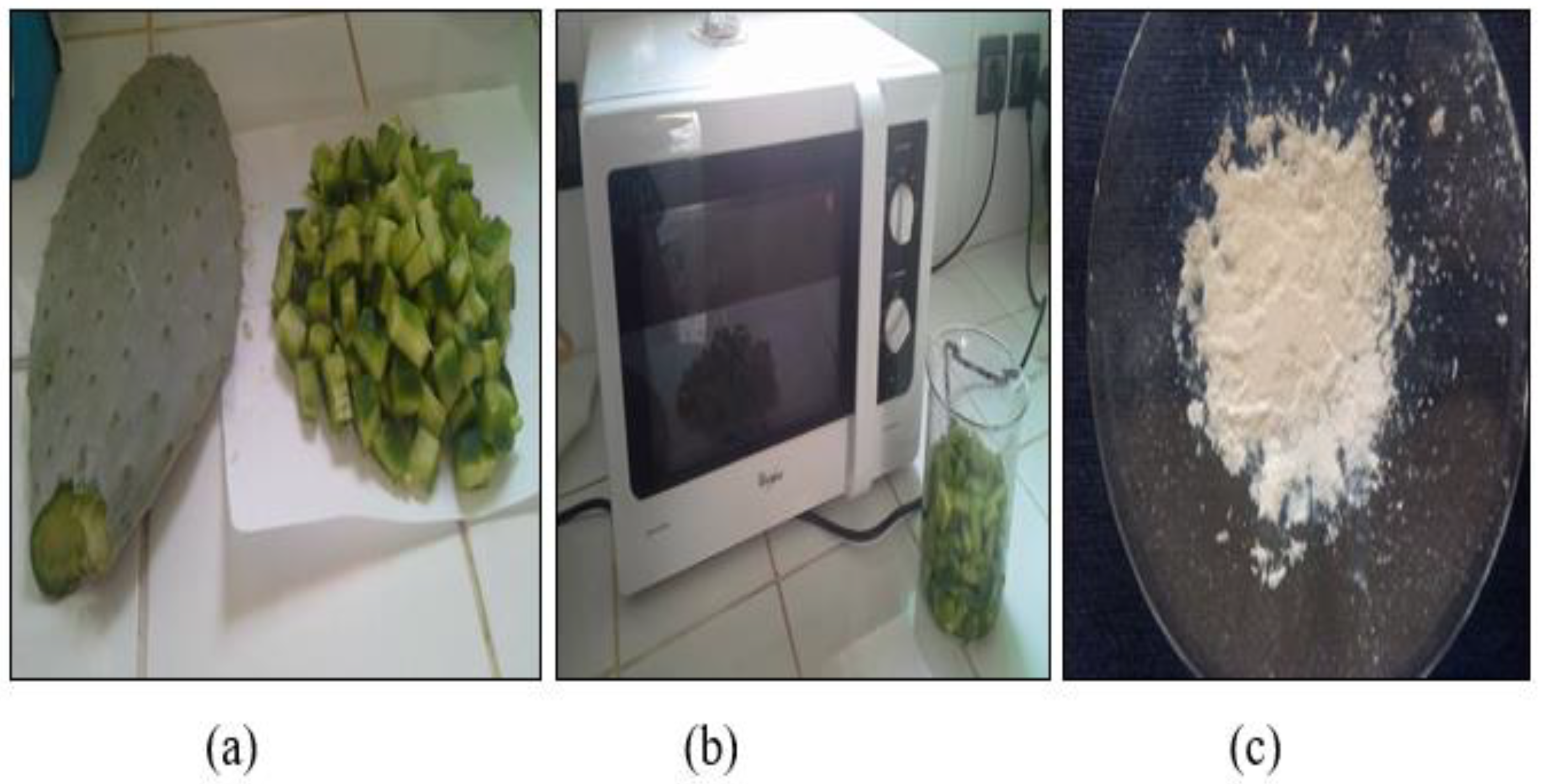
2.2. Pectin Extraction
2.3. Synthesis of Hydroxyapatite
2.4. Synthesis of Hydroxyapatite/Pectin Nanoparticles
2.5. Analyzing Methods
2.6. Study of Antibacterial Activity
2.7. Antifungal Test
3. Results and Discussions
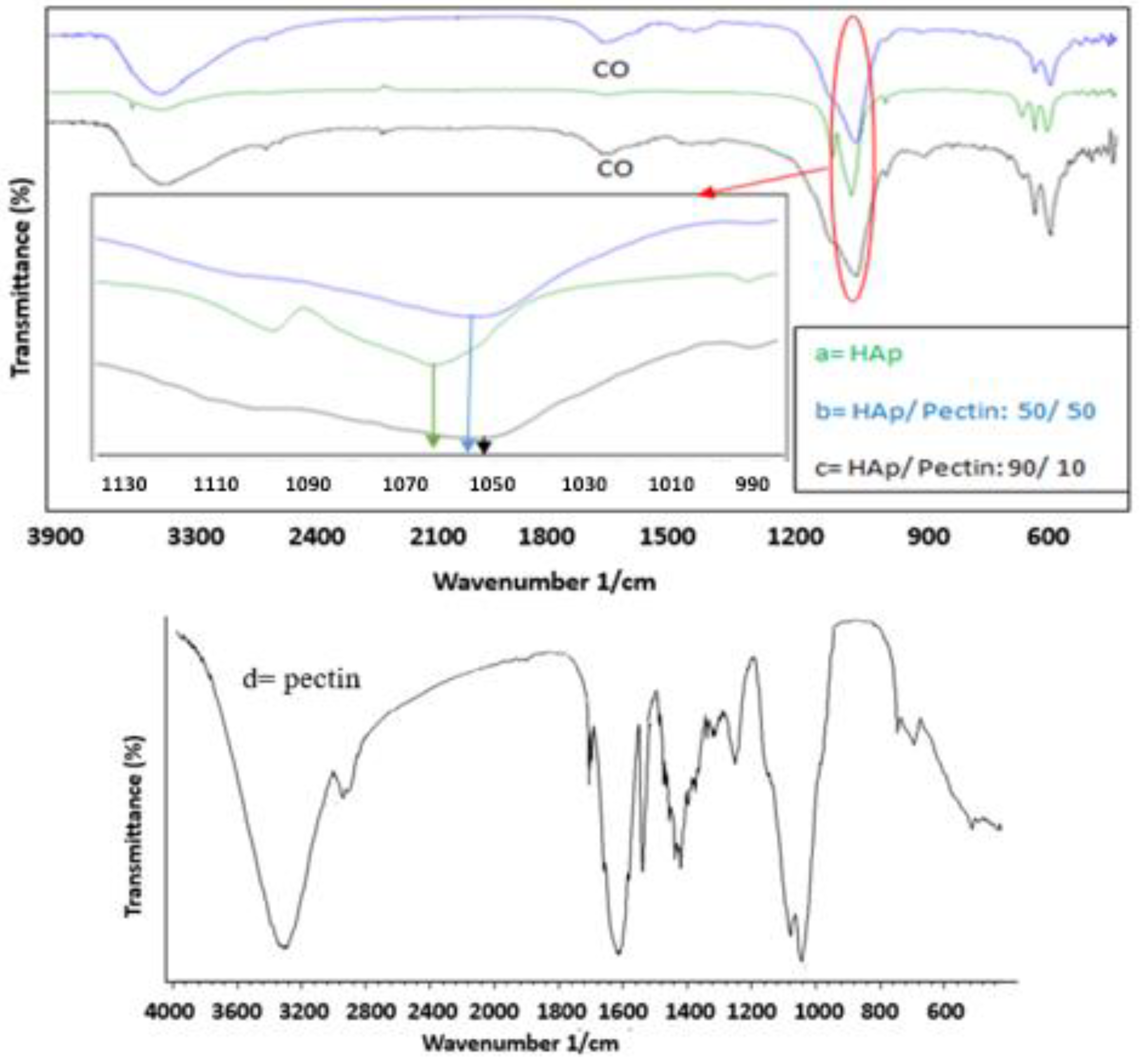
3.1. FT-IR Analysis
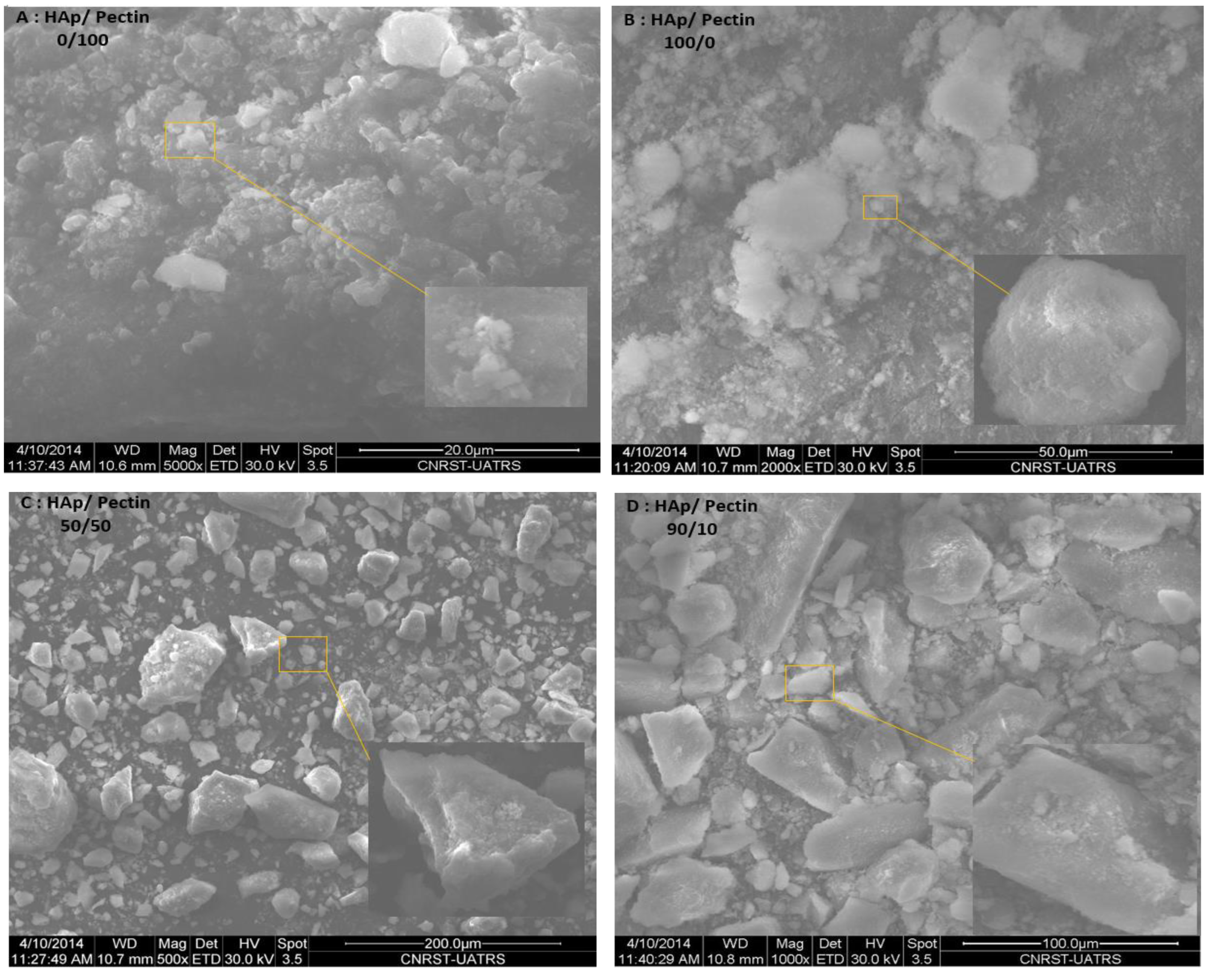
3.2. SEM Analysis
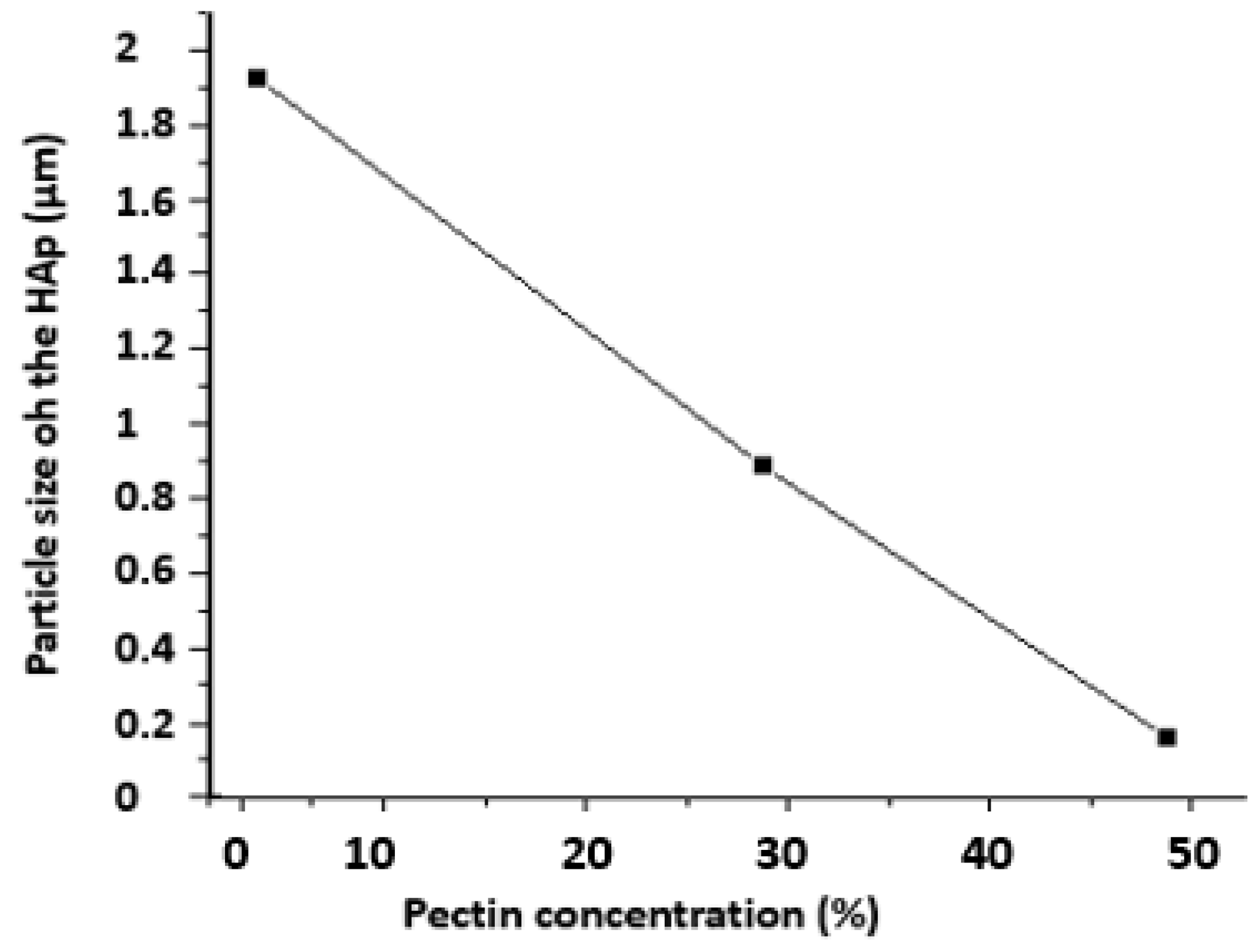
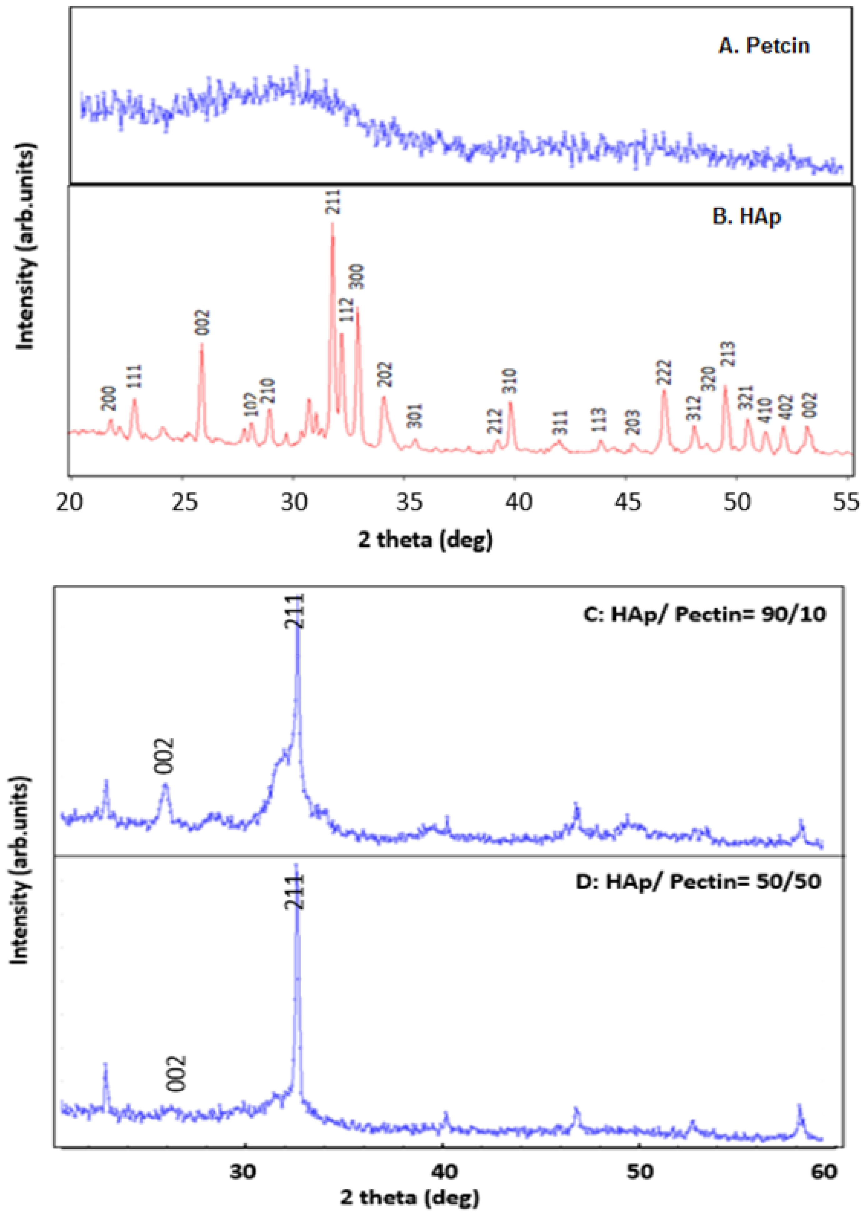
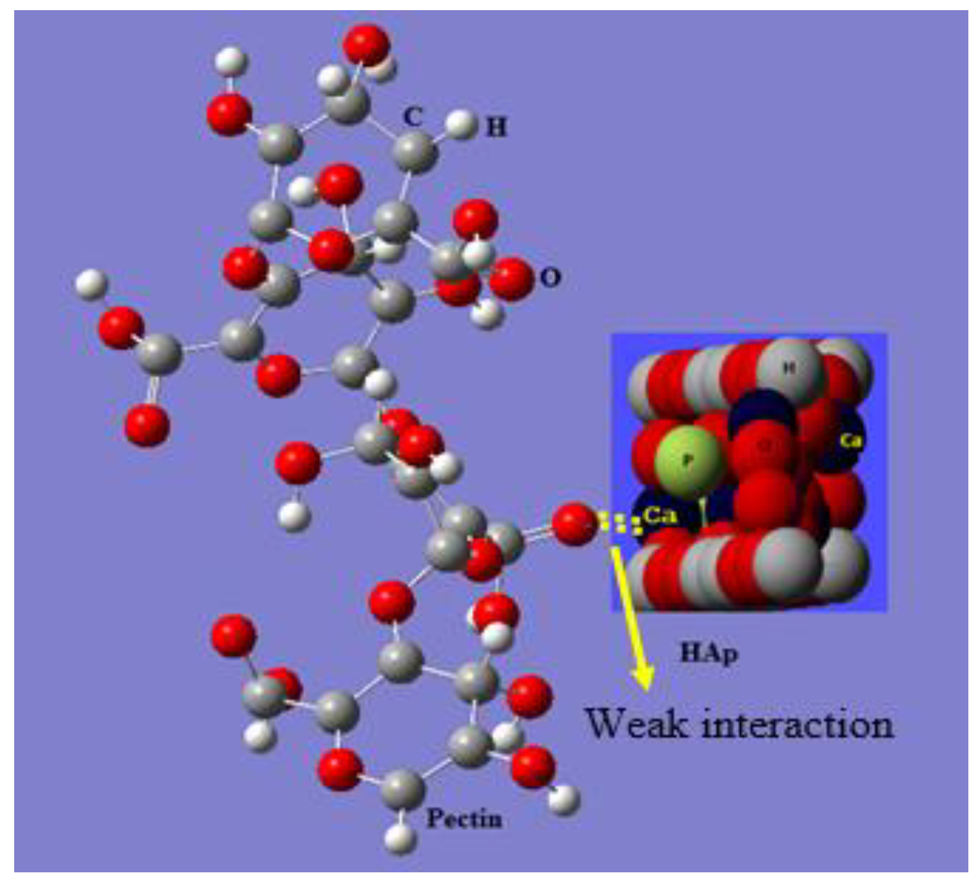
3.3. XRD Analysis of HAp/Pectin Composite
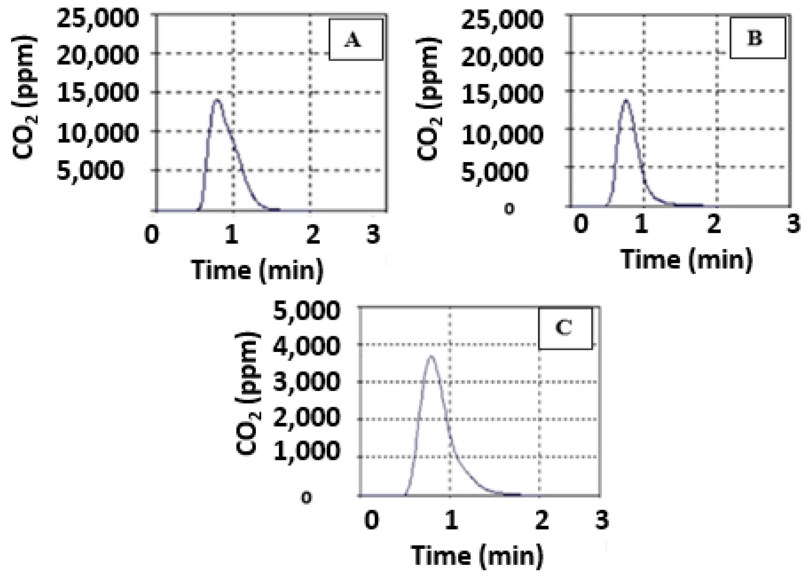
3.4. Total Organic Carbon Production
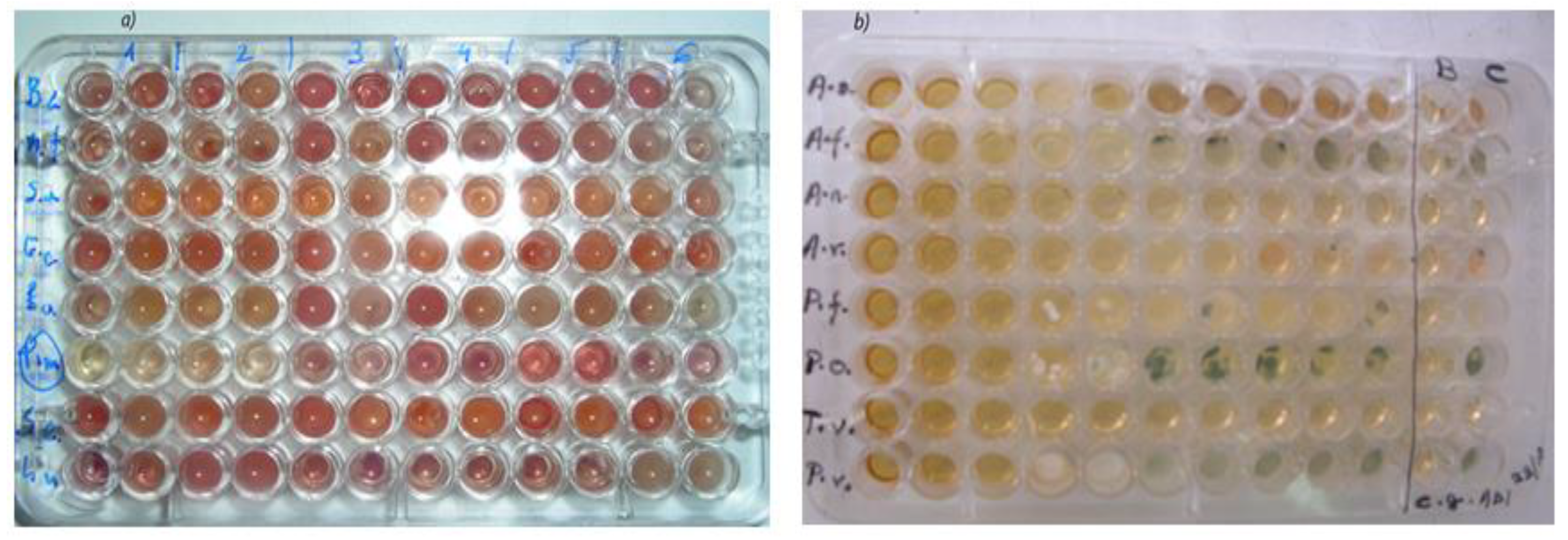
3.5. Antibacterial Efficacy
3.6. Antifungal Effect
4. Conclusions
Author Contributions
Funding
Institutional Review Board Statement
Data Availability Statement
Acknowledgments
Conflicts of Interest
References
- Shafi, A.; Hassan, F.; Zahoor, I.; Majeed, U.; Khanday, F.A. Biodiversity, Management and Sustainable Use of Medicinal and Aromatic Plant Resources. In Medicinal and Aromatic Plants: Healthcare and Industrial Applications; Aftab, T., Hakeem, K.R., Eds.; Springer International Publishing: Cham, Switzerland, 2021; pp. 85–111. [Google Scholar] [CrossRef]
- Albergamo, A.; Potortí, A.G.; Di Bella, G.; Amor, N.B.; Lo Vecchio, G.; Nava, V.; Rando, R.; Ben Mansour, H.; Lo Turco, V. Chemical Characterization of Different Products from the Tunisian Opuntia Ficus-Indica (L.) Mill. Foods 2022, 11, 155. [Google Scholar] [CrossRef] [PubMed]
- Ait Benhamou, A.; Kassab, Z.; Boussetta, A.; Salim, M.H.; Ablouh, E.-H.; Nadifiyine, M.; Qaiss, A.E.K.; Moubarik, A.; El Achaby, M. Beneficiation of Cactus Fruit Waste Seeds for the Production of Cellulose Nanostructures: Extraction and Properties. Int. J. Biol. Macromol. 2022, 203, 302–311. [Google Scholar] [CrossRef] [PubMed]
- Mishra, R.K.; Banthia, A.K.; Majeed, A.B.A. Pectin Based Formulations for Biomedical Applications: A Review. Asian J. Pharm. Clin. Res. 2012, 5, 1–7. [Google Scholar]
- Lara-Espinoza, C.; Carvajal-Millán, E.; Balandrán-Quintana, R.; López-Franco, Y.; Rascón-Chu, A. Pectin and Pectin-Based Composite Materials: Beyond Food Texture. Molecules 2018, 23, 942. [Google Scholar] [CrossRef] [PubMed] [Green Version]
- May, C.D. Industrial Pectins: Sources, Production and Applications. Carbohydr. Polym. 1990, 12, 79–99. [Google Scholar] [CrossRef]
- Gopi, D.; Kanimozhi, K.; Bhuvaneshwari, N.; Indira, J.; Kavitha, L. Novel Banana Peel Pectin Mediated Green Route for the Synthesis of Hydroxyapatite Nanoparticles and Their Spectral Characterization. Spectrochim. Acta A Mol. Biomol. Spectrosc. 2014, 118, 589–597. [Google Scholar] [CrossRef]
- Akartasse, N.; Azzaoui, K.; Mejdoubi, E.; Elansari, L.L.; Hammouti, B.; Siaj, M.; Jodeh, S.; Hanbali, G.; Hamed, R.; Rhazi, L. Chitosan-Hydroxyapatite Bio-Based Composite in Film Form: Synthesis and Application in Wastewater. Polymers 2022, 14, 4265. [Google Scholar] [CrossRef]
- Koubala, B.B.; Mbome, L.I.; Kansci, G.; Mbiapo, F.T.; Crepeau, M.-J.; Thibault, J.-F.; Ralet, M.-C. Physicochemical Properties of Pectins from Ambarella Peels (Spondias Cytherea) Obtained Using Different Extraction Conditions. Food Chem. 2008, 3, 1202–1207. [Google Scholar] [CrossRef]
- Detoxification Genes Differ between Cactus-, Fruit-, and Flower-Feeding Drosophila. J. Hered. 2019, 110, 80–91. Available online: https://academic.oup.com/jhered/article/110/1/80/5184506 (accessed on 13 October 2022). [CrossRef]
- Willats, W.G.T.; Knox, J.P.; Mikkelsen, J.D. Pectin: New Insights into an Old Polymer Are Starting to Gel. Trends Food Sci. Technol. 2006, 17, 97–104. [Google Scholar] [CrossRef]
- Gómez, L.; Ramírez, H.L.; Neira-Carrillo, A.; Villalonga, R. Polyelectrolyte Complex Formation Mediated Immobilization of Chitosan-Invertase Neoglycoconjugate on Pectin-Coated Chitin. Bioprocess Biosyst. Eng. 2006, 28, 387–395. [Google Scholar] [CrossRef] [PubMed]
- El Azzouzi, M.; Azzaoui, K.; Warad, I.; Hammouti, B.; Shityakov, S.; Sabbahi, R.; Saoiabi, S.; Youssoufi, M.H.; Akartasse, N.; Jodeh, S.; et al. Moroccan, Mauritania, and Senegalese Gum Arabic Variants as Green Corrosion Inhibitors for Mild Steel in HCl: Weight Loss, Electrochemical, AFM and XPS Studies. J. Mol. Liq. 2022, 347, 118354. [Google Scholar] [CrossRef]
- Wikiera, A.; Irla, M.; Mika, M. Health-promoting properties of pectin. Postepy Hig. Med. Dosw. 2014, 68, 590–596. [Google Scholar] [CrossRef] [PubMed]
- Benahmed, A.; Azzaoui, K.; El Idrissi, A.; Belkheir, H.; Said Hassane, S.O.; Touzani, R.; Rhazi, L. Cellulose Acetate-g-Polycaprolactone Copolymerization Using Diisocyanate Intermediates and the Effect of Polymer Chain Length on Surface, Thermal, and Antibacterial Properties. Molecules 2022, 27, 1408. [Google Scholar] [CrossRef]
- Fu, Z.; Cui, J.; Zhao, B.; Shen, S.G.F.; Lin, K. An Overview of Polyester/Hydroxyapatite Composites for Bone Tissue Repairing. J. Orthop. Transl. 2021, 28, 118–130. [Google Scholar] [CrossRef]
- Lv, M.; Lv, W.; Chen, H.; Zheng, F.; Liu, J.; Kong, F.; Liu, S.; Wang, L. Biotribological Properties of Nano Zirconium Dioxide and Hydroxyapatite-Reinforced Polyetheretherketone (HA/ZrO2/PEEK) Biocomposites. Iran. Polym. J. 2021, 30, 1127–1136. [Google Scholar] [CrossRef]
- Sumathra, M.; Sadasivuni, K.K.; Kumar, S.S.; Rajan, M. Cisplatin-Loaded Graphene Oxide/Chitosan/Hydroxyapatite Composite as a Promising Tool for Osteosarcoma-Affected Bone Regeneration. ACS Omega. 2018, 3, 14620–14633. [Google Scholar] [CrossRef] [Green Version]
- Gopi, D.; Indira, J.; Prakash, V.C.A.; Kavitha, L. Spectroscopic Characterization of Porous Nanohydroxyapatite Synthesized by a Novel Amino Acid Soft Solution Freezing Method. Spectrochim. Acta A Mol. Biomol. Spectrosc. 2009, 74, 282–284. [Google Scholar] [CrossRef]
- Subba Rao, Y.; Kotakadi, V.S.; Prasad, T.N.V.K.V.; Reddy, A.V.; Sai Gopal, D.V.R. Green Synthesis and Spectral Characterization of Silver Nanoparticles from Lakshmi Tulasi (Ocimum Sanctum) Leaf Extract. Spectrochim. Acta A Mol. Biomol. Spectrosc. 2013, 103, 156–159. [Google Scholar] [CrossRef]
- Azzaoui, K.; Lamhamdi, A.; Mejdoubi, E.M.; Berrabah, M.; Hammouti, B.; Elidrissi, A.; Fouda, M.M.G.; Al-Deyab, S.S. Synthesis and Characterization of Composite Based on Cellulose Acetate and Hydroxyapatite Application to the Absorption of Harmful Substances. Carbohydr. Polym. 2014, 111, 41–46. [Google Scholar] [CrossRef]
- Patil, S.B.; Inamdar, S.Z.; Reddy, K.R.; Raghu, A.V.; Soni, S.K.; Kulkarni, R.V. Novel biocompatible poly(acrylamide)-grafted-dextran hydrogels: Synthesis, characterization and biomedical applications. J. Microbiol. Methods. 2019, 159, 200–210. [Google Scholar] [CrossRef] [PubMed]
- Azzaoui, K.; Mejdoubi, E.; Lamhamdi, A.; Zaoui, S.; Berrabah, M.; Elidrissi, A.; Hammouti, B.; Fouda, M.M.G.; Al-Deyab, S.S. Structure and Properties of Hydroxyapatite/Hydroxyethyl Cellulose Acetate Composite Films. Carbohydr. Polym. 2015, 115, 170–176. [Google Scholar] [CrossRef] [PubMed]
- Ashrit, P.; Sadanandan, B.; Natraj, L.K.; Shetty, K.; Vaniyamparambath, V.; Raghu, A.V. A microplate-based Response Surface Methodology model for growth optimization and biofilm formation on polystyrene polymeric material in a Candida albicans and Escherichia coli co-culture. Polym. Adv. Technol. 2022, 33, 2872–2885. [Google Scholar] [CrossRef]
- Meltem Haktaniyan, M.H.; Bradley, M. Polymers Showing Intrinsic Antimicrobial Activity. Chem. Soc. Rev. 2022, 51, 8584–8611. [Google Scholar] [CrossRef]
- Santhamoorthy, M.; Vy Phan, T.T.; Ramkumar, V.; Raorane, C.J.; Thirupathi, K.; Kim, S.-C. Thermo-Sensitive Poly (N-Isopropylacrylamide-Co-Polyacrylamide) Hydrogel for PH-Responsive Therapeutic Delivery. Polymers 2022, 14, 4128. [Google Scholar] [CrossRef]
- Akartasse, N.; Azzaoui, K.; Mejdoubi, E.; Hammouti, B.; Elansari, L.L.; Abou-Salama, M.; Aaddouz, M.; Sabbahi, R.; Rhazi, L.; Siaj, M. Environmental-Friendly Adsorbent Composite Based on Hydroxyapatite/Hydroxypropyl Methyl-Cellulose for Removal of Cationic Dyes from an Aqueous Solution. Polymers 2022, 14, 2147. [Google Scholar] [CrossRef] [PubMed]
- Cuadra, J.G.; Scalschi, L.; Vicedo, B.; Guc, M.; Izquierdo-Roca, V.; Porcar, S.; Fraga, D.; Carda, J.B. ZnO/Ag Nanocomposites with Enhanced Antimicrobial Activity. Appl. Sci. 2022, 12, 5023. [Google Scholar] [CrossRef]
- Liu, D.; Zhang, C.; Wang, B.; Quan, W.; Xu, C. AgNP-AC Composite Fibers and its Adsorption and Antibacterial Properties. Front. Mater. 2022, 9, 894451. [Google Scholar] [CrossRef]
- P Baldino, L.; Aragón, J.; Mendoza, G.; Irusta, S.; Cardea, S.; Reverchon, E. Production, characterization and testing of antibacterial PVA membranes loaded with HA-Ag3PO4 nanoparticles, produced by SC-CO2 phase inversion. J. Chem. Technol. Biotechnol. 2019, 94, 98–108. [Google Scholar] [CrossRef] [Green Version]
- Landi, E.; Tampieri, A.; Celotti, G.; Sprio, S. Densification Behaviour and Mechanisms of Synthetic Hydroxyapatites. J. Eur. Ceram. Soc. 2000, 20, 2377. [Google Scholar] [CrossRef]
- Teng Chai, S.; Haydar Ali Tajuddin, A.; A Wahab, N.; Mustafa, N.; Sukor, N.; Kamaruddin, N.A. Fluconazole as a Safe and Effective Alternative to Ketoconazole in Controlling Hypercortisolism of Recurrent Cushing’s Disease: A Case Report. Int. J. Endocrinol. Metab. 2018, 16, e65233. [Google Scholar] [CrossRef] [PubMed] [Green Version]
- Funari, R.; Shen, A.Q. Detection and Characterization of Bacterial Biofilms and Biofilm-Based Sensors. ACS Sens. 2022, 7, 347–357. [Google Scholar] [CrossRef] [PubMed]
- Kasai, D.; Chougale, R.; Masti, S.; Gouripur, G.; Malabadi, R.; Chalannavar, R.; Raghu, A.V.; Radhika, D.; Dhanavant, S. Preparation, Characterization and Antimicrobial Activity of Betel-Leaf-Extract-Doped Polysaccharide Blend Films. Green Mater. 2021, 9, 49–68. [Google Scholar] [CrossRef]
- Gogoi, B.; Barua, S.; Sarmah, J.K.; Karak, N. In Situ Synthesis of a Microbial Fouling Resistant, Nanofibrillar Cellulose-Hyperbranched Epoxy Composite for Advanced Coating Applications. Prog. Org. Coat. 2018, 124, 224–231. [Google Scholar] [CrossRef]
- Peng, Q.; Xu, Q.; Yin, H.; Huang, L.; Du, Y. Characterization of an Immunologically Active Pectin from the Fruits of Lycium Ruthenicum. Int. J. Biol. Macromol. 2014, 64, 69–75. [Google Scholar] [CrossRef]
- Nagaraja, A.; Jalageri, M.D.; Puttaiahgowda, Y.M.; Raghava Reddy, K.; Raghu, A.V. A Review on Various Maleic Anhydride Antimicrobial Polymers. J. Microbiol. Methods 2019, 163, 105650. [Google Scholar] [CrossRef]
- Raghu, A.V.; Gadaginamath, G.S.; Mathew, N.T.; Halligudi, S.B.; Aminabhavi, T.M. Synthesis and Characterization of Novel Polyurethanes Based on 4,4′-[1,4-Phenylenedi-Diazene-2,1-Diyl]Bis(2-Carboxyphenol) and 4,4′-[1,4-Phenylenedi-Diazene-2,1-Diyl]Bis(2-Chlorophenol) Hard Segments. React. Funct. Polym. 2007, 67, 503–514. [Google Scholar] [CrossRef]
- Gopi, D.; Kanimozhi, K.; Kavitha, L. Opuntia Ficus Indica Peel Derived Pectin Mediated Hydroxyapatite Nanoparticles: Synthesis, Spectral Characterization, Biological and Antimicrobial Activities. Spectrochim. Acta A Mol. Biomol. Spectrosc. 2015, 141, 135–143. [Google Scholar] [CrossRef]


| Species | Gram | |
|---|---|---|
| Entero bactercloacae (Clinical isolate) | - | Negative |
| Salmonella typhimurium | ATCC 13311 | |
| Escherichia coli | ATCC 35210 | |
| Pseudomonas aeruginosa | ATCC 27853 | |
| Micrococcus flavus | ATCC 10240 | Positive |
| Listeria monocytogenes | NCTC 797 | |
| Staphylococcus aureus | ATCC 6538 | |
| Bacillus cereus (Clinical isolate) | - | |
| Pectin Concentration | Plane | θ | FWHM (°) | Xc (nm) | Xs (%) |
|---|---|---|---|---|---|
| 0 | 211 | 16.1089 | 0.1319 | 6.0241 | 10.9306 |
| 10 | 211 | 16.3294 | 0.1978 | 1.7863 | 7.2986 |
| 50 | 211 | 16.31214 | 0.1978 | 1.7863 | 7.2985 |
| S. Aureus | B. Cereus | L. Monocytogenes | M. Flavus | P. Aeruginosa | E. Coli | S. Typhimurium | En. Cloacae | ||
|---|---|---|---|---|---|---|---|---|---|
| Pectin | MIC | 0.56 | 0.56 | 0.28 | 0.56 | 0.56 | 0.56 | 0.28 | 0.56 |
| MBC | 1.14 | 1.14 | 1.14 | 1.14 | 1.14 | 1.14 | 1.14 | 1.14 | |
| Hydroxyapatite (HAp) | MIC | 0.56 | 0.28 | 0.56 | 0.56 | 0.28 | 0.56 | 0.56 | 0.28 |
| MBC | 1.14 | 0.56 | 1.14 | 1.14 | 0.56 | 1.14 | 1.14 | 0.56 | |
| HAp/pectin (90%/10%) | MIC | 0.14 | 0.14 | 0.56 | 0.28 | 0.14 | 0.28 | 0.56 | 0.28 |
| MBC | 0.28 | 0.28 | 1.14 | 0.56 | 0.28 | 0.56 | 1.14 | 0.56 | |
| HAp/pectin (50%/50%) | MIC | 0.14 | 0.14 | 0.28 | 0.28 | 0.14 | 0.28 | 0.28 | 0.28 |
| MBC | 0.28 | 0.28 | 0.56 | 0.56 | 0.28 | 0.56 | 0.56 | 0.56 | |
| Streptomycin | MIC | 0.04 | 0.09 | 0.17 | 0.17 | 0.34 | 0.26 | 0.17 | 0.17 |
| MBC | 0.09 | 0.17 | 0.34 | 0.34 | 0.68 | 0.52 | 0.34 | 0.34 | |
| Ampicillin | MIC | 0.25 | 0.25 | 0.25 | 0.37 | 0.74 | 0.37 | 0.37 | 0.25 |
| MBC | 0.37 | 0.37 | 0.37 | 0.49 | 1.24 | 0.74 | 0.49 | 0.49 | |
| A. Fumigatus | A. Versicolor | A. Ochraceus | A. Niger | P. Ochrochloron | P. Funiculosum | P. Verrucosum | T. Viride | ||
|---|---|---|---|---|---|---|---|---|---|
| Pectin | MIC | 0.55 | 1.10 | 0.55 | 1.10 | NA | 0.14 | 0.28 | 0.55 |
| MFC | 1.10 | 1.10 | 1.10 | 1.10 | NA | 0.28 | 0.55 | 1.10 | |
| Hydroxyapatite (HAp) | MIC | 0.19 | 0.19 | 0.39 | 0.78 | 0.19 | 0.19 | 0.39 | 0.39 |
| MFC | 0.38 | 0.38 | 0.78 | 1.55 | 0.39 | 0.39 | 0.78 | 0.78 | |
| HAp/pectin (90/10) | MIC | 0.098 | 0.098 | 0.048 | 0.39 | NA | 0.19 | NA | 0.39 |
| MFC | 0.19 | 0.19 | 0.098 | 0.78 | NA | 0.39 | NA | 0.78 | |
| Ketoconazole | MIC | 0.20 | 0.20 | 0.15 | 0.20 | 1.00 | 0.20 | 0.20 | 1.00 |
| MFC | 0.50 | 0.50 | 0.20 | 0.50 | 1.50 | 0.50 | 0.30 | 1.50 | |
| Bifonazole | MIC | 0.15 | 0.10 | 0.15 | 0.15 | 0.20 | 0.20 | 0.10 | 0.15 |
| MFC | 0.20 | 0.20 | 0.20 | 0.20 | 0.25 | 0.25 | 0.20 | 0.20 | |
Publisher’s Note: MDPI stays neutral with regard to jurisdictional claims in published maps and institutional affiliations. |
© 2022 by the authors. Licensee MDPI, Basel, Switzerland. This article is an open access article distributed under the terms and conditions of the Creative Commons Attribution (CC BY) license (https://creativecommons.org/licenses/by/4.0/).
Share and Cite
Saidi, N.; Azzaoui, K.; Ramdani, M.; Mejdoubi, E.; Jaradat, N.; Jodeh, S.; Hammouti, B.; Sabbahi, R.; Lamhamdi, A. Design of Nanohydroxyapatite/Pectin Composite from Opuntia Ficus-Indica Cladodes for the Management of Microbial Infections. Polymers 2022, 14, 4446. https://doi.org/10.3390/polym14204446
Saidi N, Azzaoui K, Ramdani M, Mejdoubi E, Jaradat N, Jodeh S, Hammouti B, Sabbahi R, Lamhamdi A. Design of Nanohydroxyapatite/Pectin Composite from Opuntia Ficus-Indica Cladodes for the Management of Microbial Infections. Polymers. 2022; 14(20):4446. https://doi.org/10.3390/polym14204446
Chicago/Turabian StyleSaidi, N., K. Azzaoui, M. Ramdani, E. Mejdoubi, N. Jaradat, S. Jodeh, B. Hammouti, R. Sabbahi, and A. Lamhamdi. 2022. "Design of Nanohydroxyapatite/Pectin Composite from Opuntia Ficus-Indica Cladodes for the Management of Microbial Infections" Polymers 14, no. 20: 4446. https://doi.org/10.3390/polym14204446