
Role of Oil Palm Empty Fruit Bunch-Derived Cellulose in Improving the Sonocatalytic Activity of Silver-Doped Titanium Dioxide

 , , , and
Abstract
:1. Introduction
2. Materials and Methods
2.1. Materials
2.2. Preparation of Cellulose
2.3. Preparation of TiO2, Cellulose/TiO2, Ag/TiO2, and Cellulose/Ag/TiO2
2.4. Sample Characterization
2.5. Adsorption and Sonocatalytic Degradation of Congo Red
3. Results and Discussion
3.1. Characterization Study
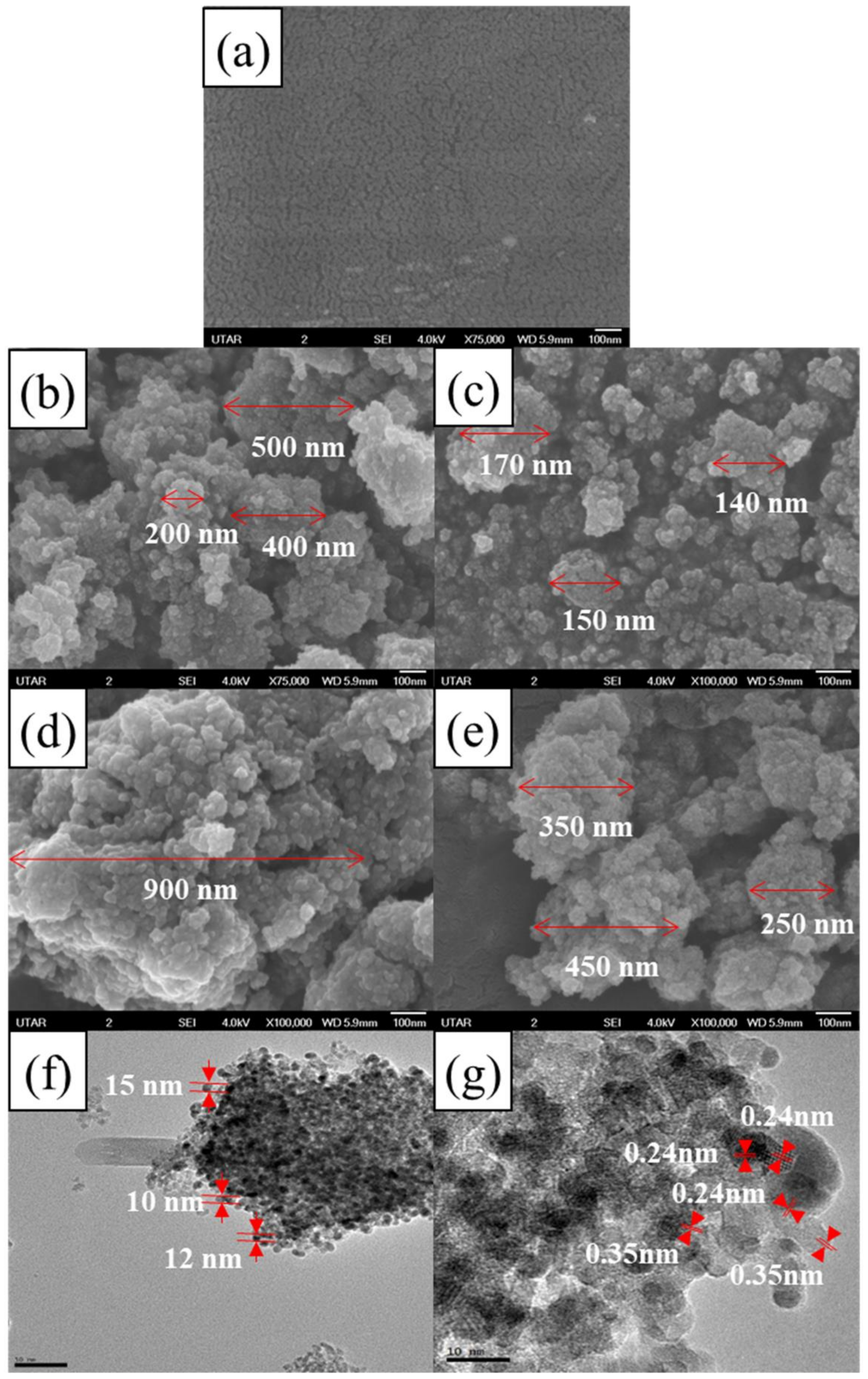
3.1.1. FESEM, HRTEM and EDX
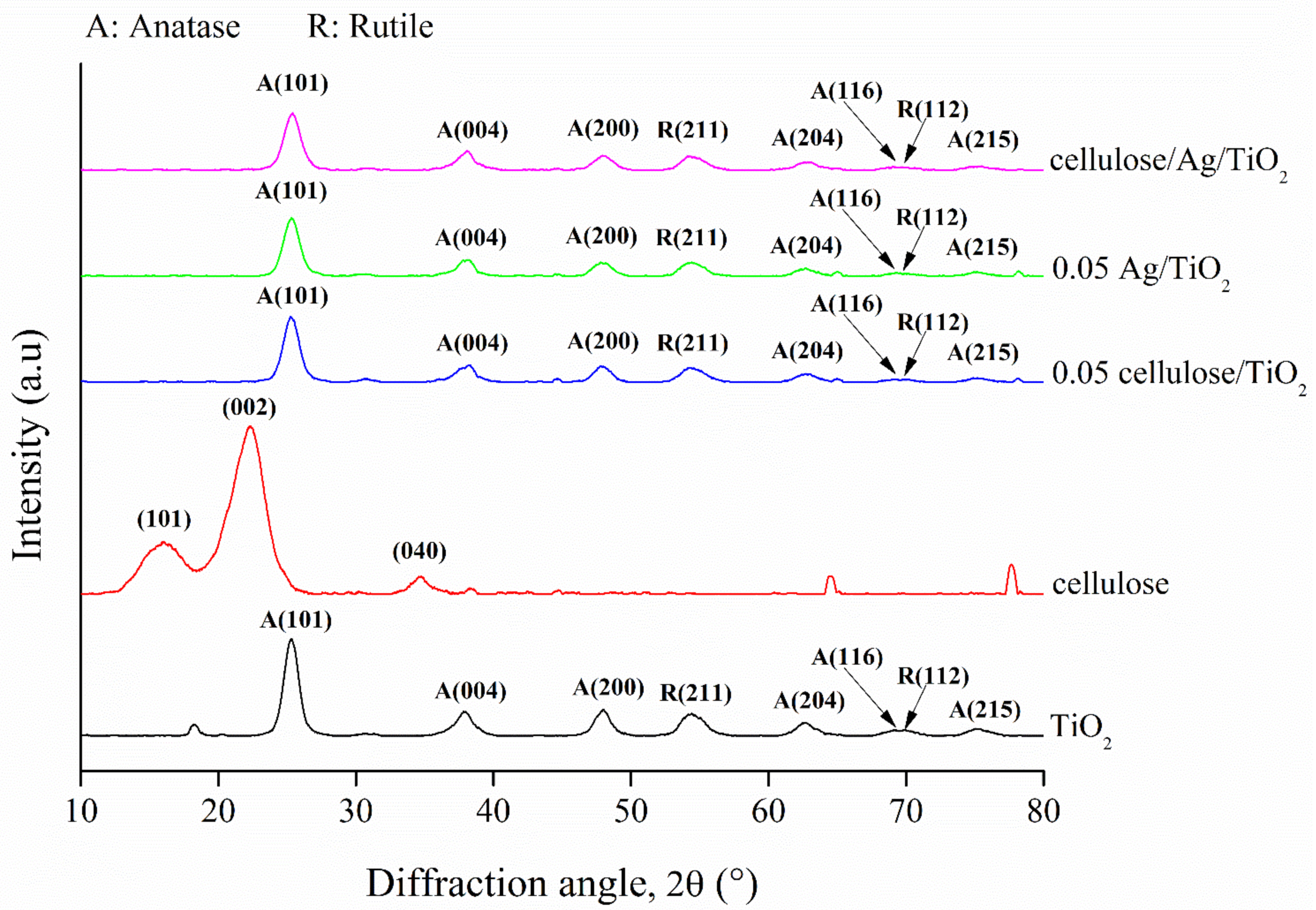
3.1.2. XRD
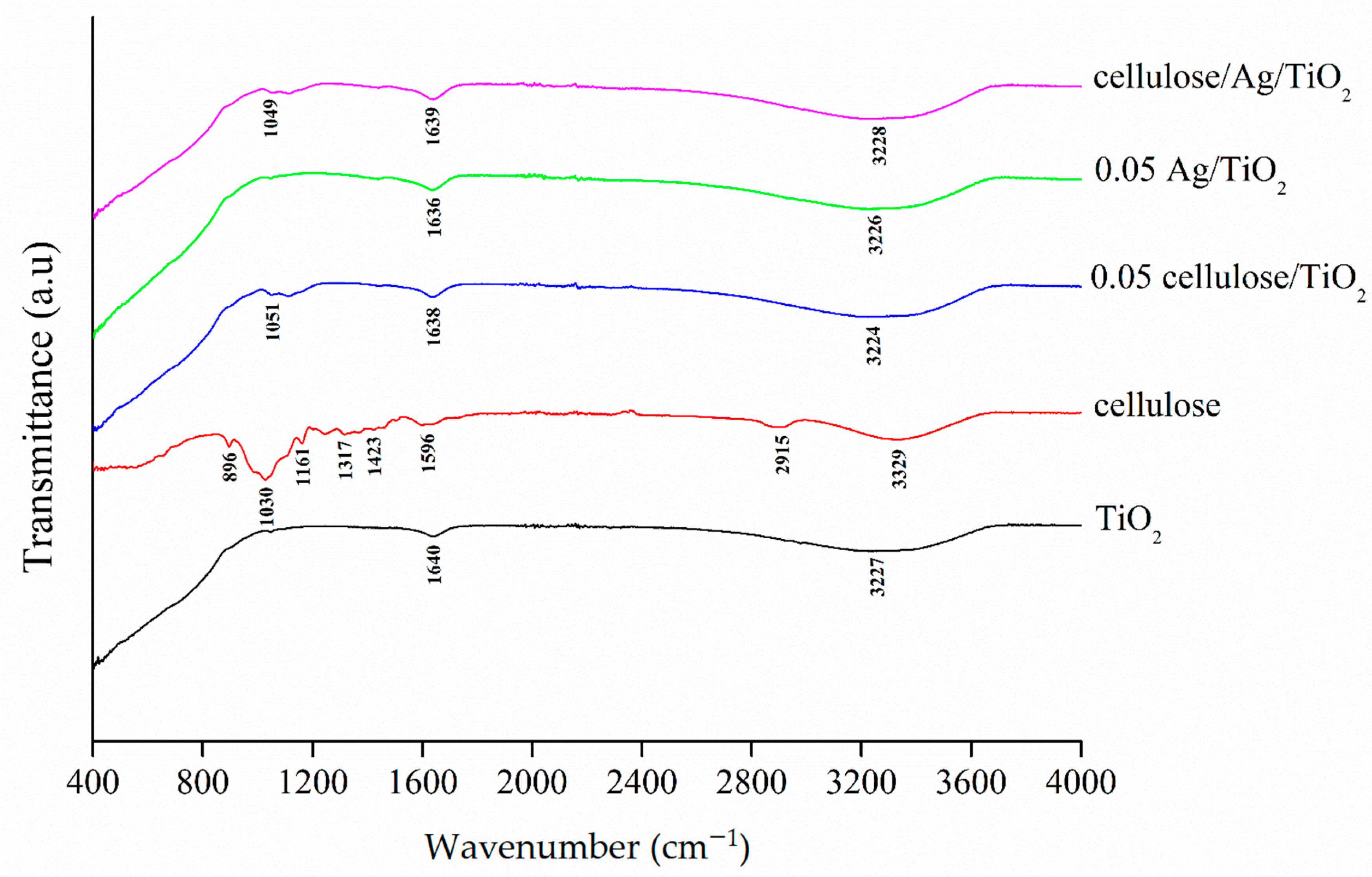
3.1.3. FTIR Analysis
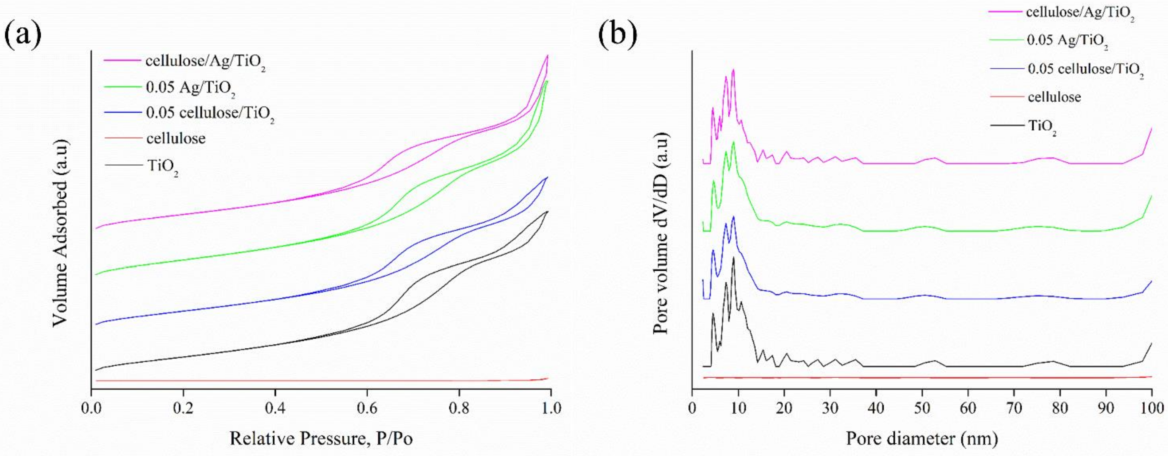
3.1.4. Surface Analysis
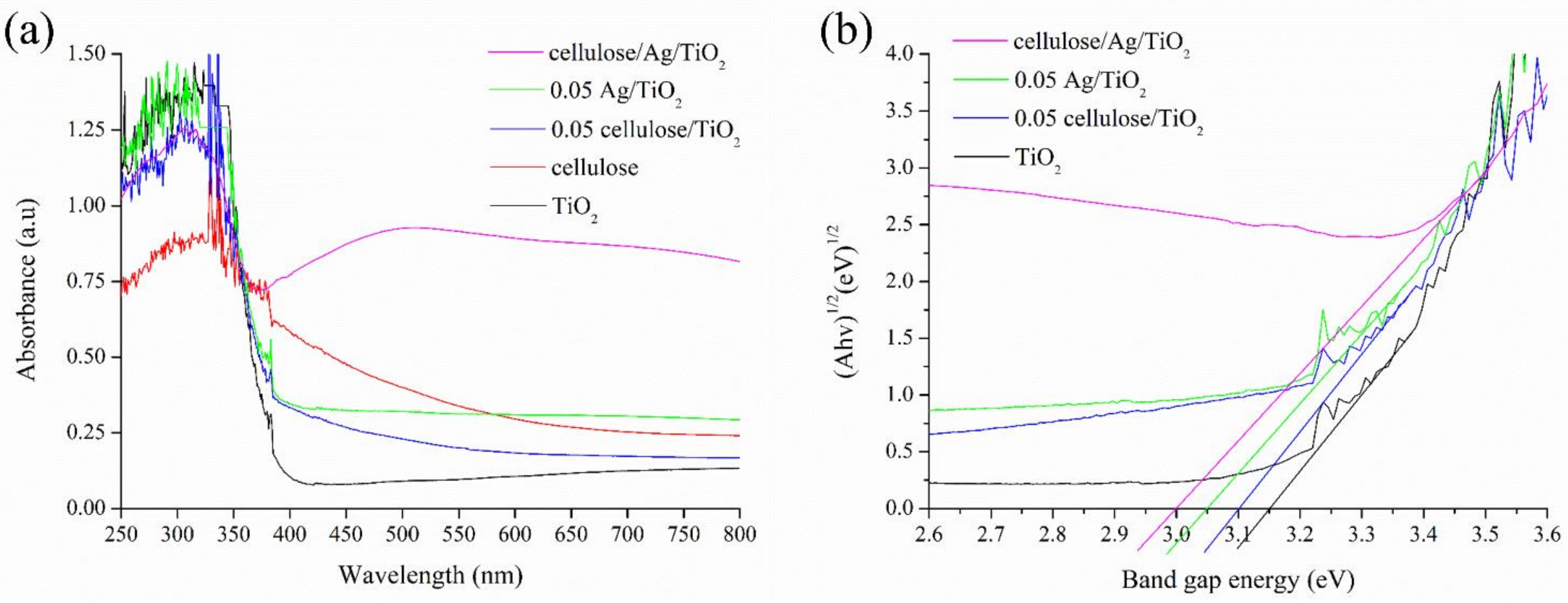
3.1.5. Optical Properties
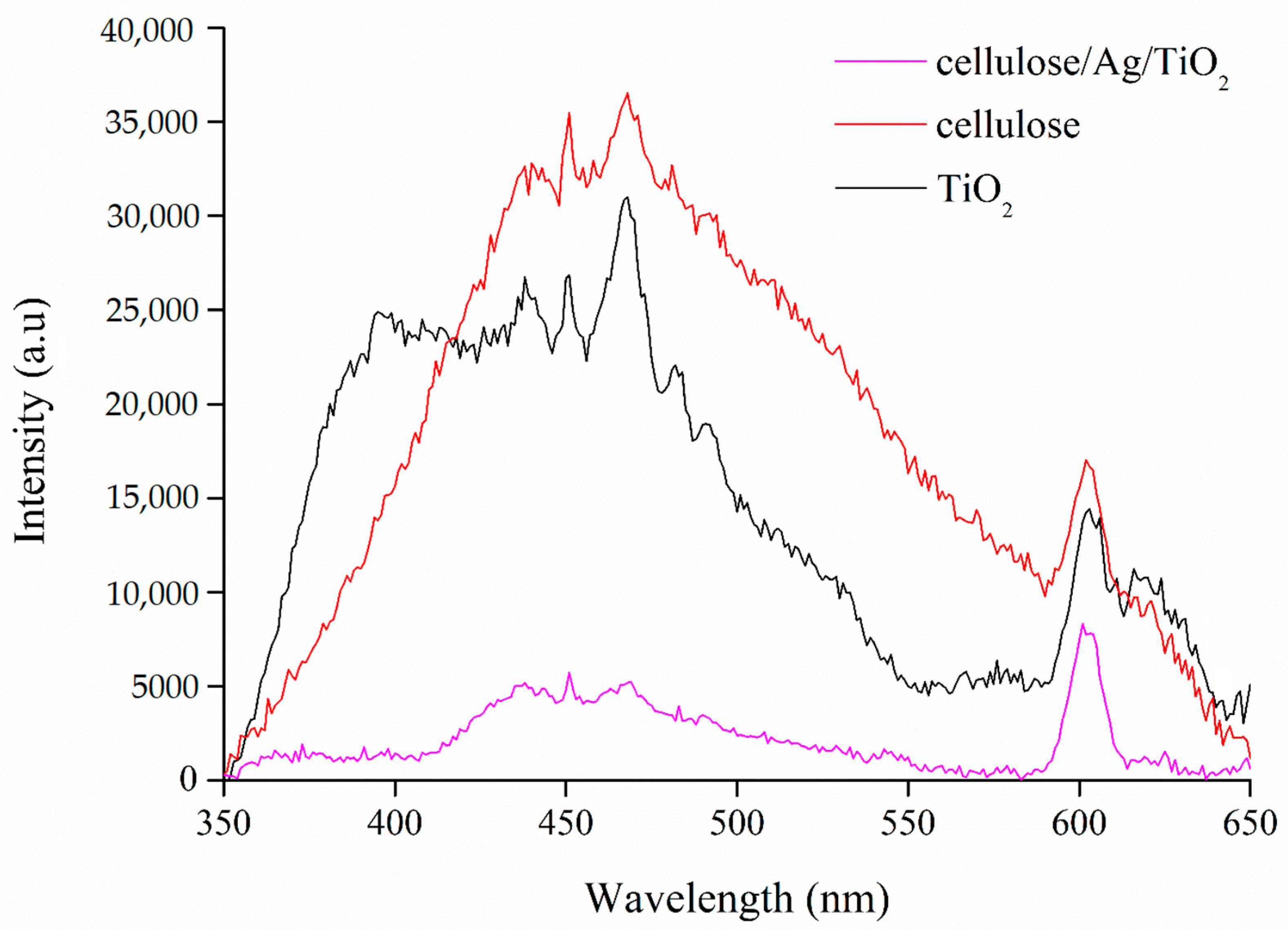
3.1.6. PL
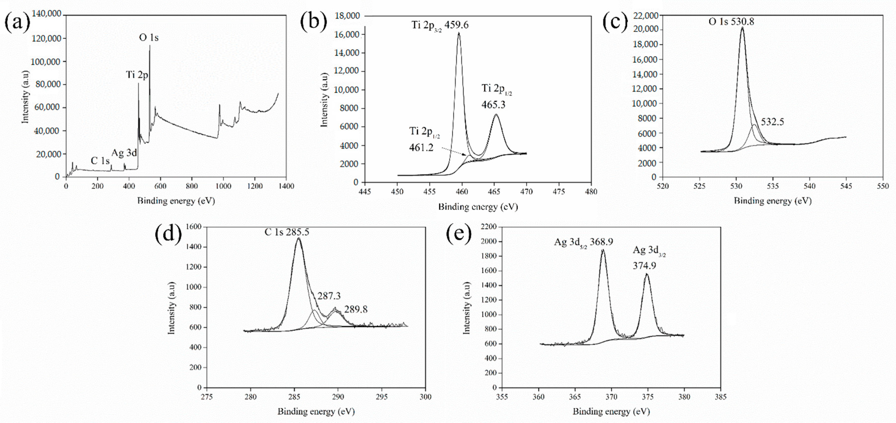
3.1.7. XPS
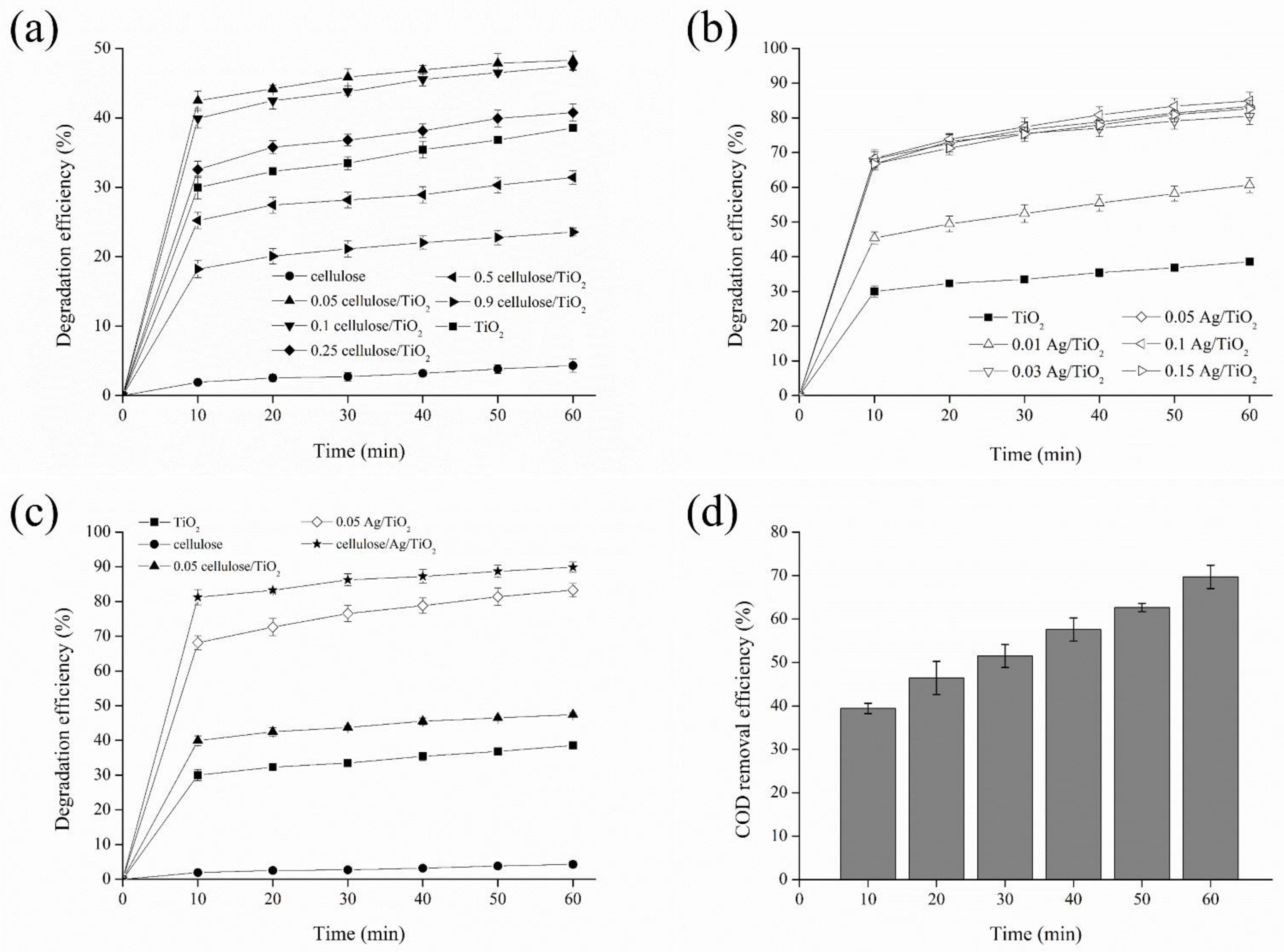
3.2. Catalytic Performance
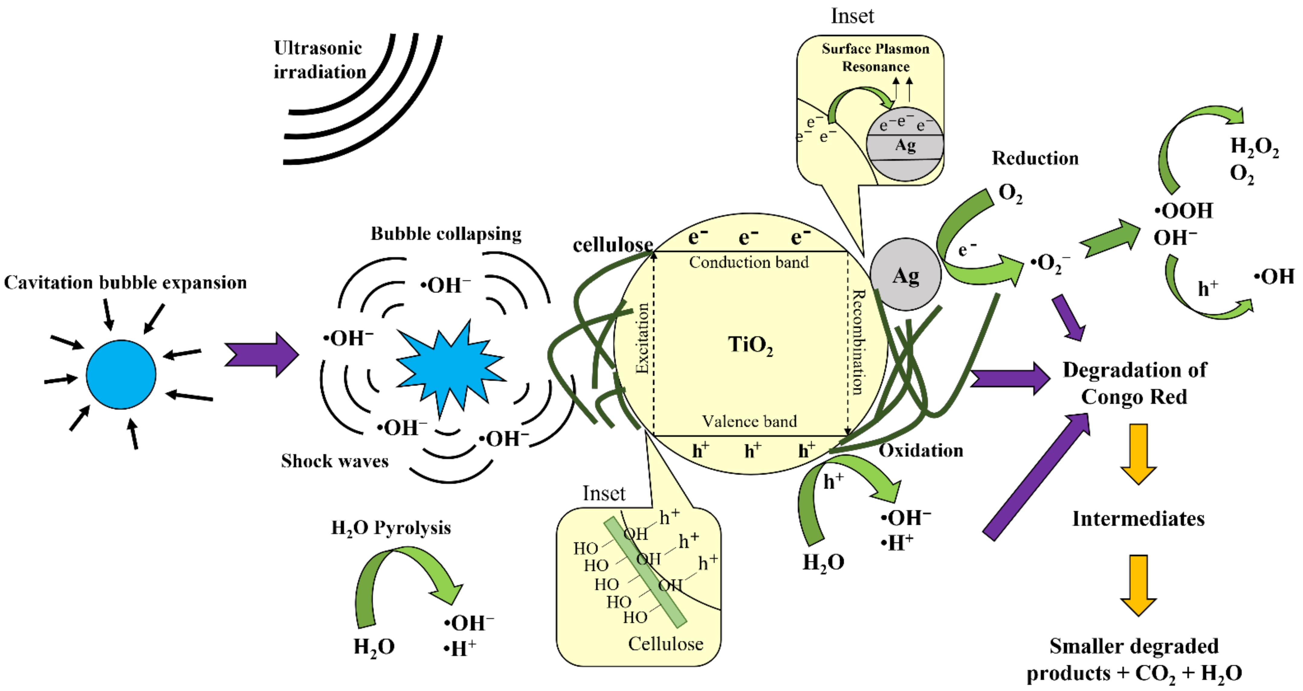
3.3. Sonocatalytic Degradation Mechanisms of Congo Red
4. Conclusions
Author Contributions
Funding
Institutional Review Board Statement
Informed Consent Statement
Data Availability Statement
Acknowledgments
Conflicts of Interest
References
- Serrano-Martínez, A.; Mercader-Ros, M.T.; Martínez-Alcalá, I.; Lucas-Abellán, C.; Gabaldón-Hernández, J.A.; Gómez-López, V.M. Degradation and toxicity evaluation of azo dye Direct red 83:1 by an advanced oxidation process driven by pulsed light. J. Water Process. Eng. 2020, 37, 101530. [Google Scholar] [CrossRef]
- Balapure, K.; Bhatt, N.; Madamwar, D. Mineralization of reactive azo dyes present in simulated textile waste water using down flow microaerophilic fixed film bioreactor. Bioresour. Technol. 2015, 175, 1–7. [Google Scholar] [CrossRef] [PubMed]
- Ahmadi, S.; Rahdar, A.; Igwegbe, C.A.; Mortazavi-Derazkola, S.; Banach, A.M.; Rahdar, S.; Singh, A.K.; Rodriguez-Couto, S.; Kyzas, G.Z. Praseodymium-doped cadmium tungstate (CdWO4) nanoparticles for dye degradation with sonocatalytic process. Polyhedron 2020, 190, 114792. [Google Scholar] [CrossRef]
- Cubas, P.d.J.; Semkiw, A.W.; Monteiro, F.C.; Weinert, P.L.; Monteiro, J.F.H.L.; Fujiwara, S.T. Synthesis of CuCr2O4 by self-combustion method and photocatalytic activity in the degradation of Azo Dye with visible light. J. Photochem. Photobiol. A Chem. 2020, 401, 112797. [Google Scholar] [CrossRef]
- Gholami, P.; Khataee, A.; Soltani, R.D.C.; Bhatnagar, A. A review on carbon-based materials for heterogeneous sonocatalysis: Fundamentals, properties and applications. Ultrason. Sonochem. 2019, 58, 104681. [Google Scholar] [CrossRef]
- Karaca, S.; Onal, E.Ç.; Açışlı, O.; Khataee, A. Preparation of chitosan modified montmorillonite biocomposite for sonocatalysis of dyes: Parameters and degradation mechanism. Mater. Chem. Phys. 2021, 260, 124125. [Google Scholar] [CrossRef]
- Liu, M.; Yin, W.; Qian, F.J.; Zhao, T.L.; Yao, Q.Z.; Fu, S.Q.; Zhou, G.T. A novel synthesis of porous TiO2 nanotubes and sequential application to dye contaminant removal and Cr(VI) visible light catalytic reduction. J. Environ. Chem. Eng. 2020, 8, 104061. [Google Scholar] [CrossRef]
- Shan, R.; Lu, L.; Gu, J.; Zhang, Y.; Yuan, H.; Chen, Y.; Luo, B. Photocatalytic degradation of methyl orange by Ag/TiO2/biochar composite catalysts in aqueous solutions. Mater. Sci. Semicond. Process. 2020, 114, 105088. [Google Scholar] [CrossRef]
- Kumaravel, V.; Mathew, S.; Bartlett, J.; Pillai, S.C. Photocatalytic hydrogen production using metal doped TiO2: A review of recent advances. Appl. Catal. B Environ. 2019, 244, 1021–1064. [Google Scholar] [CrossRef]
- Fiorenza, R.; Di Mauro, A.; Cantarella, M.; Gulino, A.; Spitaleri, L.; Privitera, V.; Impellizzeri, G. Molecularly imprinted N-doped TiO2 photocatalysts for the selective degradation of o-phenylphenol fungicide from water. Mater. Sci. Semicond. Process. 2020, 112, 105019. [Google Scholar] [CrossRef]
- Zimbone, M.; Cacciato, G.; Spitaleri, L.; Egdell, R.G.; Grimaldi, M.G.; Gulino, A. Sb-doped titanium oxide: A rationale for its photocatalytic activity for environmental remediation. ACS Omega 2018, 3, 11270–11277. [Google Scholar] [CrossRef] [PubMed] [Green Version]
- Peña-Velasco, G.; Hinojosa-Reyes, L.; Morán-Quintanilla, G.A.; Hernández-Ramírez, A.; Villanueva-Rodríguez, M.; Guzmán-Mar, J.L. Synthesis of heterostructured catalyst coupling MOF derived Fe2O3 with TiO2 for enhanced photocatalytic activity in anti-inflammatory drugs mixture degradation. Ceram. Int. 2021, 47, 24632–24640. [Google Scholar] [CrossRef]
- Baran, T.; Wojtyła, S.; Minguzzi, A.; Rondinini, S.; Vertova, A. Achieving efficient H2O2 production by a visible-light absorbing, highly stable photosensitized TiO2. Appl. Catal. B Environ. 2019, 244, 303–312. [Google Scholar] [CrossRef]
- Hamed, N.K.A.; Ahmad, M.K.; Hairom, N.H.H.; Faridah, A.B.; Mamat, M.H.; Mohamed, A.; Suriani, A.B.; Nafarizal, N.; Fazli, F.I.M.; Mokhtar, S.M.; et al. Dependence of photocatalysis on electron trapping in Ag-doped flowerlike rutile-phase TiO2 film by facile hydrothermal method. Appl. Surf. Sci. 2020, 534, 147571. [Google Scholar] [CrossRef]
- May-Lozano, M.; Lopez-Medina, R.; Mendoza Escamilla, V.; Rivadeneyra-Romero, G.; Alonzo-Garcia, A.; Morales-Mora, M.; González-Díaz, M.O.; Martinez-Degadillo, S.A. Intensification of the Orange II and Black 5 degradation by sonophotocatalysis using Ag-graphene oxide/TiO2 systems. Chem. Eng. Process. Process. Intensif. 2020, 158, 108175. [Google Scholar] [CrossRef]
- Mishra, S.; Cheng, L.; Maiti, A. The utilization of agro-biomass/byproducts for effective bio-removal of dyes from dyeing wastewater: A comprehensive review. J. Environ. Chem. Eng. 2021, 9, 104901. [Google Scholar] [CrossRef]
- Misra, N.; Rawat, S.; Goel, N.K.; Shelkar, S.A.; Kumar, V. Radiation grafted cellulose fabric as reusable anionic adsorbent: A novel strategy for potential large-scale dye wastewater remediation. Carbohydr. Polym. 2020, 249, 116902. [Google Scholar] [CrossRef] [PubMed]
- Oyewo, O.A.; Elemike, E.E.; Onwudiwe, D.C.; Onyango, M.S. Metal oxide-cellulose nanocomposites for the removal of toxic metals and dyes from wastewater. Int. J. Biol. Macromol. 2020, 164, 2477–2496. [Google Scholar] [CrossRef] [PubMed]
- Koesoemadinata, V.C.; Chou, K.; Baharin, N.S.K.; Yahya, W.J.; Yuzir, M.A.M.; Akhir, F.N.M.; Iwamoto, K.; Hata, S.; Aid, S.R.B.; Othman, N.; et al. The effectiveness of biological pretreatment of oil palm empty fruit bunch on its conversion into Bio-Coke. Bioresour. Technol. Reports 2021, 15, 100765. [Google Scholar] [CrossRef]
- Lefatshe, K.; Muiva, C.M.; Kebaabetswe, L.P. Extraction of nanocellulose and in-situ casting of ZnO/cellulose nanocomposite with enhanced photocatalytic and antibacterial activity. Carbohydr. Polym. 2017, 164, 301–308. [Google Scholar] [CrossRef]
- Zhang, Z.; Wang, C.C.; Zakaria, R.; Ying, J.Y. Role of particle size in nanocrystalline TiO2-based photocatalysts. J. Phys. Chem. B 1998, 102, 10871–10878. [Google Scholar] [CrossRef]
- Lu, W.; Gao, S.; Wang, J. One-pot synthesis of Ag/ZnO self-assembled 3D hollow microspheres with enhanced photocatalytic performance. J. Phys. Chem. C 2008, 112, 16792–16800. [Google Scholar] [CrossRef]
- Cai, Y.; Tan, F.; Qiao, X.; Wang, W.; Chen, J.; Qiu, X. Room-temperature synthesis of silica supported silver nanoparticles in basic ethanol solution and their antibacterial activity. RSC Adv. 2016, 6, 18407–18412. [Google Scholar] [CrossRef]
- Ng, H.K.M.; Leo, C.P. Translucent and adsorptive PVA thin film containing microfibrillated cellulose intercalated with TiO2 nanoparticles for dye removal. Colloids Surf. A Physicochem. Eng. Asp. 2019, 578, 123590. [Google Scholar] [CrossRef]
- Zhang, Z.; Zhu, M.; Zhang, D. A thermogravimetric study of the characteristics of pyrolysis of cellulose isolated from selected biomass. Appl. Energy 2018, 220, 87–93. [Google Scholar] [CrossRef]
- Dong, P.; Cheng, X.; Jin, Z.; Huang, Z.; Nie, X.; Wang, X.; Zhang, X. The green synthesis of Ag-loaded photocatalyst via DBD cold plasma assisted deposition of Ag nanoparticles on N-doped TiO2 nanotubes. J. Photochem. Photobiol. A Chem. 2019, 382, 111971. [Google Scholar] [CrossRef]
- Barakat, N.A.M.; Erfan, N.A.; Mohammed, A.A.; Mohamed, S.E.I. Ag-decorated TiO2 nanofibers as Arrhenius equation-incompatible and effective photocatalyst for water splitting under visible light irradiation. Colloids Surf. A Physicochem. Eng. Asp. 2020, 604, 125307. [Google Scholar] [CrossRef]
- Deng, S.; Zhang, Y.; Xie, D.; Yang, L.; Wang, G.; Zheng, X.S.; Zhu, J.; Wang, X.; Yu, Y.; Pan, G.; et al. Oxygen vacancy modulated Ti2Nb10O29-x embedded onto porous bacterial cellulose carbon for highly efficient lithium ion storage. Nano Energy 2019, 58, 355–364. [Google Scholar] [CrossRef]
- Gogoi, D.; Namdeo, A.; Golder, A.K.; Peela, N.R. Ag-doped TiO2 photocatalysts with effective charge transfer for highly efficient hydrogen production through water splitting. Int. J. Hydrog. Energy 2020, 45, 2729–2744. [Google Scholar] [CrossRef]
- Singh, M.; Pahal, V.; Ahuja, D. Isolation and characterization of microfibrillated cellulose and nanofibrillated cellulose with “biomechanical hotspots”. Carbohydr. Polym. 2020, 234, 115827. [Google Scholar] [CrossRef]
- Komaraiah, D.; Radha, E.; Sivakumar, J.; Ramana Reddy, M.V.; Sayanna, R. Photoluminescence and photocatalytic activity of spin coated Ag+ doped anatase TiO2 thin films. Opt. Mater. (Amst). 2020, 108, 110401. [Google Scholar] [CrossRef]
- Pandey, P.H.; Pawar, H.S. Cu dispersed TiO2 catalyst for direct hydrogenation of carbon dioxide into formic acid. J. CO2 Util. 2020, 41, 101267. [Google Scholar] [CrossRef]
- Messaoud, M.; Trabelsi, F.; Kumari, P.; Merenda, A.; Dumée, L.F. Recrystallization and coalescence kinetics of TiO2 and ZnO nano-catalysts towards enhanced photocatalytic activity and colloidal stability within slurry reactors. Mater. Chem. Phys. 2020, 252, 123235. [Google Scholar] [CrossRef]
- Islam, M.A.; Ong, H.L.; Villagracia, A.R.; Halim, K.A.A.; Ganganboina, A.B.; Doong, R.A. Biomass–derived cellulose nanofibrils membrane from rice straw as sustainable separator for high performance supercapacitor. Ind. Crops Prod. 2021, 170, 113694. [Google Scholar] [CrossRef]
- Jayapriya, M.; Arulmozhi, M. Beta vulgaris peel extract mediated synthesis of Ag/TiO2 nanocomposite: Characterization, evaluation of antibacterial and catalytic degradation of textile dyes-an electron relay effect. Inorg. Chem. Commun. 2021, 128, 108529. [Google Scholar] [CrossRef]
- Dey, D.; Halder, N.; Misra, K.P.; Chattopadhyay, S.; Jain, S.K.; Bera, P.; Kumar, N.; Mukhopadhyay, A.K. Systematic study on the effect of Ag doping in shaping the magnetic properties of sol-gel derived TiO2 nanoparticles. Ceram. Int. 2020, 46, 27832–27848. [Google Scholar] [CrossRef]
- Al-Mamun, M.R.; Karim, M.N.; Nitun, N.A.; Kader, S.; Islam, M.S.; Khan, M.Z.H. Photocatalytic performance assessment of GO and Ag co-synthesized TiO2 nanocomposite for the removal of methyl orange dye under solar irradiation. Environ. Technol. Innov. 2021, 22, 101537. [Google Scholar] [CrossRef]
- Reddy, K.O.; Maheswari, C.U.; Dhlamini, M.S.; Mothudi, B.M.; Zhang, J.; Zhang, J.; Nagarajan, R.; Rajulu, A.V. Preparation and characterization of regenerated cellulose films using borassus fruit fibers and an ionic liquid. Carbohydr. Polym. 2017, 160, 203–211. [Google Scholar] [CrossRef]
- Ng, H.K.M.; Leo, C.P. The coherence between TiO2 nanoparticles and microfibrillated cellulose in thin film for enhanced dispersal and photodegradation of dye. Prog. Org. Coatings 2019, 132, 70–75. [Google Scholar] [CrossRef]
- Zhou, P.; Shen, Y.; Zhao, S.; Bai, J.; Han, C.; Liu, W.; Wei, D. Facile synthesis of clinoptilolite-supported Ag/TiO2 nanocomposites for visible-light degradation of xanthates. J. Taiwan Inst. Chem. Eng. 2021, 122, 231–240. [Google Scholar] [CrossRef]
- Lopes, F.C.S.M.R.; da Rocha, M.D.G.C.; Bargiela, P.; Sousa Ferreira, H.; Pires, C.A.D.M. Ag/TiO2 photocatalyst immobilized onto modified natural fibers for photodegradation of anthracene. Chem. Eng. Sci. 2020, 227, 115939. [Google Scholar] [CrossRef]
- Kruk, M.; Jaroniec, M. Gas adsorption characterization of ordered organic-inorganic nanocomposite materials. Chem. Mater. 2001, 13, 3169–3183. [Google Scholar] [CrossRef]
- Zhang, L.; Wan, J.; Hu, Z.; Jiang, W. Preparation and photocatalytic activity of TiO2-wrapped cotton nanofiber composite catalysts. BioResources 2017, 12, 6062–6081. [Google Scholar] [CrossRef] [Green Version]
- Alothman, Z.A. A review: Fundamental aspects of silicate mesoporous materials. Materials 2012, 5, 2874–2902. [Google Scholar] [CrossRef] [Green Version]
- Lawal, A.A.; Hassan, M.A.; Zakaria, M.R.; Yusoff, M.Z.M.; Norrrahim, M.N.F.; Mokhtar, M.N.; Shirai, Y. Effect of oil palm biomass cellulosic content on nanopore structure and adsorption capacity of biochar. Bioresour. Technol. 2021, 332, 125070. [Google Scholar] [CrossRef] [PubMed]
- Zhu, Y.; Wang, X.; Li, Z.; Fan, Y.; Zhang, X.; Chen, J.; Zhang, Y.; Dong, C.; Zhu, Y. Husbandry waste derived coralline-like composite biomass material for efficient heavy metal ions removal. Bioresour. Technol. 2021, 337, 125408. [Google Scholar] [CrossRef] [PubMed]
- Li, Y.; Zhang, J.; Zhan, C.; Kong, F.; Li, W.; Yang, C.; Hsiao, B.S. Facile synthesis of TiO2/CNC nanocomposites for enhanced Cr(VI) photoreduction: Synergistic roles of cellulose nanocrystals. Carbohydr. Polym. 2020, 233, 115838. [Google Scholar] [CrossRef] [PubMed]
- Lu, L.; Wang, G.; Xiong, Z.; Hu, Z.; Liao, Y.; Wang, J.; Li, J. Enhanced photocatalytic activity under visible light by the synergistic effects of plasmonics and Ti3+-doping at the Ag/TiO2-x heterojunction. Ceram. Int. 2020, 46, 10667–10677. [Google Scholar] [CrossRef]
- Li, G.; Sun, Y.; Zhang, Q.; Gao, Z.; Sun, W.; Zhou, X. Ag quantum dots modified hierarchically porous and defective TiO2 nanoparticles for improved photocatalytic CO2 reduction. Chem. Eng. J. 2021, 410, 128397. [Google Scholar] [CrossRef]
- de M. Oliveira, A.C.; dos Santos, M.S.; Brandão, L.M.S.; de Resende, I.T.F.; Leo, I.M.; Morillo, E.S.; Yerga, R.M.N.; Fierro, J.L.G.; da S. Egues, S.M.; Figueiredo, R.T. The effect of cellulose loading on the photoactivity of cellulose-TiO2 hybrids for hydrogen production under simulated sunlight. Int. J. Hydrog. Energy 2017, 42, 28747–28754. [Google Scholar] [CrossRef]
- Yang, J.; Luo, X. Ag-doped TiO2 immobilized cellulose-derived carbon beads: One-Pot preparation, photocatalytic degradation performance and mechanism of ceftriaxone sodium. Appl. Surf. Sci. 2021, 542, 148724. [Google Scholar] [CrossRef]
- Yu, Y.; Zhu, X.; Wang, L.; Wu, F.; Liu, S.; Chang, C.; Luo, X. A simple strategy to design 3-layered Au-TiO2 dual nanoparticles immobilized cellulose membranes with enhanced photocatalytic activity. Carbohydr. Polym. 2020, 231, 115694. [Google Scholar] [CrossRef] [PubMed]
- Xue, X.; Gong, X.; Chen, X.; Chen, B.Y. A facile synthesis of Ag/Ag2O@TiO2 for toluene degradation under UV–visible light: Effect of Ag formation by partial reduction of Ag2O on photocatalyst stability. J. Phys. Chem. Solids 2021, 150, 109799. [Google Scholar] [CrossRef]
- Wang, B.B.; Zhong, X.X.; Zhu, J.; Wang, Y.; Zhang, Y.; Cvelbar, U.; Ostrikov, K. Single-step synthesis of TiO2/WO3-x hybrid nanomaterials in ethanoic acid: Structure and photoluminescence properties. Appl. Surf. Sci. 2021, 562, 150180. [Google Scholar] [CrossRef]
- Lin, Z.; Huang, J. A hierarchical H3PW12O40/TiO2 nanocomposite with cellulose as scaffold for photocatalytic degradation of organic pollutants. Sep. Purif. Technol. 2021, 264, 118427. [Google Scholar] [CrossRef]
- Liu, M.; Kuang, K.; Li, G.; Yang, S.; Yuan, Z. Photoluminescence-enhanced cholesteric films: Coassembling copper nanoclusters with cellulose nanocrystals. Carbohydr. Polym. 2021, 257, 117641. [Google Scholar] [CrossRef]
- Shanthini, G.M.; Sakthivel, N.; Menon, R.; Nabhiraj, P.Y.; Gómez-Tejedor, J.A.; Meseguer-Dueñas, J.M.; Gómez Ribelles, J.L.; Krishna, J.B.M.; Kalkura, S.N. Surface stiffening and enhanced photoluminescence of ion implanted cellulose–polyvinyl alcohol–silica composite. Carbohydr. Polym. 2016, 153, 619–630. [Google Scholar] [CrossRef]
- Mahnae, S.; Hadavi, M.S.; Azizi, H.R. Effect of silver coating on the optical, morphological, PL and crystal structure of Ag–TiO2 thin films. Opt. Mater. (Amst). 2021, 115, 111056. [Google Scholar] [CrossRef]
- Sboui, M.; Lachheb, H.; Bouattour, S.; Gruttadauria, M.; La Parola, V.; Liotta, L.F.; Boufi, S. TiO2/Ag2O immobilized on cellulose paper: A new floating system for enhanced photocatalytic and antibacterial activities. Environ. Res. 2021, 198, 111257. [Google Scholar] [CrossRef]
- Albu, S.P.; Ghicov, A.; Aldabergenova, S.; Drechsel, P.; LeClere, D.; Thompson, G.E.; Macak, J.M.; Schmuki, P. Formation of double-walled TiO2 nanotubes and robust anatase membranes. Adv. Mater. 2008, 20, 4135–4139. [Google Scholar] [CrossRef]
- Fiorenza, R.; Di Mauro, A.; Cantarella, M.; Iaria, C.; Scalisi, E.M.; Brundo, M.V.; Gulino, A.; Spitaleri, L.; Nicotra, G.; Dattilo, S.; et al. Preferential removal of pesticides from water by molecular imprinting on TiO2 photocatalysts. Chem. Eng. J. 2020, 379, 122309. [Google Scholar] [CrossRef]
- Shi, Y.; Yang, D.; Li, Y.; Qu, J.; Yu, Z.Z. Fabrication of PAN@TiO2/Ag nanofibrous membrane with high visible light response and satisfactory recyclability for dye photocatalytic degradation. Appl. Surf. Sci. 2017, 426, 622–629. [Google Scholar] [CrossRef]
- Zheng, A.L.T.; Sabidi, S.; Ohno, T.; Maeda, T.; Andou, Y. Cu2O/TiO2 decorated on cellulose nanofiber/reduced graphene hydrogel for enhanced photocatalytic activity and its antibacterial applications. Chemosphere 2022, 286, 131731. [Google Scholar] [CrossRef]
- Zhu, X.; Shen, S.; Tang, Z.; Yang, J. Ti3+-doped TiO2@C nanorods with enhanced photocatalytic performance under visible light. Compos. Interfaces 2020, 27, 263–275. [Google Scholar] [CrossRef]
- Fu, G.; Zhou, P.; Zhao, M.; Zhu, W.; Yan, S.; Yu, T.; Zou, Z. Carbon coating stabilized Ti3+-doped TiO2 for photocatalytic hydrogen generation under visible light irradiation. Dalt. Trans. 2015, 44, 12812–12817. [Google Scholar] [CrossRef] [PubMed]
- Wang, Y.; Zhang, M.; Lv, S.; Li, X.; Wang, D.; Song, C. Photogenerated oxygen vacancies in hierarchical Ag/TiO2 nanoflowers for enhanced photocatalytic reactions. ACS Omega 2020, 5, 13994–14005. [Google Scholar] [CrossRef] [PubMed]
- Zhang, Y.; Li, Y.; Yu, H.; Yu, K.; Yu, H. Interfacial defective Ti3+ on Ti/TiO2 as visible-light responsive sites with promoted charge transfer and photocatalytic performance. J. Mater. Sci. Technol. 2021. [Google Scholar] [CrossRef]
- Saroj, S.; Singh, L.; Singh, S.V. Solution-combustion synthesis of anion (iodine) doped TiO2 nanoparticles for photocatalytic degradation of Direct Blue 199 dye and regeneration of used photocatalyst. J. Photochem. Photobiol. A Chem. 2020, 396, 112532. [Google Scholar] [CrossRef]
- Lin, J.J.; Raj, R.K.; Wang, S.; Kokkonen, E.; Mikkelä, M.H.; Urpelainen, S.; Prisle, N.L. Pre-deliquescent water uptake in deposited nanoparticles observed with in situ ambient pressure X-ray photoelectron spectroscopy. Atmos. Chem. Phys. 2021, 21, 4709–4727. [Google Scholar] [CrossRef]
- Friedman, A.K.; Shi, W.; Losovyj, Y.; Siedle, A.R.; Baker, L.A. Mapping microscale chemical heterogeneity in Nafion membranes with X-ray photoelectron spectroscopy. J. Electrochem. Soc. 2018, 165, H733–H741. [Google Scholar] [CrossRef]
- Mohamed, M.A.; Salleh, W.N.W.; Jaafar, J.; Ismail, A.F.; Abd Mutalib, M.; Jamil, S.M. Incorporation of N-doped TiO2 nanorods in regenerated cellulose thin films fabricated from recycled newspaper as a green portable photocatalyst. Carbohydr. Polym. 2015, 133, 429–437. [Google Scholar] [CrossRef]
- Das, D.; Hussain, S.; Ghosh, A.K.; Pal, A.K. Studies on cellulose nanocrystals extracted from Musa sapientum: Structural and bonding aspects. Cellul. Chem. Technol. 2018, 52, 729–739. [Google Scholar]
- Ma, X.; Chen, Y. Preparation and characterization of Mn/N co-doped TiO2 loaded on wood-based activated carbon fiber and its visible light Photodegradation. Polymers 2015, 7, 1660–1673. [Google Scholar] [CrossRef] [Green Version]
- Motora, K.G.; Wu, C.; Xu, T.; Chala, T.F.; Lai, C. Photocatalytic, antibacterial, and deodorization activity of recycled triacetate cellulose nanocomposites. Mater. Chem. Phys. 2020, 240, 122260. [Google Scholar] [CrossRef]
- Xiao, G.; Zhang, X.; Zhang, W.; Zhang, S.; Su, H.; Tan, T. Visible-light-mediated synergistic photocatalytic antimicrobial effects and mechanism of Ag-nanoparticles@chitosan-TiO2 organic-inorganic composites for water disinfection. Appl. Catal. B Environ. 2015, 170–171, 255–262. [Google Scholar] [CrossRef]
- Voisin, H.; Falourda, X.; Rivard, C.; Capron, I. Versatile nanocellulose-anatase TiO2 hybrid nanoparticles in Pickering emulsions for the photocatalytic degradation of organic and aqueous dyes. JCIS Open 2021, 3, 100014. [Google Scholar] [CrossRef]
- Nsib, M.F.; Hajji, F.; Mayoufi, A.; Moussa, N.; Rayes, A.; Houas, A. In situ synthesis and characterization of TiO2/HPM cellulose hybrid material for the photocatalytic degradation of 4-NP under visible light. Comptes Rendus Chim. 2014, 17, 839–848. [Google Scholar] [CrossRef]
- Al-Mamun, M.R.; Kader, S.; Islam, M.S.; Khan, M.Z.H. Photocatalytic activity improvement and application of UV-TiO2 photocatalysis in textile wastewater treatment: A review. J. Environ. Chem. Eng. 2019, 7, 103248. [Google Scholar] [CrossRef]
- Lan, L.; Shao, Y.; Jiao, Y.; Zhang, R.; Hardacre, C.; Fan, X. Systematic study of H2 production from catalytic photoreforming of cellulose over Pt catalysts supported on TiO2. Chinese J. Chem. Eng. 2020, 28, 2084–2091. [Google Scholar] [CrossRef]
- Guillard, C.; Lachheb, H.; Houas, A.; Ksibi, M.; Elaloui, E.; Herrmann, J.M. Influence of chemical structure of dyes, of pH and of inorganic salts on their photocatalytic degradation by TiO2 comparison of the efficiency of powder and supported TiO2. J. Photochem. Photobiol. A Chem. 2003, 158, 27–36. [Google Scholar] [CrossRef]
- Zhang, H.; Wei, C.; Huang, Y.; Wang, J. Preparation of cube micrometer potassium niobate (KNbO3) by hydrothermal method and sonocatalytic degradation of organic dye. Ultrason. Sonochem. 2016, 30, 61–69. [Google Scholar] [CrossRef] [PubMed]
- Wang, J.; Guo, Y.; Liu, B.; Jin, X.; Liu, L.; Xu, R.; Kong, Y.; Wang, B. Detection and analysis of reactive oxygen species (ROS) generated by nano-sized TiO2 powder under ultrasonic irradiation and application in sonocatalytic degradation of organic dyes. Ultrason. Sonochem. 2011, 18, 177–183. [Google Scholar] [CrossRef] [PubMed]
- Wang, J.; Jiang, Y.; Zhang, Z.; Zhao, G.; Zhang, G.; Ma, T.; Sun, W. Investigation on the sonocatalytic degradation of congo red catalyzed by nanometer rutile TiO2 powder and various influencing factors. Desalination 2007, 216, 196–208. [Google Scholar] [CrossRef]
- Li, S.; Zhang, M.; Ma, X.; Qiao, J.; Zhang, H.; Wang, J.; Song, Y. Preparation of ortho-symmetric double (OSD) Z-scheme SnO2\CdSe/Bi2O3 sonocatalyst by ultrasonic-assisted isoelectric point method for effective degradation of organic pollutants. J. Ind. Eng. Chem. 2019, 72, 157–169. [Google Scholar] [CrossRef]
- Wang, J.; Wang, Z.; Vieira, C.L.Z.; Wolfson, J.M.; Pingtian, G.; Huang, S. Review on the treatment of organic pollutants in water by ultrasonic technology. Ultrason. Sonochem. 2019, 55, 273–278. [Google Scholar] [CrossRef]
- Lops, C.; Ancona, A.; Di Cesare, K.; Dumontel, B.; Garino, N.; Canavese, G.; Hérnandez, S.; Cauda, V. Sonophotocatalytic degradation mechanisms of Rhodamine B dye via radicals generation by micro- and nano-particles of ZnO. Appl. Catal. B Environ. 2019, 243, 629–640. [Google Scholar] [CrossRef]
- Samanta, M.; Mukherjee, M.; Ghorai, U.K.; Bose, C.; Chattopadhyay, K.K. Room temperature processed copper phthalocyanine nanorods: A potential sonophotocatalyst for textile dye removal. Mater. Res. Bull. 2020, 123, 110725. [Google Scholar] [CrossRef]
- Urushidate, K.; Hara, K.; Yoshiba, M.; Kojima, T.; Itoi, T.; Izumi, Y. Optimization of high voltage-type solar cell comprising thin TiO2 on anode and thin Ag–TiO2 photocatalysts on cathode. Sol. Energy 2020, 208, 604–611. [Google Scholar] [CrossRef]


| Element/Samples | TiO2 | Cellulose | 0.05 Cellulose/TiO2 | 0.05 Ag/TiO2 | Cellulose/Ag/TiO2 | |||||
|---|---|---|---|---|---|---|---|---|---|---|
| Weight % | Atomic % | Weight % | Atomic % | Weight % | Atomic % | Weight % | Atomic % | Weight % | Atomic % | |
| Ti | 58.64 | 32.13 | - | - | 49.55 | 23.71 | 56.41 | 32.17 | 42.59 | 19.60 |
| O | 32.13 | 67.87 | 46.10 | 40.03 | 41.99 | 60.15 | 39.05 | 66.68 | 40.29 | 55.52 |
| C | - | - | 50.45 | 58.35 | 8.46 | 16.15 | - | - | 19.60 | 24.06 |
| Ag | - | - | - | - | - | - | 4.54 | 1.15 | 4.00 | 0.82 |
| Si | - | - | 0.54 | 0.27 | - | - | - | - | - | - |
| Na | - | - | 0.99 | 0.60 | - | - | - | - | - | - |
| Cl | - | - | 1.93 | 0.76 | - | - | - | - | - | - |
| Sample | Pore Size (nm) | Pore Volume (cm3/g) | Specific Surface Area (m2/g) |
|---|---|---|---|
| TiO2 | 7.58 | 0.3287 | 146.46 |
| cellulose | 26.45 | 0.0038 | 0.88 |
| 0.05 cellulose/TiO2 | 7.38 | 0.3043 | 142.06 |
| 0.05 Ag/TiO2 | 8.83 | 0.3828 | 157.58 |
| cellulose/Ag/TiO2 | 8.41 | 0.3251 | 150.22 |
| Type of Catalyst | Concentration (mg/L) | Catalyst Loading (g/L) | Ultrasound Power (W) | Treatment Time (min) | Degradation (%) | Ref |
|---|---|---|---|---|---|---|
| KNbO3 | 5 | 1.0 | 300 | 300 | 69.23 | [81] |
| TiO2 | 10 | 1.0 | 50 | 120 | 25.69 | [82] |
| TiO2 | 10 | 1.5 | 50 | 180 | 100 | [83] |
| SnO2/CdSe/Bi2O3 | 10 | 1.0 | 300 | 150 | 100 | [84] |
| cellulose/Ag/TiO2 | 10 | 0.5 | 280 | 10 | 81.2 | Present work |
| 60 | 89.9 |
Publisher’s Note: MDPI stays neutral with regard to jurisdictional claims in published maps and institutional affiliations. |
© 2021 by the authors. Licensee MDPI, Basel, Switzerland. This article is an open access article distributed under the terms and conditions of the Creative Commons Attribution (CC BY) license (https://creativecommons.org/licenses/by/4.0/).
Share and Cite
Chai, Y.D.; Pang, Y.L.; Lim, S.; Chong, W.C.; Lai, C.W.; Abdullah, A.Z. Role of Oil Palm Empty Fruit Bunch-Derived Cellulose in Improving the Sonocatalytic Activity of Silver-Doped Titanium Dioxide. Polymers 2021, 13, 3530. https://doi.org/10.3390/polym13203530
Chai YD, Pang YL, Lim S, Chong WC, Lai CW, Abdullah AZ. Role of Oil Palm Empty Fruit Bunch-Derived Cellulose in Improving the Sonocatalytic Activity of Silver-Doped Titanium Dioxide. Polymers. 2021; 13(20):3530. https://doi.org/10.3390/polym13203530
Chicago/Turabian StyleChai, Yi Ding, Yean Ling Pang, Steven Lim, Woon Chan Chong, Chin Wei Lai, and Ahmad Zuhairi Abdullah. 2021. "Role of Oil Palm Empty Fruit Bunch-Derived Cellulose in Improving the Sonocatalytic Activity of Silver-Doped Titanium Dioxide" Polymers 13, no. 20: 3530. https://doi.org/10.3390/polym13203530