Article

Preparation and Characterization of Nano-Sized Co(II), Cu(II), Mn(II) and Ni(II) Coordination PAA/Alginate Biopolymers and Study of Their Biological and Anticancer Performance

by
Maged S. Al-Fakeh
1,2,*,
Munirah S. Alazmi
1 and
Yassine EL-Ghoul
1,3
1
Department of Chemistry, College of Science, Qassim University, Buraidah 51452, Saudi Arabia
2
Taiz University, Taiz 3086, Yemen
3
Textile Engineering Laboratory, University of Monastir, Monastir 5019, Tunisia
*
Author to whom correspondence should be addressed.
Crystals 2023, 13(7), 1148; https://doi.org/10.3390/cryst13071148
Submission received: 19 June 2023 / Revised: 14 July 2023 / Accepted: 21 July 2023 / Published: 23 July 2023

Abstract

:
Four of the crosslinked sodium alginate and polyacrylic acid biopolymers based nanoscale metal natural polysaccharides, [M(AG-PAA)Cl(H2O)3], where M = Co(II), Cu(II), Mn(II) and Ni(II), AG = sodium alginate and PAA = polyacrylic acid, have been synthesized and structurally characterized. Because of their numerous biological and pharmacological activities of polysaccharides, including antimicrobial, immunomodulatory, antitumor, antidiabetic, antiviral, antioxidant, hypoglycemic and anticoagulant activities, polysaccharides are one of the near-promising candidates in the biomedical and pharmaceutical fields. The complexity of the polymeric compounds has been verified by carbon and nitrogen analysis, magnetic and conductance measurements, FT-IR spectra, electronic spectral analysis and thermal analysis (DTA, TG). All the synthesized complexes were non-electrolytes with magnetic moments ranging from 1.74 to 5.94 BM. The polymeric complexes were found to be of octahedral geometry. The developed coordination polymeric was found to be crystalline using X-ray powder diffraction examinations, which is confirmed by the SEM analysis. As a result, the crystallite size of all polymeric nanocrystals was in the range of 14 - 69 nm. The test of four compounds exhibits a broad spectrum of antimicrobial activity against both Gram-positive and Gram-negative bacteria and fungal Candida albicans. Using DPPH as a substrate, studies on radical scavenging tests are carried out. The findings demonstrated the antioxidant activities of each complex. In addition, results showed that the two chosen polymeric complexes had a good ability to kill cancer cells in a dose-dependent way. The copper(II) polymeric complex showed to its superior functionality as evidenced by microbial activity. After 72 h of interaction with the normal human breast epithelial cells (MCF10A), the synthesized polymeric compounds of Cu(II) and Co(II) showed exceptional cytocompatibility with the different applied doses. Compared to poly-AG/PAA/Co(II), poly-AG/PAA/Cu(II) exhibits a greater anticancer potential at various polymeric dosages.

1. Introduction

As biopolymers (BPs) are composed of living organisms such as microbes and plants, they are a renewable materials resource, unlike most polymers which are petroleum-based coordination polymers. Generally, biopolymeric complexes are degradable. They find use in different industries from food industries to packaging, manufacturing and biomedical engineering [1,2,3]. BPs are heartening materials owing to their properties such as biocompatibility, unique properties and abundance such as non-toxicity, etc. With some nano-sized reinforcements to promote their characteristics and experimental applications, BPs are being researched for their use in more and more methods possible. Biopolymer compounds are divided into four main categories: those made from microorganisms, biomass products (agro-biopolymers), biotechnological products and petrochemical products [4]. Numerous different substances, including polysaccharides (starches, celluloses, alginates, pectin’s, gums and chitosan), are found in biopolymers made from biomass products [5,6,7,8]. Other applications of biopolymers are used in the production of nanomaterials. Nanotechnology, also known as 1 nm to 100 nm scale manipulation, is the process of changing the size and shape of structures, systems and electronics [9,10,11]. Due to their small size, they have higher reactivity than the corresponding bulk forms, more significant surface areas and a variety of properties that can be tuned [12,13,14]. These unique characteristics have accelerated the development of nanoscience and the use of nanoparticles in a diversity of industries, including cosmetics, electronics, food analysis, the environment, biomedicine and other applications [15,16,17,18,19]. Important characteristics of biopolymers include their biodegradability, biocompatibility, stability, sustainability, bioresorbability, flexibility, renewability and antibacterial and antifungal activity [20,21,22,23,24]. They are also less toxic, non-immunogenic, easier to extract, carbon-neutral, non-carcinogenic and non-thrombogenic [25,26,27]. Because of the numerous biological and pharmacological activities of polysaccharides, including antitumor, antioxidant, anticoagulant, immunomodulatory, antimicrobial, hypoglycemia activities, antidiabetic and antiviral activities, polysaccharides are one of the maximally promising candidates in pharmaceutical and biomedical applications. Polysaccharides (PSD) are fundamental macromolecules that nearly exist in all living forms and have significant biological functions. There are numerous sources of polysaccharides, including plants, microorganisms, algae and animals [28,29]. Alginates are a type of polysaccharide that is primarily derived from brown algae and bacteria found in seaweed. This bio-material is a naturally occurring polysaccharide that also occurs as capsular (PSD) in some bacteria, for example, Azotobacter and Pseudomonas, and as structural elements in the cell walls of marine brown algae such as Phaeophyceae. Alginates (AGs) constitute a strain of linear binary unbranched co-polymers consisting of α-l-guluronic acid (monomer G) and 1,4-linked β-d-mannuronic acid (monomer M) residues. Sodium alginate (Na-AG) is the sodium salt composed of alginic acid and gum mainly extracted by the cell walls of brown algae, with chelating activity (Figure 1) [30,31]. AGs, natural multifunctional polymers, have gained prominence as desirable compounds in pharmaceutical industries and biomedicine over the past few decades as a result of their distinctive physicochemical characteristics and diverse biological activities [32,33]. They are non-antigenic, biocompatible, non-toxic and biodegradable [34,35]. Additionally, alginates are used in food manufacturing as a gelling, thickening, stabilizing or emulsifying agent, as a by-product of microbial and viral protection and to coat fruits and vegetables, whereas alginate substantially contributes to the sustained release of drug-delivery products [36].
Polyacrylic acid (PAA) is the most basic acrylate polymer [37]. The structure of (PAA), which has a carbon backbone and an ionizable COOH group as a side chain in each reiterates unit, is depicted in (Figure 2). In water with a pH of 7, (PAA) is an anionic polymer, meaning that many of its COOH groups will lose their H+ and develop a negative charge. As a polyelectrolyte, PAA can therefore absorb and hold onto water, expanding to many times its initial volume [38]. In recent years, interest in PAA, a non-toxic, biodegradable and biocompatible polymer, has increased significantly. By chemically altering carboxyl groups, it is possible to create PAA nano-derivatives, which have better chemical properties than unaltered PAA [39]. An acrylic acid (AA), polymer with a (-COOH) on each of monomer unit end is known as PAA, also referred to as a carbomer. PAA, a thermoplastic polymer, has a high bioavailability due to its numerous carboxyl groups, thus serving as a surface variation for biological nano-materials [40]. Cross-linked PAA has also been used in the processing of household products, inclusively floor cleaners. The neutralized (PAA) gels are appropriate to obtain biocompatible matrices used for medical applications, for example, gels for skin disease treatment products or skin care [41].
Previously, Ni(II) and Co(II) alginate biopolymer complexes and polyacrylic acid nanoplatforms were reported to show antimicrobial, anticancer and biosensing [42,43]. Therefore, we meditate that polymeric compounds of nano-sized Co(II), Cu(II), Mn(II) and Ni(II)-PAA/alginate biopolymers will show more antioxidant, antibacterial and anticancer activity. In light of these results, we created a work on metal-PAA/alginate biopolymers compounds, which included a review of their NPs and testing for antifungal, antibacterial, antioxidative and anticancer properties. The primary goal of this paper is to synthesize and analyze the physicochemical properties of nanoscale polymeric complexes of cobalt(II), copper(II), manganese(II) and nickel(II) that have been synthesized in both the solid and solution states. CHN, UV–vis, FT-IR, spectra, molar conductance, magnetic, TG, DTA, XRD and scanning electron microscopy are used to depict the framework of the examined compounds. Further, the metal polymeric coordination compounds’ antibacterial, antifungal and antioxidant characteristics as well as their anticancer research are presented.

2. Materials and Methods

The chemicals listed below were analytical-grade substances that were used directly after delivery without further purification. Sodium Alginate, poly-(acrylic acid), methanol, dimethyl sulfoxide (DMSO), Co(II) chloride hexahydrate, Cu(II) chloride dehydrate, Mn(II) chloride tetrahydrate and Ni(II) chloride hexahydrate were all purchased from Sigma-Aldrich company.

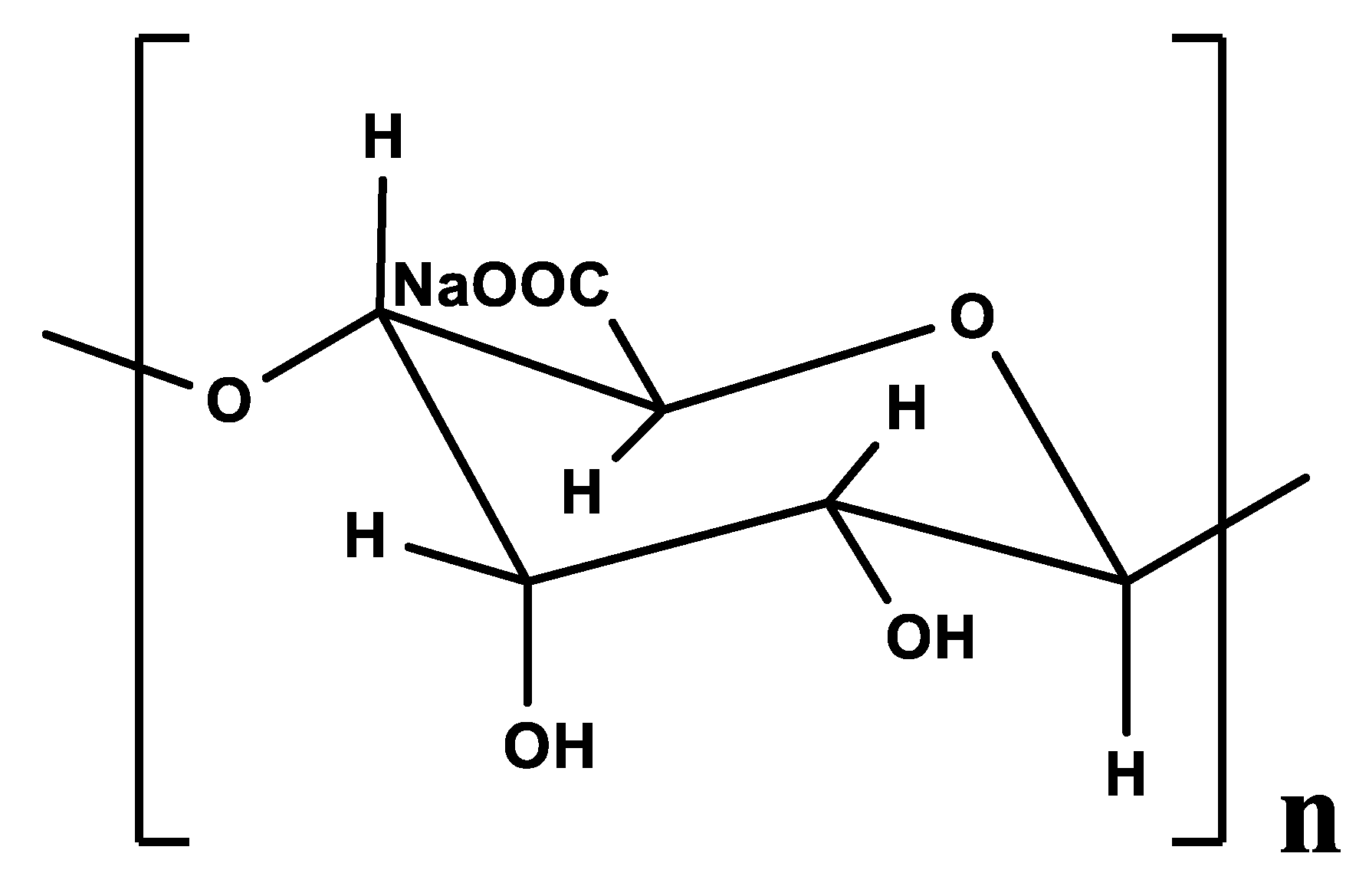
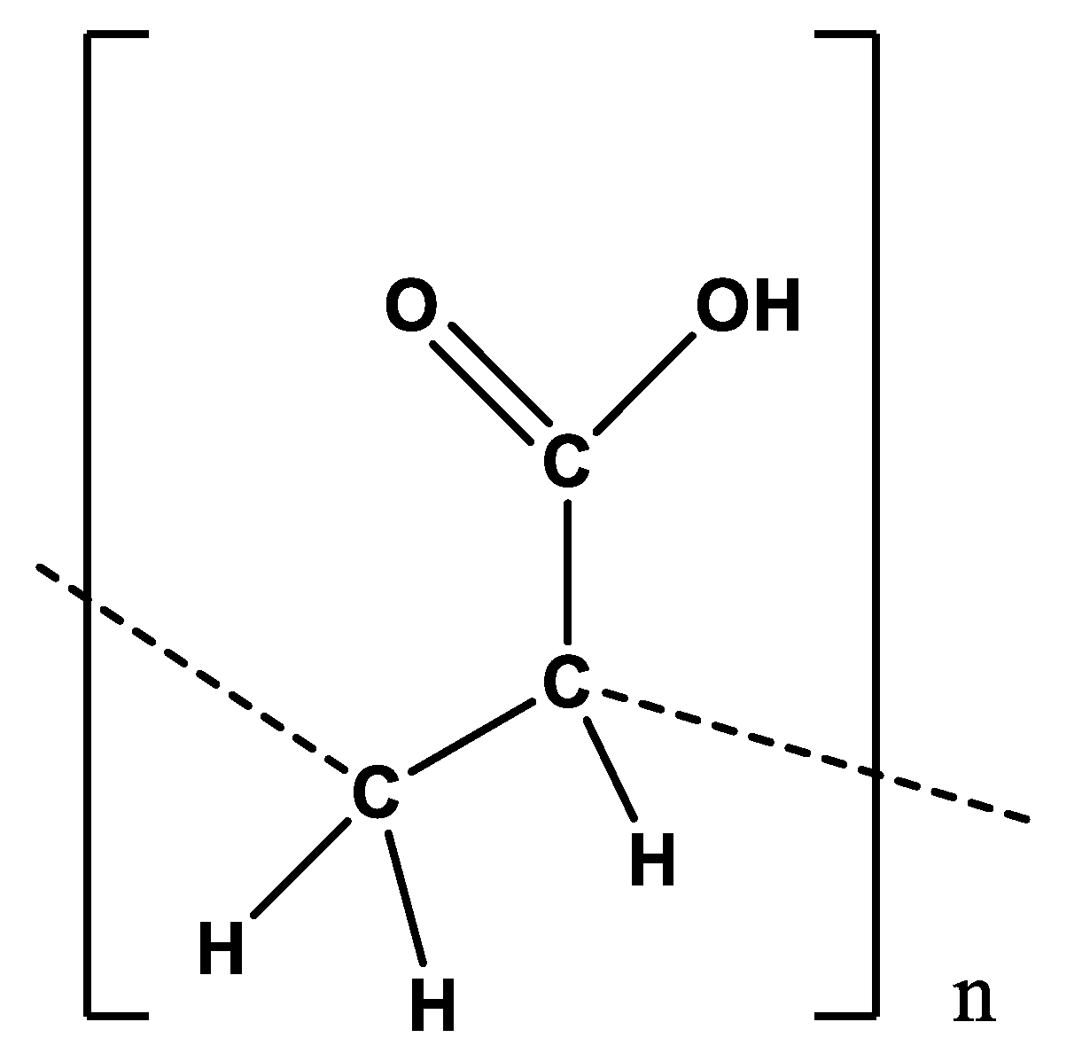
2.1. Preparation of the Crosslinked Polymeric Ligand (Poly-PAA/AG)

First, we vigorously stirred 100 mL of distilled water with 3 g of sodium alginate (AG) for an hour at 80 °C in a round bottom flask. Then, we added dropwise a polyacrylic acid (PAA) solution that had been previously made by combining 3 g of PAA with 100 mL of H2O, stirring continuously and heating at 70 °C. Under N2 pressure, the mixture was stirred at 120 °C for two hours. The reaction solution was then preserved at 90 °C overnight while being stirred. The resultant suspension was then brought to 25 °C before being concentrated at a lower pressure. The final step was to dry the obtained product at 65 °C while maintaining a high vacuum, producing a white color stable powder (90%). Anal. Calc. for C9H10O7: C, 46.97; H, 4.38. Found: C, 46.93; H, 4.98. IR data: ν(OH) 3222, ν(C-H) 2922, ν(C=O) 1725, ν(COO) 1413, ν(CO) 1263, ν(C-O-C) 1025.

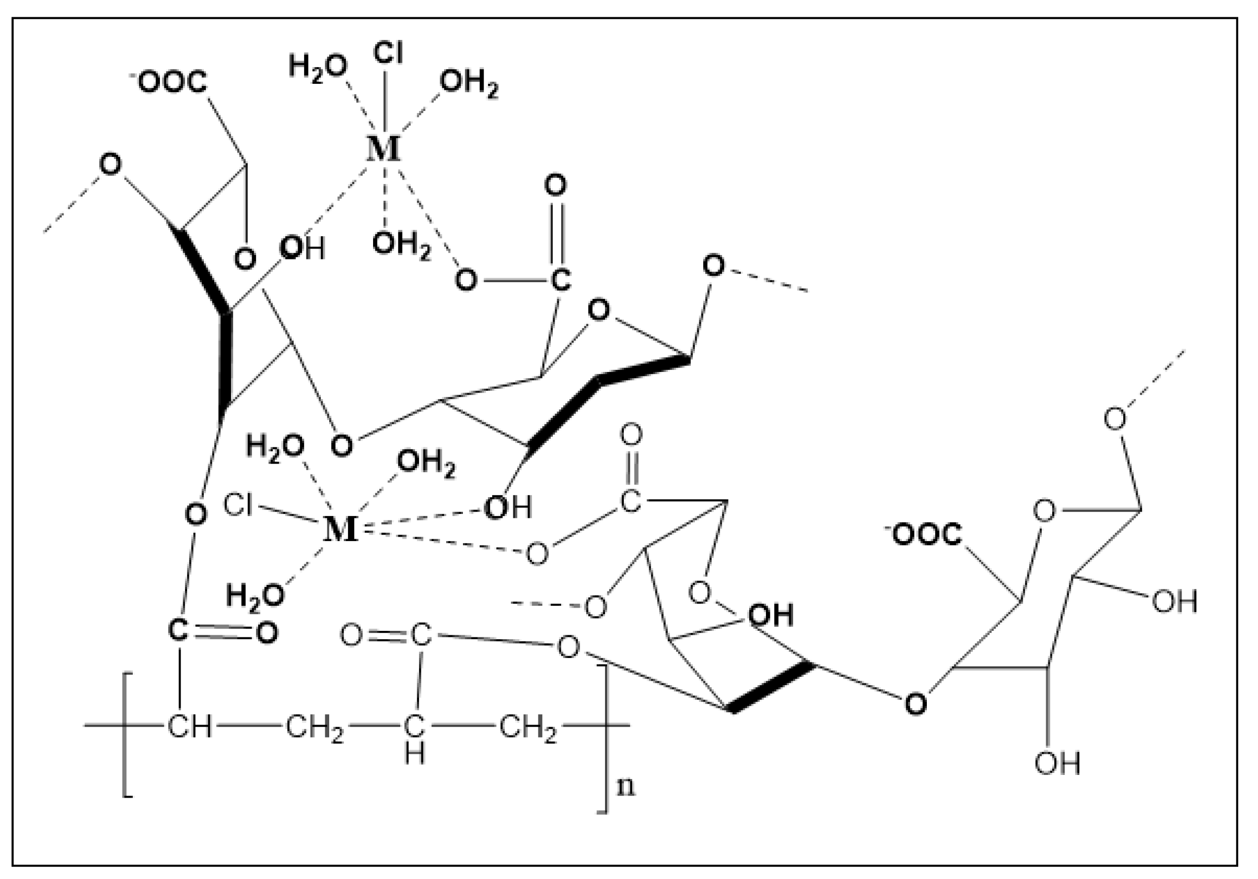
2.2. Synthesis of Metal Polymeric Complexes Nanoparticles

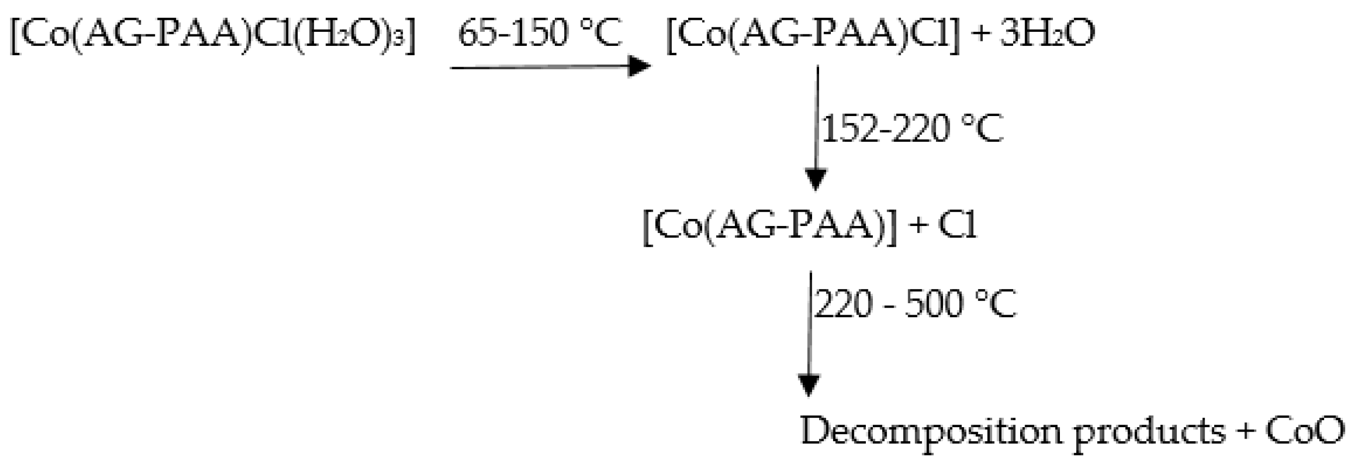
2.2.1. [Co(AG-PAA)Cl(H2O)3]

This compound was made by dissolving 1 g (0.2 mmol) of the (AG/PAA) ligand in 20 mL of methanol, boiling it for 5 min and then stirring while adding 20 mL of pure water until the ligand dispersed. We allowed the mixture to cool somewhat before adding 0.82 g (0.2 mmol) of the metal (CoCl2.6H2O), which had been dissolved in 10 mL of distilled water. Stirring continuously until the purple precipitate forms, it is then dried over CaCl2 in a desiccator. Anal. Calc. for C9H15CoClO10: C, 28.62; H, 4.00. Found: C, 29.06; H, 4.28. IR data: ν(OH) 3225, ν(C-H) 2930, ν(COO)sym 1715, ν(C=O) 1715, ν(COO)asym 1415, ν(CO) 1157, ν(C-O-C) 1022, ν(M-O) 519 cm−1, m.p. 230 °C and molar conductance 18.63 S cm2 mol−1.

2.2.2. [Cu(AG-PAA)Cl(H2O)3]

This complex was prepared by dissolving 0.5 g (0.1 mmol) of the (AG/PAA) ligand in 20 mL of methanol and heating it for 5 min before adding 20 mL of distilled water and heating and stirring until the ligand was dissolved. We allowed it to cool somewhat before adding 0.37 g (0.1 mmol) of the metal salt (CuCl2.2H2O) that was dissolved in 10 mL of distilled water. The resultant mixture was stirred, and then the mixture solution containing copper(II) complex was sonicated for 30 min; then, the green polycrystalline powder was obtained, filtered and then cooled to room temperature. The green precipitate was washed with distilled water and ethanol and then dried over CaCl2 in a desiccator. Anal. Calc. for C9H15CuClO10: C, 28.28; H, 3.95. Found: C, 28.61; H, 3.98. IR data: ν(OH) 3199, ν(C-H) 2932, ν(COO)sym 1703, ν(C=O) 1703, ν(COO)asym 1421, ν(CO) 1273, ν(C-O-C) 1026, ν(M-O) 582 cm−1, m.p. 190 °C and molar conductance 17.85 S cm2 mol−1.

2.2.3. [Mn(AG-PAA)Cl(H2O)3]

This compound was synthesized in 2 steps:
  • Step 1: In a beaker, 0.8 g (0.2 mmol) of AG-PAA was dissolved in 20 mL of methanol, and the mixture was heated for five minutes. Next, 20 mL of distilled water was added, and the mixture was stirred and heated for another 20 min.
  • Step 2: We added the metal solution (0.68 g, 0.2 mmol) of tetra-hydrated manganese chloride, which was dissolved in 10 mL of distilled water. After allowing it to cool down a bit, we stirred it until the light brown precipitate formed and then dried it in calcium chloride anhydrous. Anal. Calc. for C9H15MnClO10: C, 28.93; H, 4.04. Found: C, 29.10; H, 4.47. IR data: ν(OH) 3444, ν(C-H) 2931, ν(COO)sym 1696, ν(C=O) 1696, ν(COO)asym 1419, ν(CO) 1103, ν(C-O-C) 1027, ν(M-O) 523 cm−1, m.p. 210 °C and molar conductance 14.18 S cm2 mol−1.

2.2.4. [Ni(AG-PAA)Cl(H2O)3]

The 0.8 g of the (AG/PAA) ligand was dissolved in 20 mL of methanol, heated for 5 min, and stirred until the ligand was dissolved. Next, 20 mL of distilled H2O was added, and the mixture was heated and stirred for an additional minute. We allowed it to cool slightly before adding 0.65 g of the metal (NiCl2·6H2O), which was dissolved in 10 mL of distilled H2O. We continued stirring until the light green precipitate forms, after which it is dried in the calcium chloride anhydrous. Anal. Calc. for C9H15NiClO10: C, 28.64; H, 4.00. Found: C, 28.92; H, 4.33. IR data: ν(OH) 3216, ν(C-H) 2925, ν(COO)sym 1700, ν(C=O) 1700, ν(COO)asym 1414, ν(CO) 1164, ν(C-O-C) 1018, ν(M-O) 515 cm−1, m.p. 196 °C and molar conductance 19.15 S cm2 mol−1.

2.3. Physical Measurements

A Gmbh Vario El analyzer was used to determine the elemental analyzers. A Thermo Nicolet (6700), Fourier-transform infrared (FT-IR) spectrophotometer with a wavenumber range of (400–4000 cm−1) was used to collect structural data from FT-IR spectra. Using a Shimadzu (UV-2101) PC spectrophotometer, the UV–Vis spectra were collected. The sonication experiments were performed on sonicator type Q 700, 20 KHz, output 700 W. On a magnetic susceptibility balance of the kind (MSB-Auto), measurements of magnetic susceptibility were made. Using a conductivity meter made by JENWAY, model 4310, the complexes’ conductance was measured. On a Shimadzu (DTG 60-H) thermal analyzer heated to a rate of 10 °C per minute, thermal analysis of the polymeric complexes was performed in dynamic air. On an XRD diffractometer Model (PW 1720 Philips, Eindhoven, The Netherlands), measurements of the X-ray diffraction (XRD) were collected at room temperature with Cu-Kα radiation (λCu = 0.154059 Å). In the structural refinement procedure of our samples, we have followed the standard steps of the Rietveld method which consists of following the sequence:
(i)
Refinement of overall Scale factor + background coefficients (all other parameters are kept fixed);
(ii)
The same + refinement of detector zero offsets (or sample displacement in Bragg-Brentano geometry) + refinement of lattice parameters;
(iii)
The same + refinement of shape parameters + refinement of asymmetry parameters;
(iv)
The same + refinement of atomic positions + refinement of global DebyeWaller parameter or thermal agitation factors;
(v)
The same + refinement of site occupancy rate. In our refinements, we have taken care to respect this sequence of steps to release the different parameters. This ensures the stability of the refinement with all the parameters released.
By computing indicators such as the goodness of fit “χ2” and the R factors (Rwp = weighted profile R-factor, RB = Bragg factor and Rexp = expected R factor), the fitting quality of the experimental data is evaluated.
Using a scanning electron microscope, the morphology and structure of the prepared materials were examined.

2.4. Microbial Species and Culture Media

In this article, various metal polymeric complexes, such as Co(II), Cu(II), Mn(II) and Ni(II), were tested against Gram-positive (+) and Gram-negative (−) bacterial strains to gain insight into their broad-spectrum effect. Two Gram-positive strains, Micrococcus luteus NCIMB 8166 (S4) and Staphylococcus aureus ATCC 25923 (S1), and two Gram-negative strains, Salmonella thyphimurium ATCC 14028(S10) and Escherichia coli ATCC 35218 (S5), were the pathogenic strains that were used. A pathogenic reference strain of the yeast Candida albicans ATCC 90028 (9C) was used to test the antifungal activity. The strains were cultured on nutrient agar (Oxoid) for 24 h at 37 °C as well as in nutrient broth (Oxoid) for 24 h at 37 °C. For 24 h, the yeast strain was grown at 25 °C in Sabouraud Chloramphenicol broth (Oxoid) and grown for 24 h at 37 °C on Sabouraud Chloramphenicol agar (Oxoid). The different species are listed in Table 1.

2.5. Antimicrobial Activity

All complexes were tested for antimicrobial activity using the agar disk diffusion method. A total of 50 mg of each extract was dissolved in 1 mL of a 5% solution of dimethyl sulfoxide, or “DMSO,” prior to testing. The strains were cultured in Mueller–Hinton (MH) broth (Oxoid) at 37 °C for 24 h, and suspensions were calibrated to 0.5 McFarland standard turbidity. Afterward, 100 μL of each precultured suspension was spread onto plates containing MH agar. Sterile filter paper discs (6 mm in diameter) were impregnated with 20 μL of the different extracts and placed on agar. The treated plates were put for 1 h at 4 °C and then incubated for 24 h at 37 °C. After incubation, the diameter of the inhibition zone (clear halo) about the discs was measured. Each sample was tested in duplicate.

2.6. Antioxidant Assays

DPPH Radical Scavenging Assay

According to the method proposed by Mahdhi et al. [44], the free radical scavenging influence of the extracts was assessed using the next criteria: 1 mL of sample (5 mg/mL) was combined with 3 mL of DPPH (2,2-diphenyl-1-picrylhydrazyl) (300 µM) methanolic solution. The reaction mixture was vortexed and left to sit at 25 °C for 30 min. The solution’s absorbance was determined at 517 nm. The standard was ascorbic acid. The following Equation (1) was used to determine the inhibitory % of DPPH:
DPPH Scavenging effect (%) = [1 − (Abs sample/Abs control)] × 100

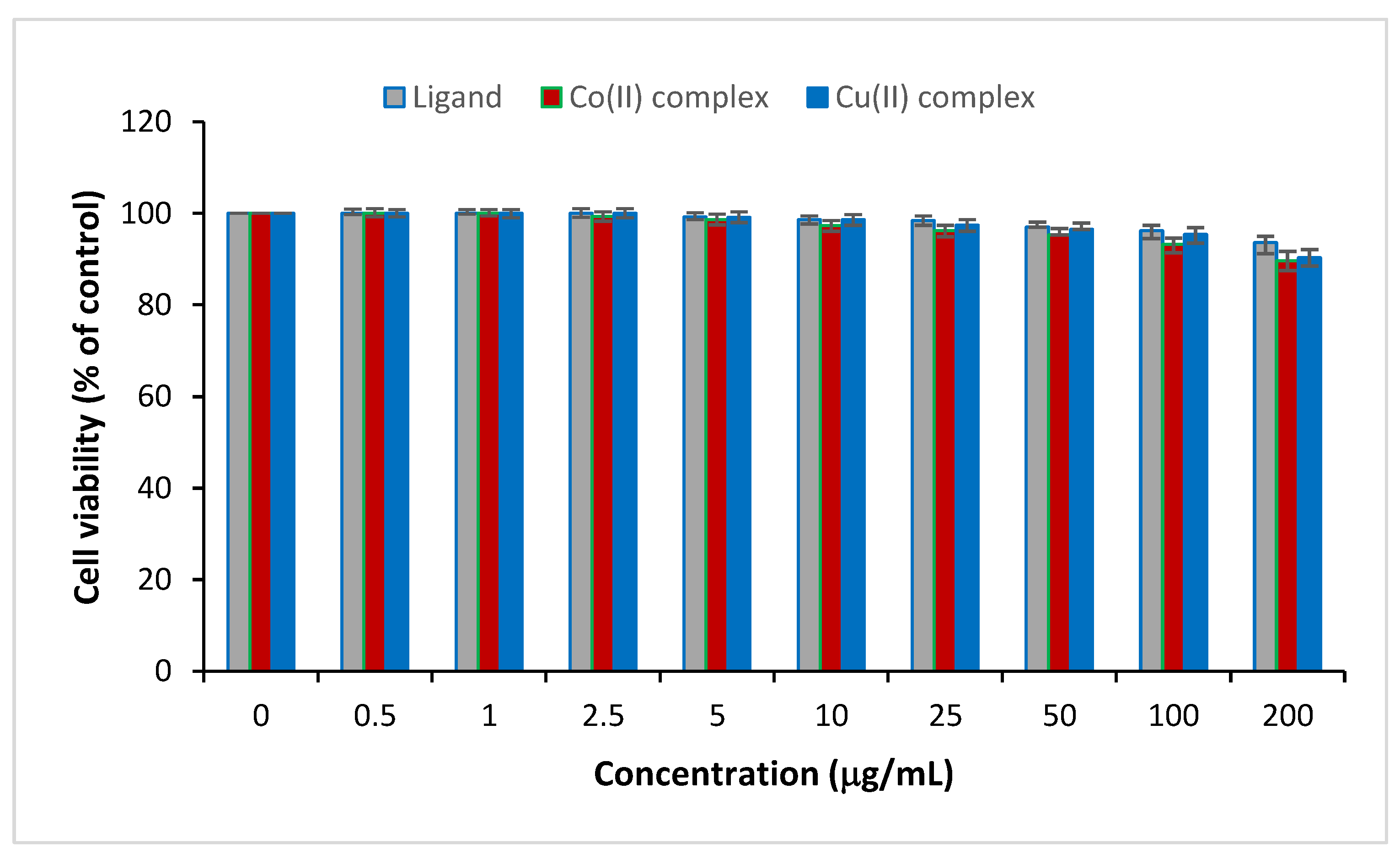
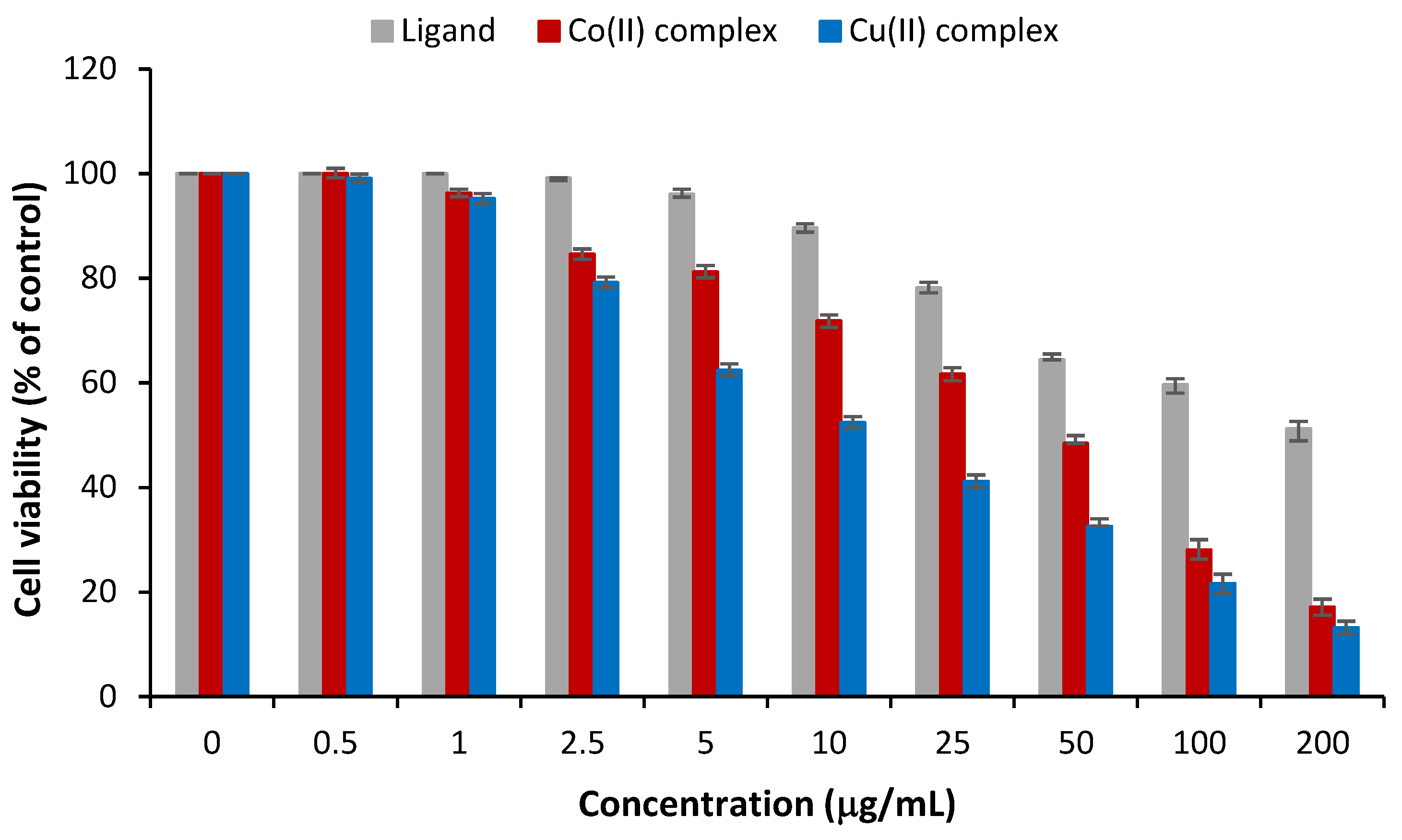
2.7. Cell Viability and Anticancer Assays

For the cell culture, human breast cancer cells (MCF-7) and human normal breast epithelial cells (MCF-10A) were obtained from American Type Culture Collection (Manassas, WV, USA). The cells were grown in Dulbecco Modified Eagle’s Medium with the addition of a synthesized solution of fetal bovine serum (10%) and a mixture of penicillin (100 IU/mL) and streptomycin (100 g/mL) as antibiotics. The medium was then incubated in a 5% CO2 atmosphere, 100% relative humidity at 37 °C. The anticancer activity and the cell viability assays were performed via the MTT tetrazolium standardized test with some modifications. This test is based on the ability of live cells to proliferate and thus metabolize and reduce the yellow MTT tetrazolium salt into purple formazan structured by a typical absorbance at 570 nm. The cultivated cells were seeded into 96-well plates at a density of 2104 cells/well. After 24 h of incubation, samples with different concentrations were added to each cell culture medium. After incubation for 72 h, we proceed with the MTT assay. Cell viability was assessed as the average % of relative formazan crystals formed taking into account the control culture. Tests were carried out in triplicate for each test.

3. Results and Discussion

The biopolymers were synthesized by the reaction of AG-PAA with Co(II), Cu(II), Mn(II) and Ni(II) chlorides (dissolved in MeOH and distilled water). The prepared compounds were found to react in the molar ratio of 1: 1: metal: AG-PAA. The polymeric complexes are air-stable and partially soluble in dimethyl sulphoxide. The electrical conductivity of the polymeric compounds adequately confirmed their non-electrolytic nature.

3.1. Elemental Analyses

The results of elemental analyses (carbon and hydrogen) of the ligand and its polymeric complexes along with the proposed molecular formula and physical characteristics were shown in the experimental section.

3.2. Molar Conductance

The molar conductivity of the polymeric complexes was measured in dimethylsulphoxide at 25 °C using 10−3 M solutions of polymeric compounds. All complexes had low conductivity readings, proving that they were nonionic, according to the results in Table 2.

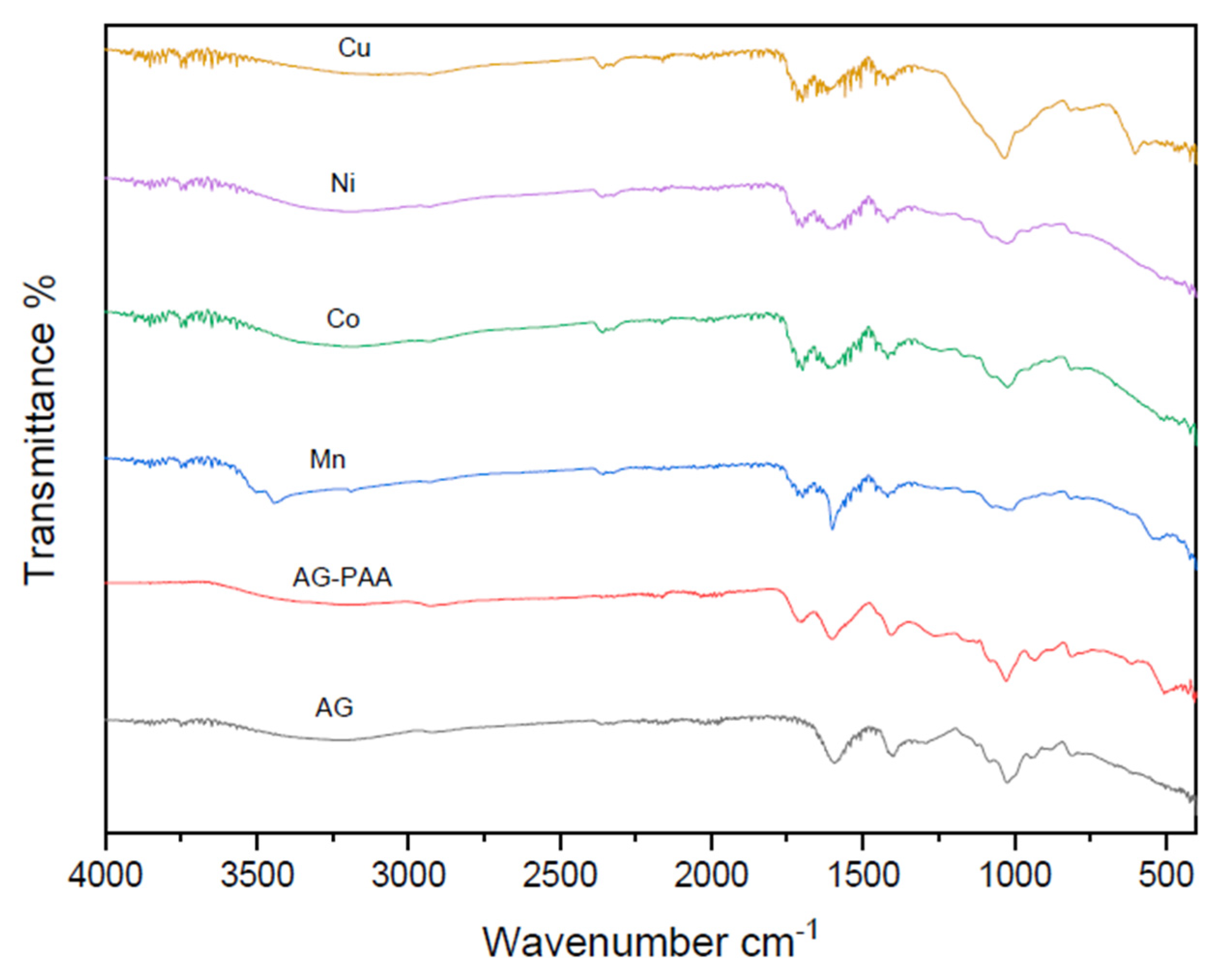
3.3. Fourier Transform Infrared Spectra

In order to pinpoint the functional groups of the components and comprehend the binding process, FT-IR spectrophotometric analysis was carried out. In this case, IR analysis was conducted to evaluate the preparation of the polymeric ligand (poly-PAA/AG) via the crosslinking process. However, this method would be crucial in establishing the existence of the various compounds between the created coordination polymer and the several metals examined in our study. Figure 3 displays the various spectra that were obtained. In relation to the synthesis of the polymeric ligand, we observe the appearance of a new band at about 1413 cm−1 indicating the fashioning of the ester group when comparing the spectra of the ligand and the alginate or polyacrylic acid biopolymer. This ester fashioning was between the COOH groups of the PAA polycarboxylic acid and the OH groups of the glycosidic moiety of the (AG) biopolymer. Therefore, we conclude that the polymeric ligand (poly-PAA/AG) was obtained throw a poly-esterification reaction between the alginate(AG) and the PAA crosslinking agent. The FT-IR spectrum of the (poly-PAA/AG) ligand showed several characteristic bands also existent in the spectra of the initial coordination polymers. Here, we notice the presence of a large band thereabout at 3222 cm−1 which is referred to as the hydroxyl group stretching (O-H). The (C-O-C) stretching vibration of the glycosidic structure was visible in a strong band near 1025 cm−1 [45]. The pyranose’s C-O-C glycosidic bonds are attributed to absorption bands with a wavelength between 1000 and 1100 cm−1. We observe some apparent shifts in the spectra of the compounds when compared to the polymeric ligand (poly-PAA/AG) alone, particularly at 3222 cm−1 (O-H), which suggests that the hydroxyl groups of the AG and the various metals have a coordinating binding (Table 2). Furthermore, for ν(COO)sym at about 1413 cm−1 and ν(COO)asym at around 1600 cm−1, we notice clear varied shifts with the used metal revealing the existence of interaction bounds between the different carboxylates of the AG and the various transition elements. In addition, at these wavenumber frequencies, the IR spectra of the complexes showed separation values Δυ (representing the difference between ν(COO)asym and ν(COO)sym) which are around 277–312 cm−1, pointing to a monodentate mode of coordination for the carboxylate group in a network of hydrogen bonds [46]. The IR spectra of the polymeric complexes show a band at 409–471 cm−1 allocated to (M-Cl) [47]. The weak bands at 409–471 cm−1 in the far IR regions were observed for complexes and are attributed to chloride anions coordinated in a trans-octahedral geometry. The existence of coordinated M-O is assured by the band at 515–582 cm−1 that appears with the four complexes [48]. We can deduce that the various functional groups of the prepared crosslinked coordination polymer, the poly-PAA/AG, which is mainly hydroxyl (OH), carboxylic (COOH) and ester groups, are able to coordinate with the different transition element ions and especially in a bidentate type.

3.4. Electronic Spectra

The electronic spectra of transition metal polymeric and their free ligands (poly-PAA/AG) are measured in the presence of DMSO (10−3 M). The outcomes show up in Table 3. The UV–Vis spectrum of (AG) peaks at 254 nm are appropriated to (π → π*) [49]. PAA displayed bands of absorbance at 270 nm appropriated to n → π* [50]. As for the electronic spectra of polymeric compounds for cobalt(II), copper(II), manganese(II) and nickel(II), it has special bands, in the visible zone of spectra assigned to d-d transition scopes at 492–580 nm. As for the bands identical to π → π* and n → π*, transitions were shown in the scopes of 258–277 nm and 273–298 nm, respectively, which indicates a bonding between the ligand (poly-PAA/AG) and the transition metals. The electronic spectrum of the Mn(II) compound in DMSO solution exhibited a peak around 510 nm assignable to the 6A1g4Eg, 4A1g transition. These data along with magnetic data are compatible with an octahedral structure around the transition metal ion [51]. The cobalt(II) polymeric complex displayed a weaker broad absorption band at 560 nm appointed to 4T1g (F) → 4T1g (P) transition of octahedral geometry [52]. The nickel(II) polymeric complex showed two bands located at 512 and 580 nm, which may be referred to as 3A2g(F) → 3T1g(P)(υ3) and 3A2g(F) → 3T1g(F)(υ2), which are characteristic of the nickel(II) ion, in an octahedral geometry [53]. The electronic spectrum of copper(II) showed a low-intensity broad band at 690 nm corresponding to the 2Eg2T2g transition, suggesting a distorted octahedral structure around Cu(II) [54].

3.5. Magnetic Moment Analysis

The determination of magnetic susceptibilities (µeff) of the polymeric compounds was carried out at 25 °C. The data of the effective magnetic moment (µeff) were specified by the data of diamagnetic corrections and Pascal’s constants according to Equation (2):
µeff = 2828(χMT)1/2 (in B.M.)
where µeff is the effective magnetic moment (in Bohr Magneton, B.M.), T is the temperature (K) and ᵡM is the molar magnetic susceptibility after correction. The value of µeff for the Mn(II) complex was 5.94 µB, which suggested an octahedral geometry, whereas, in the case of the Co(II) complex, the magnetic measurements illustrated a 4.82 µB value, which is sufficient for octahedral geometry [55,56]. As for the Ni(II) compound, magnetic measurements revealed that the compound has a µeff value of 2.88 µB, which is compatible with the range of the expected octahedral geometry of the nickel(II) compound [57]. Moreover, it has been found that the Cu(II) compound stabilizes in an octahedral geometry with a value of 1.74 µB [58].
The suggested structures for nano-sized Co(II), Cu(II), Mn(II) and Ni(II) coordination PAA/alginate biopolymers are shown in Figure 4.
Where M = Cobalt(II), copper(II), manganese(II) or nickel(II).

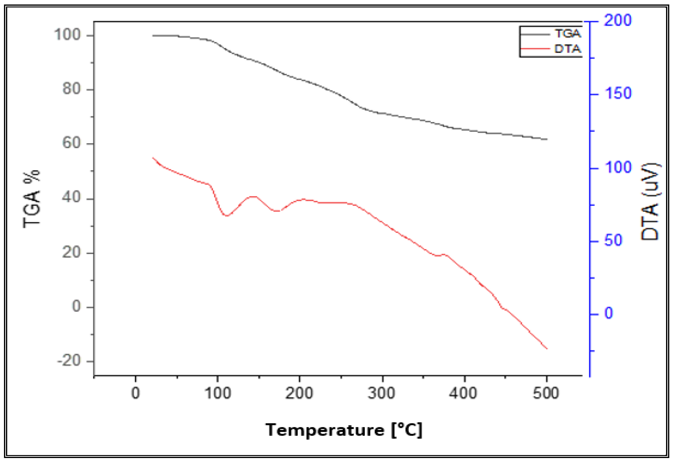
3.6. Thermal Analysis

Thermal analyses of the manufactured compounds were performed up to 500 °C. Using this method, complex composition, temperature stability and the presence or absence of water molecules (if any) inside or outside the compounds inner coordination sphere can all be determined. TGA thermograms of the synthesized ligand and transition metal polymeric complexes showed a gradual weight reduction, indicating disintegration by fragmentation as the temperature increased. The findings revealed a good agreement between the calculated data, and the suggested formulae for weight loss (Table 4).

3.6.1. [Co(AG-PAA)Cl(H2O)3] Complex

Four decomposition stages were observed for the thermolysis curve of the Co(II) polymeric complex. These occur in the temperature ranges 65–150, 152–220, 222–350 and 352–500 °C. The first mass loss correlates well with the release of three H2O molecules. This may be referred to as an ion-dipole interaction between cobalt and water. The corresponding mass loss was found to 13.94% (calc. 14.32%). For this step, a DTG midpoint appears at 92 °C with an endothermic peak in the DTA curve, at 94 °C. In the subsequent steps, the decomposition of the complex proceeds. The ultimate product was characterized as cobalt oxide (calc. 19.84%, found 17.18%). Scheme 1 depicts these decompositions.

3.6.2. [Cu(AG-PAA)Cl(H2O)3] Complex

The TG thermogram of [Cu(AG-PAA)Cl(H2O)3] exhibits four inflection points corresponding to four decomposition steps. Three water molecules are released in the first step, (calc. 14.15%, found 13.98%). For this step (thermal gravimetric analysis) (peak at 96 °C), an endothermic effect is observed at 98 °C in the differential thermal analysis trace. The observed mass loss in the 2nd step (120–205 °C) agrees well with the expected loss of chloride atoms (calc. 9.27%, found 9.18%) (DTG peak at 180 °C). This step is marked on the differential thermal analysis (DTA) curve as an exothermic peak at 182 °C. Then, the decomposition products are produced in the rest of the steps. The 3rd and 4th steps are consistent with the decomposition of rest products (calc. 60.20%, found 59.32%) (thermal gravimetric analysis) (peaks at 290 and 386 °C), for which exothermic peaks at 292 and 388 °C are recorded in the DTA trace. The residue was suggested to be CuO on the basis of mass loss consideration (calc. 20.80%, found 17.52%).

3.6.3. [Mn(AG-PAA)Cl(H2O)3] Complex

There were four distinct stages of decomposition visible in the Mn(II) complex thermograms. The first stage of decomposition occurs between 70 °C and 142 °C, where the loss of three H2O molecules results in a rated mass loss of 13.90% (calculated to be 14.46%) (thermal gravimetric analysis TG) (peak at 98 °C), for which a broad, endothermic peak manifests in the DTA curve at 100 °C. In the second stage, chloride atoms are lost with a mass loss of 9.12% percent (calculated as 9.48 percent) between 144 and 202 °C. At this stage, a DTG peak manifests at 182 °C, and a broad endothermic effect is listed in the differential thermal analysis trace at 184 °C. The third and fourth stages of decomposition took place at 204–295 °C and 297–500 °C, respectively, as a result of the remain of the organic ligand AG-PAA decomposing and forming the final product (MnO), which experienced mass losses of 59.88% (calculated at 61.59%) and 17.10% (calculated at 18.98%). These stages are manifested in the DTG curve as peaks at 244 and 384 °C, and the DTA trace furnishes a broad exothermic effect at 246 and 386 °C. (Figure 5).

3.6.4. [Ni(AG-PAA)Cl(H2O)3] Complex

The decomposition of [Ni(AG-PAA)Cl(H2O)3] proceeds in four distinct steps in the temperature ranges 64–138, 140–230, 232–368 and 370–500 °C. The 1st mass loss correlates well with the relief of three H2O molecules (calc. 14.31%, found 13.65%) (thermal gravimetric analysis) (peak at 98 °C) with an endothermic broad peak in the differential thermal analysis trace at 100 °C. The second mass loss is compatible with the expulsion of the chloride atom (calc. 9.39%, found 9.08%), A thermal gravimetric analysis peak at 202 °C and an exothermic effect at 204 °C in the differential thermal analysis trace are noticed. The third and fourth mass loss represents the release of the rest of organic (AG-PAA) ligands (calc. 60.98%, found 59.72%). The final product is consistent with NiO (calc. 19.79%, found 17.55%).

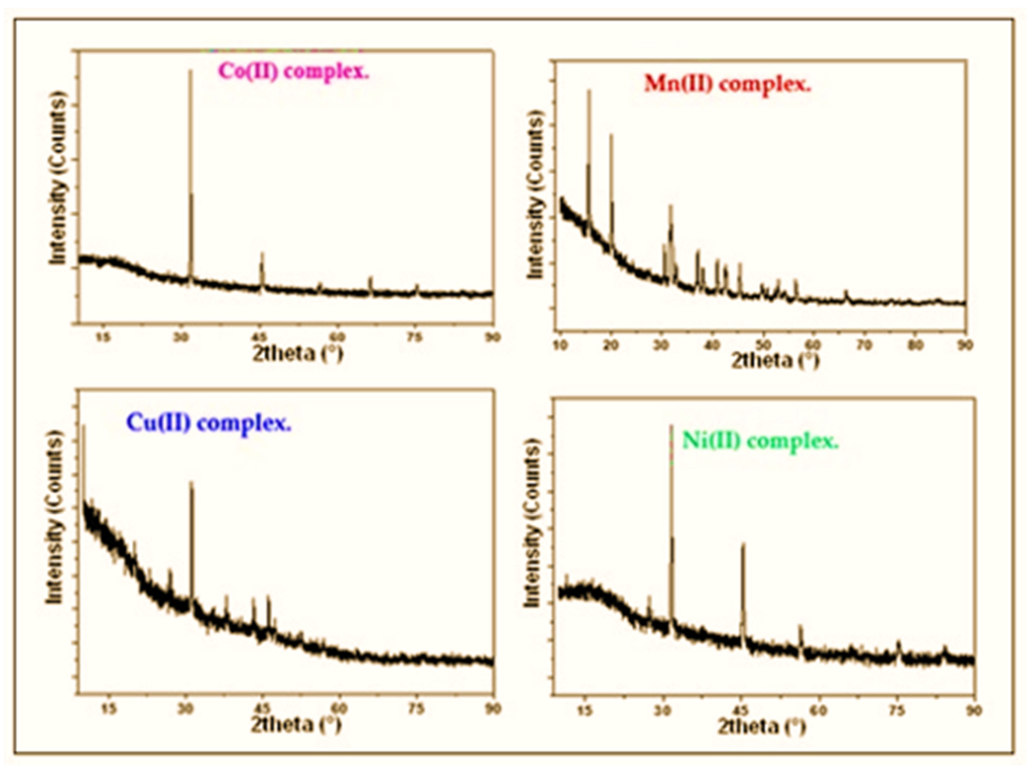
3.7. X-ray Powder Diffraction

This XRD study is another piece of proof for the emergence of metal–ligand complexes. The compounds’ XRD patterns were documented. Table 5 lists the crystal structures of various compounds. The comparison of the diffraction patterns of the ligands with the obtained complexes.
The XRD diffractogram of the ligand film indicates the presence of an amorphous structure, while the prepared Co(II), Cu(II), Mn(II) and Ni(II) compounds were crystalline. All polymeric complexes have sharp peaks in Figure 6. On the other hand, the Mn(II) compound has a monoclinic crystal system while the Co(II) compound has a cubic crystal system. The Ni(II) and Cu(II) compounds have a triclinic, and a hexagonal crystal system, respectively. The significant broadening of the peaks illustrates that the particles are of nm dimensions. Scherrer’s Equation (3) was used to estimate the particle size of the polymeric complexes.
D = Kλ/βcosθ
where K is the shape factor, λ is the X-ray wavelength typically 1.54 Å, β is the line broadening at half the maximum intensity in radians and θ is Bragg angle. D is the mean size of the ordered (crystalline) domains, which may be smaller or equal to the grain size. Scherrer’s equation is limited to nanoscale particles. The average size of the particles lies in the range of 14–68 nm which is in agreement with that noticed by scanning electron microscopy.

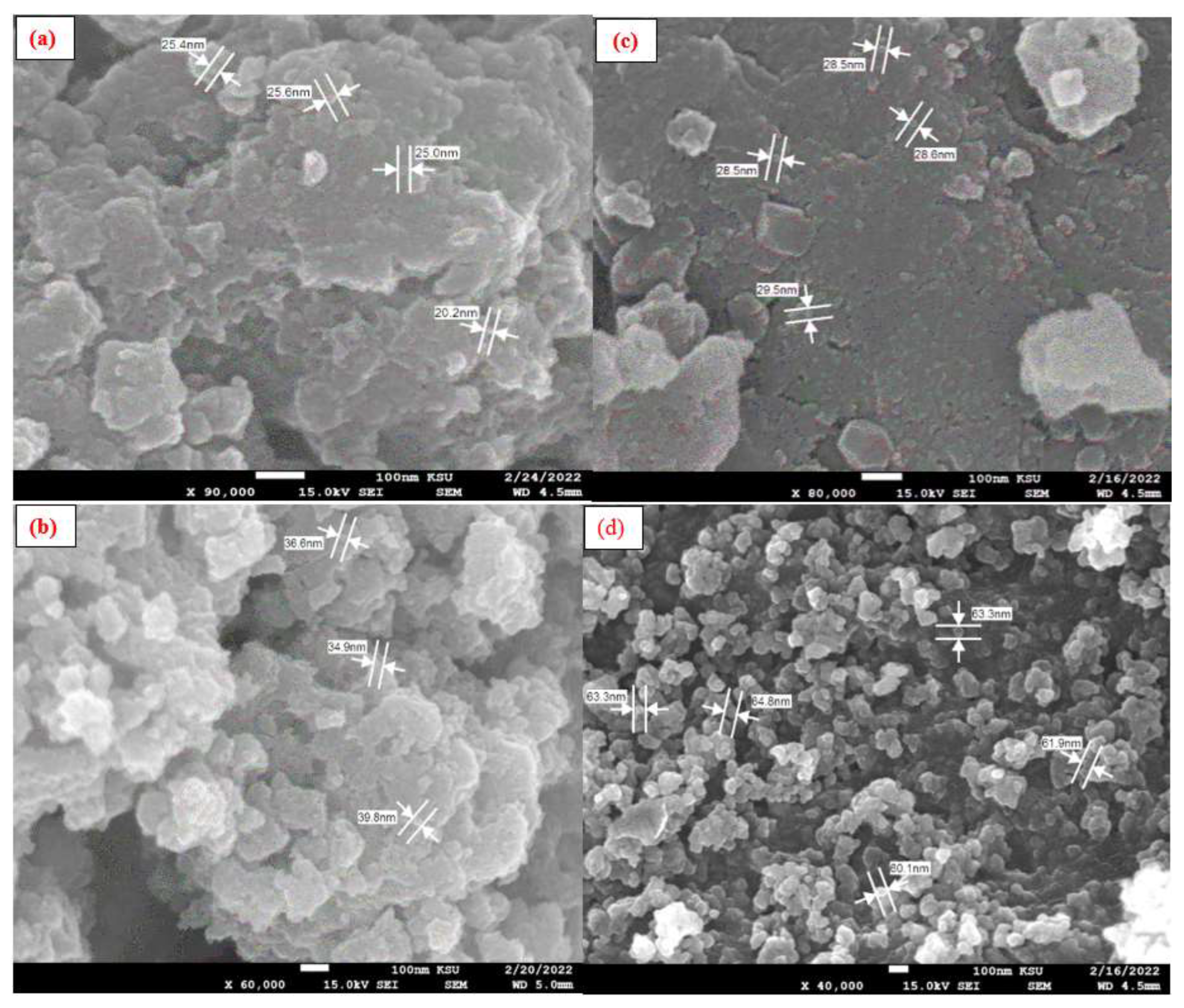
3.8. SEM Morphological Analysis

Electron microscopy (EM) has played a vital function in polymeric complexes properties and analysis. In addition to extremely more magnification levels, it can provide us with significant information containing composition and novel materials’ morphology. It can also show distinguishing contaminant materials that are only existent in polymers in trace amounts. Figure 7 displays as a general note an evident difference in surface morphology which is diverse depending on the type of transition element used in prepared polymeric complexes. The crosslinked polymeric ligand (poly-PAA/AG) revealed a porous and irregular surface reflecting its considerable hydrophilic property. This surface property could be efficient in the fashioning of ligand (poly-PAA/AG-metal coordination) polymers, and given that polymeric complexes are readily available in solution, continuous crosslinking into nanostructures is expected. While such particles have been used in the biomedical industry [59], their presence does support the aim to synthesize homogenous Co(II), Cu(II), Mn(II) and Ni(II) coordination PAA/alginate biopolymer NPs. A microscopic image of Mn(II) and Co(II) polymeric compounds illustrates small particles with a diameter between 20 and 29 nm. The surface morphology of the Ni(II) compound appears as uniform spherical particles with a diameter between 60 and 65 nm. In the Cu(II) complex, the SEM shows different shapes with diameters between 34 and 40 nm.

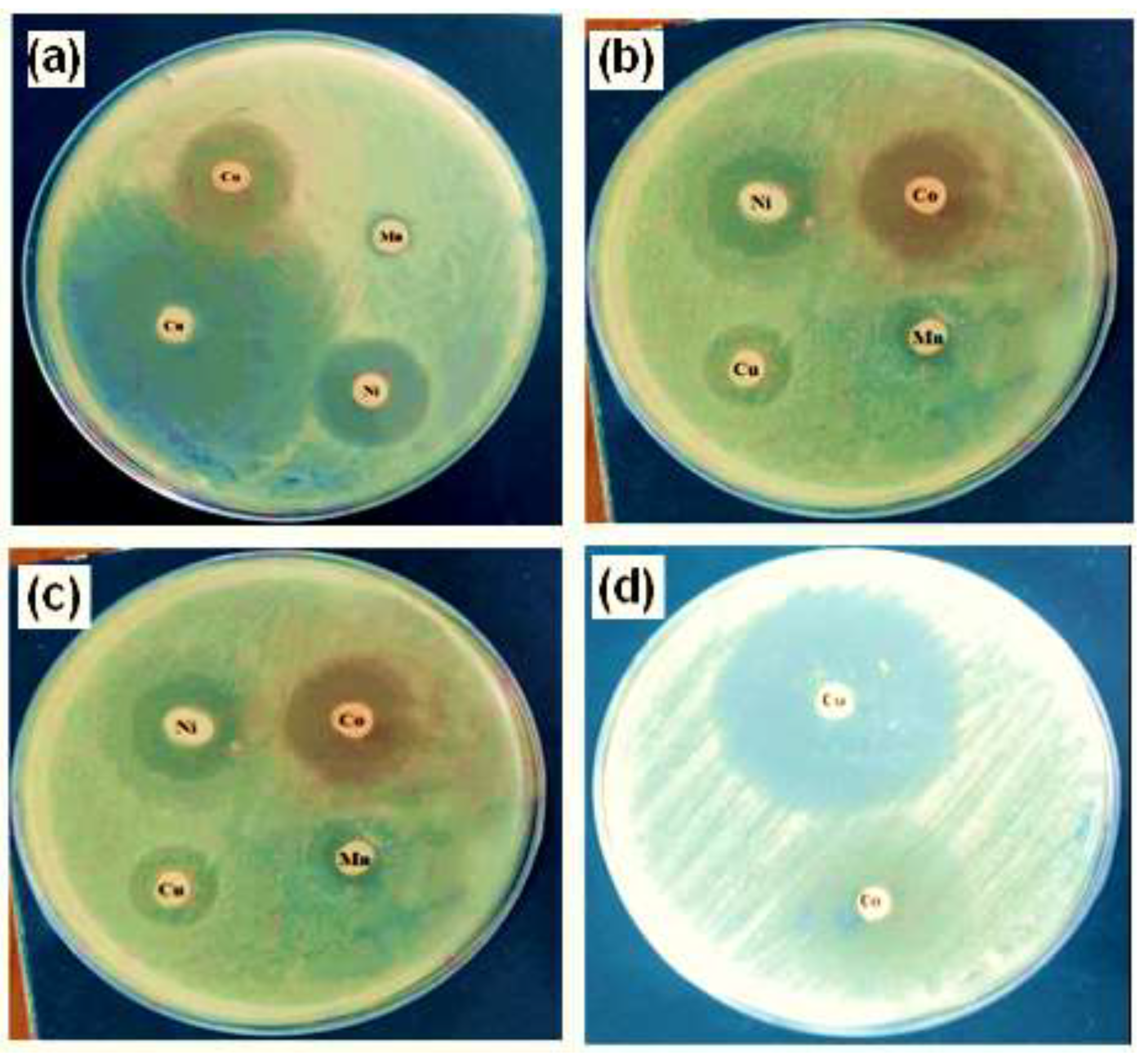
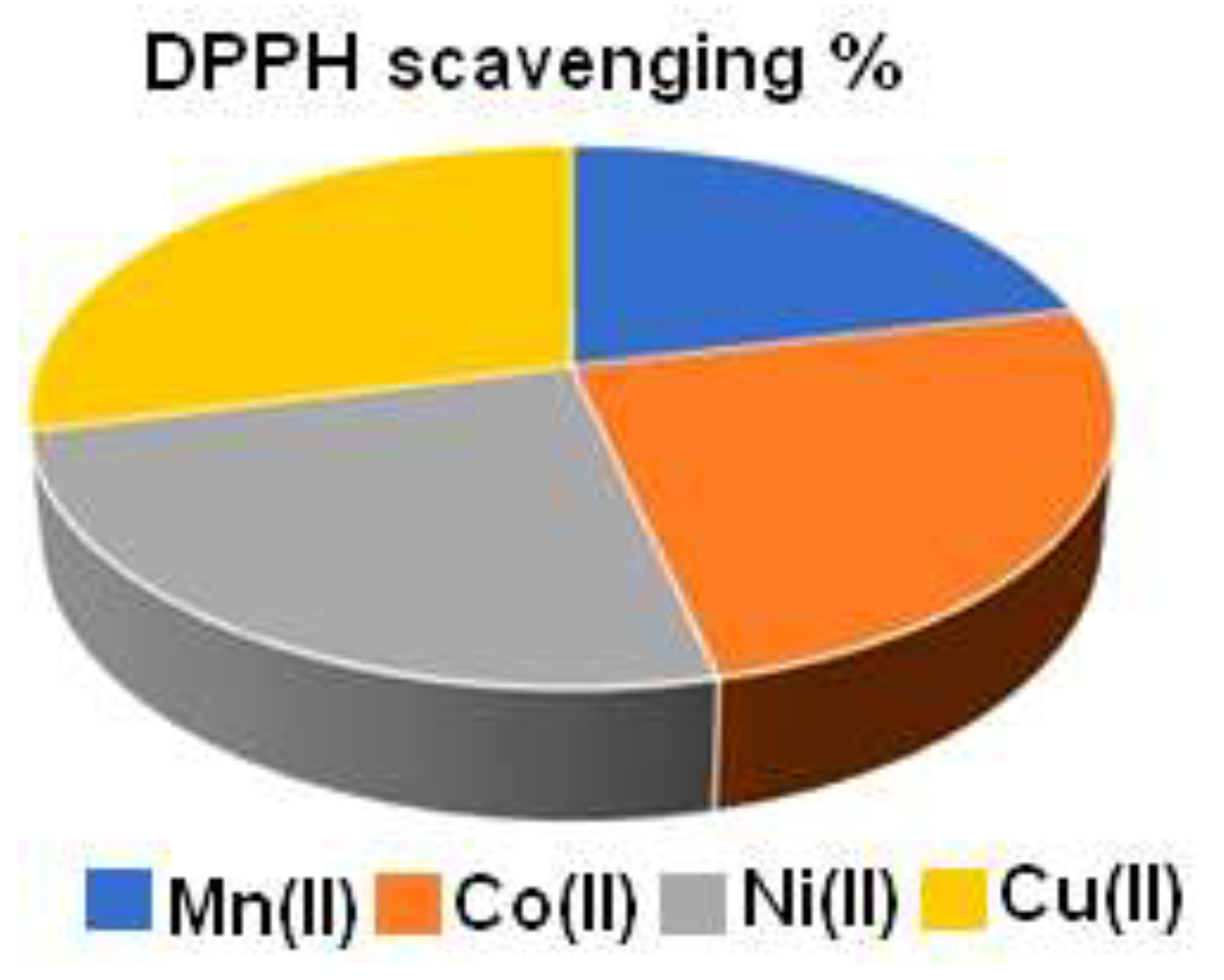
3.9. Antimicrobial and Antioxidant Assays

The test polymeric complex NPs exhibit a broad spectrum of antimicrobial activity because they are active against both Gram-positive and Gram-negative bacteria, but especially against Gram-positive strains, according to the antimicrobial results summarized in Table 6 and Figure 8a–d. The inhibitory zone diameter of the other extracts reached 5 cm, demonstrating their strong antimicrobial activity. The strongest are copper(II) and cobalt(II) compounds, which have a strain-dependent antimicrobial effect. The antioxidant effect of the different polymeric complexes is displayed in Figure 9.

3.10. In Vitro Anticancer Assay

In vitro, cell assays proceeded to investigate both the biocompatibility and anticancer activity of the prepared polymeric compounds. Human breast cancer cells (MCF-7) were used to assess the anticancer potential of the crosslinked polysaccharide ligand (poly-AG/PAA) and its two metal coordination polymers, the poly-AG/PAA/copper(II) and the poly-AG/PAA/cobalt(II). The assessment of the anticancer activity was carried out via the treatment of the cancer cells using the MTT assay by varying the concentration of the different polymeric vectors. The outcomes in Figure 10 displayed no cytotoxicity of the polymeric ligand in the approach with normal epithelial cells. This was reported previously by various research investigations that showed the excellent biocompatibility of the naturally extracted polysaccharides [60,61]. Therefore, the crosslinked polymeric ligand based on alginate biopolymer can be considered as a safe drug delivery carrier and other pharmaceutical applications. In addition, the synthesized polymeric compounds of Cu(II) and Co(II) revealed excellent cytocompatibility after 72 h in contact with the normal human breast epithelial cells(MCF10A), with the various applied concentrations. This was an important stage; meanwhile, the potential of anticancer drugs significantly concentrated on their biocompatibility with the normal counterparts of the analyzed cancer cells. The anticancer valuation in Figure 11 exhibited that the polymeric ligand without metals has the ability to kill cancer cells, and this is from a dose of 50 g/mL. The anticancer performance increases by sample concentration, to reach 51% at a concentration of 200 g/mL, which is in concordance with some previous studies [62].
Concerning the two selected polymeric complexes, results showed a good ability to kill the cancer cells in a dose-dependent method. The copper(II) polymeric compound confirms its excellent performance shown in microbial activity and demonstrates a higher anticancer potential compared to the Co(II) complex with the different polymeric doses. After a period of incubation fixed at 72 h and at a dose of 200 g/mL, the transition metal complexation significantly lowered the viability of the MCF-7 cancer cells up to 17.1 ± 1.21% and 13.2 ± 1.52% for the copper(II) and the cobalt(II) polymeric compounds, respectively. In summary, both the polymeric ligand (poly-AG/PAA) and the Cu(II) and Co(II) compounds exhibited good cytocompatibility with normal human epithelial cells. Furthermore, the in vitro anticancer evaluation showed the high anticancer performance of the Cu(II) and Co(II) polymeric compounds with a potential rising with the used polymeric concentration.

4. Conclusions

Polymeric complex NPs were prepared from polyacrylic acid (PAA) and sodium alginate (AG) with cobalt(II), copper(II), manganese(II) and nickel(II) chlorides. The various physico-chemical analyses included elemental analysis (C and H), FT-IR, UV–Vis spectra, TG, DTA, XRD and SEM. The magnetic studies suggested the octahedral geometrical structure for all produced polymeric complexes. Alginate and polyacrylic acid were used because they are biocompatible materials. As it is known, (PAA) gels are suitable biocompatible matrices for medical applications, such as gels for skin care products. PAA films can be deposited on orthopedic implants to protect them from corrosion. Crosslinked hydrogels of PAA and gelatin have also been used as in medical glue. In addition, Alginate (AG), which is rich in sources, is non-toxic and has excellent biocompatibility, biodegradability and safety, has been widely used in medical wound dressings, drug delivery carriers and delivery of bioactive substances in tissue engineering and skeleton materials. Therefore, nano-sized metallic polymers were prepared from these biopolymers, which really gave excellent results with the microbes that were tested, whether Gram-positive or Gram-negative bacteria or fungi. In summary, both the polymeric ligand (poly-AG/PAA) and the two metal complexes exhibited good cytocompatibility with normal human epithelial cells. Furthermore, the in vitro anticancer evaluation showed the high anticancer performance of the poly-AG/PAA/Cu(II) and poly-AG/PAA/Co(II) compounds with a potential rise with the used polymeric concentration.

Author Contributions

Data curation, M.S.A.-F., M.S.A. and Y.E.-G.; formal analysis, M.S.A.-F., M.S.A. and Y.E.-G.; investigation, M.S.A.-F., M.S.A. and Y.E.-G.; methodology, M.S.A.-F. and Y.E.-G.; project administration, M.S.A.-F.; software, M.S.A.-F. and M.S.A.; supervision, M.S.A.-F. and Y.E.-G.; validation, M.S.A.-F. and Y.E.-G.; writing—original draft, M.S.A.-F. and M.S.A.; writing—review and editing, M.S.A.-F. All authors have read and agreed to the published version of the manuscript.

Funding

This research received no external funding.

Data Availability Statement

The data are contained within the article.

Conflicts of Interest

The authors declare no conflict of interest.

References

  1. Ezeoha, S.L. Production of Biodegradable Plastic Packaging Film from Cassava Starch. IOSR J. Eng. 2013, 3, 14–20. [Google Scholar] [CrossRef]
  2. Rebelo, R.; Fernandes, M.; Fangueiro, R. Biopolymers in medical implants: A brief review. Procedia Eng. 2017, 200, 236–243. [Google Scholar] [CrossRef]
  3. Bala, I.A.; Abdullahi, M.R.; Bashir, S.S. A review on formulation of enzymatic solution for biopolymer hydrolysis. J. Chem. 2017, 6, 9–13. [Google Scholar]
  4. Pattanashetti, N.A.; Heggannavar, G.B.; Kariduraganavar, M.Y. Smart biopolymers and their biomedical applications. Procedia Manuf. 2017, 12, 263–279. [Google Scholar] [CrossRef]
  5. Qasim, U.; Osman, A.I.; Al-Muhtaseb, A.H.; Farrell, C.; Al-Abri, M.; Ali, M.; Vo, D.V.N.; Jamil, F.; Rooney, D.W. Renewable cellulosic nanocomposites for food packaging to avoid fossil fuel plastic pollution: A review. Environ. Chem. Lett. 2021, 19, 613–641. [Google Scholar] [CrossRef]
  6. Nessi, V.; Falourd, X.; Maigret, J.-E.; Cahier, K.; D’Orlando, A.; Descamps, N.; Gaucher, V.; Chevigny, C.; Lourdin, D. Cellulose nanocrystals-starch nanocomposites produced by extrusion: Structure and behavior in physiological conditions. Carbohydr. Polym. 2019, 225, 115123. [Google Scholar] [CrossRef]
  7. Barclay, T.G.; Day, C.M.; Petrovsky, N.; Garg, S. Review of polysaccharide particle-based functional drug delivery. Carbohydr. Polym. 2019, 221, 94–112. [Google Scholar] [CrossRef]
  8. Reis, A.V.; Guilherme, M.R.; Cavalcanti, O.A.; Rubira, A.F.; Muniz, E.C. Synthesis and characterization of pH-responsive hydrogels based on chemically modified Arabic gum polysaccharide. Polymer 2006, 47, 2023–2029. [Google Scholar] [CrossRef]
  9. Farooqi, Z.U.R.; Qadeer, A.; Hussain, M.M.; Zeeshan, N.; Ilic, P. Characterization and physicochemical properties of nanomaterials. In Nanomaterials: Synthesis, Characterization, Hazards and Safety; Elsevier: Amsterdam, The Netherlands, 2021; pp. 97–121. [Google Scholar] [CrossRef]
  10. Salem, S.S.; Fouda, A. Green Synthesis of Metallic Nanoparticles and Their Prospective Biotechnological Applications: An Overview. Biol. Trace Elem. Res. 2021, 199, 344–370. [Google Scholar] [CrossRef]
  11. Nikalje, A.P. Nanotechnology and its applications in medicine. Med. Chem. 2015, 5, 081–089. [Google Scholar] [CrossRef]
  12. Barik, T.K.; Maity, G.C.; Gupta, P.; Mohan, L.; Santra, T.S. Nanomaterials: An Introduction. Nanomater. Their Biomed. Appl. 2021, 16, 1–11. [Google Scholar]
  13. Singh, B.K.; Lee, S.; Na, K. An overview on metal-related catalysts: Metal oxides, nanoporous metals and supported metal nanoparticles on metal organic frameworks and zeolites. Rare Metals 2020, 39, 751–766. [Google Scholar] [CrossRef]
  14. Salem, S.S.; Fouda, M.M.G.; Fouda, A.; Awad, M.A.; Al-Olayan, E.M.; Allam, A.A.; Shaheen, T.I. Antibacterial, Cytotoxicity and Larvicidal Activity of Green Synthesized Selenium Nanoparticles Using Penicillium corylophilum. J. Clust. Sci. 2021, 32, 351–361. [Google Scholar] [CrossRef]
  15. Khan, S.; Mansoor, S.; Rafi, Z.; Kumari, B.; Shoaib, A.; Saeed, M.; Alshehri, S.; Ghoneim, M.M.; Rahamathulla, M.; Hani, U.; et al. A review on nanotechnology: Properties, applications, and mechanistic insights of cellular uptake mechanisms. J. Mol. Liquids 2021, 347, 118008. [Google Scholar] [CrossRef]
  16. Pérez-Hernández, H.; Pérez-Moreno, A.; Sarabia-Castillo, C.; García-Mayagoitia, S.; Medina-Pérez, G.; López-Valdez, F.; Campos-Montiel, R.; Jayanta-Kumar, P.; Fernández-Luqueño, F. Ecological Drawbacks of Nanomaterials Produced on an Industrial Scale: Collateral Effect on Human and Environmental Health. Water Air Soil Pollut. 2021, 232, 435. [Google Scholar] [CrossRef] [PubMed]
  17. Singh, R.; Singh, S. Nanomanipulation of consumer goods: Effects on human health and environment. In Nanotechnology in Modern Animal Biotechnology; Springer: Berlin/Heidelberg, Germany, 2019; pp. 221–254. [Google Scholar] [CrossRef]
  18. Pathakoti, K.; Goodla, L.; Manubolu, M.; Hwang, H.-M. Nanoparticles and Their Potential Applications in Agriculture, Biological Therapies, Food, Biomedical, and Pharmaceutical Industry: A Review. In Nanotechnology and Nanomaterial Applications in Food, Health, and Biomedical Sciences; Apple Academic Press: Palm Bay, FL, USA, 2019; pp. 121–162. ISBN 9780429425660. [Google Scholar]
  19. Elkodous, M.A.; El-Husseiny, H.M.; El-Sayyad, G.S.; Hashem, A.H.; Doghish, A.S.; Elfadil, D.; Radwan, Y.; El-Zeiny, H.M.; Bedair, H.; Ikhdair, O.A.; et al. Recent advances in waste-recycled nanomaterials for biomedical applications: Waste-to-wealth. Nanotechnol. Rev. 2021, 10, 1662–1739. [Google Scholar] [CrossRef]
  20. Parker, G. Measuring the environmental performance of food packaging: Life cycle assessment. In Environmentally Compatible Food Packaging; Elsevier: Amsterdam, The Netherlands, 2008; pp. 211–237. [Google Scholar]
  21. Shankar, S.; Rhim, J.-W. Bionanocomposite films for food packaging applications. In Reference Module in Food Science; Elsevier: Amsterdam, The Netherlands, 2018; pp. 1–10. [Google Scholar]
  22. Hassan, M.E.; Bai, J.; Dou, D.Q. Biopolymers; Definition, classification and applications. Egypt. J. Chem. 2019, 62, 1725–1737. [Google Scholar] [CrossRef]
  23. Soldo, A.; Mileti’c, M.; Auad, M.L. Biopolymers as a sustainable solution for the enhancement of soil mechanical properties. Sci. Rep. 2020, 10, 267. [Google Scholar] [CrossRef] [Green Version]
  24. Song, J.; Winkeljann, B.; Lieleg, O. Biopolymer-based coatings: Promising strategies to improve the biocompatibility and functionality of materials used in biomedical engineering. Adv. Mater. Interfaces 2020, 7, 2000850. [Google Scholar] [CrossRef]
  25. Jummaat, F.; Yahya, E.B.; Khalil H.P.S., A.; Adnan, A.S.; Alqadhi, A.M.; Abdullah, C.K.; A.K., A.S.; Olaiya, N.G.; Abdat, M. The Role of Biopolymer-Based Materials in Obstetrics and Gynecology Applications: A Review. Polymers 2021, 13, 633. [Google Scholar] [CrossRef] [PubMed]
  26. Reddy, M.S.B.; Ponnamma, D.; Choudhary, R.; Sadasivuni, K.K. A Comparative Review of Natural and Synthetic Biopolymer Composite Scaffolds. Polymers 2021, 13, 1105. [Google Scholar] [CrossRef]
  27. Olivia, M.; Jingga, H.; Toni, N.; Wibisono, G. Biopolymers to improve physical properties and leaching characteristics of mortar and concrete: A review. In IOP Conference Series: Materials Science and Engineering; Institute of Physics Publishing: Bristol, UK, 2018; Volume 345, p. 012028. [Google Scholar]
  28. Darge, H.F.; Andrgie, A.T.; Tsai, H.C.; Lai, J.Y. Polysaccharide and polypeptide based injectable thermo-sensitive hydrogels for local biomedical applications. Int. J. Biol. Macromol. 2019, 133, 545–563. [Google Scholar] [CrossRef]
  29. Mohammed, A.S.A.; Naveed, M.; Jost, N. Polysaccharides; classification, chemical properties, and future perspective applications in fields of pharmacology and biological medicine (a review of current applications and upcoming potentialities). J. Polym. Environ. 2021, 29, 2359–2371. [Google Scholar] [CrossRef]
  30. Grasdalen, H.; Larsen, B.; Smidsrød, O. 13C-NMR studies of alginate. Carbohydr. Res. 1977, 56, C11–C15. [Google Scholar] [CrossRef]
  31. Grasdalen, H.; Larsen, B.; Smidsrød, O. A pmr study of the composition and sequence of uronate residues in alginates. Carbohydr. Res. 1979, 68, 23–31. [Google Scholar] [CrossRef]
  32. Kothale, D.; Verma, U.; Dewangan, N.; Jana, P.; Jain, A.; Jain, D. Alginate as Promising Natural Polymer for Pharmaceutical, Food, and Biomedical Applications. Curr. Drug Deliv. 2020, 17, 755–775. [Google Scholar] [CrossRef]
  33. EL-Ghoul, Y.; Al-Fakeh, M.S.; Al-Subai, N. Synthesis and Characterization of a New Alginate/Carrageenan crosslinked biopolymer and Study of the Antibacterial, Antioxidant, and Anticancer Performance of its Mn(II), Fe(III), Ni(II), and Cu(II) polymeric complexes. Polymers 2023, 15, 2511. [Google Scholar] [CrossRef]
  34. da Silva, T.L.; Vidart, J.M.M.; da Silva, M.G.C.; Gimenes, M.L.; Vieira, M.G.A. Alginate and sericin: Environmental and pharmaceutical applications. Biol. Act. Appl. Mar. Polysacch. 2017, 57–86. [Google Scholar] [CrossRef] [Green Version]
  35. Draget, K.I.; Taylor, C. Chemical, physical and biological properties of alginates and their biomedical implications. Food Hydrocolloids 2011, 25, 251–256. [Google Scholar] [CrossRef]
  36. Gheorghita Puscaselu, R.; Lobiuc, A.; Dimian, M.; Covasa, M. Alginate: From food industry to biomedical applications and management of metabolic disorders. Polymers 2020, 12, 2417. [Google Scholar] [CrossRef]
  37. Orwoll, R.A.; Chong, Y.S. Poly(acrylic acid). In Polymer Data Handbook; James, M., Ed.; Oxford University Press: Oxford, UK, 1999; pp. 252–253. [Google Scholar]
  38. Zhuo, W. Synthesis, Characterization, and Self-Assembly of Amphiphilic Copolymer Based on Poly (Acrylic Acid); National University of Singapore: Singapore, 2015. [Google Scholar]
  39. Arkaban, H.; Barani, M.; Akbarizadeh, M.R.; Pal Singh Chauhan, N.; Jadoun, S.; Dehghani Soltani, M.; Zarrintaj, P. Polyacrylic Acid Nanoplatforms: Antimicrobial, Tissue Engineering, and Cancer Theranostic Applications. Polymers 2022, 14, 1259. [Google Scholar] [CrossRef] [PubMed]
  40. Xu, M.; Zhu, J.; Wang, F.; Xiong, Y.; Wu, Y.; Wang, Q.; Weng, J.; Zhang, Z.; Chen, W.; Liu, S. Improved in vitro and in vivo biocompatibility of graphene oxide through surface modification: Poly (acrylic acid)-functionalization is superior to PEGylation. ACS Nano 2016, 10, 3267–3281. [Google Scholar] [CrossRef] [PubMed]
  41. Mahon, R.; Balogun, Y.; Oluyemi, G.; Njuguna, J. Swelling performance of sodium polyacrylate and poly(acrylamide-co-acrylic acid) potassium salt. SN Appl. Sci. 2020, 2, 117–132. [Google Scholar] [CrossRef] [Green Version]
  42. Dumitrașcu, A.-M.; Caraș, I.; Țucureanu, C.; Ermeneanu, A.-L.; Tofan, V.-C. Nickel (II) and Cobalt (II) Alginate Biopolymers as a “Carry and Release” Platform for Polyhistidine-Tagged Proteins. Gels 2022, 8, 66. [Google Scholar] [CrossRef] [PubMed]
  43. 43. Sharafshadeh, M.S.; Tafvizi, F.; Khodarahmi, P.; Ehtesham, S. Preparation and physicochemical properties of cisplatin and doxorubicin encapsulated by niosome alginate nanocarrier for cancer therapy. Int. J. Biol. Macromol. 2023, 235, 123686. [Google Scholar] [CrossRef]
  44. Mahdhi, A.; Leban, N.; Chakroun, I.; Chaouch, M.A.; Hafsa, J.; Fdhila, K.; Mahdouani, K.; Majdoub, H. Extracellular polysaccharide derived from potential probiotic strain with antioxidant and antibacterial activities as a prebiotic agent to control pathogenic bacterial biofilm formation. Microb. Pathogenesis 2017, 109, 214–220. [Google Scholar] [CrossRef]
  45. Ammar, C.; Alminderej, F.M.; EL-Ghoul, Y.; Jabli, M.; Shafiquzzaman, M. Preparation and Characterization of a New Polymeric Multi-Layered Material Based K-Carrageenan and Alginate for Efficient Bio-Sorption of Methylene Blue Dye. Polymers 2021, 13, 411. [Google Scholar] [CrossRef]
  46. Deacon, G.B.; Phillips, R.J. Relationships between the carbon-oxygen stretching frequencies of carboxylato complexes and the type of carboxylate coordination. Coord. Chem. Rev. 1980, 33, 227–250. [Google Scholar] [CrossRef]
  47. Al-Fakeh, M.S.; Alsikhan, M.A.; Alnawmasi, J.S. Physico-Chemical Study of Mn(II), Co(II), Cu(II), Cr(III), and Pd(II) Complexes with Schiff-Base and Aminopyrimidyl Derivatives and Anti-Cancer, Antioxidant, Antimicrobial Applications. Molecules 2023, 28, 2555. [Google Scholar] [CrossRef]
  48. Al-Fakeh, M.S.; Messaoudi, S.; Alresheedi, F.I.; Albadri, A.E.; El-Sayed, W.A.; Saleh, E.E. Preparation, Characterization, DFT Calculations, Antibacterial and Molecular Docking Study of Co(II), Cu(II), and Zn(II) Mixed Ligand Complexes. Crystals 2023, 13, 118. [Google Scholar] [CrossRef]
  49. Yue, W.; Zhang, H.H.; Yang, Z.N.; Xie, Y. Preparation of low-molecular-weight sodium alginate by ozonation. Carbohydr. Polym. 2021, 251, 117104. [Google Scholar] [CrossRef] [PubMed]
  50. Vetriselvi, V.; Santhi Raj, R.J.J. Synthesis and characterization of poly acrylic acid modified with dihydroxy benzene-redox polymer. Res. J. Chem. Sci. 2014, 4, 78–86. [Google Scholar]
  51. Parihari, R.K.; Patel, R.K.; Patel, R.N. Synthesis and characterization of metal complexes of manganese-, cobalt-and zinc (II) with Schiff base and some neutral ligand. J. Indian Chem. Soc. 2000, 77, 339. [Google Scholar]
  52. Mohanan, K.; Murukan, B. Complexes of manganese (II), iron (II), cobalt (II), nickel (II), copper (II), and zinc (II) with a bishydrazone. Synth. React. Inorg. Met. Org. Nano Met. Chemistry 2005, 35, 837–844. [Google Scholar] [CrossRef]
  53. Gupta, L.K.; Bansal, U.; Chandra, S. Spectroscopic and physicochemical studies on nickel(II) complexes of isatin-3,2′-quinolylhydrazones and their adducts. Spectrochim. Acta Part A Mol. Biomol. Spectro. 2007, 66, 972–975. [Google Scholar] [CrossRef]
  54. Ajaykumar, D. Kulkarni, et, Synthesis, spectral, electrochemical and biological studies of Co(II), Ni(II) and Cu(II) complexes with Schiff bases of 8-formyl-7-hydroxy-4-methyl coumarin. J. Coord. Chem. 2009, 62, 481–492. [Google Scholar]
  55. Al-Fakeh, M.S.; Allazzam, G.A.; Yarkandi, N.H. Ni (II), Cu (II), Mn (II), and Fe (II) Metal Complexes Containing 1, 3-Bis (diphenylphosphino) propane and Pyridine Derivative: Synthesis, Characterization, and Antimicrobial Activity. Int. J. Biomater. 2021, 2021, 4981367. [Google Scholar] [CrossRef]
  56. Al-Fakeh, M.S.; Alsaedi, R.O. Synthesis, characterization, and antimicrobial activity of CoO nanoparticles from a Co (II) complex derived from polyvinyl alcohol and aminobenzoic acid derivative. Sci. World J. 2021, 2021, 6625216. [Google Scholar] [CrossRef]
  57. Al-Fakeh, M.S. Synthesis, characterization and anticancer activity of NiO nanoparticles from a Ni (II) complex derived from chitosan and pyridine derivative. Bulg. Chem. Commun. 2021, 53, 321–326. [Google Scholar]
  58. Al-Fakeh, M.S. Synthesis, thermal stability and kinetic studies of copper (II) and cobalt (II) complexes derived from 4-aminobenzohydrazide and 2-mercaptobenzothiazole. Eur. Chem. Bulletin 2020, 9, 403–409. [Google Scholar] [CrossRef]
  59. Zhang, H.; Cheng, J.; Ao, Q. Preparation of Alginate-Based Biomaterials and Their Applications in Biomedicine. Mar. Drugs 2021, 19, 264. [Google Scholar] [CrossRef] [PubMed]
  60. Geetha Bai, R.; Tuvikene, R. Potential Antiviral Properties of Industrially Important Marine Algal Polysaccharides and Their Significance in Fighting a Future Viral Pandemic. Viruses 2021, 13, 1817. [Google Scholar] [CrossRef] [PubMed]
  61. Liu, C.; Jiang, F.; Xing, Z.; Fan, L.; Li, Y.; Wang, S.; Ling, J.; Ouyang, X.-K. Efficient Delivery of Curcumin by Alginate Oligosaccharide Coated Aminated Mesoporous Silica Nanoparticles and In Vitro Anticancer Activity against Colon Cancer Cells. Pharmaceutics 2022, 14, 1166. [Google Scholar] [CrossRef] [PubMed]
  62. Gutiérrez-Rodríguez, A.G.; Juárez-Portilla, C.; Olivares-Bañuelos, T.; Zepeda, R.C. Anticancer activity of seaweeds. Drug Discov. Today 2018, 23, 434–447. [Google Scholar] [CrossRef]
Figure 1. Chemical structure of sodium alginate (Na-AG).
Crystals 13 01148 g001
Figure 2. Chemical structure of polyacrylic acid (PAA).
Crystals 13 01148 g002
Figure 3. FT-IR spectra of the alginate (AG), polymeric ligand (poly-PAA/AG) and the different synthesized Co(II), Cu(II), Mn(II) and Ni(II) polymeric compounds.
Crystals 13 01148 g003
Figure 4. Suggested structure of metallic coordination polymers.
Crystals 13 01148 g004
Scheme 1. Steps for decomposition of Co(II) polymeric complex.
Crystals 13 01148 sch001
Figure 5. Thermal gravimetric analysis and differential thermal analysis thermograms of Mn(II) compound.
Crystals 13 01148 g005
Figure 6. X-ray diffraction patterns of the different synthesized polymeric complexes (Cu-Kα radiation (λCu= 0.154059 Å)).
Crystals 13 01148 g006
Figure 7. SEM of the different prepared polymeric complexes, (a) Co(II) complex, (b) Cu(II) complex, (c) Mn(II) complex and (d) Ni(II) complex.
Crystals 13 01148 g007
Figure 8. Microbiological activity against (a) Staphylococcus aureus, (b) Escherichia coli, (c) Salmonella typhimurium and (d) Candida albicans.
Crystals 13 01148 g008
Figure 9. Antioxidant effect of the different polymeric complexes.
Crystals 13 01148 g009
Figure 10. Cell viability assessments in human normal beast cells of the ligand and the polymeric compounds via cell growth inhibition rates by varying the concentration after 72 h incubation on MCF-10A and MCF-7 cell lines.
Crystals 13 01148 g010
Figure 11. Anticancer activity in human breast cancer cells of the ligand and the polymeric compounds via cell growth inhibition rates by varying the concentrate ion after 72 h incubation on MCF-10A and MCF-7 cell lines.
Crystals 13 01148 g011
Table 1. The used microbial strains.
Microbe TypeStrainReference
Gram-positive bacteriaS1Staphylococcus aureus ATCC 25923
S4Micrococcus luteus NCIMB 8166
Gram-negative bacteriaS5Escherichia coli ATCC 35218
S10Salmonella thyphimurium ATCC 14028
Yeast9CCandida albicans ATCC 90028
Table 2. FTIR spectral bands and their assignments of the ligand and its metal complexes.
Compoundν(O-H)ν(C-H)ν(COO)symν(COO)asymυ∆ν(C=C)ν(CO)ν(C-O-C)ν(M-O)ν(M-Cl)
AG-PAA3222292217251413312160012631025--
Co(II) complex3225293017151415300159911571022519417
Cu(II) complex3199293217031421282159212731026582471
Mn(II) complex3444293116961419277159511031027523409
Ni(II) complex3216292517001414286160411641018515422
ν(C=O) represents the frequencies of the ester carbonyl obtained after condensation of the AG and the PAA.
Table 3. Electronic spectral data of free ligand and its polymeric compounds and magnetic moments.
Ligands and the Complexesλmax.
(nm)

(cm−1)
Assignmentµeff
(B.M.)
Geometry
PAA27037,037n→π*- -
AG25439,370π→π*- -
Co(II) complex56017,8574T1g (F)→4T1g (P) 4.82Octahedral
27336,630n→π*
25838,759π→π*
Cu(II) complex69014,4902Eg2T2g1.74Octahedral
27736,101n→π*
26338,022π→π*
Mn(II) complex51019,6076A1g4Eg, 4A1g5.94Octahedral
27835,971n→π*
26338,022π→π*
Ni(II) complex51219,5313A2g(F)→3T1g(P)(υ3) 2.88Octahedral
58017,2413A2g(F)→3T1g(F)(υ2)
28634,965n→π*
25139,840π→π*
Table 4. Thermal analysis of polymeric complexes.
CompoundsStageTemp. Range
(°C)
TGA (Wt. Loss) (%)
Found Calcd.
Assignment

Co(II)
complex
1st
2nd
3rd
4th
65–150
152–220
13.94
9.05
59.83
14.32
9.38
60.94
Loss of three water molecules
Loss of chloride atom
Decomposition rest of the organic ligand AG-PAA with the formation of cobalt oxide
222–350
352–500
Crystals 13 01148 i001

Cu(II)
complex
1st
2nd
3rd
4th
60–118
120–205
13.98
9.18
59.32
14.15
9.27
60.20
Loss of three water molecules
Loss of chloride atom
Decomposition rest of the organic ligand AG-PAA with the formation of copper oxide
207–326
328–500
Crystals 13 01148 i001

Mn(II)
complex
1st
2nd
3rd
4th
70–142
144–202
13.90
9.12
59.88
14.46
9.48
61.59
Loss of three water molecules
Loss of chloride atom
Decomposition rest of the organic ligand AG-PAA with the formation of manganese oxide
204–295
297–500
Crystals 13 01148 i001

Ni(II)
complex
1st
2nd
3rd
4th
64–13813.65
9.08
59.72
14.31
9.39
60.98
Loss of three water molecules
Loss of chloride atom
Decomposition rest of the organic ligand AG-PAA with the formation of nickel oxide
140–230
232–368
Crystals 13 01148 i001
370–500
Table 5. X-ray powder diffraction data of the different polymeric compounds (Cu-Kα radiation (λCu = 0.154059 Å)).
ParametersCrosslinked Polymeric
Ligand (Poly-PAA/AG)
Co(II)
Complex
Cu(II)
Complex
Mn(II) ComplexNi(II) Complex
Empirical formulaC9H10O7C9H15CoClO10C9H15CuClO10C9H15MnClO10C9H15NiClO10
Formula Weight230.12377.60382.21373.60377.36
a (Å)20.227.9677.84477.40145.673
b (Å)11.587.9677.84478.79015.655
c (Å)20.747.96726.3583.68898.004
Alfa (◦)90.0090.0090.0090.0090.35
Beta (◦)110.6490.0090.0098.16590.72
gamma (◦)90.0090.00120.0090.0089.99
Crystal systemMonoclinicCubicHexagonalMonoclinicTriclinic
Space groupC12/m1Fm-3mR-3mC2/mI-1
Volume of unit cell (Å3)4498 505.81404.7237.57256.7
Particle size (nm)17114383368
Table 6. Antimicrobial and antioxidant effect (Inhibitory zone expressed in cm ± SD).
CompoundAntimicrobialAntioxidant
S1S4S5S109C
Co(II) complex2.11.32.21.43.171 ± 1.4
Cu(II) complex5.41.41.72.94.182 ± 1.4
Mn(II) complex11.651.41.41.361 ± 0.6
Ni(II) complex1.91.31.91.21.3570.5 ± 0.7
SD: Standard Deviation.
Disclaimer/Publisher’s Note: The statements, opinions and data contained in all publications are solely those of the individual author(s) and contributor(s) and not of MDPI and/or the editor(s). MDPI and/or the editor(s) disclaim responsibility for any injury to people or property resulting from any ideas, methods, instructions or products referred to in the content.

