Article

Processing and Investigation of Cd0.5Zn0.5Fe2−xCrxO4 (0 ≤ x ≤ 2) Spinel Nanoparticles

1
Department of Chemistry, College of Science, Qassim University, Buraydah 51452, Saudi Arabia
2
Department of Physics, College of Sciences and Arts at ArRass, Qassim University, Buraidah 51452, Saudi Arabia
3
Faculty of Science and Technology of Sidi Bouzid, University Campus Agricultural City, University of Kairouan, Sidi Bouzid 9100, Tunisia
*
Authors to whom correspondence should be addressed.
Crystals 2023, 13(7), 1121; https://doi.org/10.3390/cryst13071121
Submission received: 14 June 2023 / Revised: 2 July 2023 / Accepted: 5 July 2023 / Published: 18 July 2023

Abstract

:
This study presents the synthesis of Cd0.5Zn0.5Fe2−xCrxO4 nanoparticles via the sol–gel method, along with a comprehensive characterization of their morphological, structural, infrared, and magnetic properties. The X-ray diffraction pattern confirms the formation of the spinel structure, and the cation distribution is estimated using X-ray analysis and confirmed by magnetization measurements. The crystalline size, ranging from 152 to 189 nm, and lattice parameter, varying from 8.51134 Å to 8.42067 Å, decrease with increasing Cr content. The saturation magnetization decreases from 55 emu/g to 10.8 emu/g, while the remanent magnetization increases (3.5 emu/g ≤ Mr ≤ 6.27 emu/g), and the coercivity increases (82 Oe ≤ HC ≤ 422.15 Oe) with the addition of Cr ions. Fourier transform infrared (FTIR) spectroscopy reveals two absorption bands at ν1 and ν2, located near 600 and 400 cm−1, respectively, which correspond to the vibrations of the metal–oxygen bonds in the spinel structure.

1. Introduction

Ferrites with spinel structures of MFe2O4 (M = Cd, Zn, Ni, and Co) are among the most extensively investigated oxides in recent years. Physico-chemical investigations of these materials have drawn upon various disciplines, including magnetism, optics, electronics, and mechanics. These materials, which can exist as nanoparticles, aggregates, and nanostructured powders consisting of grains separated by grain boundaries, offer distinct advantages for manipulation and utilization in various applications, such as recording heads, antenna rods, loading coils, microwave devices, and core materials for power transformers in electronics and telecommunication applications [1,2,3].
Numerous synthesis techniques, such as electrochemical [4], hydrothermal [5], co-precipitation [6], sol–gel [7], plasma synthesis [8], citrate precursor [9], and reverse micelle [10] techniques, have been developed to produce ferrite materials. Among these methods, the sol–gel route is an efficient technique due to its simplicity and ability to regulate the properties of the final product, leading to a homogeneous material with a stoichiometric composition and nanoscale grain size [11,12,13,14]. The sol–gel method enables the adjustment of various parameters to enhance the physical and chemical characteristics of spinel ferrites, including pH, citric acid content, calcination temperature, and grain size [11,12].
Scientists can change ferrite materials by adding different ions or using different processes to make them better for specific uses. For example, the substitution of Cr3+ can improve magnetic properties like remanence magnetization and coercivity, which are essential for technology [15]. When natural chromite materials form in an environment with oxygen, they can mix Fe2+ and Fe3+ in different places, affecting their properties [16]. Lee and other researchers have studied how magnetic properties change when Cr3+ is substituted and found that magnetic moment and Curie temperature decrease with this substitution [17]. Other researchers have also looked at what happens when Fe3+ is replaced by Cr3+ [18,19]. The effect of Al3+ and Cr3+ substitution in cobalt ferrite has also been reported [20,21]. It was observed that substituting Al3+ and Cr3+ ions in the cobalt ferrite lattice leads to a decrease in the saturation magnetization values due to the lower magnetic moment of Al3+ and Cr3+ compared to Fe3+ ions.
Due to their potential applications, the Cd-Zn ferrites have gained significant attention in nanoscience and nanotechnology [22]. Various compositions of Cd-Zn ferrites have been extensively investigated and characterized in the literature [23,24,25,26,27], showing intriguing electrical, magnetic, and optical properties. Enhancing their properties is of great interest given the numerous applications of Cd-Zn ferrites. To this end, mixed Cd-Zn (Cd0.5Zn0.5Fe2−xCrxO4 with x ranging from 0 to 2) ferrites substituted with Cr were examined in this work. Equal concentrations of Cd and Zn were maintained. Cr3+ ions preferentially occupy the octahedral B-sites in Cd0.5Zn0.5Fe2−xCrxO4 ferrites, leading to their selection. The substitution of Fe3+ ions by Cr3+ ions with a different ionic radius alters the crystal geometry and modifies the materials’ magnetic and dielectric characteristics. In this study, we report the synthesis of Cd0.5Zn0.5Fe2−xCrxO4 (0 ≤ x ≤ 2) samples using the sol–gel method and their morphological and structural characterization. In addition, the infrared and magnetic properties at room temperature were investigated. Our results showed that the prepared Cd0.5Zn0.5Fe2−xCrxO4 materials maintained a regular spinel cubic structure. These samples present several advantages, such as their good infrared and magnetic properties, low cost, and, above all, their easy synthesis. These features make the Cd0.5Zn0.5Fe2−xCrxO4 spinels a good candidate for magnetic devices and can be studied in perspective for other potential applications.

2. Experimental Section

2.1. Materials Synthesis

Cadmium, zinc, iron, and chromium nitrates were precursors to synthesize Cd0.5Zn0.5Fe2−xCrxO4 (0 ≤ x ≤ 2) nanoparticles. Stoichiometric amounts of the nitrates were weighed and dissolved in distilled water, which was heated to 90 °C. The metal cations were complexed with citric acid, which was added to each solution. Next, the pH was adjusted to around seven by adding ammonia to the solutions. Ethylene glycol, a polymerization agent, was added at this stage. After approximately 4 h, a viscous liquid (gel) began to form. To create a soft powder, the magnetic stirring temperature gradually increased to 250 °C. After grinding and annealing in the air for 12 h, the powders were subjected to an annealing temperature range of 700 °C to 1200 °C. All characterizations of Cd0.5Zn0.5Fe2−xCrxO4 spinels annealed at 1200 °C are presented in this study.

2.2. Materials Characterization Technics

The samples’ X-ray diffraction (XRD) patterns were collected using the “Panalytical X’Pert Pro System” diffractometer, operating at a copper wavelength of 1.5406 Å. The measurements ranged from 10° to 80° with a step size of 0.02° and a counting period of 18 s per step. The morphology of the materials in the form of pellets was studied using Philips XL 30 scanning electron microscopy (SEM) equipped with an electron gun and a 15 kV accelerating voltage. The FTIR spectra in a wavenumber range of 400–1000 cm−1 were recorded using a Shimadzu Fourier Transform Infrared Spectrophotometer (FTIR-8400S).

3. Results and Discussions

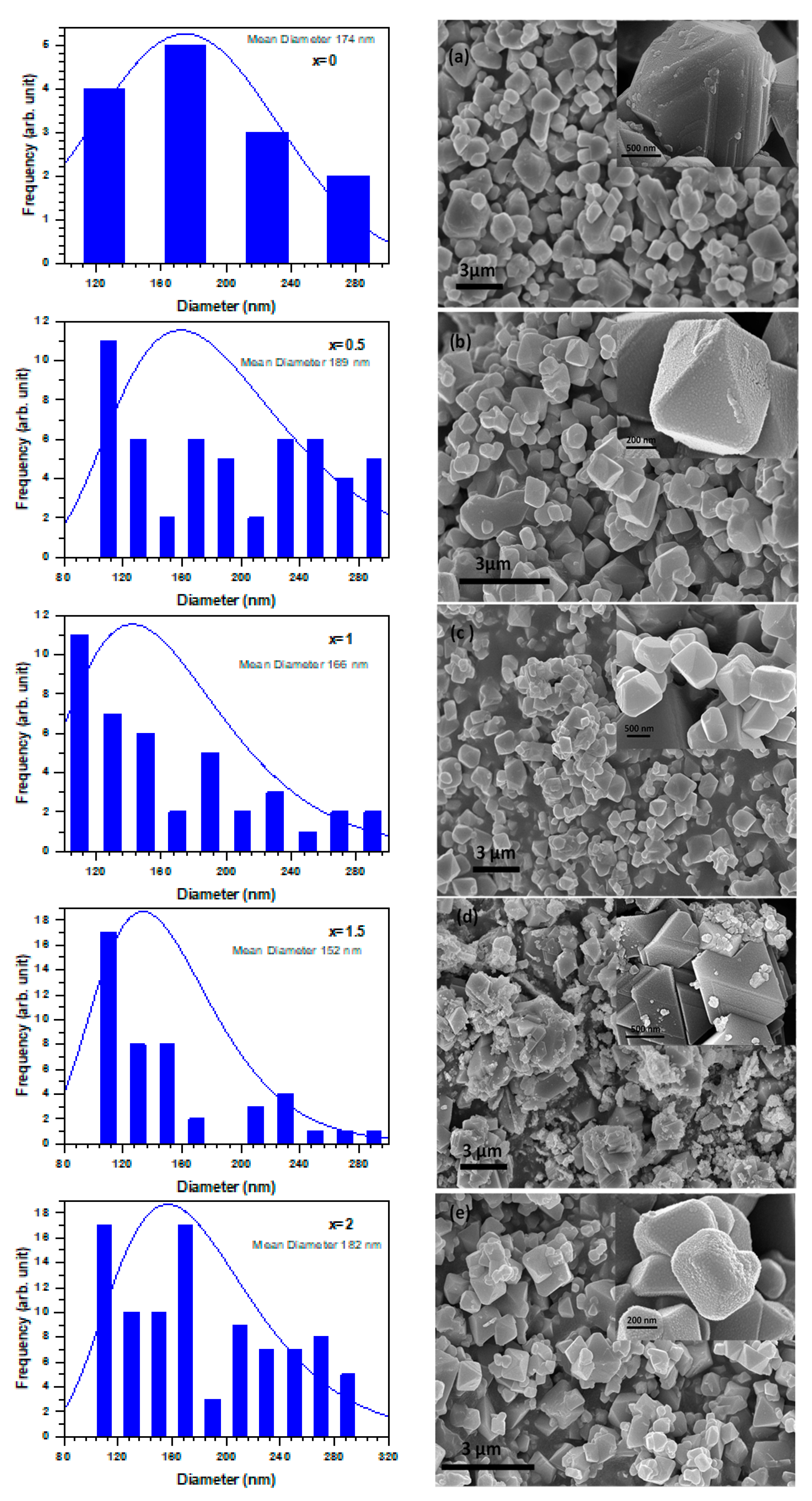
3.1. SEM Micrographs

The samples were characterized using scanning electron microscopy (SEM). The resulting images and their corresponding grain size distributions are shown in Figure 1a–e. The microscopic structure and morphology of Cd0.5Zn0.5Fe2−xCrxO4 with Cr substitution were also investigated. The SEM images revealed that the synthesized materials comprised an irregularly shaped group of tailed grains with a non-uniform grain size distribution. The particles exhibited a prismatic and pyramidal morphology.
Moreover, they were non-uniformly distributed, agglomerated, and inhomogeneous. Some massive particles were observed, along with smaller particles and increased agglomeration. All samples’ average grain size values varied from 152 nm to 189 nm and were found to be random with a high Cr content [28,29].

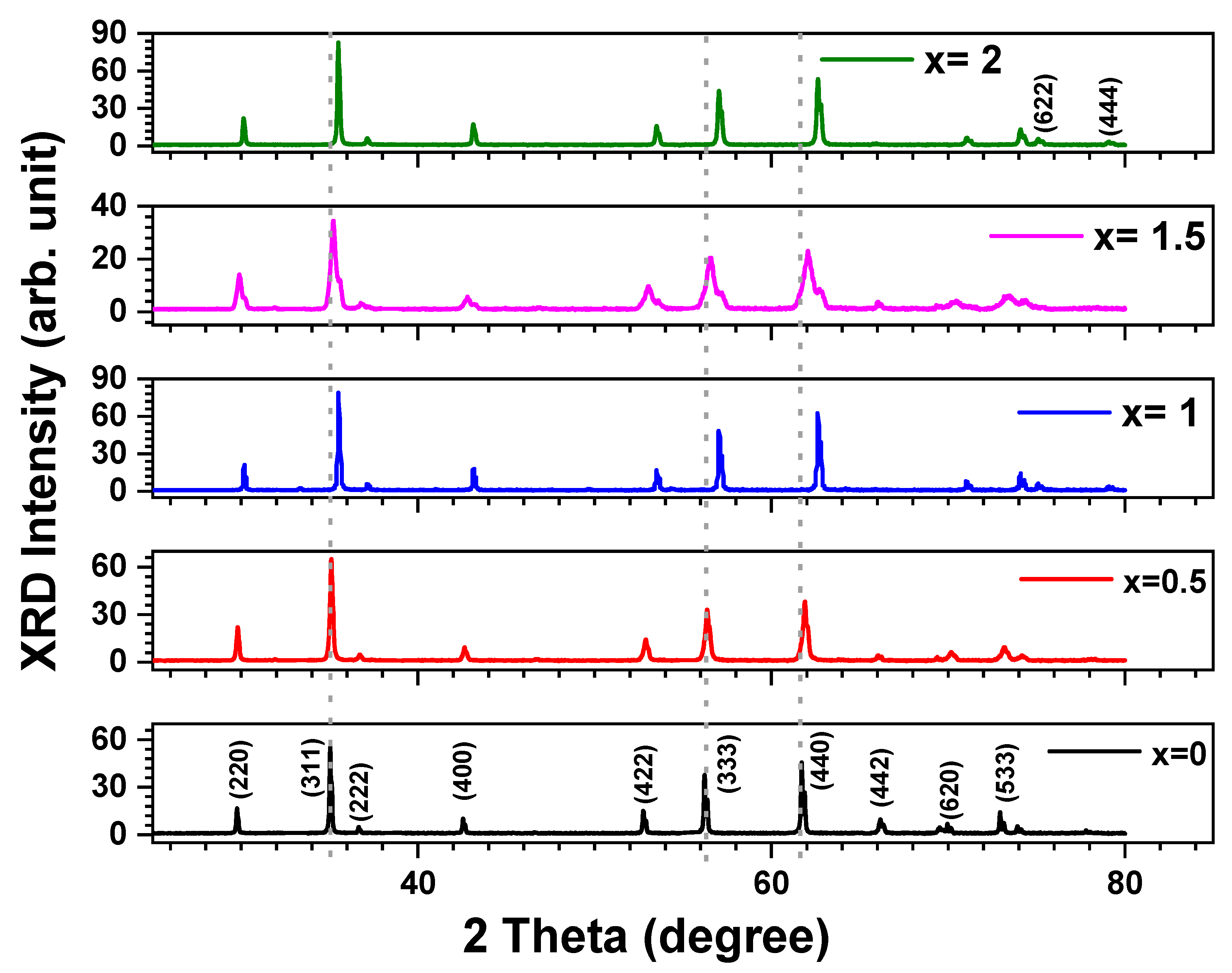
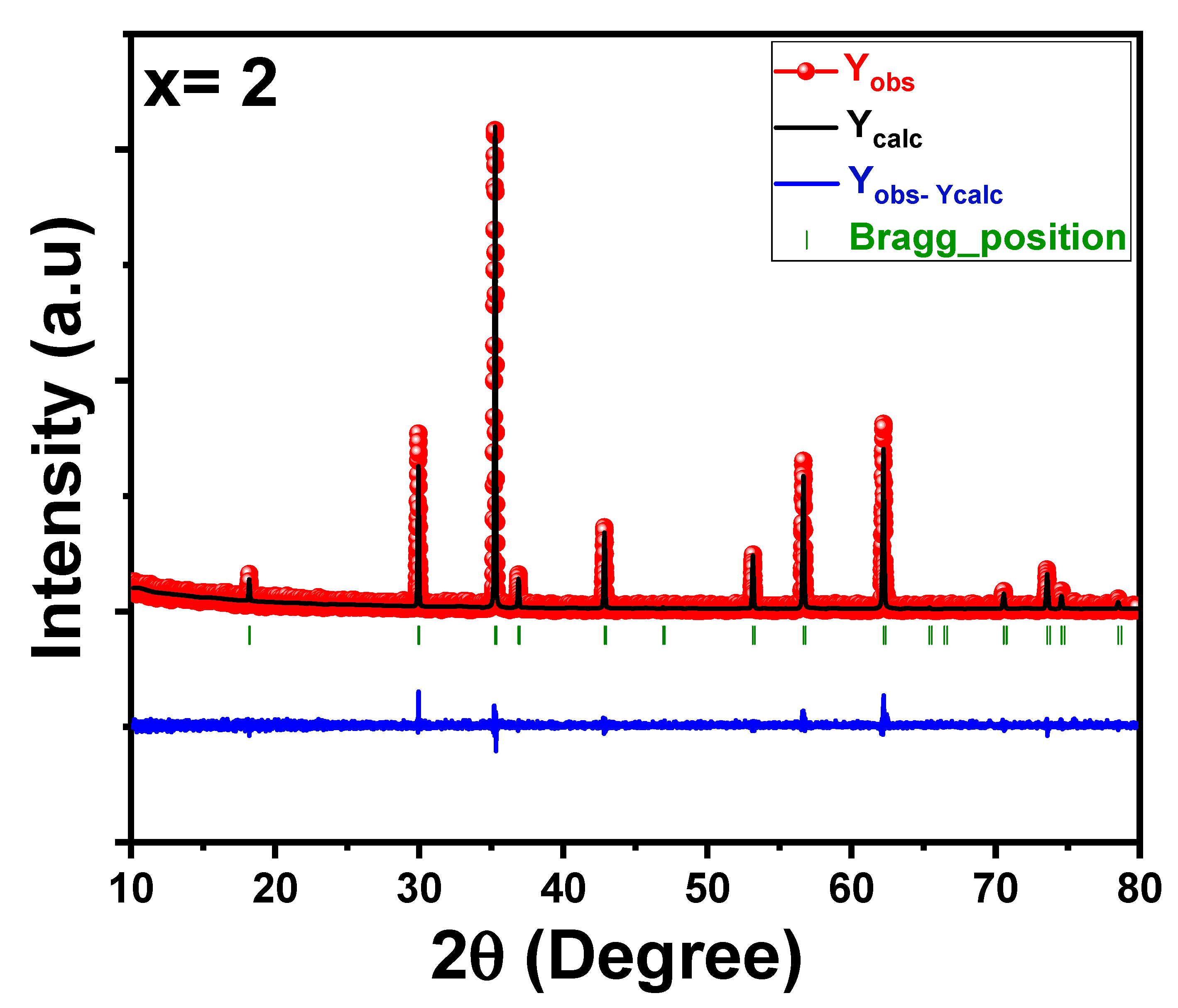
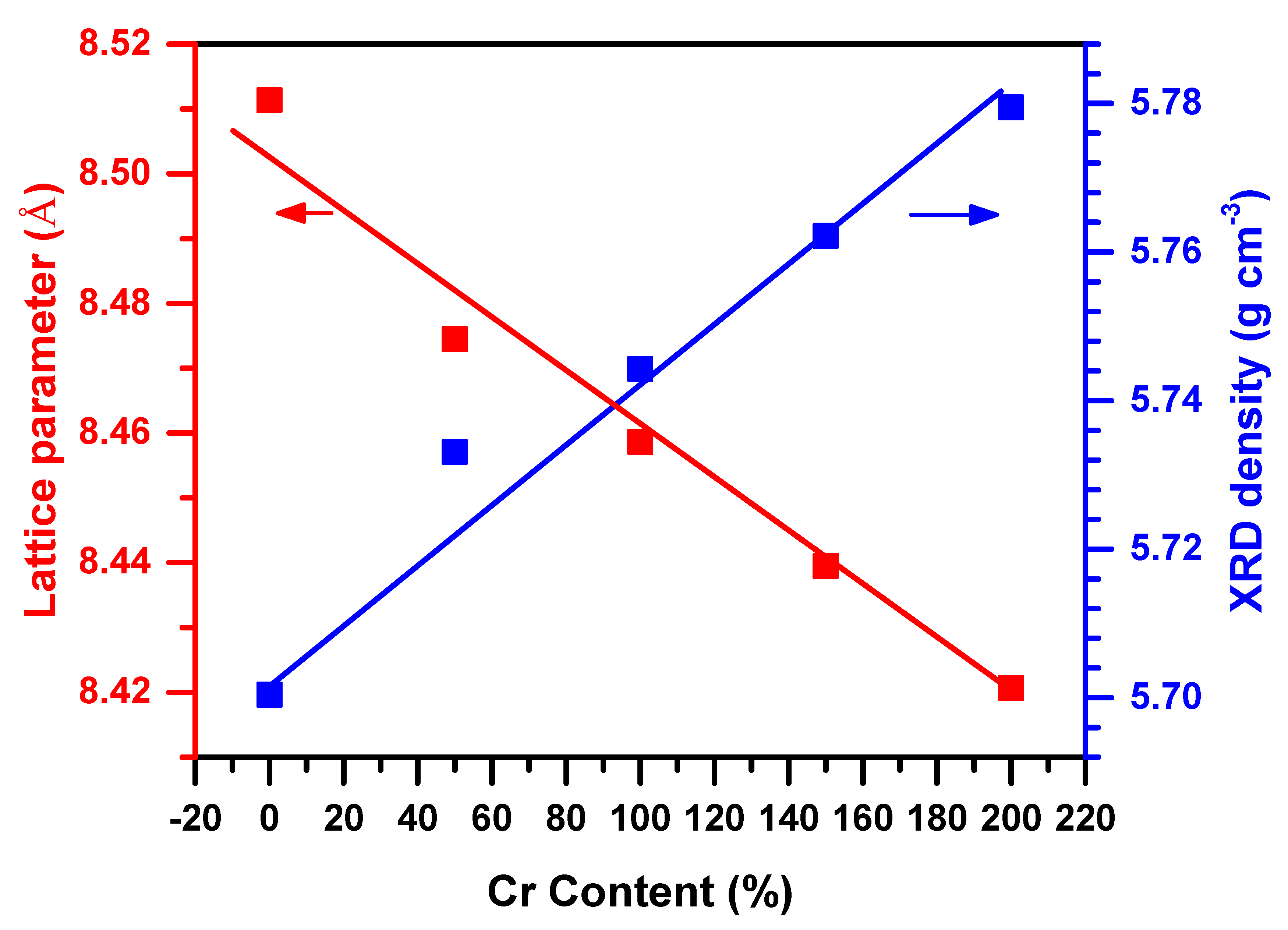
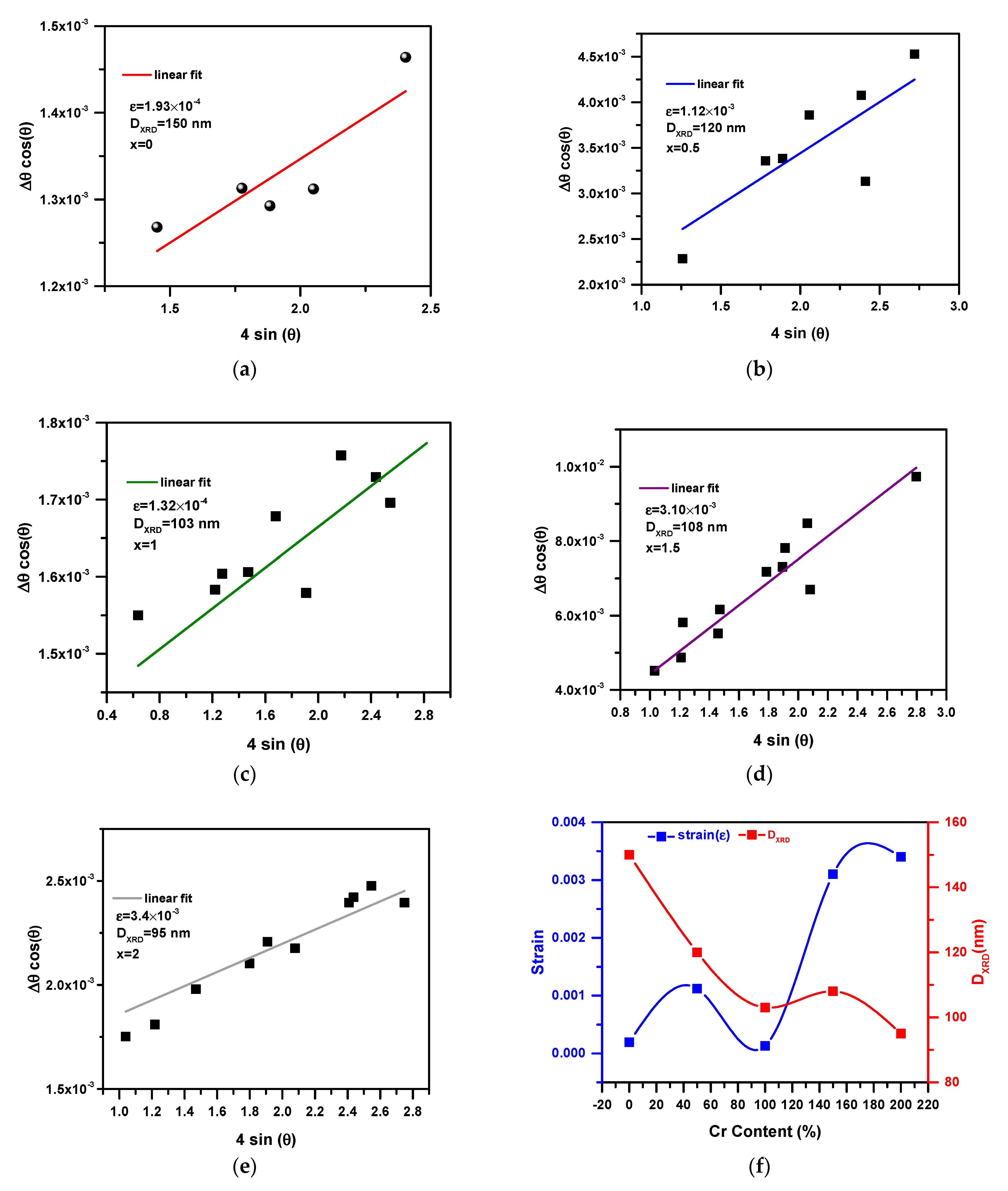
3.2. Structural Properties and Cation Distributions
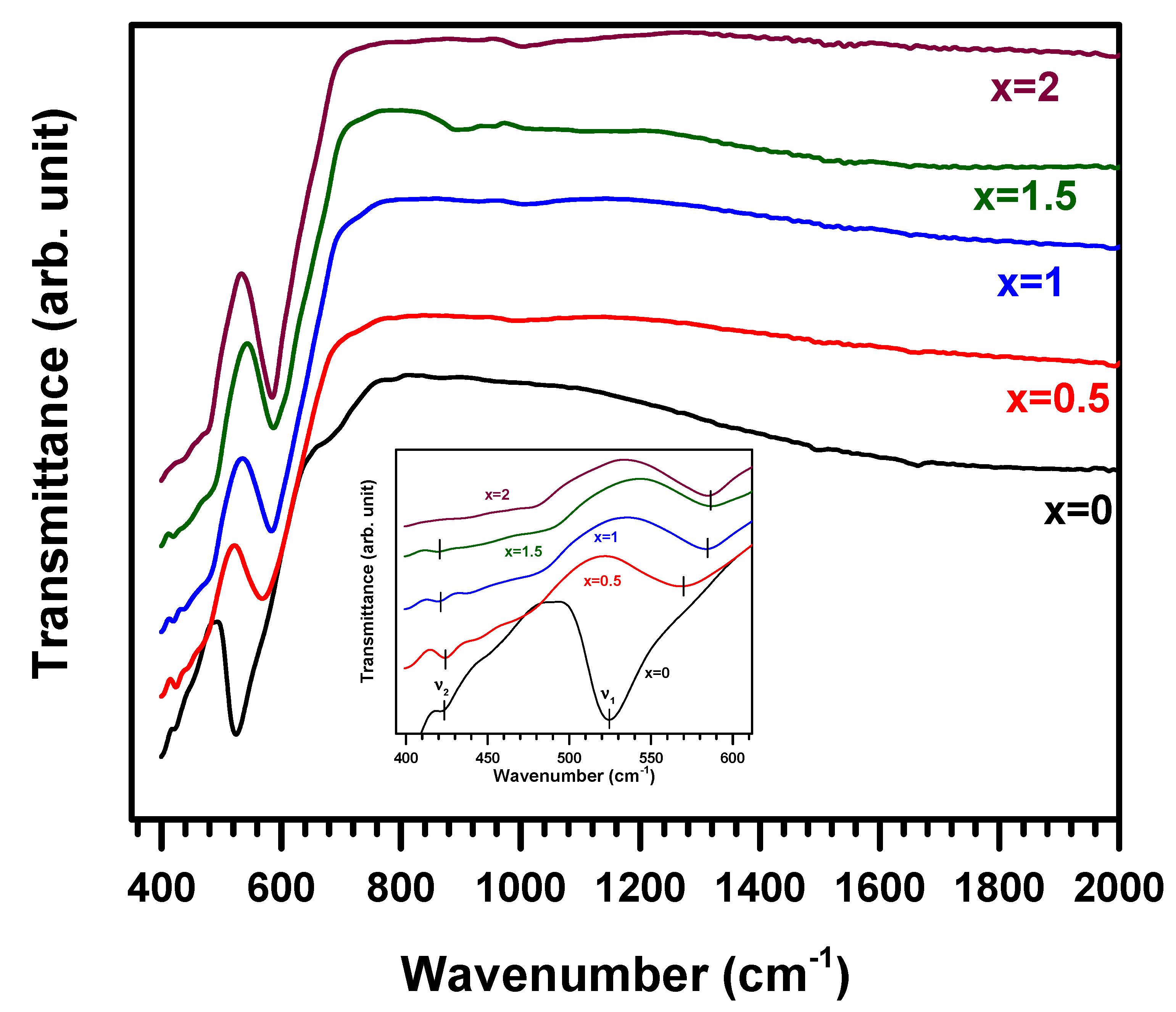
The XRD patterns of Cd0.5Zn0.5Fe2−xCrxO4 ferrites in Figure 2 reveal that a cubic spinel structure occurs in one phase. All diffraction lines are indexed in the cubic spinel structure, indicating the well-crystalline nature of the compounds. In addition, the absence of any reflection peak related to secondary phases confirms the purity of the samples. The cation distribution in the system was determined based on previous studies [30]. Mössbauer spectroscopic investigations have determined the cation distribution in ferrites with the general formula AB2O4. An investigation of Cd-Zn ferrites [24] and Cr-substituted ferrites [25] revealed that the tetrahedral A-sites were preferably occupied by Cd2+ and Zn2+ ions. In contrast, Cr3+ ions are distributed over the octahedral B-sites, and Fe3+ ions are distributed over both sites. This cation distribution has been confirmed in other studies [31,32]. Hence, the Rietveld refinement for Cd0.5Zn0.5Fe2−xCrxO4 samples was performed using the ( C d 0.5 2 + Z n 0.5 2 + ) A [ F e 2 x 3 + C r x 3 + ] B O 4 2 cation distribution model. In this cation distribution, the A-sites are completely occupied by both Cd2+ and Zn2+ cations with equal concentrations (50 atom%). Hence, in the case of our samples, the Fe3+ and Cr3+ ions are distributed only over the octahedral B-sites. Furthermore, this cation distribution model confirms the absence of the inversion phenomenon and the non-occupation of the A-site by Fe3+ cations [33]. Figure 3 shows a typical example of the Rietveld refinement of Cd0.5Zn0.5Fe2−xCrxO4 spinel (x = 2). Table 1 outlines the various properties of the prepared compositions. The reliability factors (Bragg RBragg, profile Rp, experimental Rexp, and weighted profile Rwp) are all less than 10% in all cases. Rietveld fittings tend to be good, as shown by the χ2 = Rwp/Rexp (goodness of fit) tendency towards unity. As a result, the refined occupancy factors for (Cd/Zn) and (Fe/Cr) at the A- and B-sites corresponded with the nominal values, supporting the suggested hypothesis. According to Table 1 and Figure 4, the decrease in lattice constant (a) and volume (V) appears to be caused by the replacement of a smaller radius of the Cr3+ ( r C r 3 + = 0.63 Å ) ion for the Fe3+ ion radius ( r F e 3 + = 0.67 Å ) [34]. Moreover, other Cr-doped ferrites have shown similar reductions in lattice parameters [35]. Furthermore, the atomic positions of oxygen exhibit the characteristic features of the spinel structure [13]. Alternatively, the cation–oxygen bond at the octahedral sites (dB-O) is shorter with Cr substitution because of the decrease in the average ionic radius of the B-site <rB>. Since the ionic radius of the A-site (<rA>) remains the same, the length of the cation–oxygen bonds (dA-O) remains almost constant. Table 1 also shows the bond angle values (φA-O-B) associated with A-O-B interactions in the produced samples. The bond angle for A-O-B is greater than that of B-O-B, according to Table 1. Thus, A-B exchange interactions are more potent than B-B exchange interactions [36,37]. Furthermore, the observed decrease in the bond angle (φA-O-B) indicates that A-B exchange interactions become less intense when Cr replacement is conducted. The XRD density was calculated using the following formula [14]:
d x = 8 M N a 3
where M is the molar mass, a is the cell parameter, and N is the Avogadro number (6.022 × 10²³). Table 1 (also Figure 4) shows that the XRD density increases with Cr substitution. This finding is consistent with previous reports in the literature [38]. The increase in XRD density may be due to the reduction in oxygen vacancies, which significantly impact densification kinetics [29]. It can also be attributed to the dominant effect of the reduction in the lattice parameter compared to the relatively small variation in molar mass resulting from the lower molar mass of Cr3+ ions (51.996 g/mol) compared to Fe3+ ions (55.847 g/mol).
The values of the crystallite size (DXRD) and the lattice strain (ε) were determined by the Williamson–Hall method as a function of Cr content. This method, developed by G.K. Williamson and his student W.H. Hall [39], utilizes the full width at half maximum (FWHM) of Bragg peaks (Δθ, in radians) and the angle of peak position (θ), as well as the X-ray wavelength (λ = 1.5406 Å), to calculate the average crystallite size (D) and lattice strain (ε). The relationship is given by Δθcosθ = k ʎ D + 4 ε s i n θ , where k is a constant value (0.94) obtained by assuming the spherical nature of the powders. By plotting Δθ × cosθ versus 4sinθ, the strain component (ε) can be determined from the slope, and the size component can be determined from the intercept ( k ʎ D ). This plot is known as a Williamson–Hall plot. Figure 5a–e depicts the variations in ((Δθhkl)cosθhkl) as a function of (4sinθhkl) for Cd0.5Zn0.5Fe2−xCrxO4 spinels (x = 0; x = 0.5; x = 1; x = 1.5; x = 2). The estimated values of DXRD and ε are (150 nm, 1.93 × 10⁻⁴), (120 nm, 1.12 × 10⁻³), (103 nm, 1.32 × 10⁻⁴), (108 nm, 3.10 × 10⁻³), and (95 nm, 3.4 × 10⁻³) for Cd0.5Zn0.5Fe2−xCrxO4 spinels (x = 0; x = 0.5; x = 1; x = 1.5; x = 2), respectively. These results suggest a small variation in crystallite size due to Cr substitution, consistent with the values obtained from SEM analysis. The lattice strain increases while the crystallite size decreases approximately with an increasing Cr content.

3.3. FTIR Spectra

The infrared (IR) spectra provide valuable information about the crystal lattice’s valence state and vibrational modes. Table 2 presents the band positions obtained from the IR spectra of the Cd0.5Zn0.5CrxFe2−xO4 series. Figure 6 shows the IR spectra of this series, with the high-frequency band υ1 observed in the 524–586 cm−1 range and a small band in the low-frequency band υ2 in the 420–424 cm−1 range. These absorption bands indicate the formation of a single-phase spinel structure. The two major absorption bands at υ1 and υ2 are due to vibrations of the oxygen bonds with positive ions at A- and B-sites [40]. The small band at low-frequency band υ2 is constant for all samples except for x = 2, where it disappears. The vibrational bands υ1 and υ2 are assigned to intrinsic vibrations of the tetrahedral and octahedral sites, respectively [41].
The infrared spectra of the Cr-substituted ferrite system prepared through the ceramic route exhibit similar features, as reported in the literature [42]. The intensity of the absorption band corresponding to the tetrahedral complex (υ1) increases and shifts towards a higher frequency with an increased Cr content, while the octahedral complex (υ2) exhibits weaker absorption bands. This behavior can be attributed to the first selection rule, which states that transitions between d orbitals in a complex with a center of symmetry are forbidden. As the tetrahedral complex possesses a center of symmetry, its absorption bands are more intense than those of the octahedral complex, which lacks a center of symmetry and thus allows more transitions to occur between d orbitals [43].
The observed shift in the band position in the IR spectra is attributed to the change in the Fe3+-O22− distance for the tetrahedral and octahedral complexes. The slight frequency change in band υ2 and the significant shift of band υ1 towards a higher frequency are due to the substitution of Cr3+ ions, which replace Fe3+ ions only at the octahedral B-site, leading to no significant change in the size of the octahedral site. As the FeB3+-O22− complex numbers decrease, metal–oxygen vibrational energies increase, prompting a decrease in the FeB3+-O22− intermolecular distance. This phenomenon is observed due to the increased number of Cr3+-O22− complexes [38] and the creation of Me3+O22− complexes at A-sites. As Cd2+-O2− and Zn2+-O2− bonds are stretched at the A-sites and Fe3+-O2− and Cr3+-O2− bonds are stretched at the B-sites, these bands are produced. The two bands may exhibit different positions for various reasons, including differences in ionic radius, the average distance between metal and oxygen, and electronegativity. It has been found that similar results have been obtained for other ferrite systems [44,45,46]. Assuming that the other independent parameters are constant, the force constant would be the second derivative of the potential energy based on the site radius. Based on Waldron’s method [41], we calculated force constants for tetrahedral and octahedral sites. For each site, Waldron gives the force constants KT and KO as follows:
K T = 7.62 M 1 ν 1 2 10 3 d y n e c m
K O = 10.62 M 2 2 ν 2 2 10 3 d y n e c m
Assuming that M1 and M2 refer to the molecular weight of the cations at sites A and B, respectively. Based on the cation distribution for the prepared samples, tetrahedral M1 and octahedral M2 molecular weights have been calculated. Table 2 contains the force constants KT and KO. With an increasing Cr content, force constants KT and KO increase. According to IR studies, bond length and the force constant inversely relate [47].

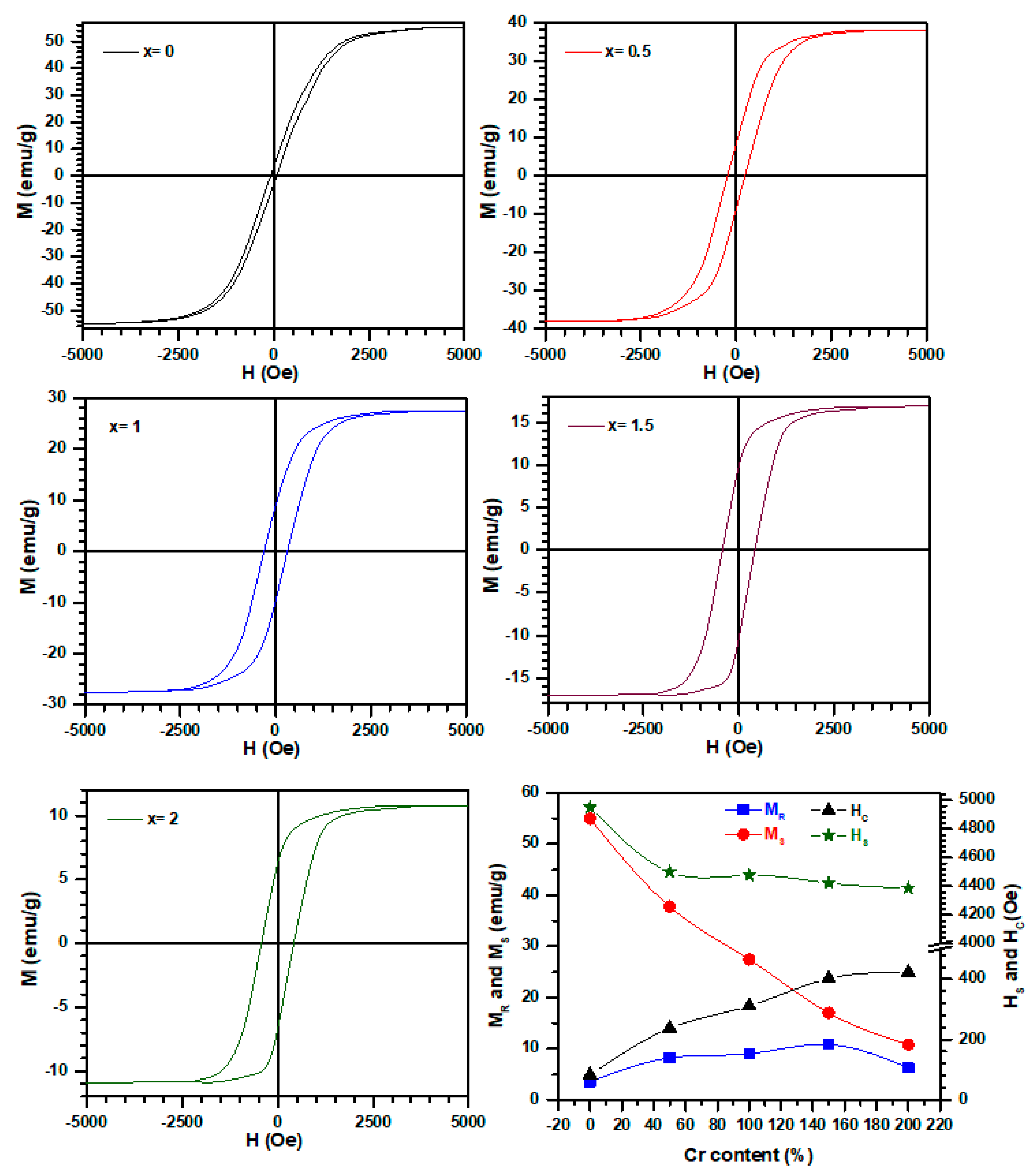
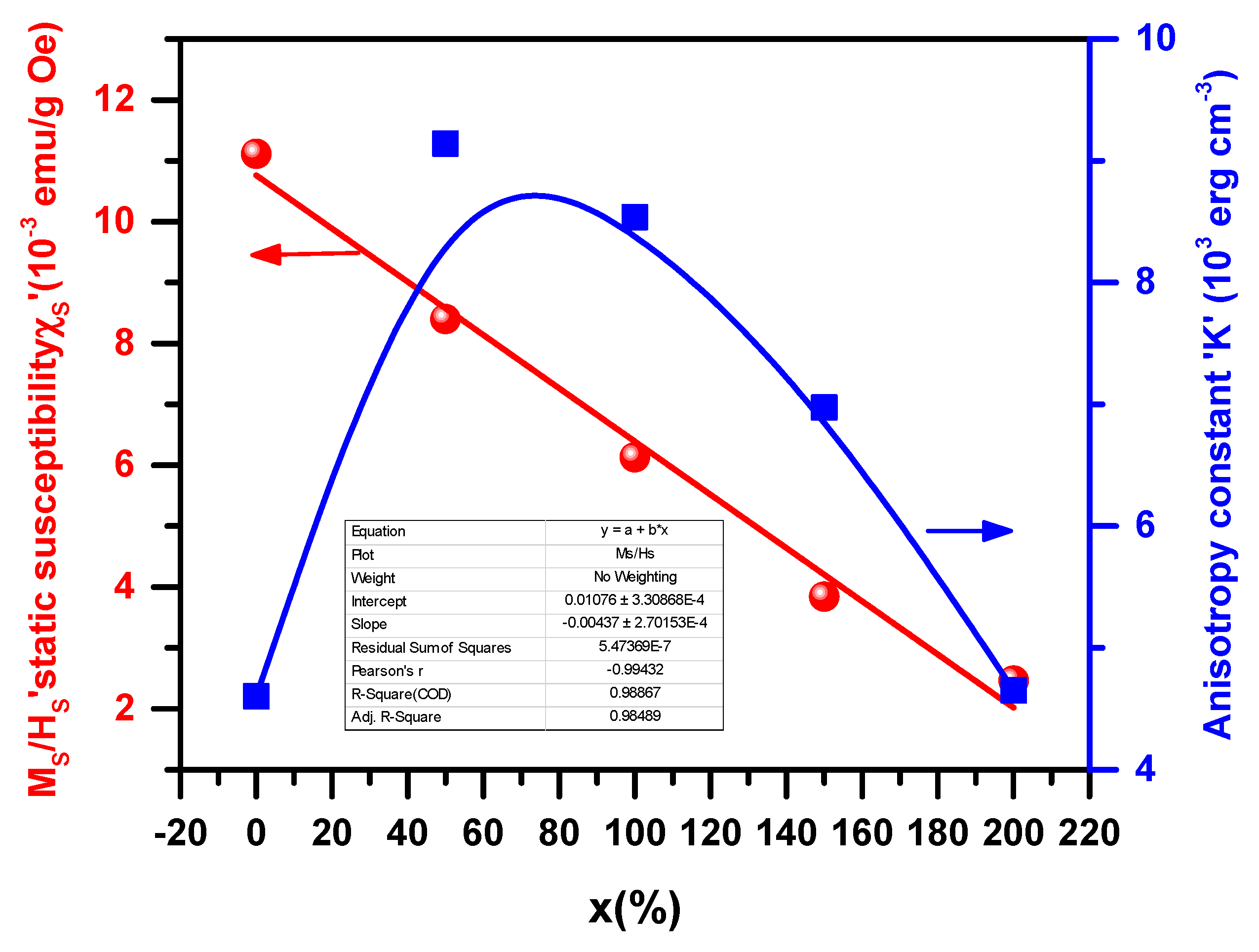
3.4. Magnetic Properties

To obtain the magnetic hysteresis curves, a magnetic field (±50 kOe) is applied to the prepared samples at room temperature (see Figure 7). Samples at low magnetic fields exhibit nonlinear magnetization and become saturated at high magnetic fields, revealing ferromagnetism. Table 3 summarizes saturation magnetization (Ms), remanent magnetization (Mr), and coercivity (Hc) results. The synthesized samples have low Hc values. Therefore, the samples could be classified as soft magnetic spinels. As a result, the Cd0.5Zn0.5CrxFe2−xO4 spinels have the potential to be applied in some magnetic applications such as recording heads, spintronic devices, microwave devices, transformers, induction cores, telecommunication systems, electromagnetic devices, and magnetic recording field sensors [48,49,50]. As the Cr content increases, the Hc also increases, indicating an increase in the resistive nature against spin inversion. The anisotropy constant increases with an increasing Cr content but decreases when the Cr content is more significant than 0.5. The anisotropy constant K depends on the substituted ion concentration [51], which can be evaluated using the corresponding relation.
H c = 0.98   K M S
Furthermore, saturation magnetization is related to Hc through Brown’s relation [52], and Hc = 2 K μ 0 M S , states that Hc is inversely proportional to Ms. This is consistent with our experimental results.
Table 3 illustrates the decrease in the Ms value with Cr replacement, consistent with other spinel systems [53,54]. There is a correlation between the increase in Ms values and Neel’s theory [55] and the cations distribution between A- and B-sites. According to Neel’s model, ferrimagnet materials interact in three ways: A-A, B-B, and A-B sublattices. A-A and B-B interactions within the sublattice are dominated by the super-exchange interaction between A- and B-sites. Consequently, the net magnetic moment consists of the vector sum of magnetic moments on sublattices A and B [56]:
n B c a l = M B M A
MB and MA represent B and A sublattice magnetic moments in Bohr magneton (μB), respectively. When Cr3+ replaces Fe3+ at the octahedral site, saturation magnetization decreases since the Cr3+ ion (3 μB) has a smaller magnetic moment than Fe3+(5 μB) [57]. Accordingly, the magnetic properties of the prepared samples are closely related to their predicted cation distribution.
The inset of Figure 8 shows variations in the anisotropy constant “K” and static susceptibility ‘’χS” with Cr. The ratio Ms/Hs, named static susceptibility χS, increases linearly with the Cr content. In contrast, the anisotropic coefficient increases with the Cr content. It reaches a maximum when the Cr composition is equal to that of Fe and regains the same value as the beginning ferrite when the material becomes chromite.

4. Conclusions

Cd0.5Zn0.5Fe2−xCrxO4 (0 ≤ x ≤ 2) ferrites synthesized via the sol–gel method exhibit a cubic F d 3 ¯ m spinel structure. Substituting Cr for Fe reduces cell parameters, average grain size, spontaneous magnetization, and conductivity compared to the parent compound Cd0.5Zn0.5Fe2O4. The FTIR spectra reveal two principal absorption bands that increase with Cr substitution. These materials show potential for use in various magnetic and electronic applications. The significant findings of this work show that the examined materials have regular spinel cubic structures and low coercive fields, allowing them to be used in magnetic devices.

Author Contributions

Conceptualization, S.H., A.M. and L.B.; Methodology, R.K.A., N.K. and A.M.; Software, A.S.A. and H.T.; Formal analysis, L.B., S.H. and A.S.A.; Investigation, L.B. and S.H.; Writing—original draft, R.K.A., L.B., A.A., A.S.A. and S.H.; Writing—review & editing, N.K., A.A. and H.T.; Supervision, N.K. and A.M. All authors have read and agreed to the published version of the manuscript.

Funding

This research received no external funding.

Data Availability Statement

Not applicable.

Conflicts of Interest

The authors declare no conflict of interest.

References

  1. Adam, J.D.; Davis, L.E.; Dionne, G.F.; Schloemann, E.F.; Stitzer, S.N. Ferrite Devices and Materials. IEEE Trans. Microw. Theory Tech. 2002, 50, 721–737. [Google Scholar] [CrossRef] [Green Version]
  2. Kulikowski, J. Soft Magnetic Ferrites—Development or Stagnation? J. Magn. Magn. Mater. 1984, 41, 56–62. [Google Scholar] [CrossRef]
  3. Harris, V.G.; Geiler, A.; Chen, Y.; Yoon, S.D.; Wu, M.; Yang, A.; Chen, Z.; He, P.; Parimi, P.V.; Zuo, X. Recent Advances in Processing and Applications of Microwave Ferrites. J. Magn. Magn. Mater. 2009, 321, 2035–2047. [Google Scholar] [CrossRef]
  4. Mazarío, E.; Herrasti, P.; Morales, M.P.; Menéndez, N. Synthesis and Characterization of CoFe2O4 Ferrite Nanoparticles Obtained by an Electrochemical Method. Nanotechnology 2012, 23, 355708. [Google Scholar] [CrossRef] [PubMed]
  5. Chen, K.; Jia, L.; Yu, X.; Zhang, H. A Low Loss NiZnCo Ferrite, Prepared Using a Hydrothermal Method, for Antenna Applications. J. Appl. Phys. 2014, 115, 17A520. [Google Scholar] [CrossRef]
  6. Rahman, S.; Nadeem, K.; Anis-ur-Rehman, M.; Mumtaz, M.; Naeem, S.; Letofsky-Papst, I. Structural and Magnetic Properties of ZnMg-Ferrite Nanoparticles Prepared Using the Co-Precipitation Method. Ceram. Int. 2013, 39, 5235–5239. [Google Scholar] [CrossRef]
  7. Sun, L.; Zhang, R.; Ni, Q.; Cao, E.; Hao, W.; Zhang, Y.; Ju, L. Magnetic and Dielectric Properties of MgxCo1−XFe2O4 Ferrites Prepared by the Sol-Gel Method. Phys. B Condens. Matter 2018, 545, 4–11. [Google Scholar] [CrossRef]
  8. Safari, A.; Gheisari, K.; Farbod, M. Structural, Microstructural, Magnetic and Dielectric Properties of Ni-Zn Ferrite Powders Synthesized by Plasma Arc Discharge Process Followed by Post-Annealing. J. Magn. Magn. Mater. 2019, 488, 165369. [Google Scholar] [CrossRef]
  9. Soibam, I.; Phanjoubam, S.; Prakash, C. Mössbauer and Magnetic Studies of Cobalt Substituted Lithium Zinc Ferrites Prepared by Citrate Precursor Method. J. Alloys Compd. 2009, 475, 328–331. [Google Scholar] [CrossRef]
  10. Singh, C.; Jauhar, S.; Kumar, V.; Singh, J.; Singhal, S. Synthesis of Zinc Substituted Cobalt Ferrites via Reverse Micelle Technique Involving in Situ Template Formation: A Study on Their Structural, Magnetic, Optical and Catalytic Properties. Mater. Chem. Phys. 2015, 156, 188–197. [Google Scholar] [CrossRef]
  11. Hankare, P.P.; Jadhav, S.D.; Sankpal, U.B.; Chavan, S.S.; Waghmare, K.J.; Chougule, B.K. Synthesis, Characterization and Effect of Sintering Temperature on Magnetic Properties of MgNi Ferrite Prepared by Co-Precipitation Method. J. Alloys Compd. 2009, 475, 926–929. [Google Scholar] [CrossRef]
  12. Ebrahimi, S.A.S.; Masoudpanah, S.M. Effects of PH and Citric Acid Content on the Structure and Magnetic Properties of MnZn Ferrite Nanoparticles Synthesized by a Sol–Gel Autocombustion Method. J. Magn. Magn. Mater. 2014, 357, 77–81. [Google Scholar] [CrossRef]
  13. Kouki, N.; Hcini, S.; Boudard, M.; Aldawas, R.; Dhahri, A. Microstructural Analysis, Magnetic Properties, Magnetocaloric Effect, and Critical Behaviors of Ni0.6Cd0.2Cu0.2Fe2O4 Ferrites Prepared Using the Sol–Gel Method under Different Sintering Temperatures. RSC Adv. 2019, 9, 1990–2001. [Google Scholar] [CrossRef] [PubMed]
  14. Hcini, S.; Kouki, N.; Omri, A.; Dhahri, A.; Bouazizi, M.L. Effect of Sintering Temperature on Structural, Magnetic, Magnetocaloric and Critical Behaviors of Ni-Cd-Zn Ferrites Prepared Using Sol-Gel Method. J. Magn. Magn. Mater. 2018, 464, 91–102. [Google Scholar] [CrossRef]
  15. Mane, D.R.; Patil, S.; Birajdar, D.D.; Kadam, A.B.; Shirsath, S.E.; Kadam, R.H. Sol–Gel Synthesis of Cr3+ Substituted Li0.5Fe2.5O4: Cation Distribution, Structural and Magnetic Properties. Mater. Chem. Phys. 2011, 126, 755–760. [Google Scholar] [CrossRef]
  16. Mitra, S.; Bidyananda, M.; Samanta, A.K. Cation Distribution in Cr-Spinels from the Sittampundi Layered Complex and Their Intracrystalline Thermodynamics. Curr. Sci. 2006, 90, 435–439. [Google Scholar]
  17. Lee, S.H.; Yoon, S.J.; Lee, G.J.; Kim, H.S.; Yo, C.H.; Ahn, K.; Lee, D.H.; Kim, K.H. Electrical and Magnetic Properties of NiCrxFe2−XO4 Spinel (0 ≤ X ≤ 0.6). Mater. Chem. Phys. 1999, 61, 147–152. [Google Scholar] [CrossRef]
  18. Ikehara, T.; Yamaguchi, H.; Hosokawa, K.; Miyamoto, H.; Aizawa, K. Effects of ELF Magnetic Field on Membrane Protein Structure of Living HeLa Cells Studied by Fourier Transform Infrared Spectroscopy. Bioelectromagn. J. Bioelectromagn. Soc. Soc. Phys. Regul. Biol. Med. Eur. Bioelectromagn. Assoc. 2003, 24, 457–464. [Google Scholar] [CrossRef]
  19. Gismelseed, A.M.; Yousif, A.A. Mössbauer Study of Chromium-Substituted Nickel Ferrites. Phys. B Condens. Matter 2005, 370, 215–222. [Google Scholar] [CrossRef]
  20. Singhal, S.; Sharma, R.; Namgyal, T.; Jauhar, S.; Bhukal, S.; Kaur, J. Structural, Electrical and Magnetic Properties of Co0.5Zn0.5AlxFe2−XO4 (X = 0, 0.2, 0.4, 0.6, 0.8 and 1.0) Prepared via Sol–Gel Route. Ceram. Int. 2012, 38, 2773–2778. [Google Scholar] [CrossRef]
  21. Singhal, S.; Jauhar, S.; Singh, J.; Chandra, K.; Bansal, S. Investigation of Structural, Magnetic, Electrical and Optical Properties of Chromium Substituted Cobalt Ferrites (CoCrxFe2−XO4, 0 ≤ X ≤ 1) Synthesized Using Sol Gel Auto Combustion Method. J. Mol. Struct. 2012, 1012, 182–188. [Google Scholar] [CrossRef]
  22. Valan, M.F.; Manikandan, A.; Antony, S.A. Microwave Combustion Synthesis and Characterization Studies of Magnetic Zn1−xCdxFe2O4 (0 ≤ X ≤ 0.5) Nanoparticles. J. Nanosci. Nanotechnol. 2015, 15, 4543–4551. [Google Scholar] [CrossRef] [PubMed]
  23. Harish, K.N.; Naik, H.S.B.; Viswanath, R. Synthesis, Enhanced Optical and Photocatalytic Study of Cd–Zn Ferrites under Sunlight. Catal. Sci. Technol. 2012, 2, 1033–1039. [Google Scholar] [CrossRef]
  24. Gupta, M.; Gupta, M.; Mudsainiyan, R.K.; Randhawa, B.S. Physico-Chemical Analysis of Pure and Zn Doped Cd Ferrites (Cd1−XZnxFe2O4) Nanofabricated by Pechini Sol–Gel Method. J. Anal. Appl. Pyrolysis 2015, 116, 75–85. [Google Scholar] [CrossRef]
  25. Chakrabarti, M.; Sanyal, D.; Chakrabarti, A. Preparation of Zn(1−x)CdxFe2O4 (X = 0.0, 0.1, 0.3, 0.5, 0.7 and 1.0) Ferrite Samples and Their Characterization by Mössbauer and Positron Annihilation Techniques. J. Phys. Condens. Matter 2007, 19, 236210. [Google Scholar] [CrossRef]
  26. Siddique, M.; Anwar-ul-Islam, M.; Butt, N.M.; Abbas, T. Composition Dependence of Quadrupole Splitting in Cd—Zn Ferrites. Phys. Status Solidi 1999, 216, 1069–1072. [Google Scholar] [CrossRef]
  27. Arean, C.O.; Diaz, E.G.; Gonzalez, J.M.R.; Garcia, M.A.V. Crystal Chemistry of Cadmium-Zinc Ferrites. J. Solid State Chem. 1988, 77, 275–280. [Google Scholar] [CrossRef]
  28. Weil, L.; Bertaut, F.; Bochirol, L. Propriétés Magnétiques et Structure de La Phase Quadratique Du Ferrite de Cuivre. J. Phys. le Radium 1950, 11, 208–212. [Google Scholar] [CrossRef]
  29. Mane, D.R.; Birajdar, D.D.; Shirsath, S.E.; Telugu, R.A.; Kadam, R.H. Structural and Magnetic Characterizations of Mn—Ni—Zn Ferrite Nanoparticles. Phys. Status Solidi 2010, 207, 2355–2363. [Google Scholar] [CrossRef]
  30. Cvejic, Z.; Rakic, S.; Kremenovic, A.; Antic, B.; Jovalekic, C.; Colomban, P. Nanosize Ferrites Obtained by Ball Milling: Crystal Structure, Cation Distribution, Size-Strain Analysis and Raman Investigations. Solid State Sci. 2006, 8, 908–915. [Google Scholar] [CrossRef]
  31. Hakim, M.A.; Nath, S.K.; Sikder, S.S.; Maria, K.H. Cation Distribution and Electromagnetic Properties of Spinel Type Ni–Cd Ferrites. J. Phys. Chem. Solids 2013, 74, 1316–1321. [Google Scholar] [CrossRef]
  32. Khalaf, K.A.M.; Al Rawas, A.D.; Gismelssed, A.M.; Al Jamel, A.; Al Ani, S.K.J.; Shongwe, M.S.; Al Riyami, K.O.; Al Alawi, S.R. Influence of Cr Substitution on Debye-Waller Factor and Related Structural Parameters of ZnFe2−XCrxO4 Spinels. J. Alloys Compd. 2017, 701, 474–486. [Google Scholar] [CrossRef]
  33. Hossain, A.K.M.A.; Mahmud, S.T.; Seki, M.; Kawai, T.; Tabata, H. Structural, Electrical Transport, and Magnetic Properties of Ni1−XZnxFe2O4. J. Magn. Magn. Mater. 2007, 312, 210–219. [Google Scholar] [CrossRef]
  34. Shannon, R.D. Revised Effective Ionic Radii and Systematic Studies of Interatomic Distances in Halides and Chalcogenides. Acta Crystallogr. Sect. A Cryst. Phys. Diffr. Theor. Gen. Crystallogr. 1976, 32, 751–767. [Google Scholar] [CrossRef]
  35. Patange, S.M.; Shirsath, S.E.; Lohar, K.S.; Algude, S.G.; Kamble, S.R.; Kulkarni, N.; Mane, D.R.; Jadhav, K.M. Infrared Spectral and Elastic Moduli Study of NiFe2−XCrxO4 Nanocrystalline Ferrites. J. Magn. Magn. Mater. 2013, 325, 107–111. [Google Scholar] [CrossRef]
  36. Kumar, G.; Kotnala, R.K.; Shah, J.; Kumar, V.; Kumar, A.; Dhiman, P.; Singh, M. Cation Distribution: A Key to Ascertain the Magnetic Interactions in a Cobalt Substituted Mg–Mn Nanoferrite Matrix. Phys. Chem. Chem. Phys. 2017, 19, 16669–16680. [Google Scholar] [CrossRef]
  37. Sharma, R.; Thakur, P.; Kumar, M.; Thakur, N.; Negi, N.S.; Sharma, P.; Sharma, V. Improvement in Magnetic Behaviour of Cobalt Doped Magnesium Zinc Nano-Ferrites via Co-Precipitation Route. J. Alloys Compd. 2016, 684, 569–581. [Google Scholar] [CrossRef]
  38. Hemeda, O.M.; Amer, M.A.; Aboul-Enein, S.; Ahmed, M.A. Effect of Sintering on X-Ray and IR Spectral Behaviour of the MnAlxFe2-xO4 Ferrite System. Phys. Status Solidi 1996, 156, 29–38. [Google Scholar] [CrossRef]
  39. Williamson, G.K.; Hall, W.H. X-Ray Line Broadening from Filed Aluminium and Wolfram. Acta Metall. 1953, 1, 22–31. [Google Scholar] [CrossRef]
  40. AlArfaj, E.; Hcini, S.; Mallah, A.; Dhaou, M.H.; Bouazizi, M.L. Effects of Co Substitution on the Microstructural, Infrared, and Electrical Properties of Mg0.6−xCoxZn0.4Fe2O4 Ferrites. J. Supercond. Nov. Magn. 2018, 31, 4107–4116. [Google Scholar] [CrossRef]
  41. Waldron, R.D. Infrared Spectra of Ferrites. Phys. Rev. 1955, 99, 1727. [Google Scholar] [CrossRef]
  42. Amer, M.A.; Ahmed, M.A.; El-Nimr, M.K.; Mostafa, M.A. Mössbauer and Infrared Studies of the Cu-Cr Ferrites. Hyperfine Interact. 1995, 96, 91–98. [Google Scholar] [CrossRef]
  43. Shaeel, A.; Al-Thabaiti. Communications de la Facult´e des Sciences de l’Universit´e d’Ankara B; La Faculté: Leiden, The Netherlands, 2003; Volume 49, pp. 5–14. [Google Scholar]
  44. Mohammed, K.A.; Al-Rawas, A.D.; Gismelseed, A.M.; Sellai, A.; Widatallah, H.M.; Yousif, A.; Elzain, M.E.; Shongwe, M. Infrared and Structural Studies of Mg1−XZnxFe2O4 Ferrites. Phys. B Condens. Matter 2012, 407, 795–804. [Google Scholar] [CrossRef]
  45. Khalaf, K.A.M.; Al-Rawas, A.D.; Widatallah, H.M.; Al-Rashdi, K.S.; Sellai, A.; Gismelseed, A.M.; Hashim, M.; Jameel, S.K.; Al-Ruqeishi, M.S.; Al-Riyami, K.O. Influence of Zn2+ Ions on the Structural and Electrical Properties of Mg1−XZnxFeCrO4 Spinels. J. Alloys Compd. 2016, 657, 733–747. [Google Scholar] [CrossRef]
  46. Yoon, S.J.; Lee, S.H.; Kim, K.H.; Ahn, K.S. Electrical and Magnetic Properties of Spinel ZnCr2−XFexO4 (0 ≤ X ≤ 1.0). Mater. Chem. Phys. 2002, 73, 330–334. [Google Scholar] [CrossRef]
  47. Pradeep, A.; Priyadharsini, P.; Chandrasekaran, G. Sol–Gel Route of Synthesis of Nanoparticles of MgFe2O4 and XRD, FTIR and VSM Study. J. Magn. Magn. Mater. 2008, 320, 2774–2779. [Google Scholar] [CrossRef]
  48. Shokrollahi, H.; Janghorban, K. Soft Magnetic Composite Materials (SMCs). J. Mater. Process. Technol. 2007, 189, 1–12. [Google Scholar] [CrossRef]
  49. Chakrabarti, P.K.; Nath, B.K.; Brahma, S.; Das, S.; Das, D.; Ammar, M.; Mazaleyrat, F. Magnetic and Hyperfine Properties of Chemically Synthesized Nanocomposites of (Al2O3)x(Ni0.2Zn0.6Cu0.2Fe2O4)(1−x) (X = 0.15, 0.30, 0.45). Solid State Commun. 2007, 144, 305–309. [Google Scholar] [CrossRef]
  50. Modak, S.; Ammar, M.; Mazaleyrat, F.; Das, S.; Chakrabarti, P.K. XRD, HRTEM and Magnetic Properties of Mixed Spinel Nanocrystalline Ni–Zn–Cu-Ferrite. J. Alloys Compd. 2009, 473, 15–19. [Google Scholar] [CrossRef]
  51. Kambale, R.C.; Shaikh, P.A.; Bhosale, C.H.; Rajpure, K.Y.; Kolekar, Y.D. The Effect of Mn Substitution on the Magnetic and Dielectric Properties of Cobalt Ferrite Synthesized by an Autocombustion Route. Smart Mater. Struct. 2009, 18, 115028. [Google Scholar] [CrossRef]
  52. Coey, J.M.D. Rare Earth Permanent Magnetism; John Wiley and Sons: Hoboken, NJ, USA, 1996. [Google Scholar] [CrossRef]
  53. Patange, S.M.; Shirsath, S.E.; Jadhav, S.S.; Jadhav, K.M. Cation Distribution Study of Nanocrystalline NiFe2−XCrxO4 Ferrite by XRD, Magnetization and Mössbauer Spectroscopy. Phys. Status Solidi 2012, 209, 347–352. [Google Scholar] [CrossRef]
  54. Patange, S.M.; Shirsath, S.E.; Toksha, B.G.; Jadhav, S.S.; Jadhav, K.M. Electrical and Magnetic Properties of Cr3+ Substituted Nanocrystalline Nickel Ferrite. J. Appl. Phys. 2009, 106, 023914. [Google Scholar] [CrossRef]
  55. Néel, L. Magnetism and Local Molecular Field. Science 1971, 174, 985–992. [Google Scholar] [CrossRef] [PubMed] [Green Version]
  56. Torkian, S.; Ghasemi, A.; Razavi, R.S. Cation Distribution and Magnetic Analysis of Wideband Microwave Absorptive CoxNi1− XFe2O4 Ferrites. Ceram. Int. 2017, 43, 6987–6995. [Google Scholar] [CrossRef]
  57. Satyanarayana, G.; Nageswara Rao, G.; Babu, K.V.; Santosh Kumar, G.V.; Dinesh Reddy, G. Effect of Cr3+ Substitution on the Structural, Electrical and Magnetic Properties of Ni0.7Zn0.2Cu0.1Fe2−xCrxO4 Ferrites. J. Korean Phys. Soc. 2019, 74, 684–694. [Google Scholar] [CrossRef]
Figure 1. SEM micrographs and particle size distributions of Cd0.5Zn0.5Fe2−xCrxO4 spinels, elaborated by sol–gel method. Images labeled (ae) correspond to x = 0, 0.5, 1, 1.5, and 2 Cr compositions, respectively. The inset images are the higher magnifications of micrographs.
Crystals 13 01121 g001
Figure 2. XRD patterns of Cd0.5Zn0.5Fe2−xCrxO4 spinels with (0 ≤ x ≤ 2).
Crystals 13 01121 g002
Figure 3. Typical example for the structural refinement of the XRD patterns using the Rietveld method for Cd0.5Zn0.5 Fe2−xCrxO4 spinels with (x = 2).
Crystals 13 01121 g003
Figure 4. Lattice parameter and XRD density of Cd0.5Zn0.5Fe2−xCrxO4 (0 ≤ x ≤ 2) spinels as a function of Cr content.
Crystals 13 01121 g004
Figure 5. (ae) Williamson–Hall plots of (Δθcosθ) vs. (4sinθ) of Cd0.5Zn0.5Fe2−xCrxO4 spinels (x = 0; x = 0.5; x = 1; x = 1.5; x = 2). (f) Values of the crystallite’s size (DXRD) and the lattice strain (ε) calculated using the Williamson–Hall methods.
Crystals 13 01121 g005
Figure 6. FTIR spectra at room temperature of Cd0.5Zn0.5Fe2−xCrxO4 spinel ferrites. Spectra is vertically translated for more clarity details. The inset figure is the higher magnification of the region between 400 and 600 cm−1 represented in semi-log scale.
Crystals 13 01121 g006
Figure 7. Magnetization loop M-H of Cd0.5Zn0.5Fe2−xCrxO4 spinels recorded at 300 K The values Mr, Ms, Hc, and Hs were extracted from M-H curves and plotted as a function of Cr content.
Crystals 13 01121 g007
Figure 8. Variation in anisotropy constant “K” and static susceptibility ‘’χS” with Cr content.
Crystals 13 01121 g008
Table 1. Structural parameters for Cd0.5Zn0.5Fe2−xCrxO4 spinels with (0 ≤ x ≤ 2) obtained following the structural refinement by the Rietveld method. a: cell parameter; V: cell volume; Biso: isotropic thermal agitation parameter. Definitions of structural parameters are given in the text.
Cr Content00.511.52
Space group F d 3 ¯ m
Cell parametersa (Å)8.5113 (4)8.4745 (4)8.4586 (4)8.4395 (4)8.4207 (4)
V (Å3)616.59 (4)608.61 (5)605.20 (4)601.10 (4)597.09 (4)
AtomsTetrahedral A-site (Cd/Zn)Wyckoff
positions
4c4c4c4c4c
Site
symmetry
−43m−43m−43m−43m−43m
Atomic positionsx = y = z1/81/81/81/81/8
Occupancy
factors
0.50 (1)/
0.49 (1)
0.51 (1)/
0.50 (1)
0.50 (1)/
0.50 (1)
0.49 (1)/
0.50 (1)
0.50 (1)/
0.50 (1)
Biso2)1.191.221.351.181.27
Octahedral B-site
[Fe/Cr]
Wyckoff
positions
16d16d16d16d16d
Site
symmetry
−3m−3m−3m−3m−3m
Atomic positionsx = y = z1/21/21/21/21/2
Occupancy
factors
2.01 (2)/
0
1.51 (2)/0.49 (2)1.01 (2)/1.02 (2)0.50 (2)/1.48 (2)0/2.02(2)
Biso2)1.461.141.220.941.34
OWyckoff
positions
32e32e32e32e32e
Site
symmetry
3m3m32e32e32e
Atomic positionsx = y = z0.2553 (1)0.2551 (8)0.2548 (8)0.2545 (8)0.2541 (8)
Occupancy
factors
44444
Biso2)1.421.541.421.651.58
Structural parametersdA-O (Å)1.905 (8)1.903 (7)1.901 (9)1.898 (7)1.896 (8)
dB-O (Å)2.058 (9)2.053 (7)2.045 (8)2.041 (7)2.036 (7)
φA-O-B (°)124.8 (5)124.5 (3)123.7 (4)123.4 (3)123.1 (3)
φB-O-B (°)92.4 (5)91.2 (3)91.0 (4)90.8 (3)90.3 (3)
dx (g. cm−3)5.70045.73315.74435.76225.7795
Agreement factorsRp (%)6.415.475.445.635.48
Rwp (%)8.257.527.357.427.25
Rexp (%)7.147.337.477.127.04
RBragg (%)3.833.343.832.942.72
χ2 (%)1.131.191.231.321.18
Table 2. Band positions (υ1 and υ2) and force constants (KO and KT) of Cd0.5Zn0.5Fe2−xCrxO4.
xν1ν2KT × 105 (dyne cm−1)KO × 105 (dyne cm−1)
05244231.861.06
0.55694242.191.05
15844212.311.02
1.55864202.331.01
25864202.331.00
Table 3. Values of the spontaneous magnetization (Ms), remanent magnetization (Mr), coercivity (Hc), Hc magnetic field, and anisotropy constant K.
x.Mr (emu/g)Ms (emu/g)Hc (Oe)HsK (erg/cm3)
03.5558249504602
0.58.237.823745009141
1927.45311.3544788536
1.510.81740244236973
26.2710.8422.1543874652
Disclaimer/Publisher’s Note: The statements, opinions and data contained in all publications are solely those of the individual author(s) and contributor(s) and not of MDPI and/or the editor(s). MDPI and/or the editor(s) disclaim responsibility for any injury to people or property resulting from any ideas, methods, instructions or products referred to in the content.

