
Colloidal Synthesis and Ultraviolet Luminescence of Rb2AgI3 Nanocrystals

 and
and 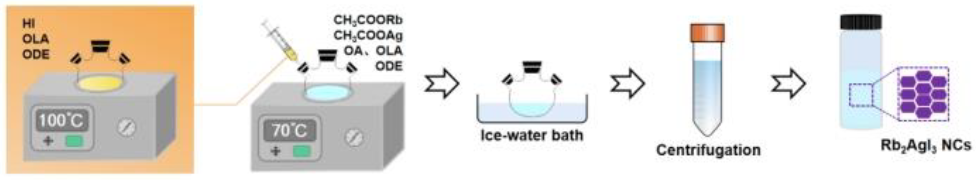
Abstract
:1. Introduction
2. Materials and Methods
2.1. Materials
2.2. Synthesis of Rb2AgI3 NCs
2.3. Characterizations
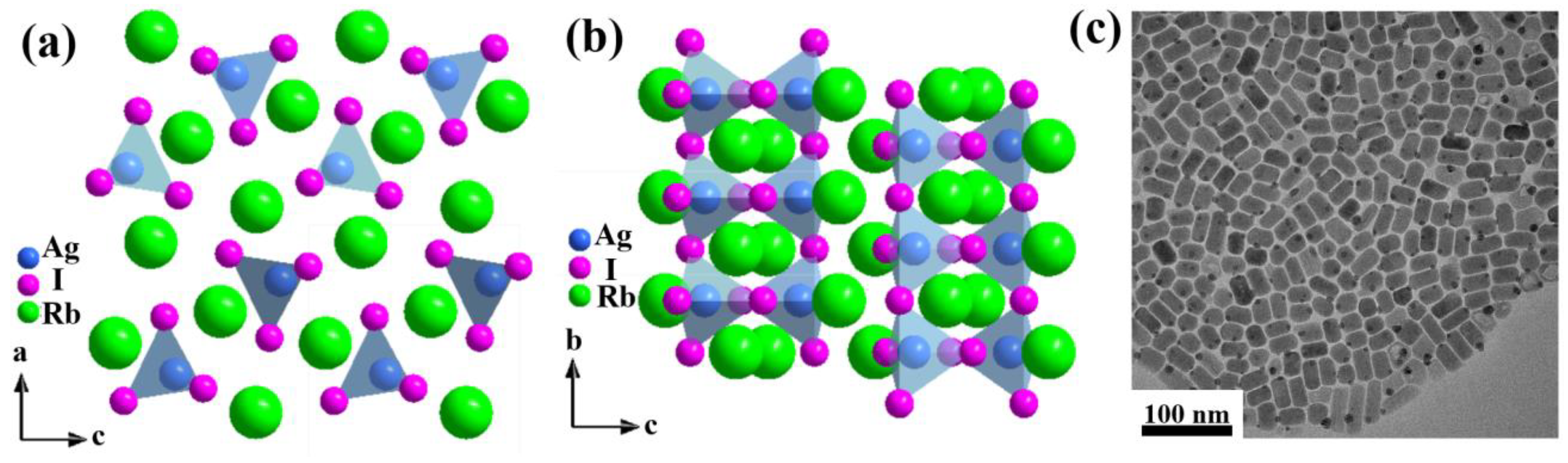
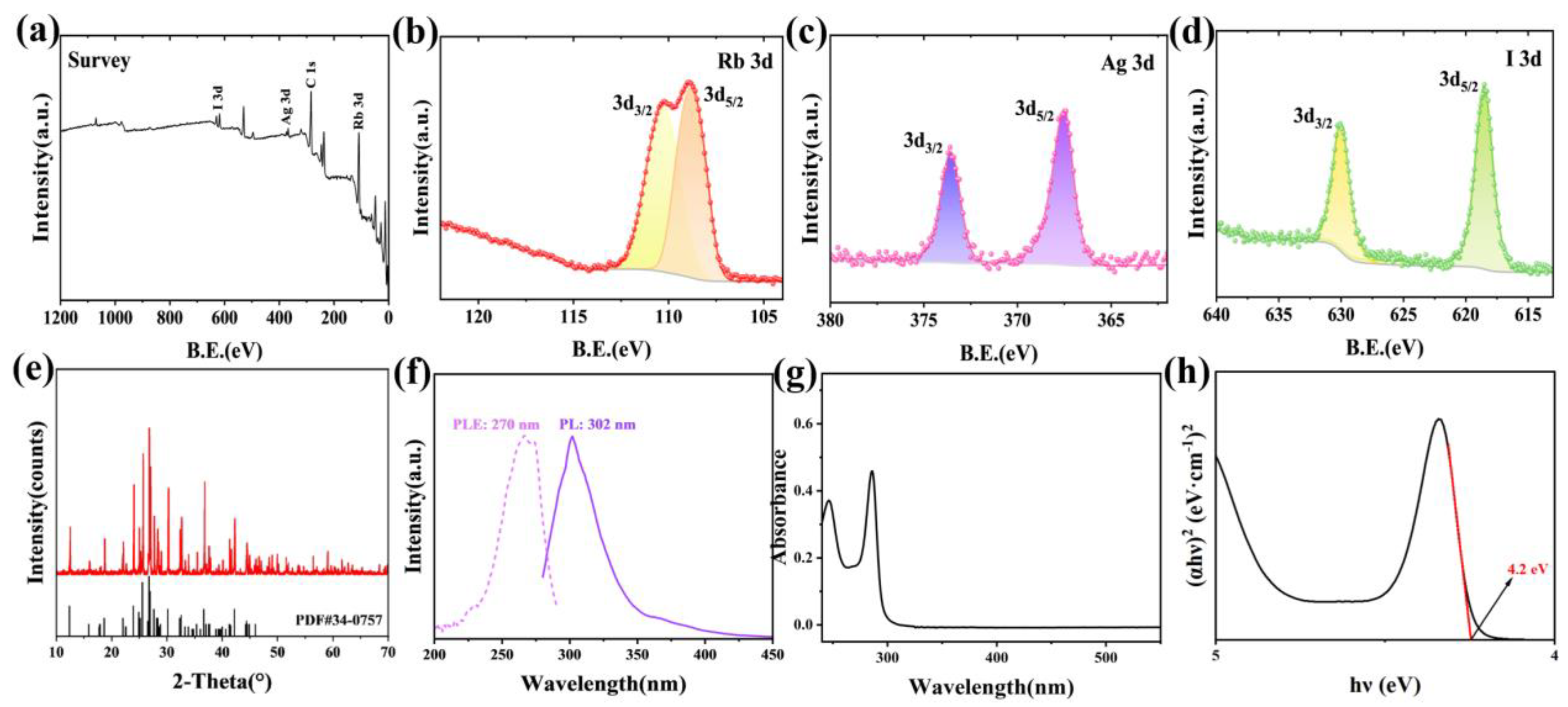
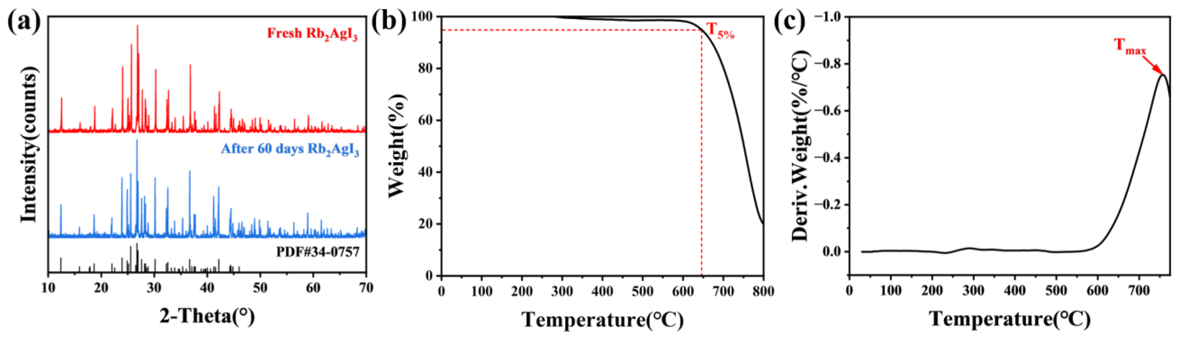
3. Results and Discussion
4. Conclusions
Supplementary Materials
Author Contributions
Funding
Data Availability Statement
Conflicts of Interest
References
- Ren, A.; Wang, H.; Zhang, W.; Wu, J.; Wang, Z.; Penty, R.V.; White, I.H. Emerging light-emitting diodes for next-generation data communications. Nat. Electron. 2021, 4, 559–572. [Google Scholar] [CrossRef]
- Chen, J.; Xiang, H.; Wang, J.; Wang, R.; Li, Y.; Shan, Q.; Xu, X.; Dong, Y.; Wei, C.; Zeng, H. Perovskite White Light Emitting Diodes: Progress, Challenges, and Opportunities. ACS Nano 2021, 15, 17150–17174. [Google Scholar] [CrossRef]
- Lin, K.; Xing, J.; Quan, L.N.; de Arquer, F.P.G.; Gong, X.; Lu, J.; Xie, L.; Zhao, W.; Zhang, D.; Yan, C.; et al. Perovskite light-emitting diodes with external quantum efficiency exceeding 20 per cent. Nature 2018, 562, 245–248. [Google Scholar] [CrossRef] [PubMed]
- Matafonova, G.; Batoev, V. Recent advances in application of UV light-emitting diodes for degrading organic pollutants in water through advanced oxidation processes: A review. Water Res. 2018, 132, 177–189. [Google Scholar] [CrossRef] [PubMed]
- Liang, S.; Sun, W. Recent Advances in Packaging Technologies of AlGaN-Based Deep Ultraviolet Light-Emitting Diodes. Adv. Mater. Technol. 2022, 7, 2101502. [Google Scholar] [CrossRef]
- Kim, C.; Ji, T.; Eom, J.B. Determination of organic compounds in water using ultraviolet LED. Meas. Sci. Technol. 2018, 29, 045802. [Google Scholar] [CrossRef]
- Moser, R.; Kunzer, M.; Goßler, C.; Köhler, K.; Pletschen, W.; Schwarz, U.T.; Wagner, J. Laser processing of gallium nitride–based light-emitting diodes with ultraviolet picosecond laser pulses. Opt. Eng. 2012, 51, 114301. [Google Scholar] [CrossRef]
- Matsumoto, T.; Tatsuno, I.; Hasegawa, T. Instantaneous Water Purification by Deep Ultraviolet Light in Water Waveguide: Escherichia Coli Bacteria Disinfection. Water 2019, 11, 968. [Google Scholar] [CrossRef] [Green Version]
- Hirayama, H.; Maeda, N.; Fujikawa, S.; Toyoda, S.; Kamata, N. Recent progress and future prospects of AlGaN based high-efficiency deep-ultraviolet light-emitting diodes. Jpn. J. Appl. Phys. 2014, 53, 100209. [Google Scholar] [CrossRef]
- Wang, C.; Jin, Y.; Zhang, J.; Li, X.; Wu, H.; Zhang, R.; Yao, Q.; Hu, Y. Linear charging-discharging of an ultralong UVA per-sistent phosphor for advanced optical data storage and wide-wavelength-range detector. Chem. Eng. J. 2023, 453, 139558. [Google Scholar] [CrossRef]
- Chen, M.H.; Xing, D.; Su, V.C.; Lee, Y.C.; Ho, Y.L.; Delaunay, J.J. GaN Ultraviolet Laser based on Bound States in the Continuum (BIC). Adv. Opt. Mater. 2023, 11, 2201906. [Google Scholar] [CrossRef]
- Liu, M.; Jiang, M.; Zhao, Q.; Tang, K.; Sha, S.; Li, B.; Kan, C.; Shi, D.N. Ultraviolet Exciton-Polariton Light-Emitting Diode in a ZnO Microwire Homojunction. ACS Appl. Mater. Interfaces 2023, 15, 13258–13269. [Google Scholar] [CrossRef]
- Pan, S.; Lu, W.; Chu, Z.; Li, G. Deep Ultraviolet Emission from Water-Soluble SnO2 Quantum Dots Grown via a Facile “Top-Down” Strategy. J. Mater. Sci. Technol. 2015, 31, 670–673. [Google Scholar] [CrossRef]
- Wang, J.; Feng, M.; Zhou, R.; Sun, Q.; Liu, J.; Huang, Y.; Zhou, Y.; Gao, H.; Zheng, X.; Ikeda, M.; et al. GaN-based ultraviolet microdisk laser diode grown on Si. Photonics Res. 2019, 7, B32. [Google Scholar] [CrossRef]
- Chen, F.; Ji, X.; Lau, S.P. Recent progress in group III-nitride nanostructures: From materials to applications. Mater. Sci. Eng. R Rep. 2020, 142, 100578. [Google Scholar] [CrossRef]
- Kwak, J.; Lim, J.; Park, M.; Lee, S.; Char, K.; Lee, C. High-Power Genuine Ultraviolet Light-Emitting Diodes Based On Colloidal Nanocrystal Quantum Dots. Nano Lett. 2015, 15, 3793–3799. [Google Scholar] [CrossRef]
- Lim, K.-G.; Han, T.-H.; Lee, T.-W. Engineering electrodes and metal halide perovskite materials for flexible/stretchable perovskite solar cells and light-emitting diodes. Energy Environ. Sci. 2021, 14, 2009–2035. [Google Scholar] [CrossRef]
- Zhou, Z.; Qiao, H.W.; Hou, Y.; Yang, H.G.; Yang, S. Epitaxial halide perovskite-based materials for photoelectric energy conversion. Energy Environ. Sci. 2021, 14, 127–157. [Google Scholar] [CrossRef]
- Cheng, L.; Jiang, T.; Cao, Y.; Yi, C.; Wang, N.; Huang, W.; Wang, J. Multiple-Quantum-Well Perovskites for High-Performance Light-Emitting Diodes. Adv. Mater. 2020, 32, 1904163. [Google Scholar] [CrossRef] [PubMed]
- Bae, S.R.; Heo, D.Y.; Kim, S.Y. Recent progress of perovskite devices fabricated using thermal evaporation method: Perspective and outlook. Mater. Today Adv. 2022, 14, 100232. [Google Scholar] [CrossRef]
- Zhou, C.; Lin, H.; He, Q.; Xu, L.; Woru, M.; Chaaban, M.; Lee, S.; Shi, X.; Du, M.; Ma, B. Low dimensional metal halide perovskites and hybrids. Mat. Sci. Eng. R Rep. 2019, 137, 38–65. [Google Scholar] [CrossRef]
- Han, Y.; Yue, S.; Cui, B. Low-Dimensional Metal Halide Perovskite Crystal Materials: Structure Strategies and Luminescence Applications. Adv. Sci. 2021, 8, 2004805. [Google Scholar] [CrossRef]
- Kumar, P.; Creason, T.D.; Fattal, H.; Sharma, M.; Du, M.H.; Saparov, B. Composition-Dependent Photoluminescence Properties and Anti-Counterfeiting Applications of A2AgX3 (A = Rb, Cs; X = Cl, Br, I). Adv. Funct. Mater. 2021, 31, 2104941. [Google Scholar] [CrossRef]
- Yao, M.; Zhang, Q.; Wang, D.; Chen, R.; Yin, Y.; Xia, J.; Tang, H.; Xu, W.; Yu, S. Lead-Free Halide CsAg2I3 with 1D Electronic Structure and High Stability for Ultraviolet Photodetector. Adv. Funct. Mater. 2022, 32, 2202894. [Google Scholar] [CrossRef]
- Zhang, Z.; Guo, X.; Huang, K.; Sun, X.; Li, X.; Zeng, H.; Zhu, X.; Zhang, Y.; Xie, R. Lead-free bright yellow emissive Rb2AgCl3 scintillators with nanosecond radioluminescence. J. Lumin. 2022, 241, 118500. [Google Scholar] [CrossRef]
- Zhang, Z.; Zhao, R.; Teng, S.; Huang, K.; Zhang, L.; Wang, D.; Yang, W.; Xie, R.; Pradhan, N. Color Tunable Self-Trapped Emissions from Lead-Free All Inorganic IA-IB Bimetallic Halides Cs-Ag-X (X = Cl, Br, I). Small 2020, 16, 2004272. [Google Scholar] [CrossRef] [PubMed]
- The Materials Project. Materials Data on Rb2AgI3 by Materials Project; The Materials Project: Washington, DC, USA, 2020. [Google Scholar]
- Zeng, Y.; Chen, W.; Deng, Y.; Gu, W.; Wu, C.; Guo, Y.; Huang, P.; Liu, F.; Li, H. FAPbBr3/Cs4PbBr6 Core/Shell Perovskite Nanocrystals with Enhanced Stability and Emission: Implications for LEDs. ACS Appl. Nano Mater. 2022, 5, 9534–9543. [Google Scholar] [CrossRef]
- Ansari, S.M.; Sinha, B.B.; Phase, D.; Sen, D.; Sastry, P.U.; Kolekar, Y.D.; Ramana, C.V. Particle Size, Morphology, and Chemical Composition Controlled CoFe2O4 Nanoparticles with Tunable Magnetic Properties via Oleic Acid Based Solvothermal Synthesis for Application in Electronic Devices. ACS Appl. Nano Mater. 2019, 2, 1828–1843. [Google Scholar] [CrossRef]
- Muro-Cruces, J.; Roca, A.G.; Lopez-Ortega, A.; Fantechi, E.; Del-Pozo-Bueno, D.; Estrade, S.; Peiro, F.; Sepulveda, B.; Pineider, F.; Sangregorio, C.; et al. Precise Size Control of the Growth of Fe3O4 Nanocubes over a Wide Size Range Using a Rationally Designed One-Pot Synthesis. ACS Nano 2019, 13, 7716–7728. [Google Scholar] [CrossRef] [Green Version]
- Wu, Z.; Zhao, Y.; Chen, X.; Guo, Y.; Wang, H.; Jin, Y.; He, P.; Wei, Q.; Wang, B. Preparation of polymeric carbon nitride/TiO2 heterostructure with NH4Cl as template: Structural and photocatalytic studies. J. Phys. Chem. Solids 2022, 164, 110629. [Google Scholar] [CrossRef]
- Fu, W.; Zhao, Y.; Wang, H.; Chen, X.; Liu, K.; Zhang, K.; Wei, Q.; Wang, B. Study on preparation, photocatalytic performance and degradation mechanism of polymeric carbon nitride/Pt/nano-spherical MoS2 composite. J. Phys. Chem. Solids 2022, 166, 110700. [Google Scholar] [CrossRef]
- Xie, L.; Chen, B.; Zhang, F.; Zhao, Z.; Jiang, T.; Wang, M.; Wu, Y.; Huang, L.; Song, W.; Liu, Y.; et al. Stability enhancement of Cs3Cu2I5 powder with high blue emission realized by Na+ doping strategy. J. Lumin. 2021, 239, 118333. [Google Scholar] [CrossRef]
- Sharma, M.; Yangui, A.; Whiteside, V.R.; Sellers, I.R.; Han, D.; Chen, S.; Du, M.H.; Saparov, B. Rb4Ag2BiBr9: A Lead-Free Visible Light Absorbing Halide Semiconductor with Improved Stability. Inorg. Chem. 2019, 58, 4446–4455. [Google Scholar] [CrossRef] [PubMed]
- Nairui, X.; Yehua, T.; Yali, Q.; Duoduo, L.; Ke-Fan, W. One-step solution synthesis and stability study of inorganic perovskite semiconductor Cs2SnI6. Sol Energy 2020, 204, 429–439. [Google Scholar] [CrossRef]
- Jiang, Y.; Zhang, H.; Qiu, X.; Cao, B. The air and thermal stabilities of lead-free perovskite variant Cs2SnI6 powder. Mater. Lett. 2017, 199, 50–52. [Google Scholar] [CrossRef]


Disclaimer/Publisher’s Note: The statements, opinions and data contained in all publications are solely those of the individual author(s) and contributor(s) and not of MDPI and/or the editor(s). MDPI and/or the editor(s) disclaim responsibility for any injury to people or property resulting from any ideas, methods, instructions or products referred to in the content. |
© 2023 by the authors. Licensee MDPI, Basel, Switzerland. This article is an open access article distributed under the terms and conditions of the Creative Commons Attribution (CC BY) license (https://creativecommons.org/licenses/by/4.0/).
Share and Cite
Deng, Y.; Zeng, Y.; Gu, W.; Huang, P.; Jin, G.; Liu, F.; Wei, J.; Li, H. Colloidal Synthesis and Ultraviolet Luminescence of Rb2AgI3 Nanocrystals. Crystals 2023, 13, 1110. https://doi.org/10.3390/cryst13071110
Deng Y, Zeng Y, Gu W, Huang P, Jin G, Liu F, Wei J, Li H. Colloidal Synthesis and Ultraviolet Luminescence of Rb2AgI3 Nanocrystals. Crystals. 2023; 13(7):1110. https://doi.org/10.3390/cryst13071110
Chicago/Turabian StyleDeng, Yuan, Yicheng Zeng, Wanying Gu, Pan Huang, Geyu Jin, Fangze Liu, Jing Wei, and Hongbo Li. 2023. "Colloidal Synthesis and Ultraviolet Luminescence of Rb2AgI3 Nanocrystals" Crystals 13, no. 7: 1110. https://doi.org/10.3390/cryst13071110