Article

Surface-Enhanced Raman Spectroscopy of Benzylpenicillin Using Silver Nanocrystals Modified with Moroccan Plant Extracts

by
Yasmine Fernine
1,2,*,
Natércia C. T. Martins
2,
Mustapha Taleb
1 and
Tito Trindade
2
1
Laboratory of Engineering Electrochemistry, Modeling and Environment, Department of Chemistry, Faculty of Sciences Dhar El Mahraz, Sidi Mohamed Ben Abdellah University, Fez 30000, Morocco
2
Department of Chemistry and CICECO—Aveiro Institute of Materials, University of Aveiro, 3810-193 Aveiro, Portugal
*
Author to whom correspondence should be addressed.
Crystals 2023, 13(7), 1105; https://doi.org/10.3390/cryst13071105
Submission received: 26 June 2023 / Revised: 5 July 2023 / Accepted: 11 July 2023 / Published: 16 July 2023
(This article belongs to the Special Issue Raman Spectroscopy of Crystalline Materials and Nanostructures)

Abstract

:
Green chemical routes for the synthesis of colloidal metal nanocrystals have been of great interest, namely in the context of nanosciences associated with biological applications. Among these methods, the synthesis of metal colloids using medicinal plant extracts originates nanocrystals having surfaces modified with chemical compounds of biological origin, which can be further explored in association with conventional pharmaceutics. In this context, the development of spectroscopic methods that seeks for understanding the potential benefits of using formulations that contain natural compounds and metal nanoparticles with therapeutic properties is of relevance. This research describes the chemical synthesis of silver colloids via the reduction of Ag(I) in the presence of distinct aqueous plant extracts. The selected extracts were obtained from Moroccan plants that have been used in traditional therapeutic practices over the centuries. The method led to stable colloids comprising polydispersed Ag nanocrystals that show surface-enhanced Raman scattering (SERS) activity. As an illustrative scenario, these colloids have been applied to the SERS detection of the natural β-lactam antibiotic benzylpenicillin, also known as penicillin G (PG). Our results indicate that all the Ag colloids tested with the different plant extracts are SERS-active for PG without showing detrimental interference from chemical adsorbates originated from the extracts. Therefore, this spectroscopic method can be further explored for monitoring nanoformulations of pharmaceuticals and metal colloids obtained using biological synthesis.

1. Introduction

Over the last two decades, there has been significant advancements in the study of colloidal inorganic nanocrystals using green chemistry techniques [1,2,3]. Among these systems, Ag colloids have been intensively investigated owing to their unique properties that render them attractive for a wide range of applications, such as in medical applications [1,2,3,4,5,6]. In fact, the therapeutic use of silver and its compounds (e.g., AgNO3) has been recognized for a long time, namely as an antimicrobial agent and cauterizing agent [4,5,6]. Additionally, Ag nanoparticles have been synthesized using various plant extracts that contain phytochemicals of recognized medical value in their chemical composition, such as antioxidant and anti-inflammatory substances [7]. Examples of green syntheses of Ag colloids using plant extracts include the reduction of Ag(I) using extracts of Ocimum sanctum, Azadirachta indica, and Aloe vera [8]. In another study, Ag nanoparticles were synthesized using the aqueous extract of Acacia cyanophyte, which contains reducing agents and also capping agents [9]. Similarly, the aqueous leaf extract of Cucumis sativus was used to synthesize Ag nanoparticles in a facile, eco-friendly, reliable, and cost-effective manner [10]. The aqueous stem extract of E. spiralis was also used to synthesize Ag colloids, and the physicochemical parameters were varied to control their morphology, stability, particle size distribution, and surface charge [11].
The use of plant extracts in those synthesis techniques offers a sustainable and cost-effective alternative for the synthesis of Ag colloids, but there are also several drawbacks associated with their reproducible application, namely, the diversity and complexity of the chemical compositions of the extracts, which are further influenced by geographical factors and crop seasons. Although there has been progress on the morphological control of the particles obtained by such processes, the colloids generally obtained in such syntheses lack the level of size monodispersity and shape control that can be found with conventional chemical synthesis [12,13]. On the other hand, the use of phytochemicals of recognized medical value as coatings of metal colloids (e.g., Ag, Au), which can be found in several emergent nanomedicines and analytical methods [14,15,16,17], offers an undeniable field for the development of new agents for medicine [7]. Moreover, conventional pharmaceutics can be associated with such nanoformulations, which enriches this field of research. In this regard, the development of methods for monitoring the surface chemistry of biosynthesized metal colloids is important to gain scientific understanding of the potential therapeutic benefits.
In the above context, Raman spectroscopic studies can be particularly helpful by taking advantage on the surface-enhanced Raman scattering effect observed when certain molecules are close to metal surfaces, such as colloidal Ag nanocrystals [16,18]. The SERS effect occurs when a molecule is adsorbed onto a metal surface, resulting in an enhancement of its Raman scattering signal due to local electromagnetic field amplification and, with a less contribution, also due to charge transfer occurring in the formed surface metal–adsorbate complex. The observed Raman enhancement depends on several factors, such as the metal substrates’ characteristics, instrumentation, chemical nature of the samples under analysis, among others. In colloidal Ag nanoparticles, the SERS signal of molecular adsorbates can increase many orders of magnitude allowing for trace detection due to increased sensitivity and selectivity. As such, the SERS effect has been explored as a powerful tool in studies of analytes of biomedical interest, such as nanoformulations containing pharmaceutical compounds.
In this study, Moroccan plant extracts of rosemary, marrube, and ephedra were used to prepare Ag colloids. The effect of various experimental parameters on the optical and morphological properties of the Ag nanocrystals was systematically studied. The Ag colloids were then investigated as SERS substrates for the detection of penicillin G (PG), which was deliberately added to the colloidal nanoformulation. This type of research is of relevance for SERS applications of biologically synthesized metal nanoparticles because such an effect depends strongly on the nanoparticles employed as the substrates, namely, on their surface chemistry. Our main goal was to demonstrate that SERS is a valuable spectroscopic method for detecting pharmaceutics, even when the Ag colloids employed as substrates were generated in the presence of a complex mixture of phytochemical compounds.

2. Materials and Methods

2.1. Materials

Silver acetate (AgC2H3O2, 99.9%) was purchased from Sigma-Aldrich, Sofia, Bulgaria, and penicillin G sodium salt (PG: C16H17N2NaO4S) was obtained from Alfa Aesar. The harvesting of the three medicinal plant samples was conducted in the Fez-Meknes region during the period of March 2022, which is in the central north of Morocco. Rosemary was collected from the El Hajeb city area, marrube from the Sefrou city area, and ephedra from the Taounate city area. Subsequently, the samples were dried in the shade until a constant humidity level was achieved, followed by grinding to obtain a fine powder. Ultra-pure water (18.2 MΩ·cm, 25 °C, MilliQ Millipore, Burlington, MA, USA) was employed for the preparation of aqueous plant extracts, silver colloids, and aqueous solutions of analytes.

2.2. Preparation of Plant Extracts

The harvested plants were washed thoroughly with distilled water, dried in the shade at room temperature for two weeks, and then crushed into a fine powder using an electric mixer. The extraction was carried out using ultra-pure water. For this purpose, 10 g of the powder obtained from each plant was mixed with 100 mL of ultra-pure water, and the mixture was sonicated at room temperature for 30 min. The obtained aqueous extracts underwent centrifugation at 9000 rpm for 10 min, followed by filtration. They were then refrigerated in a sealed, light- and oxygen-protected container until ready for use.

2.3. Green Synthesis of Ag Colloids

The colloidal Ag nanocrystals were obtained by treating 1 mM aqueous solution of AgC2H3O2 with a volume of the aqueous plant extract to reduce cationic silver to the elemental form [18]. Typically, a 42 mL aqueous solution of Ag(I) (1 mM) was brought to reflux and vigorously stirred for 10 min. Then, 7 mL of the aqueous plant extract was added dropwise to the boiling solution. All Ag colloids were prepared using this procedure, but for each plant extract, the respective amount in the starting solution varied as follows: 2, 3, 4, and 5 mg/mL. After a few minutes of reaction, a color change was observed, indicating the formation of the silver colloid. The mixture was refluxed for 60 min at 80 °C and then slowly cooled to room temperature. The obtained colloid was centrifuged at 9000 rpm for 10 min, and the collected solid was re-dispersed in ultra-pure water for subsequent experiments.

2.4. Raman Spectroscopic Measurements

The Ag colloids were investigated as SERS substrates for the detection of PG dissolved in aqueous samples. Also, control experiments were performed for the Ag colloids and for PG, both in solid and solution. To prepare the SERS samples, a volume of 0.3 mL of the Ag colloid was mixed with 30 µL of PG solution (0.01 mol.dm−3). The resulting mixtures were then incubated for 10 min at room temperature using a mini-rotor. After the incubation period, an aliquot of the solution was transferred into a glass tube for Raman measurements. Raman spectra were measured using a Bruker MultiRAM FT-Raman spectrometer (Nd:YAG 1064 nm laser at 350 mW) with the resolution set at 4 cm−1 using 1000 scans per spectrum.

2.5. Instrumentation

The UV/VIS absorption spectra of the colloids were measured using a Jasco V-560 spectrophotometer in the range of 300–800 nm. Fourier-transform infrared (FTIR) spectra of the colloids were acquired over the range of 4000–200 cm−1 using a Bruker Optics Tensor 27 spectrometer coupled with a horizontal attenuated total reflectance (ATR) cell. Scanning transmission electron microscopy (STEM) images were obtained with a SU-70 Hitachi instrument fitted with energy-dispersive spectroscopy (EDS) accessory, operated at 15 kV or 4 kV. To prepare the STEM samples, a drop of silver colloids was placed on carbon-coated copper grids and dried at room temperature. Zeta potential measurements were performed using a Zeta Sizer Nano Series (Malvern) equipped with a standard 633 nm laser.

3. Results

A series of colloidal Ag nanocrystals were prepared by the reduction of silver (I) acetate in the presence of aqueous extracts of three selected Moroccan plants: rosemary, marrube, and ephedra. It is well known that the experimental parameters in the biosynthesis of metal nanocrystals have strong influence not only on their morphology and average size but also on the colloids’ stability [19,20]. Hence, the increase in the reaction temperature was reported to favor small-sized Ag nanocrystals and the amount of extract had an overall influence on the biosynthesis of Ag nanocrystals [21,22]. In order to establish a systematic use of the Ag colloids for the SERS studies, all the green syntheses were performed at 80 °C over 1 h for different amounts of each plant aqueous extract. The ensuing Ag hydrosols showed long-term colloidal stability. The metal nanocrystals were characterized for their optical and morphological properties, as detailed below according to the plant extract used in the biosynthesis.

3.1. Characterization of Colloidal Ag Nanocrystals

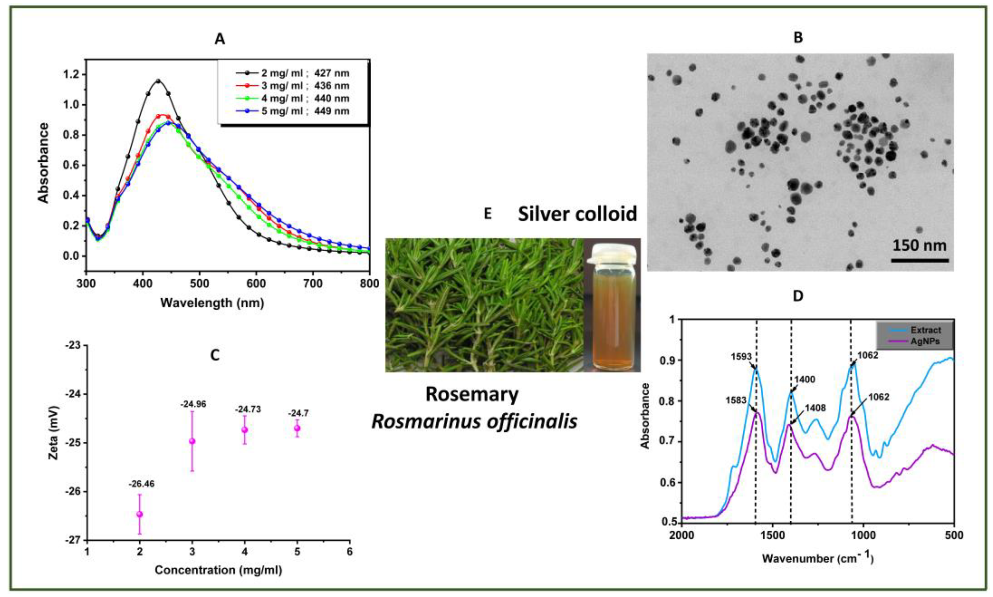
3.1.1. Rosemary Extract

Figure 1 shows the characterization data for samples of Ag nanocrystals obtained in the presence of the rosemary extract. The UV/VIS spectra of the colloidal samples show the characteristic localized surface plasmon resonance (LSPR) band of Ag hydrosols, which, depending on the extract amount used in the synthesis, shows the absorption maximum localized within the 427–449 nm wavelength range. As the amount of extract increased, the wavelength of maximum absorption for the LSPR red-shifted and a slight band broadening was observed. This observation agrees with the formation of larger Ag nanocrystals, as confirmed by the TEM images shown, which revealed polydispersed particles (ranging 30–40 nm average size) for the different amounts of rosemary extract used for the samples analyzed. The zeta potential of the Ag colloids prepared with less amounts of extract was −26.5 mV and just varied slightly with the increase in extract and kept the pH at 4.5. The high negative zeta potential of the colloids accounts for the stability of the colloids due to inter-particle electrostatic repulsions. This is a first indication for the presence of anionic species at the colloidal Ag nanocrystals’ surfaces, originated from ionizable organic compounds of the plant extract. The nanocrystalline powders collected from the Ag colloids were characterized by ATR-FTIR spectroscopy that showed common vibrational bands with the FTIR spectra of the rosemary extract, also shown for comparative purposes and as indicated in the spectra.

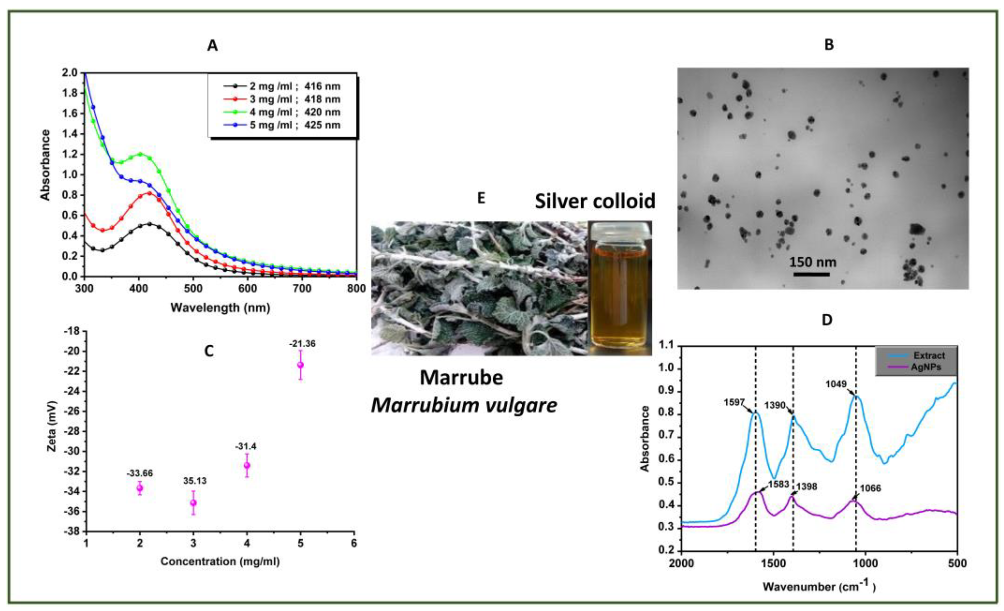
3.1.2. Marrube Extract

Figure 2 shows the characterization data for samples of Ag nanocrystals obtained in the presence of the marrube extract. Similarly to the Ag colloids prepared in the presence of the rosemary extract, the UV/VIS spectra also show the LSPR band at a wavelength that varies depending on the amount of extract used, nonetheless, slightly shifted to a lower wavelength range (416–425 nm). The TEM analysis also shows polydispersed particles which ranged 25–75 nm average size, depending on the amount of extract employed in the synthesis. Regardless of the amount of extract used, the zeta potential of the Ag colloids was observed as negative, varying between −35.1 mV and −21.4 mV, at a pH of 4.5. This is in line with the observation of stable colloids, as already mentioned above. The ATR-FTIR spectra of the Ag nanocrystals show strong vibrational bands in the 500–2000 cm−1 region, which are also observed in the corresponding spectrum of the marrube extract.

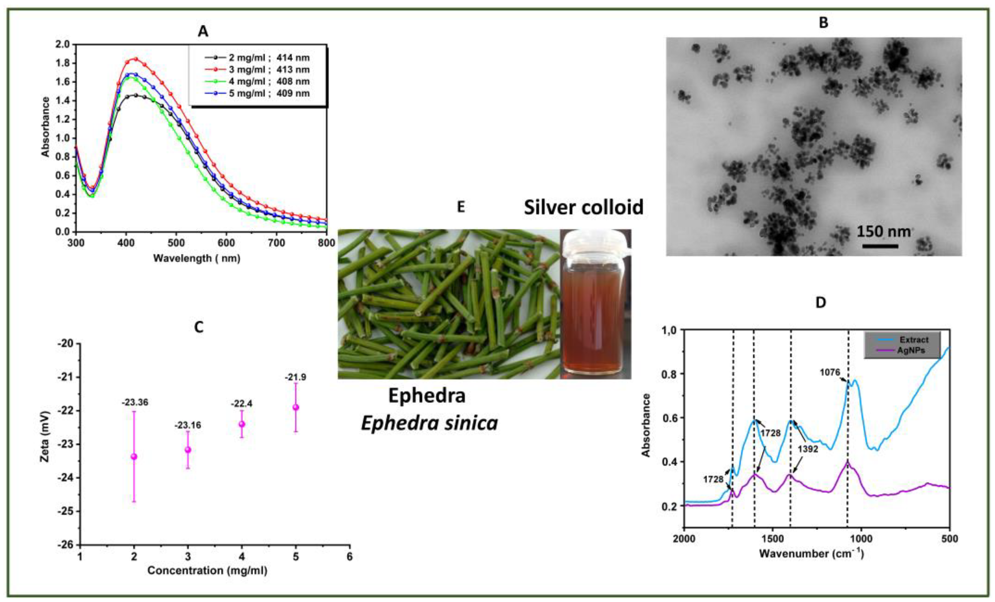
3.1.3. Ephedra Extract

Figure 3 shows the characterization data for samples of Ag nanocrystals obtained in the presence of the ephedra extract. As noted above for the Ag samples obtained in the presence of the other plant extracts, the UV/VIS spectra of the colloidal samples also show the LSPR band maximum at a wavelength that depends on the amount of extract employed in the synthesis. In this case, the wavelength range (408–414 nm) is observed at a lower wavelength region as compared both to the rosemary and marrube syntheses. Furthermore, there is not a marked shift on the LSPR band by varying the amount of extract. Conversely, band broadening is more pronounced in this case for all the samples analyzed, which might indicate that the Ag nanocrystals tend to form agglomerates, as observed in the TEM image. In fact, for each extract plant used in the Ag colloids synthesis, the TEM analysis performed on the Ag nanocrystals obtained for a higher amount of extract (5 mg/mL) showed polydispersed nanoparticles forming agglomerates, among which the largest were observed when using the ephedra extract (Figure 4). As observed for the rosemary and marrube extracts, stable Ag colloids were prepared in the presence of the ephedra extract, showing a high negative zeta potential and vibrational bands in the ATR-FTIR spectra, which are consistent with the presence of organic anionic adsorbates at the surfaces.

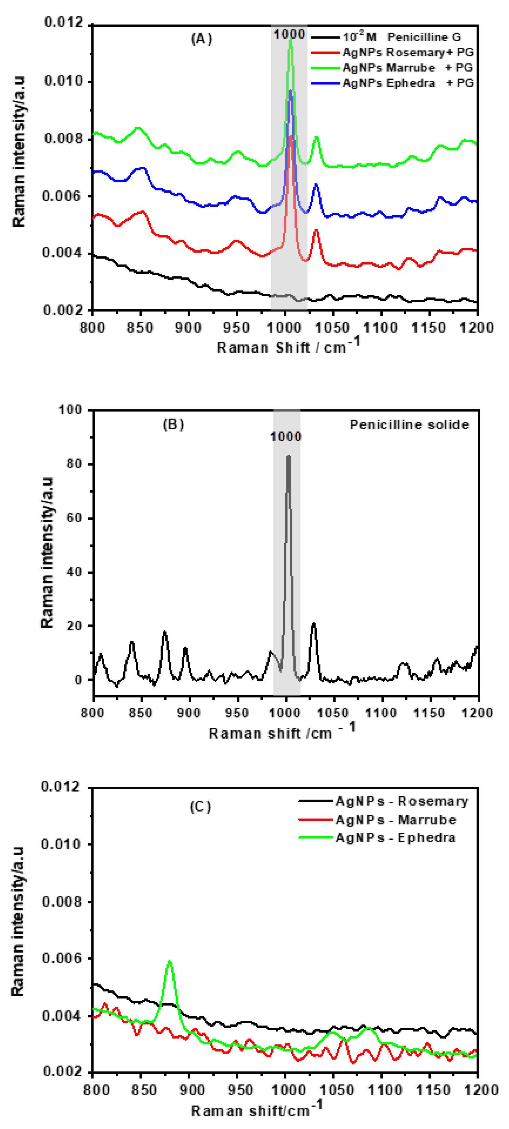
3.2. SERS Detection of Penicillin G Using Ag Colloids Prepared in Plant Extracts

The Ag colloids characterized above were then used as SERS substrates for sensing PG dissolved in aqueous solutions. The recorded ATR-FTIR spectra were consistent with the presence of organic adsorbates at the metal surfaces. Figure 5 shows the Raman spectrum of PG in an aqueous solution (0.01 M, pH = 5) when contacted with the distinct Ag colloids. For comparative purposes, Figure 5 also shows the Raman spectra of solid PG and of the Ag colloids modified with the plant extracts.
The more intense vibrational band observed at 1000 cm−1 in the Raman spectrum of solid PG was used as a spectroscopic signature for this compound [23,24,25]. The Raman spectra of the Ag colloids do not show any enhanced Raman signals of the extract compounds, which suggests that, in principle, the extracts themselves do not interfere with the PG detection. Indeed, the Raman spectra of PG in the Ag colloids clearly show this band, which are not observed for a PG solution of equal concentration but in the absence of Ag colloids.

4. Discussion

In this research, a series of aqueous colloids containing dispersed Ag nanocrystals were prepared by reducing silver (I) acetate in the presence of the plant extracts rosemary, marrube, and ephedra. All the aqueous extracts resulted in long-term stable metal colloids after the reduction of Ag(I) due to phytochemicals that act as reducing agents. Plant extracts comprise a complex mixture of phytochemicals even by considering only those mainly soluble in the aqueous phase during extraction. Among these phytochemicals, aqueous plant extracts typically contain polyphenols (flavonoids and phenolic acids), organic acids, glycosides, and some alkaloids [12,26]. Although the discussion of the chemical composition of the aqueous extracts used here is out of the scope of this research, it is important to note that polyphenols are well known for their antioxidant properties and citric acid, a reducing agent widely employed in metal nanoparticles syntheses, can be also found in certain plant extracts among other organic acids, such as malic acid and tartaric acid.
The aqueous plant extracts employed in the colloidal synthesis contain phytochemicals that have the ability to reduce dissolved cationic silver to form elemental Ag. As shown above (Figure 1, Figure 2 and Figure 3), the three extracts investigated led to the formation of metal colloids characterized by strong absorption in the visible due to the LSPR band observed in the respective UV/VIS spectrum. The LSPR band is observed at a wavelength and with a profile that depend on the sample under analysis, such as particle size, shape, and dispersing medium [27,28]. This well-known optical behavior has been widely reported for colloids containing Ag nanocrystals dispersed in a dielectric medium, such as water. The TEM images confirm the presence of polydispersed Ag nanocrystals in aliquots of the colloidal samples, whose average dimensions (21–25 nm) vary slightly depending on the extract used. However, both the type of extract and its amount in the synthesis reaction had a marked effect on the polydispersity of the Ag colloids, thus altering the LSPR band profile observed in the UV/VIS spectra (Figure 1, Figure 2 and Figure 3). The band broadening was observed mostly for the Ag colloids obtained in the presence of the rosemary and ephedra extracts and as the amount of extract in the reaction medium increased. The colloidal synthesis in the presence of the marrube extract led to more uniform Ag nanocrystals, though far from monodispersed as those normally obtained by conventional chemical synthesis. Regardless of the type of extract used, an increase in the amount of plant extract in the reaction medium caused an increase in the polydispersity and particles’ average size. An increase in the amount of reducing agent in relation to the concentration of metal precursor is usually associated to a decrease in the average size of the resulting metal nanocrystals, which was not observed in these experiments. A possible explanation for this counter-intuitive result relies on the effect of the extract in the agglomeration state of the particles, which increased as the amount of extract increased. This is in line with the TEM images depicted in Figure 4, which show bigger Ag nanocrystals when these were obtained using a high amount of plant extract. Note the presence of numerous agglomerates of the primary particles for the sample obtained in the presence of the ephedra extract (5 mg/mL).
The aqueous plant extracts contain phytochemicals that not only act as reducing agents but also as colloidal stabilizers. As pointed above, a common organic acid found in certain plant extracts is citric acid, which was employed as the reducing and capping agent in the synthesis of metal hydrosols. Besides organic acids, other phytochemicals can play a similar dual role, thus reducing the metal salt and originating anionic species that protect the metal nanocrystals against agglomeration. The presence of such species at the particles’ surfaces accounts for the observed colloidal stability of all the Ag colloids prepared with the different extracts. The zeta potential measurements (Figure 1, Figure 2 and Figure 3) were performed to monitor the surface charge of the Ag colloids, which, despite some slight variations, showed a negative surface charge below −22 mV for all the samples. This agrees with the presence of anionic species as capping agents over the colloidal Ag nanocrystals, which originate from phytochemicals used in the reduction chemical reaction. Furthermore, the ATR-FTIR spectra of Ag nanocrystals treated with the plant extracts show strong vibrational bands characteristic of organic capping agents, such as polyphenols, organic acids, and alkaloids. Although this is an open issue due to the complexity of the chemical composition of the different extracts, tentative assignments can be made to the vibrational bands depicted in Figure 1, Figure 2 and Figure 3, as illustrative of the presence of the organic capping agents at the particles’ surfaces. Hence, the bands observed in the 1579–1608 cm−1 range can be associated with C-H and C=C-C (aromatic) stretching modes. The broad bands observed between 1382 and 1408 cm−1 are possibly due to the CO-H in-plane bending mode coupled with ring-stretching and C-H bending vibrations. Lastly, the strong bands observed in the 1047–1263 cm−1 region are characteristic of stretching vibrations (C-O; C-C) and in-plane bending modes (C-H). All the ATR-FTIR spectra showed strong and broad vibrational bands in the 2800–3600 cm−1 region, characteristic of stretching modes of the O-H and -CHn groups [26,29,30].
The presence of phytochemical adsorbates at the Ag nanocrystals’ surfaces raises the question about the SERS activity of these metal colloids, which is a central aspect in this research. The Ag colloids prepared in the presence of different plant extracts were tested for the SERS detection of PG dissolved in aqueous samples, originating characteristic Raman bands of this antibiotic (Figure 5). In the 975–1025 cm−1 spectral region, none of the Ag colloids showed Raman bands that could interfere with PG detection in this spectral window with the exception of the ephedra extract, which showed a medium band at 875 cm−1 and weak bands in the 1025–1100 cm−1 region. No other significant bands were observed in the Raman spectra of the colloids. The Raman spectra of aliquots of PG solution dissolved in the Ag colloids were dominated by a strong and sharp band at 1000 cm−1, which was strongly enhanced compared to the Raman spectrum of PG in an aqueous solution of equal concentration. This is an important observation because it indicates that, in principle, Ag colloids stabilized by phytochemical adsorbates can be employed as SERS substrates for monitoring other chemical species, such as pharmaceutics, as exemplified here for the case of PG.
The chemical form of PG present in an aqueous solution depends on the solution pH, among other factors that can also contribute to the degradation of this compound, namely, into the allergen compound penicilloic acid [31,32]. The latter can be formed by alkaline hydrolysis of PG, due to carboxylation and opening of the β-lactam ring. Here, the SERS detection of PG was performed using aqueous samples at a pH of 5. In these conditions, the carboxylic group of PG is deprotonated, and the resulting ionized form predominates. The Raman band observed (Figure 5A) at about 1000 cm−1 was used as a diagnosis band for PG [25,32,33,34]. Although the literature reports different assignments for this band, in fact, it is not observed in the Raman spectrum of penicilloic acid, unlike its observation in the Raman spectrum of PG, which is in line with the β-lactam ring vibration. This indicates that PG still predominates as a molecular adsorbate in the Ag colloids reported here. Additionally, penicilloic acid shows a strong band at 882 cm−1 due to a carboxylic group, which was not detected in the SERS spectra of PG in the Ag colloids containing the plant extracts.
The above discussion suggests that the use of Ag nanoparticle substrates modified with plant extracts for SERS detection, namely for identifying PG dissolved in water, is a method with potential but still requires further investigation. One of the advantages of using this type of colloid is the possibility of up-scaling substrates production by greener and cheaper methods as compared to conventional synthesis. Another advantage worth mentioning is the use of a detection method using SERS substrates that are themselves modified on the surface with compounds of biological interest. While this could also be a limitation in its use because their presence on the surface of the particles could limit the SERS activity of the Ag nanoparticles, the results presented above show that the detection of PG was still observed at least in the indicated experimental conditions. On the other hand, there are limitations to consider, such as the variability in the characteristics of plant extracts which, via the biological synthesis of the colloids, results in SERS substrates with different properties and therefore with enhancement factors that can vary considerably. The last point reinforces the importance of conducting more detailed studies on the nature of the surface of metal nanocrystals obtained by green synthesis methods, for which the SERS effect is also a valuable tool.

5. Conclusions

In summary, this research demonstrated the use of aqueous extracts of rosemary, marrube, and ephedra plants in the green chemical synthesis of colloidal Ag nanocrystals. The Ag colloids show colloidal stability due to surface capping by organic molecules present in the plant extracts. This type of organic coating does not preclude the use of the Ag colloids as active SERS substrates for the detection of certain molecules, such as penicillin G. Although our studies focus on the detection of PG dissolved in aqueous solutions, in principle, the Ag nanocrystals modified with plant extracts can be used for the SERS identification of other chemical compounds in other types of matrices. However, several challenges should be considered, such as interference due to the complexity of the medium and enhancement factor improvement. Still, these findings are particularly relevant because they demonstrate the potential of Raman methods applied to medical approaches that explore phytochemicals with therapeutic value together with well-known antimicrobial agents, such as silver and β-lactam antibiotics. The intersection of scientific knowledge in conventional pharmaceutical products and chemical compounds of natural origin has lately been explored for the potential of new discoveries. In this sense, this research is not only of interest in the specific domain of Raman spectroscopy applied to nanocrystals but might also contribute to other scientific fields by following a translational approach.

Author Contributions

Conceptualization, Y.F., M.T. and T.T.; methodology, Y.F. and T.T.; validation, Y.F. and N.C.T.M.; investigation, Y.F. and N.C.T.M.; resources, M.T. and T.T.; writing—original draft preparation, Y.F.; writing—review and editing, Y.F., N.C.T.M., M.T. and T.T.; visualization, Y.F. and T.T.; supervision, M.T. and T.T.; project administration, M.T. and T.T.; funding acquisition, T.T. All authors have read and agreed to the published version of the manuscript.

Funding

N.C.T. Martins is funded by National funds (OE), through FCT- Fundação para a Ciência e Tecnologia, I.P., in the scope of the framework contract foreseen in the numbers 4, 5 and 6 of the article 23, of the Decree-Law 57/2016, of August 29, changed by the law 57/2017,of July 19. This work was developed within the scope of the project CICECO-Aveiro Institute of Materials, UIDB/50011/2020, UIDP/50011/2020 and LA/P/0006/2020, financed by national funds through the FCT/MEC (PIDDAC).

Conflicts of Interest

The authors declare no conflict of interest.

References

  1. Shukla, A.K. Green Synthesis, Characterization and Applications of Nanoparticles; Iravani, S., Ed.; Elsevier: Amsterdam, The Netherlands, 2019. [Google Scholar]
  2. Iravani, S. Green synthesis of metal nanoparticles using plants. Green Chem. 2011, 13, 2638–2650. [Google Scholar] [CrossRef]
  3. Duan, H.; Wang, D.; Li, Y. Green chemistry for nanoparticle synthesis. Chem. Soc. Rev. 2015, 44, 5778–5792. [Google Scholar] [CrossRef]
  4. Klasen, H.J. A historical review of the use of silver in the treatment of burns. II. Renewed interest for silver. Burns 2000, 26, 131–138. [Google Scholar] [CrossRef]
  5. Rai, M.; Yadav, A.; Gade, A. Silver nanoparticles as a new generation of antimicrobials. Biotechnol. Adv. 2009, 27, 76–83. [Google Scholar] [CrossRef] [PubMed]
  6. Bernd, N.; Harald, F.; Krug, H.F.; Murray, H. 120 Years of Nanosilver History: Implications for Policy Makers. Environ. Sci. Technol. 2011, 45, 1177–1183. [Google Scholar]
  7. Shreyash, N.; Bajpai, S.; Khan, M.A.; Vijay, Y.; Tiwary, S.K.; Sonker, M. Green synthesis of nanoparticles and their biomedical applications: A review. ACS Appl. Nano Mater. 2021, 4, 11428–11457. [Google Scholar] [CrossRef]
  8. Panda, S.K.; Sen, S.; Roy, S.; Moyez, A. Synthesis of colloidal silver nanoparticles by reducing aqueous AgNO3 Using Green Reducing Agents. Mater. Today Proc. 2018, 5, 10054–10061. [Google Scholar] [CrossRef]
  9. Jalab, J.; Abdelwahed, W.; Kitaz, A.; Al-Kayali, R. Green synthesis of silver nanoparticles using aqueous extract of Acacia cyanophylla and its antibacterial activity. Heliyon 2021, 7, e08033. [Google Scholar] [CrossRef]
  10. Hemlata; Meena, P.R.; Singh, A.P.; Tejavath, K.K. Biosynthesis of silver nanoparticles using Cucumis prophetarum aqueous leaf extract and their antibacterial and antiproliferative activity against cancer cell lines. ACS Omega 2020, 5, 5520–5528. [Google Scholar] [CrossRef] [PubMed] [Green Version]
  11. Wan Mat Khalir, W.K.A.; Shameli, K.; Jazayeri, S.D.; Othman, N.A.; Che Jusoh, N.W.; Hassan, N.M. Biosynthesized silver nanoparticles by aqueous stem extract of Entada spiralis and screening of their biomedical activity. Front. Chem. 2020, 8, 620. [Google Scholar] [CrossRef]
  12. Perotti, G.F.; Pereira da Costa, L. Biological Materials in Reducing Agents in Colloidal Nanoparticles Synthesis; Nanoscience & Nanotechnology, Series; Mourdikoudis, S., Ed.; The Royal Society of Chemistry: London, UK, 2021; pp. 316–332. [Google Scholar]
  13. Ying, S.; Guan, Z.; Ofoegbu, P.C.; Clubb, P.; Rico, C.; He, F.; Hong, J. Green synthesis of nanoparticles: Current developments and limitations. Environ. Technol. Innov. 2022, 26, 102336. [Google Scholar] [CrossRef]
  14. Pereira, S.O.; Barros-Timmons, A.; Trindade, T. Biofunctionalisation of colloidal gold nanoparticles via polyelectrolytes assemblies. Colloid Polym. Sci. 2014, 292, 33–50. [Google Scholar] [CrossRef]
  15. Fateixa, S.; Raposo, M.; Nogueira, H.I.S.; Trindade, T. A general strategy to prepare SERS active filter membranes for extraction and detection of pesticides in water. Talanta 2018, 182, 558–566. [Google Scholar] [CrossRef] [PubMed]
  16. Chandrakala, V.; Aruna, V.; Angajala, G. Review on metal nanoparticles as nanocarriers: Current challenges and perspectives in drug delivery systems. Emergent Mater. 2022, 5, 1593–1615. [Google Scholar] [CrossRef]
  17. Xu, J.J.; Zhang, W.C.; Guo, Y.W.; Chen, X.Y.; Zhang, Y.N. Metal nanoparticles as a promising technology in targeted cancer treatment. Drug Deliv. 2022, 29, 664–678. [Google Scholar] [CrossRef]
  18. Al-Dhabi, N.A.; Esmail, G.A.; Duraipandiyan, V.; Valan Arasu, M.; Salem-Bekhit, M.M. Isolation, identification and screening of antimicrobial thermophilic Streptomyces sp. Al-Dhabi-1 isolated from Tharban hot spring, Saudi Arabia. Extremophiles 2016, 20, 79–90. [Google Scholar] [CrossRef]
  19. Jayapriya, M.; Dhanasekaran, D.; Arulmozhi, M.; Nandhakumar, E.; Senthilkumar, N.; Sureshkumar, K. Green synthesis of silver nanoparticles using Piper longum catkin extract irradiated by sunlight: Antibacterial and catalytic activity. Res. Chem. Intermed. 2019, 45, 3617–3631. [Google Scholar] [CrossRef]
  20. Al-Rajhi, A.M.; Salem, S.S.; Alharbi, A.A.; Abdelghany, T.M. Ecofriendly synthesis of silver nanoparticles using Kei-apple (Dovyalis caffra) fruit and their efficacy against cancer cells and clinical pathogenic microorganisms. Arab. J. Chem. 2022, 15, 103927. [Google Scholar] [CrossRef]
  21. Verma, A.; Mehata, M.S. Controllable synthesis of silver nanoparticles using Neem leaves and their antimicrobial activity. J. Radiat. Res. Appl. Sci. 2016, 9, 109–115. [Google Scholar] [CrossRef] [Green Version]
  22. Nasar, M.Q.; Shah, M.; Khalil, A.T.; Kakar, M.Q.; Ayaz, M.; Dablool, A.S.; Shinwari, Z.K. Ephedra intermedia mediated synthesis of biogenic silver nanoparticles and their antimicrobial, cytotoxic and hemocompatability evaluations. Inorg. Chem. Commun. 2022, 137, 109252. [Google Scholar] [CrossRef]
  23. El-Zahry, M.R.; Refaat, I.H.; Mohamed, H.A.; Rosenberg, E.; Lendl, B. Utility of surface enhanced Raman spectroscopy (SERS) for elucidation and simultaneous determination of some penicillins and penicilloic acid using hydroxylamine silver nanoparticles. Talanta 2015, 144, 710–716. [Google Scholar] [CrossRef]
  24. Filgueiras, A.L.; Paschoal, D.; Dos Santos, H.F.; Sant’Ana, A.C. Adsorption study of antibiotics on silver nanoparticle surfaces by surface-enhanced Raman scattering spectroscopy. Spectrochim. Acta Part A Mol. Biomol. Spectrosc. 2015, 136, 979–985. [Google Scholar] [CrossRef]
  25. Pinheiro, P.C.; Fateixa, S.; Trindade, T. SERS detection of penicillin G using magnetite decorated with gold nanoparticles. Magnetochemistry 2017, 3, 32. [Google Scholar] [CrossRef] [Green Version]
  26. Raja, S.; Ramesh, V.; Thivaharan, V. Green biosynthesis of silver nanoparticles using Calliandra haematocephala leaf extract, their antibacterial activity and hydrogen peroxide sensing capability. Arab. J. Chem. 2017, 10, 253–261. [Google Scholar] [CrossRef] [Green Version]
  27. Wiley, B.J.; Im, S.H.; Li, Z.Y.; McLellan, J.; Siekkinen, A.; Xia, Y. Maneuvering the surface plasmon resonance of silver nanostructures through shape-controlled synthesis. J. Phys. Chem. B 2006, 110, 15666–15675. [Google Scholar] [CrossRef] [PubMed]
  28. Wu, C.; Zhou, X.; Wei, J. Localized Surface Plasmon Resonance of Silver Nanotriangles Synthesized by a Versatile Solution Reaction. Nanoscale Res. Lett. 2015, 10, 354. [Google Scholar] [CrossRef] [PubMed] [Green Version]
  29. Espina, A.; Sanchez-Cortes, S.; Jurašeková, Z. Vibrational study (Raman, SERS, and IR) of plant gallnut polyphenols related to the fabrication of iron gall inks. Molecules 2022, 27, 279. [Google Scholar] [CrossRef]
  30. Wongsa, P.; Phatikulrungsun, P.; Prathumthong, S. FT-IR characteristics, phenolic profiles and inhibitory potential against digestive enzymes of 25 herbal infusions. Sci. Rep. 2022, 12, 6631. [Google Scholar] [CrossRef] [PubMed]
  31. Deshpande, A.D.; Baheti, K.G.; Chatterjee, N.R. Degradation of β-lactam antibiotics. Curr. Sci. 2004, 87, 1684–1695. [Google Scholar]
  32. Zhang, L.; Jin, Y.; Mao, H.; Zheng, L.; Zhao, J.; Peng, Y.; Zhang, Z. Structure-selective hot-spot Raman enhancement for direct identification and detection of trace penicilloic acid allergen in penicillin. Biosens. Bioelectron. 2014, 58, 165–171. [Google Scholar] [CrossRef]
  33. Clarke, S.J.; Littleford, R.E.; Smith, W.E.; Goodacre, R. Rapid monitoring of antibiotics using Raman and surface enhanced Raman spectroscopy. Analyst 2005, 130, 1019–1026. [Google Scholar] [CrossRef] [PubMed]
  34. Chen, Y.; Li, X.; Yang, M.; Yang, L.; Han, X.; Jiang, X.; Zhao, B. High sensitive detection of penicillin G residues in milk by surface-enhanced Raman scattering. Talanta 2017, 167, 236–241. [Google Scholar] [CrossRef] [PubMed]
Figure 1. Characterization data for colloidal Ag nanocrystals obtained in the presence of aqueous extracts of the rosemary plant. (A) UV/VIS spectra of colloids prepared with different amounts of extract, as indicated together with the wavelength for the LSPR band maximum; (B) TEM image for a sample prepared using an amount of 2 mg/mL in extract; (C) zeta potential measurements at pH 4.5 for a different amount of extract in the colloids; and (D) ATR-FTIR spectra of the Ag nanocrystals and rosemary extract; (E) Digital photographs: plant that originated the extract used in the synthesis of the indicated Ag colloid.
Crystals 13 01105 g001
Figure 2. Characterization data for colloidal Ag nanocrystals obtained in the presence of aqueous extracts of the marrube plant. (A) UV/VIS spectra of colloids prepared with different amounts of extract, as indicated together with the wavelength for the LSPR band maximum; (B) TEM image for a sample prepared using an amount of 2 mg/mL in extract; (C) zeta potential measurements at pH 4.5 for a different amount of extract in the colloids; and (D) ATR-FTIR spectra of the Ag nanocrystals and the rosemary extract; (E) Digital photographs: plant that originated the extract used in the synthesis of the indicated Ag colloid.
Crystals 13 01105 g002
Figure 3. Characterization data for colloidal Ag nanocrystals obtained in the presence of aqueous extracts of the ephedra plant. (A) UV/VIS spectra of colloids prepared with different amounts of extract, as indicated together with the wavelength for the LSPR band maximum; (B) TEM image for a sample prepared using an amount of 2 mg/mL in extract; (C) zeta potential measurements at pH 5.8 for a different amount of extract in the colloids; and (D) ATR-FTIR spectra of the Ag nanocrystals and the ephedra extract; (E) Digital photographs: plant that originated the extract used in the synthesis of the indicated Ag colloid.
Crystals 13 01105 g003
Figure 4. TEM images of Ag nanocrystals (scale bar 150 nm) obtained for the plant extract amount 5 mg/mL: (A) rosemary, (B) marrube, and (C) ephedra.
Crystals 13 01105 g004
Figure 5. Raman spectra: (A) PG (0.01 mol.dm−3) dissolved in Ag colloids prepared using distinct plant extracts and the Raman spectrum of an aqueous solution of PG 0.01 mol.dm−3; (B) solid PG; and (C) Ag colloids in different plant extracts.
Crystals 13 01105 g005
Disclaimer/Publisher’s Note: The statements, opinions and data contained in all publications are solely those of the individual author(s) and contributor(s) and not of MDPI and/or the editor(s). MDPI and/or the editor(s) disclaim responsibility for any injury to people or property resulting from any ideas, methods, instructions or products referred to in the content.

