
Real-Time In-Situ Investigation of the Neutron Irradiation Resistance Ability of Nd3+-Doped Gd3Sc2Al3O12 Laser Crystal

Abstract
:1. Introduction
2. Experimental Section
2.1. Crystal Growth
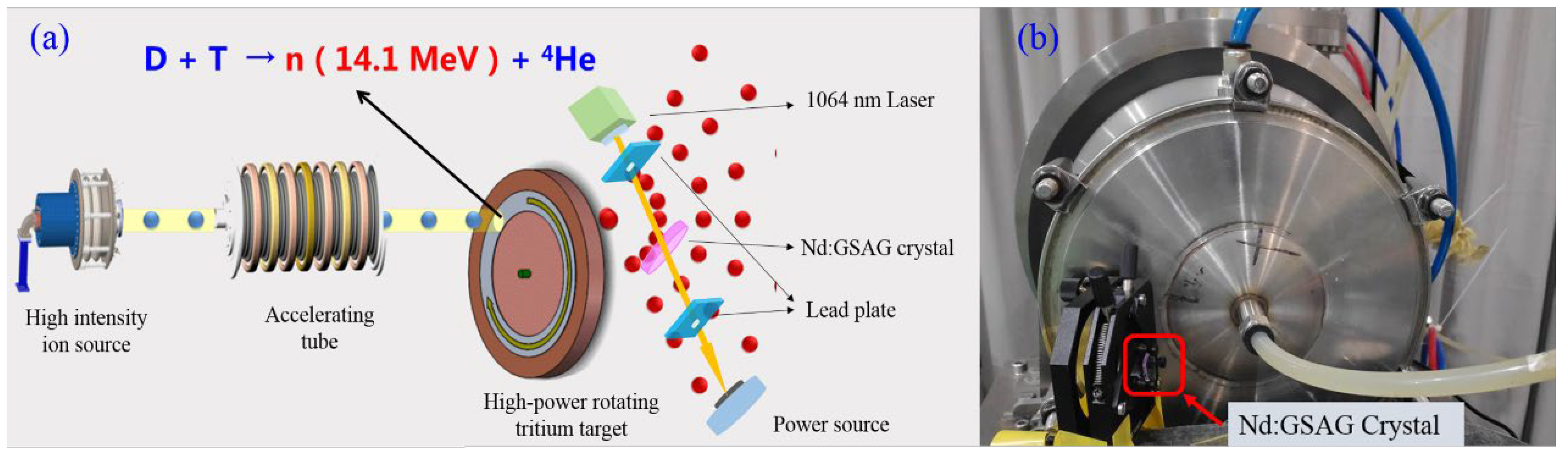
2.2. In-Situ Setup
2.3. Characterization
3. Results and Discussion
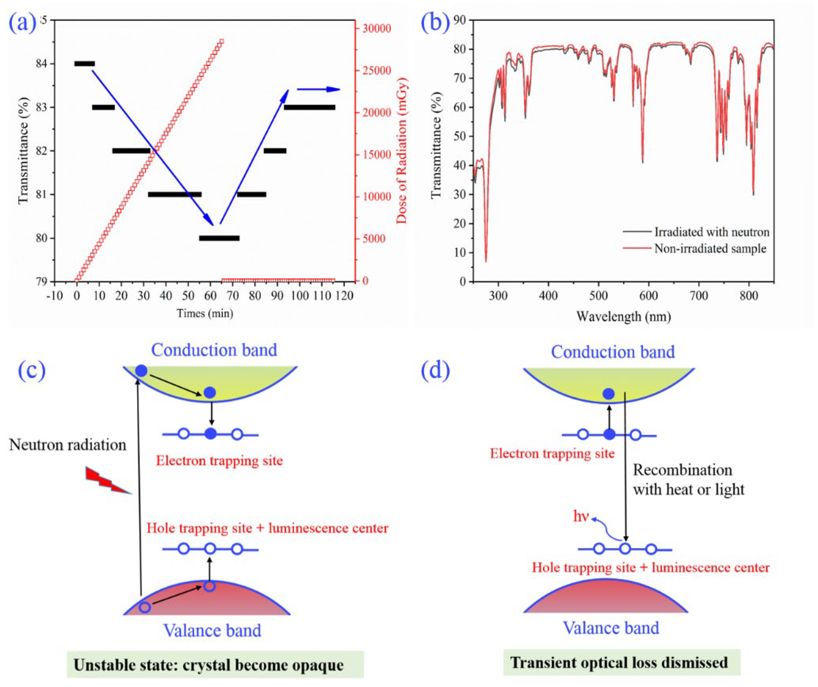
3.1. In-Situ and Real-Time Transmittance
3.2. Optical Properties
3.3. Crystal Structure
3.4. XPS Spectra
4. Conclusions
Author Contributions
Funding
Data Availability Statement
Conflicts of Interest
References
- Vaddigiri, A.; Potter, K.; Thomes, W.; Meister, D. Ionizing radiation effects in single-crystal and polycrystalline YAG. IEEE Trans. Nucl. Sci. 2006, 53, 3882. [Google Scholar] [CrossRef]
- Ding, S.; Wang, H.; Liu, W.; Luo, J.; Ma, Y.; Zhang, Q. Enhanced radiation resistant properties of Nd: GSAG laser crystal by co-doping of Cr3+. J. Lumin. 2019, 213, 249. [Google Scholar] [CrossRef]
- Takaichi, K.; Yagi, H.; Lu, J.; Bisson, J.-F.; Shirakawa, A.; Ueda, K.-I.; Yanagitani, T.; Kaminskii, A.A. Highly efficient continuous-wave operation at 1030 and 1075 nm wavelengths of LD-pumped Yb3+: Y2O3 ceramic lasers. Appl. Phys. Lett. 2004, 84, 317. [Google Scholar] [CrossRef]
- Girard, S.; Alessi, A.; Richard, N.; Martin-Samos, L.; De Michele, V.; Giacomazzi, L.; Agnello, S.; Francesca, D.; Morana, A.; Winkler, B.; et al. Overview of radiation induced point defects in silica-based optical fibers. Rev. Phys. 2019, 4, 100032. [Google Scholar] [CrossRef]
- Schupp, R.; Torretti, F.; Meijer, R.; Bayraktar, M.; Sheil, J.; Scheers, J.; Kurilovich, D.; Bayerle, A.; Schafgans, A.; Purvis, M.; et al. Radiation transport and scaling of optical depth in Nd: YAG laser-produced microdroplet-tin plasma. Appl. Phys. Lett. 2019, 115, 124101. [Google Scholar] [CrossRef] [Green Version]
- Rai, V.; Sekhar, B.R.; Kher, S.; Deb, S. 1 micrometer high-efficient radiation resistant laser crystal: Nd: YSAG. J. Lumin. 2010, 130, 582. [Google Scholar] [CrossRef]
- Sun, D.L.; Luo, J.Q.; Xiao, J.Z.; Zhang, Q.L.; Jiang, H.H.; Yin, S.T.; Wang, Y.F.; Ge, X.W. Effects of annealing treatment and gamma irradiation on the absorption and fluorescence spectra of Cr: GSGG laser crystal. Appl. Phys. B 2008, 92, 529. [Google Scholar] [CrossRef]
- Ding, S.; Chen, Y.; Liu, W.; Luo, J.; Chen, Y.; Li, X.; Zhang, Q. A promising high-efficient radiation resistant laser crystal Nd: GSAG. Infrared Phys. Technol. 2019, 102, 103005. [Google Scholar] [CrossRef]
- Ding, S.; Liu, W.; Chen, Y.; Li, X.; Zhang, Q. Effects of the gamma-ray irradiation on the structure, spectral and laser damage threshold of Nd: GSAG crystal. Opt. Mater. 2019, 95, 109259. [Google Scholar] [CrossRef]
- Gromov, V.V.; Karaseva, L.G.; Saunin, E.I. Study of physico-chemical processes in yttrium-aluminium garnet under gamma-rays irradiation. J. Radioanal. Chem. 1976, 30, 441. [Google Scholar] [CrossRef]
- Wu, Y. Development of high intensity D–T fusion neutron generator HINEG. Int. J. Energy Res. 2018, 42, 68. [Google Scholar] [CrossRef]
- Yuan, G.; Hu, G.; Shan, W.; Jin, S.; Gu, Q.; Chen, J. Structural and luminescence modulation in 8-hydroxyquinolinate-based coordination polymers by varying the dicarboxylic acid. Dalton Trans. 2015, 44, 17774. [Google Scholar] [CrossRef]
- Sheldrick, G. PLATON SQUEEZE: A tool for the calculation of the disordered solvent contribution to the calculated structure factors. Acta Crystallogr. Sect. C 2015, 71, 3. [Google Scholar] [CrossRef] [Green Version]
- Sheldrick, G. Redetermination of the crystal structure of β-zinc molybdate from single-crystal X-ray diffraction data. Acta Crystallogr. Sect. A 2015, 71, 3. [Google Scholar] [CrossRef] [Green Version]
- Compton, D.M.J.; Cesena, R.A. Mechanisms of radiation effects on lasers. IEEE Trans. Nucl. Sci. 1967, 14, 55. [Google Scholar] [CrossRef]
- Koshimizu, M.; Yanagida, T.; Shinsho, K.; Yanagisawa, S.; Fujimoto, Y.; Yagi, H.; Yanagitani, T.; Asai, K. Similarity of trap state and thermoluminescence processes of Y3Al5O12 (YAG): Ce for X-ray and UV irradiation. Nucl. Instrum. Methods Phys. Res. Sect. B Beam Interact. Mater. At. 2018, 435, 285. [Google Scholar] [CrossRef]
- Sun, T.; Lu, M. Band-structure modulation of SrTiO3 by hydrogenation for enhanced photoactivity. Appl. Phys. A 2012, 108, 171. [Google Scholar] [CrossRef]
- Tan, H.; Zhao, Z.; Zhu, W.-B.; Coker, E.N.; Li, B.; Zheng, M.; Yu, W.; Fan, H.; Sun, Z. A two-dimensional CdO/CdS heterostructure used for visible light photocatalysis. ACS Appl. Mater. Interfaces 2014, 6, 19184–19190. [Google Scholar] [CrossRef]
- Barreca, D.; Gasparotto, A.; Milanov, A.; Tondello, E.; Devi, A.; Fischer, R.A. Gd2O3 nanostructured thin films analyzed by XPS. Surf. Sci. Spectra 2007, 14, 60. [Google Scholar] [CrossRef]
- Datta, P.; Majewski, P.; Aldinger, F. Study of gadolinia-doped ceria solid electrolyte surface by XPS. Mater. Charact. 2009, 60, 138. [Google Scholar] [CrossRef]
- Li, Y.-L.; Chen, N.-F.; Zhou, J.-P.; Song, S.-L.; Liu, L.-F.; Yin, Z.-G.; Cai, C.-L. Effect of the oxygen concentration on the properties of Gd2O3 thin films. J. Cryst. Growth 2004, 265, 548. [Google Scholar] [CrossRef]
- Holgado, J.P.; Munuera, G.; Espinós, J.P.; González-Elipe, A.R. XPS study of oxidation processes of CeOx defective layers. Appl. Surf. Sci. 2000, 158, 164. [Google Scholar] [CrossRef]
- El Kazzi, M.; Merckling, C.; Delhaye, G.; Arzel, L.; Grenet, G.; Bergignat, E.; Hollinger, G. Measurement of wurtzite ZnO/rutile TiO2 heterojunction band offsets by X-ray photoelectron spectroscopy. Mater. Sci. Semicond. Process. 2006, 9, 954. [Google Scholar] [CrossRef]
- Kaichev, V.V.; Ivanova, E.V.; Zamoryanskaya, M.V.; Smirnova, T.P.; Yakovkina, L.V.; Gritsenko, V.A. XPS and cathodoluminescence studies of HfO2, Sc2O3 and (HfO2) 1-x (Sc2O3) x films. Eur. Phys. J. Appl. Phys. 2013, 64, 10302. [Google Scholar] [CrossRef]
- Talik, E.; Szubka, M.; Skrzypek, D.; Zarek, W.; Kusz, J.; Łukasiewicz, T. SQUID magnetometry, EPR and XPS characterization of Y3Al5O12: Co, Si single crystals. Cryst. Res. Technol. 2009, 44, 823. [Google Scholar] [CrossRef]
- Vitanov, P.; Stefanov, P.; Harizanova, A.; Ivanova, T. XPS characterization of thin (Al2O3) x (TiO2)1−x films deposited on silicon. J. Phys. Conf. Ser. 2008, 113, 012036. [Google Scholar] [CrossRef]
- Reddy, N.; Bera, P.; Reddy, V.R.; Sridhara, N.; Dey, A.; Anandan, C.; Sharma, A.K. XPS study of sputtered alumina thin films. Ceram. Int. 2014, 40, 11099. [Google Scholar] [CrossRef]
- Shen, Z.; Zhang, F.; Chen, J.; Fu, Z.; Liu, X.; Yan, G.; Lv, B.; Wang, Y.; Wang, L.; Zhao, W. Effects of annealing on the interfacial properties and energy-band alignment of AlN dielectric on 4H–SiC. Appl. Phys. Lett. 2020, 117, 102105. [Google Scholar] [CrossRef]


| Formula | Non-Irradiated Sample | Irradiated with Neutrons |
|---|---|---|
| System and space group | Cubic, Ia-3d | Cubic, Ia-3d |
| a [Å] | 12.4265 | 12.4247 |
| V [Å3] | 1918.87 | 1918.04 |
| F (000) | 1696 | 1696 |
| Reflections: measured/unique | 5263/187 | 2291/185 |
| R (int) | 0.0893 | 0.1018 |
| R (sigma) | 0.0308 | 0.0542 |
Disclaimer/Publisher’s Note: The statements, opinions and data contained in all publications are solely those of the individual author(s) and contributor(s) and not of MDPI and/or the editor(s). MDPI and/or the editor(s) disclaim responsibility for any injury to people or property resulting from any ideas, methods, instructions or products referred to in the content. |
© 2023 by the authors. Licensee MDPI, Basel, Switzerland. This article is an open access article distributed under the terms and conditions of the Creative Commons Attribution (CC BY) license (https://creativecommons.org/licenses/by/4.0/).
Share and Cite
Gao, Y.; Liu, W.; Ding, S.; Chen, Y.; Zhang, Q. Real-Time In-Situ Investigation of the Neutron Irradiation Resistance Ability of Nd3+-Doped Gd3Sc2Al3O12 Laser Crystal. Crystals 2023, 13, 136. https://doi.org/10.3390/cryst13010136
Gao Y, Liu W, Ding S, Chen Y, Zhang Q. Real-Time In-Situ Investigation of the Neutron Irradiation Resistance Ability of Nd3+-Doped Gd3Sc2Al3O12 Laser Crystal. Crystals. 2023; 13(1):136. https://doi.org/10.3390/cryst13010136
Chicago/Turabian StyleGao, Yuxi, Wenpeng Liu, Shoujun Ding, Yuanzhi Chen, and Qingli Zhang. 2023. "Real-Time In-Situ Investigation of the Neutron Irradiation Resistance Ability of Nd3+-Doped Gd3Sc2Al3O12 Laser Crystal" Crystals 13, no. 1: 136. https://doi.org/10.3390/cryst13010136