Review

Structural Changes as a Tool for Affinity Recognition: Conformational Switch Biosensing

by
Viviana Scognamiglio
* and
Amina Antonacci
Institute of Crystallography, National Research Council, Department of Chemical Sciences and Materials Technologies, Via Salaria km 29.300, 00015 Monterotondo, Italy
*
Author to whom correspondence should be addressed.
Crystals 2022, 12(9), 1209; https://doi.org/10.3390/cryst12091209
Submission received: 3 August 2022 / Revised: 24 August 2022 / Accepted: 25 August 2022 / Published: 27 August 2022
(This article belongs to the Special Issue Feature Papers in Biomolecular Crystals in 2022-2023)

Abstract

:
Biosensors draw inspiration from natural chemosensing based on molecular switches between different bond-induced conformational states. Proteins and nucleic acids can be adapted into switch-based biosensors with a wide plethora of different configurations, taking advantage of the variety of transduction systems, from optical to electrochemical or electrochemiluminescence, as well as from nanomaterials for signal augmentation. This review reports the latest trends in conformational switch biosensors reported in the literature in the last 10 years, focusing on the main representative and recent examples of protein-based switching biosensors, DNA nanomachines, and structure-switched aptamers being applied for the detection of a wide range of target analytes with interest in biomedical and agro-environmental sectors.

1. Introduction

In nature, tasks related to real-time monitoring and detection in complex media have been already solved through biomolecular interaction mechanisms. Macromolecules can sense chemical signals through molecular interactions and transduce them into undergoing specific, binding-induced conformational changes. Proteins and nucleic acids can switch between distinct conformational states to drive cellular processes, with “impressive specificity, affinity and versatility of biomolecular recognition” as described by Vallée-Bélisle and Plaxco [1]. This is due to a delicate molecular dialogue between macromolecules established through non-covalent bonds capable of conferring unique molecule–ligand interfaces, including hydrophobic interactions, hydrogen bonds, and van der Waals forces [1].
Biosensing technology arises from this biological concept. The biological recognition element, or biocomponent, changes its shape upon binding target analytes, inspired by the modular design of natural signaling. As the binding event is very specific, the conformational equilibria of molecular switches are quantitatively associated with target concentration. In contrast to other biocomponents (e.g., antibodies), macromolecules as proteins and nucleic acids, as well as aptamers, can undergo conformational switches upon target interaction, discriminating between the authentic target and non-specific ones [2]. This fine-tuned mechanism furnishes a reliable way of transducing the conformational isomerism of the involved macromolecules into optical, electrochemical, or coupled outputs without incurring undesirable events. Furthermore, nanotechnology has been demonstrated to be a valid support for the design of similar switchable biosensors, both as signal enhancers and loading supports. In the following sections, the principles relying on switch conformational biosensing, based on the optical and electrochemical readout, are described, as well as noteworthy examples reported from the literature, including protein-based switching biosensors, DNA nanomachines, and structure-switched aptamers.

2. Main Conformational Switch Biosensing Readouts

2.1. Optical Read-Out

Optical-based switch biosensing entails variations in the fluorescence signals upon target binding through different configurations. The first entails the use of a commercial fluorophore labeled to a strategic portion of the biocomponent and able to sense changes in the microenvironment due to variations in its conformation (Figure 1A). This is the case of the sugar-binding protein from the Archaeon Pyrococcus horikoshii, covalently labeled at the Cys residue located in the C-terminal domain with the fluorophore 2-(4′-(iodoacetamido)anilino)naphthalene-6-sulfonic acid (IAANS), which is known to be a molecule sensitive to its local environment. The probe, placed in a strategical position of the protein, 10 Å away from the sugar-binding groove, senses variations in its conformation upon sugar binding, thus transducing these changes into detectable fluorescence signals [2].
A second configuration relies on the use of a donor/acceptor pair of commercial fluorophores labeled to selected sites of the biocomponent, where the distance between the two permits the fluorescence emission of the donor to overlap with the absorption/excitation spectrum of the acceptor. Indeed, the Förster resonance energy transfer (FRET) between two fluorophores allows for fluorescence spectral variations through space interaction that occurs whenever the donor and the acceptor are within the Forster distance (R0). This is the case of the glucose-/galactose-binding protein from Escherichia coli labeled with rhodamine as an acceptor probe at the N-terminal amino acid and with acrylodan as a donor probe in a Cys residue, specially inserted in position 182 of the protein via site-directed mutagenesis (Figure 1B). Upon glucose binding, the protein undergoes a conformational change, which provides a target-dependent spatial realignment of the two probes and a consequent change in their distance from 46.27 Å to 38.27 Å, in the absence and the presence of glucose, respectively. This rational placement of the probes allows for the determination of glucose by increasing rhodamine fluorescence emission intensity [3]. Other FRET-based configurations have been recently proposed as highly sensitive and high-throughput detection systems for diverse targets, such as the system described by Xu et al. [4]. In this case, an aptamer-induced “switch on” FRET biosensor enables the detection of tumor markers (AFP and CEA), merging MoS2 nanosheets, used as energy receptors, with multicolored gold nanoclusters, used as energy donors.
A third configuration involves a fluorophore/quencher pair to provide electron-transfer-based fluorescence quenching. This is the case of the fluorescent aptamer-based lateral flow biosensor proposed by Cheng and co-workers [5], which integrated aptamers with fluorophore–quencher nanopairs to perform “signal-on” sensing. In detail, streptavidin-biotinylated complementary sequences were conjugated with quantum dot nanobeads as fluorophores and gold nanostars as quenchers for the sensitive and specific detection of selected pesticides (Figure 1C). In this case, the fluorophore–quencher system is composed of gold nanosheets used as acceptors and QD nanobeads used as donors, and it exploits the matched fluorescence quenching–unquenching abilities of such nanomaterials for the “turn-on” fluorescence mechanism.
These last two methods do not require a change in the probe microenvironment and are exceptionally sensitive. Angstrom-level measures of change in molecular distance are monitored because the rate of energy transfer for dipole–dipole interactions is inversely proportional to r6, according to the equation E = Ro6/(Ro6 + r6), where Ro is the Förster distance at which half the energy is transferred and r is the actual distance between donor and acceptor [6].

2.2. Electrochemical Read-Out

Electrochemical-based switch biosensing involves electroactive mediators, attached to a specific site of the biocomponent immobilized onto an interrogating electrode, which produces redox currents. Changes in the biocomponent conformation, induced by the binding event, hamper the mediator to reach the electrode surface, thus varying the redox currents that can be recorded by electrochemical transductions [7,8]. A crucial example of such fine configuration is described by Zhao and colleagues [9], in which a biosensor was constructed to analyze the Nosema bombycis gene DNA (PTP1) by recording PTP1-based LAMP (loop-mediated isothermal amplification), transducing the chemical stimuli H+ into a ferrocene electrochemical signal. The biosensing principles rely on the ability of H+ to induce a conformational switch of DNA nanostructure, assembled by the ferrocene-labeled signal probe and DNA-based receptor hybridization, and it is responsive to pH. In detail, as the pH decreases, this DNA-based receptor undergoes a structural variation and changes into a stable triplex, causing the release of the ferrocene-labeled signal probe and thus generating amplified electrochemical signals (Figure 2).
A different arrangement was described as a label-free electrochemical biosensor to detect nereistoxin (NRT)-related insecticides, based on a target-binding-triggered conformational switching of a DNAzyme [10]. First, a pretreatment step provides conversion of NRT-related insecticides into NRT by the addition of two sulfhydryl groups. Successively, the assembly of DNAzyme into functional catalytic structures is achieved through conformational change induction due to thymine-Hg2+-thymine (T-Hg2+-T) mismatch. This activates Mg2+-DNAzyme and hemin/G-quadruplex DNAzyme, respectively, which are switched off by the uptake of Hg2+ from Mg2+-DNAzyme, upon which converted NRTs are introduced. This “signal-off” strategy, registered by a block of the electrochemical signals, was revealed to be very sensitive for the analysis of bensultap with a 6.9  ng/L detection limit and 0.01–2000 μg/L linear range.

2.3. Coupled Read-Out

Photoelectrochemical-based switch biosensing is a “recently emerged but rapidly developing method based on a total separation of the detection signal and excitation source” as described by Hu et al. [11]. These authors elaborated an “on-off-on” signal-switchable photoelectrochemical strategy to cope with the main drawbacks in terms of low sensitivity, non-negligible background signal, and false-positive errors. A prerequisite of such transduction is coupling switchable biocomponents (i.e., aptamers) to photoactive materials with good photoelectric properties and chemical stability, such as nanomaterials (i.e., quantum dots). Several photoelectrochemical switch biosensors have been described in the literature [12]. As an example, Wang et al. [13] employed photoelectrochemical detection. In this case, hairpin triple-helix DNA, for the detection of human immunodeficiency virus type 1 (HIV-1) nucleic acid, was conjugated to a cascaded photoactive material formed by CdTe quantum dots (QDs) and sensitized ZnO nanorods (ZnO NRs) to amplify the photocurrent signal. Then, gold nanoparticles functionalized with single-stranded DNA labeled with alkaline phosphatase were used to hybridize with the hairpin DNA to form a triple-helix conformation. This conformational switch determined a photocurrent variation due to the conversion of ascorbic acid 2-phosphate to generate ascorbic acid catalyzed by the alkaline phosphatase, allowing for the reproducible and specific detection of the target DNA with a limit of detection of 0.65 fM.
Electrochemiluminescence-based switch biosensing is also available in many switch biosensing systems [14,15,16]. For example, Gao et al. [17] reported an “on-off” detection of lipopolysaccharides with a multiple amplification strategy, exploiting, as target recognition, a specific aptamer immobilized on magnetic beads. Amplified DNA fragments (a1) were then inserted into a triple-helix sensing system and their interaction generates the opening of the aptamer triple-helix structure. Exploiting this aptamer structure, the authors were able to sense lipopolysaccharides by electrochemiluminescence within a wide linear range from 0.1 fg/mL to 0.1 ng/mL, as well as with a low detection limit of 0.012 fg/mL.

3. Main Conformational Switch Case Studies

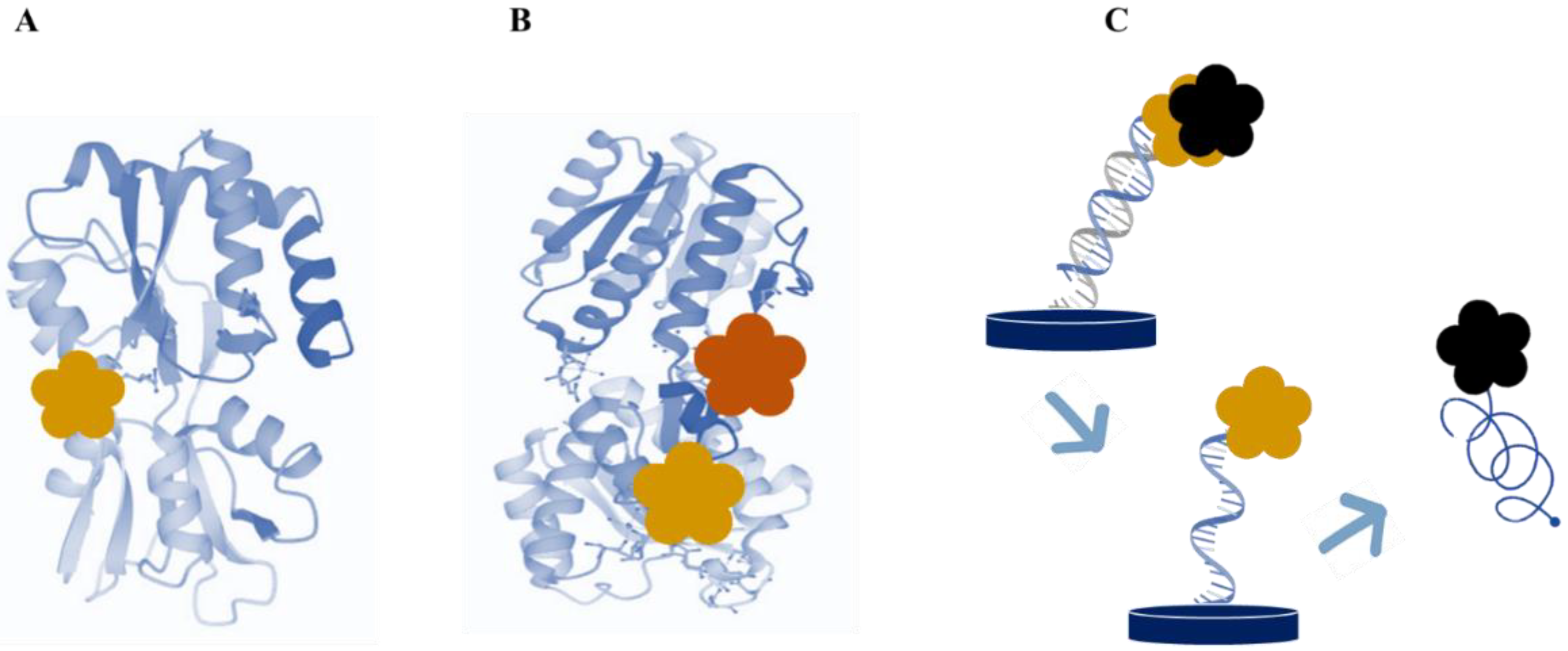
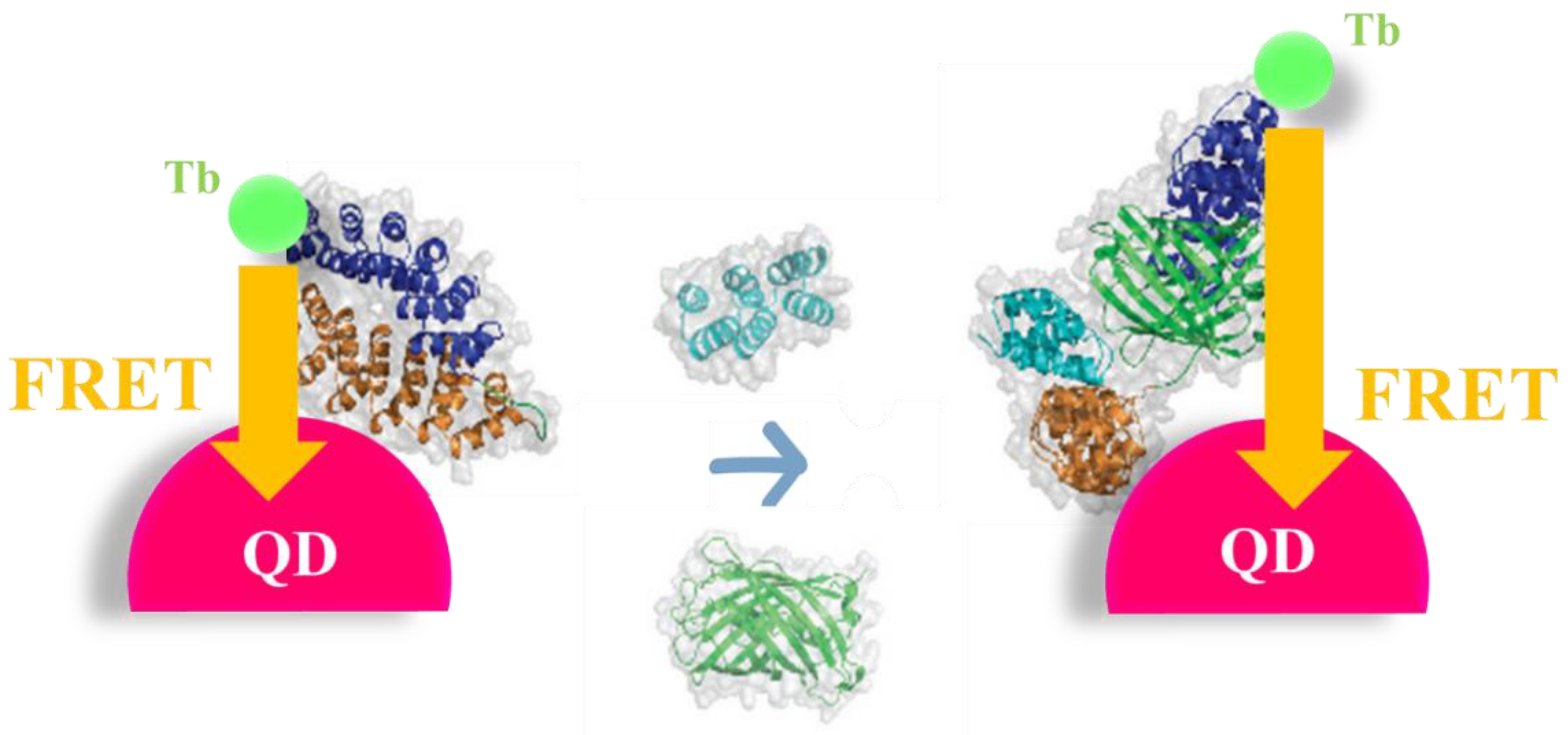
3.1. Protein-Based Switch Biosensing

In recent decades, “bioengineers have been struggling to expand the toolkit of molecular switches recognition repertoire utilizing periplasmic binding proteins”, according to Ribeiro and colleagues [18]. A very large amount of the literature describes the expression, purification, and characterization of proteins belonging to the periplasmic binding family by bioinformatics as well as fluorescence, circular dichroism, and Fourier transform infrared spectroscopy [19,20,21,22,23], to exploit them as switch bioreceptors to detect small molecules [24,25,26,27,28]. The members of this family undergo a hinge-bending motion when the target analyte is bound to the cleft that separates the two domains of the protein [29,30,31]. An example of a huge conformational change upon target binding comes from the glutamine binding protein from E. coli, which was largely investigated by steady-state and time-resolved fluorescence, as well as circular dichroism spectroscopy [32]. These techniques highlighted that the interaction with glutamine results in a marked change in the structure and conformational dynamics of the protein. A recent study reports the use of a genetically modified arginine-binding protein from Thermotoga maritima to create a reagentless fluorescent protein biosensor [33]. In detail, a single site-directed cysteine mutant of the protein was produced, successively labeled with different fluorescent probes sensitive to environmental changes in response to arginine.
Several computational studies have also been conducted for the engineering of these proteins, with the aim of solving steric conflicts within the structure and manipulating the intrinsic equilibrium between the open and closed state to improve the conformational switch of the protein upon ligand binding, in view of a better sensitivity towards the target [24].
These studies illustrate both the versatility and the remarkable adaptability of the periplasmic binding proteins. A given scaffold can indeed be manipulated to improve their affinity towards the specific ligand, as well as diversified to bind different ligands, as is demonstrated with the ribose-binding protein (RBP) (Figure 3).
Besides proteins, artificial scaffold proteins have been also described in the literature as switch probes, overcoming the main limitations of antibodies arising from their size, stability, and structure complexity. For example, Leger et al. [34] described the development of two bidomain artificial proteins (αRep) for the design of a FRET-based biosensor. In detail, such bidomains were labeled with a Tb complex (Tb) donor on the C-terminus and a semiconductor quantum dot (QD) acceptor on the N-terminus, to quantify different protein targets at sub-nanomolar concentrations. In the absence of the targets, the bidomains are in a “closed” conformation, with domains facing each other. In the presence of the target, the bidomains switch to an “open” conformation, which can be detected by time-resolved FRET (Figure 4). This FRET biosensor was able to achieve sub-nanomolar detection limits in phosphate-buffered saline and low nanomolar ones in serum-containing samples.
Antibodies have also been employed for the realization of switch biosensors, as in the case of the fluorescence immunosensor for the detection of lysozyme [35]. The detection is based on the quenching of quantum dots labeled with lysozyme by gold nanoparticle-labeled lysozyme antibodies through a FRET mechanism. Then, in the presence of lysozyme, the fluorescence is recovered thanks to the specific competitive interaction between the antibodies and the enzyme, which can be detected with a linear range of 50–1000 ng/mL and a detection limit of 33.43 ng/mL.

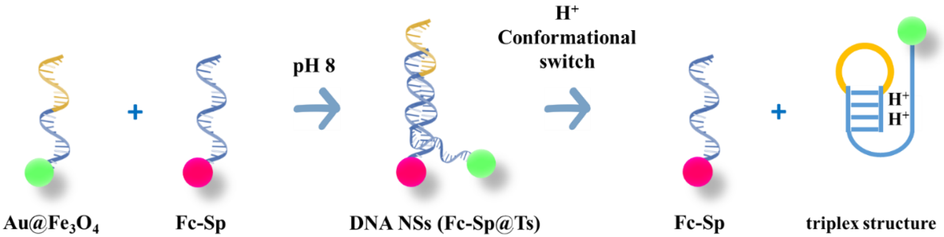
3.2. DNA Nanomachines

As stated by Angell et al. [36], “DNA nanotechnology is a branch of nanoengineering that takes advantage of the precise, elegant base-pairing nature of self-assembly, geometry, and structure of DNA to engineer synthetic structures for various applications including drug delivery, detection, and computing”. DNA nanotechnology, indeed, allows for the realization of customized molecules with intelligent targeting and motion capabilities, which can find numerous applications such as drug delivery and biosensing. Examples of similar molecules include DNA nanomachines, capable of conformational and positional actions in response to environmental stimuli. Such nanomachines take inspiration from biological nanomotors, for example, actin/myosin, kinesin, and the adenosine triphosphate (ATP) F0/F1 motor, or the asymmetric propulsion movement of prokaryotes such as E. coli, which deliver cargo, move in biological environments and can discern specific targets.
Zhang et al. [37] introduced the concept of binding-induced DNA nanomachines activated by the interaction with proteins and nucleic acids, which induce nanomachine assembly otherwise unable to spontaneously assemble. In particular, the authors constructed 3D DNA tracks of high density on the surface of gold nanoparticles functionalized with single-stranded oligonucleotides and an affinity ligand. In the absence of the target, a DNA swing arm interacts with a second affinity ligand in solution. In the presence of the target, both the affinity ligands cause the swing arm to move around the gold nanoparticle surface, powered by enzymatic cleavage of conjugated oligonucleotides. In particular, the authors describe three case studies, exploiting streptavidin, platelet-derived growth factor, and the Smallpox gene; indeed, simply substituting the ligands allows the nanomachine to bind specific targets.
Zhang et al. [38] constructed a DNA nanomachine-based biosensor organized as a one-step regenerated sensing platform. In this configuration, the DNA nanomachine was labeled with Alexa Fluor 488 as the donor and CdSe@ZnS QDs as the acceptor, and an electrochemiluminescence resonance energy transfer (ERET) strategy was developed to sensitively determine the presence of miRNAs associated with cancer cells. The DNA nanomachine, kept in an “off” state in the absence of the target analyte, was able to switch into an “on” state when in the presence of DNA previously converted and amplified by a dual amplification strategy (i.e., target recycling and signal transformation). In the “on” state, the donor/acceptor pair was close enough to generate ERET, and thus enhance QD emission intensity, allowing for the detection of the target at femtomolar level (detection limit of 0.03 fM for miRNA).
A similar configuration was described by Chen et al. [39] who designed a structure-convertible DNA switch for the detection of miRNA by ratiometric fluorescence. In detail, a single-strand DNA with a stem-loop structure was labeled with Cy3 and Cy5 fluorophores and with a quencher at specific sites of the switch, to interact with amplicon fragments obtained by the exponential amplification reaction. This interaction drives the structure of the switch to convert into a specific conformational cue, which causes the movements between Cy5 and Cy3, and thus the transfer of their fluorescence resonance energy. This helped to detect miRNAs quantitatively and rapidly with high sensitivity (detection limit of 70.9 fM) in a concentration range from 100 fM to 100 nM.
Xiong et al. [40] projected a triple-helix molecular switch for the realization of an electrochemical dual-signaling ratiometric biosensor for nucleic acids. A hairpin DNA was labeled with methylene blue and exploited for the functionalization of a gold electrode. Successively, a single-strand DNA labeled with two ferrocenes on each end was hybridized with the methylene blue DNA to form a triple-helix conformation. In the presence of the target, it interacts with the methylene blue DNA causing a conformational switch of the biocomponent from the triple-helix conformation to its hairpin structure. This generates the ferrocene tag displacing, resulting in a decrease in its peak current as well as a concomitant increase in the methylene blue DNA. This allowed for a target detection within the linear range from 0.5 to 80 pM, with a detection limit of 0.12 pM.
A DNA nanotweezer coupled with a split G-quadruplex was also designed for the development of a label-free electrochemical biosensor for DNA [41]. In detail, a long single-strand DNA nanotweezer was constructed, with two hybridized hairpin structures in the middle working as a recognition element and two short single strands with split G-rich sequences on the outside working as a signal reporter. The hybridized hairpin structures can fold into both open and closed cues upon target binding, generating variations in the electrochemical signals. On the other hand, the split G-rich sequences put outside of the hairpin structure allowed for an improved signal–background ratio. This assay was capable of high-sensitivity circulating tumor DNA determination with a detection limit of 22 aM within a wide linear range from 1 fM to 1 nM.

3.3. Structure-Switching Aptamers

Nucleic acid aptamers are single-stranded DNA or RNA sequences that specifically bind a complementary target ligand [42]; as stated by Feagin et al. [43], “aptamer biosensor that can switch its structure upon target binding offers a powerful strategy for molecular detection”. Indeed, despite the intrinsic difficulty of converting aptamers into structure-switching biosensors, similar sensing approaches have largely demonstrated their potential for sensitive and effective diagnostic. As an example, Lackey et al. [44] converted an L-tyrosinamide-binding oligonucleotide aptamer into a structure-switching biosensor by integrating a fluorescently labeled oligonucleotide complementary with the L-tyrosinamide target binding portion. In this way, the interaction between the aptamer and the target induces the disruption of the hybridization equilibrium and thus the occurrence of a target concentration-dependent signal. In detail, the authors shed light on the kinetics and mechanism of target interaction with the structure-switching aptamer, providing a general means of measuring the rates of unlabeled analyte-binding kinetics in aptamer-based biosensors.
Zhou et al. [45] described the redesign of an RNA aptamer from Spinach for the conformational switch-based fluorescence detection of telomerase activity, as research on the activity and inhibition of telomerase is crucial for cancer diagnosis and therapy. This redesigned aptamer has an extended 5′ end and arranges as an inactive structure not able to interact with 5-difluoro-4-hydroxybenzylidene-imidazolinone (DFHBI) fluorophore. In detail, the stem loop 3 was split into two arms, where the new extended 5′-end fold into a new hairpin structure (a/b/a*), thus preventing hybridization with the split branch on the 3′ end (a**). When telomerase is present, a telomerase substrate primer likewise extended to the 3′ end with (TTAGGG)n repeats can hybridize with the inactive redesigned aptamer, which folds into an active conformation (Figure 5). This structure switch allowed for the monitoring of telomerase activity from 100 to 5 × 10⁴ Hela cells with a detection limit of 100 cells, furnishing a valid tool for the evaluation of the differentiation of cancer cell lines from normal cell lines as well as of the inhibition efficiency of telomere-binding ligands, crucial for telomerase-related cancer diagnosis and therapy.
Besides sensitivity and robustness, aptamer switching biosensors show high versatility in being capable of revealing diverse targets, from cancer biomarkers to environmental pollutants. Wang et al. [46] realized a chemiluminescence biosensor for ochratoxin A detection, using a triple-helix aptamer probe and a luminol-H2O2 solution. The aptamer probe consisted of a loop, two DNA segment stems, and a triplex-forming oligonucleotide working as the signal probe. The interaction between the aptamer and the ochratoxin A, occurring with very high affinity, generates a structural modification of the aptamers, which leads to the release of the signal probe. This latter folds into a G-quadruplex structure able to catalyze luminol and thus produce chemiluminescence H2O2 mediated. A linear response towards ochratoxin A was observed between 0.1 ng/mL and 2.0 ng/mL, with a detection limit of 0.07 ng/mL.
A further advantage of structure-switching DNA aptamers is coupling them with DNA amplification methods to improve their sensitivity. Liu et al. [47] described the combination of rolling circle amplification (RCA) based on ϕ29 DNA polymerase to carry out repetitive rounds of DNA synthesis using a circular DNA template, with a DNA aptamer able to trigger a structure-switching event upon target binding, demonstrating a massive production of DNA amplicons and thus a dramatically enhanced sensitivity.
More recently, a triple-helix aptamer switch was proposed to reveal Staphylococcus aureus, combined with a signal amplification system to increase the sensitivity [48]. In detail, Bsm DNA polymerase and Nb.bpu10I endonuclease were provided for the target amplification of single-strand DNA fragments. These were able to interact with the molecular switch, causing the dissociation of the triple-helix DNA structure on the electrode and thus forming an electroactive G-quadruplex/hemin complex that turns on the generation of electrochemical signals (Figure 6). This configuration enabled the detection of S. aureus in standard solution with a detection limit of 8 CFU/mL as well as in lake water, tap water, and honey samples.

4. Conclusions

Switch biosensors take inspiration from nature to solve molecular detection, exploiting proteins and nucleic acids able to structurally switch between different binding-induced conformational variations. Such sensing configurations represent a promising strategy toward the development of versatile and effective diagnostics to be applied in agro-environmental and biomedical sectors. Indeed, conformational switch sensing demonstrated high detection capability in terms of specificity and sensitivity. However, some drawbacks still need to be overcome, for example, the need for high stringency of the experimental conditions to minimize cross-reactivity with target analogs showing similar structures and thus to guarantee the maximum specificity. Additional research in this sense is contributing to further improving the performances of switch biosensors, as highlighted by Plaxco group [49], which proposes novel strategies based on structure-switching and allostery to tailor the biocomponent affinity towards the specific target and thus “to optimize the placement of their specificity windows without the need to alter measurement conditions” including pH, ionic strength, or temperature. Structure-switching and allosteric control can tune the range of target concentration over which a biocomponent shows its optimum in specificity. This could help expand the versatility of switch biosensors as well as their applicability in many different fields where the range of target concentrations varies for a given application.

Author Contributions

V.S. Conceptualization, writing—review and editing, A.A.; writing—review and editing. All authors have read and agreed to the published version of the manuscript.

Funding

This work was supported by AdSWiM Interreg Project Italy-Croatia 2019/2021 and NanoSWS project EraNetMed-RQ3-2016.

Institutional Review Board Statement

Not applicable.

Informed Consent Statement

Not applicable.

Data Availability Statement

Not applicable.

Conflicts of Interest

The authors declare no conflict of interest.

References

  1. Plaxco, K.W.; Tom Soh, H. Switch-based biosensors: A new approach towards real-time; in vivo molecular detection. Trends Biotechnol. 2011, 29, 1–5. [Google Scholar] [CrossRef] [PubMed]
  2. Staiano, M.; Sapio, M.; Scognamiglio, V.; Marabotti, A.; Facchiano, A.M.; Bazzicalupo, P.; Rossi, M.; D’Auria, S. A Thermostable Sugar-Binding Protein from the Archaeon Pyrococcus horikoshii as a Probe for the Development of a Stable Fluorescence Biosensor for Diabetic Patients. Biotechnol. Prog. 2004, 20, 1572–1577. [Google Scholar] [CrossRef] [PubMed]
  3. Scognamiglio, V.; Aurilia, V.; Cennamo, N.; Ringhieri, P.; Iozzino, L.; Tartaglia, M.; Staiano, M.; Ruggiero, G.; Orlando, P.; Labella, T.; et al. D-galactose/D-glucose-binding Protein from Escherichia coli as Probe for a Non-consuming Glucose Implantable Fluorescence Biosensor. Sensors 2007, 7, 2484–2491. [Google Scholar] [CrossRef] [PubMed]
  4. Xu, S.; Feng, X.; Gao, T.; Liu, G.; Mao, Y.; Lin, J.; Yu, X.; Luo, X. Aptamer induced multicoloured Au NCs-MoS2 “switch on” fluorescence resonance energy transfer biosensor for dual color simultaneous detection of multiple tumor markers by single wavelength excitation. Anal. Chim. Acta 2017, 983, 173–180. [Google Scholar] [CrossRef]
  5. Cheng, N.; Song, Y.; Fu, Q.; Du, D.; Luo, Y.; Wang, Y.; Xu, W.; Lin, Y. Aptasensor based on fluorophore-quencher nano-pair and smartphone spectrum reader for on-site quantification of multi-pesticides. Biosens. Bioelectron. 2018, 117, 75–83. [Google Scholar] [CrossRef]
  6. Lakowicz, J.R. Principles of Fluorescence Spectroscopy; Springer Science & Business Media: Berlin/Heidelberg, Germany, 2013. [Google Scholar]
  7. Wang, Q.; Gao, F.; Ni, J.; Liao, X.; Zhang, X.; Lin, Z. Facile construction of a highly sensitive DNA biosensor by in-situ assembly of electro-active tags on hairpin-structured probe fragment. Sci. Rep. 2016, 6, 22441. [Google Scholar] [CrossRef]
  8. Campuzano, S.; Yáñez-Sedeño, P.; Pingarrón, J.M. Reagentless and reusable electrochemical affinity biosensors for near real-time and/or continuous operation. Advances and prospects. Curr. Opin. Electrochem. 2019, 16, 35–41. [Google Scholar] [CrossRef]
  9. Zhao, J.; Gao, J.; Zheng, T.; Yang, Z.; Chai, Y.; Chen, S.; Yuan, R.; Xu, W. Highly sensitive electrochemical assay for Nosema bombycis gene DNA PTP1 via conformational switch of DNA nanostructures regulated by H+ from LAMP. Biosens. Bioelectron. 2018, 106, 186–192. [Google Scholar] [CrossRef]
  10. Xie, S.; Yuan, W.; Wang, P.; Tang, Y.; Teng, L.; Peng, Q. Target-induced conformational switch of DNAzyme for homogeneous electrochemical detection of nereistoxin-related insecticide on an ultramicroelectrode. Sens. Actuators B Chem. 2019, 292, 64–69. [Google Scholar] [CrossRef]
  11. Hu, M.; Yang, H.; Li, Z.; Zhang, L.; Zhu, P.; Yan, M.; Yu, J. Signal-switchable lab-on-paper photoelectrochemical aptasensing system integrated triple-helix molecular switch with charge separation and recombination regime of type-II CdTe@CdSe core-shell quantum dots. Biosens. Bioelectron. 2020, 147, 111786. [Google Scholar] [CrossRef]
  12. Geng, W.; Yang, R. A triple-helix molecular switch photoelectrochemical biosensor for ultrasensitive microRNA detection based on position-controllable CdS//CdTe signal enhancement and switching. Chem. Commun. 2020, 56, 2909–2912. [Google Scholar] [CrossRef] [PubMed]
  13. Wang, S.; Zhao, J.; Zhang, Y.; Yan, M.; Zhang, L.; Ge, S.; Yu, J. Photoelectrochemical biosensor of HIV-1 based on cascaded photoactive materials and triple-helix molecular switch. Biosens. Bioelectron. 2019, 139, 111325. [Google Scholar] [CrossRef] [PubMed]
  14. Kun, Q.; Lin, Y.; Peng, H.; Cheng, L.; Cui, H.; Hong, N.; Xiong, J.; Fan, H. A “signal-on” switch electrochemiluminescence biosensor for the detection of tumor cells. J. Electroanal. Chem. 2018, 808, 101–106. [Google Scholar] [CrossRef]
  15. Zhou, Y.; Wang, H.; Zhuo, Y.; Chai, Y.; Yuan, R. Highly Efficient Electrochemiluminescent Silver Nanoclusters/Titanium Oxide Nanomaterials as a Signal Probe for Ferrocene-Driven Light Switch Bioanalysis. Anal. Chem. 2017, 89, 3732–3738. [Google Scholar]
  16. Zhuo, Y.; Wang, H.-J.; Lei, Y.-M.; Zhang, P.; Liu, J.-L.; Chai, Y.-Q.; Yuan, R. Electrochemiluminescence biosensing based on different modes of switching signals. Analyst 2018, 143, 3230–3248. [Google Scholar] [CrossRef]
  17. Gao, X.; Li, H.; Zhao, Y.; Jie, G. Triple-helix molecular switch-based versatile “off-on” electrochemiluminescence and fluorescence biosensing platform for ultrasensitive detection of lipopolysaccharide by multiple-amplification strategy. Biosens. Bioelectron. 2019, 143, 111602. [Google Scholar] [CrossRef]
  18. Ribeiro, L.F.; Amarelle, V.; Ribeiro, L.F.C.; Guazzaroni, M.-E. Converting a Periplasmic Binding Protein into a Synthetic Biosensing Switch through Domain Insertion. BioMed Res. Int. 2019, 2019, e4798793. [Google Scholar] [CrossRef]
  19. Kuznetsova, I.M.; Stepanenko, O.V.; Turoverov, K.K.; Staiano, M.; Scognamiglio, V.; Rossi, M.; D’Auria, S. Fluorescence Properties of Glutamine-Binding Protein from Escherichia coli and Its Complex with Glutamine. J. Proteome Res. 2005, 4, 417–423. [Google Scholar] [CrossRef]
  20. D’Auria, S.; Scognamiglio, V.; Rossi, M.; Staiano, M.; Campopiano, S.; Cennamo, N.; Zeni, L. Odor binding protein as probe for a refractive index-based biosensor: New perspectives in biohazard assessment; in: Biomedical Vibrational Spectroscopy and Biohazard Detection Technologies. SPIE 2004, 5321, 258–264. [Google Scholar]
  21. Herman, P.; Vecer, J.; Barvik, I., Jr.; Scognamiglio, V.; Staiano, M.; de Champdoré, M.; Varriale, A.; Rossi, M.; D’Auria, S. The role of calcium in the conformational dynamics and thermal stability of the D-galactose/D-glucose-binding protein from Escherichia coli. Proteins Struct. Funct. Bioinform. 2005, 61, 184–195. [Google Scholar] [CrossRef]
  22. D’Auria, S.; Ausili, A.; Marabotti, A.; Varriale, A.; Scognamiglio, V.; Staiano, M.; Bertoli, E.; Rossi, M.; Tanfani, F. Binding of Glucose to the d-Galactose/d-Glucose–Binding Protein from Escherichia coli Restores the Native Protein Secondary Structure and Thermostability That Are Lost upon Calcium Depletion. J. Biochem. 2006, 139, 213–221. [Google Scholar] [CrossRef] [PubMed]
  23. Scognamiglio, V.; Scirè, A.; Aurilia, V.; Staiano, M.; Crescenzo, R.; Palmucci, C.; Bertoli, E.; Rossi, M.; Tanfani, F.; D’Auria, S. A Strategic Fluorescence Labeling of d-Galactose/d-Glucose-Binding Protein from Escherichia coli Helps to Shed Light on the Protein Structural Stability and Dynamics. J. Proteome Res. 2007, 6, 4119–4126. [Google Scholar] [CrossRef] [PubMed]
  24. Dwyer, M.A.; Hellinga, H.W. Periplasmic binding proteins: A versatile superfamily for protein engineering. Curr. Opin. Struct. Biol. 2004, 14, 495–504. [Google Scholar] [CrossRef] [PubMed]
  25. Salins, L.L.E.; Shrestha, S.; Daunert, S. Fluorescent Biosensing Systems Based on Analyte-Induced Conformational Changes of Genetically Engineered Periplasmic Binding Proteins; in: Chemical and Biological Sensors for Environmental Monitoring. Am. Chem. Soc. 2000, 762, 87–101. [Google Scholar]
  26. De Stefano, L.; Rotiroti, L.; Rendina, I.; Moretti, L.; Scognamiglio, V.; Rossi, M.; D’Auria, S. Porous silicon-based optical microsensor for the detection of l-glutamine. Biosens. Bioelectron. 2006, 21, 1664–1667. [Google Scholar] [CrossRef]
  27. D’Auria, S.; Staiano, M.; Varriale, A.; Scognamiglio, V.; Rossi, M.; Parracino, A.; Campopiano, S.; Cennamo, N.; Zeni, L. The Odorant-Binding Protein from Canis familiaris: Purification; Characterization and New Perspectives in Biohazard Assessment. Protein Pept. Lett. 2006, 13, 349–352. [Google Scholar] [CrossRef]
  28. De Stefano, L.; Rossi, M.; Staiano, M.; Mamone, G.; Parracino, A.; Rotiroti, L.; Rendina, I.; Rossi, M.; D’Auria, S. Glutamine-Binding Protein from Escherichia coli Specifically Binds a Wheat Gliadin Peptide Allowing the Design of a New Porous Silicon-Based Optical Biosensor. J. Proteome Res. 2006, 5, 1241–1245. [Google Scholar] [CrossRef]
  29. Sharma, B.V.; Shrestha, S.S.; Deo, S.K.; Daunert, S. Biosensors Based on Periplasmic Binding Proteins, in: Fluorescence Sensors and Biosensors; CRC Press/Taylor & Francis Group: Raton, FL, USA, 2006; pp. 45–65. [Google Scholar]
  30. D’Auria, S.; Scirè, A.; Varriale, A.; Scognamiglio, V.; Staiano, M.; Ausili, A.; Marabotti, A.; Rossi, M.; Tanfani, F. Binding of glutamine to glutamine-binding protein from Escherichia coli induces changes in protein structure and increases protein stability. Proteins Struct. Funct. Bioinform. 2005, 58, 80–87. [Google Scholar] [CrossRef]
  31. Staiano, M.; Scognamiglio, V.; Rossi, M.; D’Auria, S.; Stepanenko, O.V.; Kuznetsova, I.M.; Turoverov, K.K. Unfolding and Refolding of the Glutamine-Binding Protein from Escherichia coli and Its Complex with Glutamine Induced by Guanidine Hydrochloride. Biochemistry 2005, 44, 5625–5633. [Google Scholar] [CrossRef]
  32. Herman, P.; Vecer, J.; Scognamiglio, V.; Staiano, M.; Rossi, M.; D’Auria, S. A Recombinant Glutamine-Binding Protein from Escherichia coli: Effect of Ligand-Binding on Protein Conformational Dynamics. Biotechnol. Prog. 2004, 20, 1847–1854. [Google Scholar] [CrossRef]
  33. Donaldson, T.; Iozzino, L.; Deacon, L.J.; Billones, H.; Ausili, A.; D’Auria, S.; Dattelbaum, J.D. Engineering a switch-based biosensor for arginine using a Thermotoga maritima periplasmic binding protein. Anal. Biochem. 2017, 525, 60–66. [Google Scholar] [CrossRef] [PubMed]
  34. Léger, C.; Yahia-Ammar, A.; Susumu, K.; Medintz, I.L.; Urvoas, A.; Valerio-Lepiniec, M.; Minard, P.; Hildebrandt, N. Picomolar Biosensing and Conformational Analysis Using Artificial Bidomain Proteins and Terbium-to-Quantum Dot Förster Resonance Energy Transfer. ACS Nano 2020, 14, 5956–5967. [Google Scholar] [CrossRef] [PubMed]
  35. Fu, X.; Fu, X.; Wang, Q.; Sheng, L.; Huang, X.; Ma, M.; Cai, Z. Fluorescence switch biosensor based on quantum dots and gold nanoparticles for discriminative detection of lysozyme. Int. J. Biol. Macromol. 2017, 103, 1155–1161. [Google Scholar] [CrossRef] [PubMed]
  36. Angell, C.; Kai, M.; Xie, S.; Dong, X.; Chen, Y. Bioderived DNA Nanomachines for Potential Uses in Biosensing; Diagnostics; and Therapeutic Applications. Adv. Healthc. Mater. 2018, 7, 1701189. [Google Scholar] [CrossRef] [PubMed]
  37. Zhang, H.; Lai, M.; Zuehlke, A.; Peng, H.; Li, X.-F.; Le, X.C. Binding-Induced DNA Nanomachines Triggered by Proteins and Nucleic Acids. Angew. Chem. Int. Ed. 2015, 54, 14326–14330. [Google Scholar] [CrossRef]
  38. Zhang, P.; Li, Z.; Wang, H.; Zhuo, Y.; Yuan, R.; Chai, Y. DNA nanomachine-based regenerated sensing platform: A novel electrochemiluminescence resonance energy transfer strategy for ultra-high sensitive detection of microRNA from cancer cells. Nanoscale 2017, 9, 2310–2316. [Google Scholar] [CrossRef]
  39. Chen, X.; Xu, K.; Li, J.; Yang, M.; Li, X.; Chen, Q.; Lu, C.; Yang, H. Switch-conversional ratiometric fluorescence biosensor for miRNA detection. Biosens. Bioelectron. 2020, 155, 112104. [Google Scholar] [CrossRef]
  40. Xiong, E.; Li, Z.; Zhang, X.; Zhou, J.; Yan, X.; Liu, Y.; Chen, J. Triple-Helix Molecular Switch Electrochemical Ratiometric Biosensor for Ultrasensitive Detection of Nucleic Acids. Anal. Chem. 2017, 89, 8830–8835. [Google Scholar] [CrossRef]
  41. Guo, J.; Feng, C.; Liu, Z.; Ye, B.; Li, G.; Zou, L. A label-free electrochemical biosensor based on novel DNA nanotweezer coupled with G-quadruplex for sensitive DNA detection. Sens. Actuators B Chem. 2021, 331, 129437. [Google Scholar] [CrossRef]
  42. Munzar, J.D.; Ng, A.; Juncker, D. Duplexed aptamers: History; design; theory; and application to biosensing. Chem. Soc. Rev. 2019, 48, 1390–1419. [Google Scholar] [CrossRef]
  43. Feagin, T.A.; Maganzini, N.; Soh, H.T. Strategies for Creating Structure-Switching Aptamers. ACS Sens. 2018, 3, 1611–1615. [Google Scholar] [CrossRef] [PubMed]
  44. Lackey, H.H.; Peterson, E.M.; Harris, J.M.; Heemstra, J.M. Probing the Mechanism of Structure-Switching Aptamer Assembly by Super-Resolution Localization of Individual DNA Molecules. Anal. Chem. 2020, 92, 6909–6917. [Google Scholar] [CrossRef] [PubMed]
  45. Zhou, Y.; Shen, S.; Lau, C.; Lu, J. A conformational switch-based fluorescent biosensor for homogeneous detection of telomerase activity. Talanta 2019, 199, 21–26. [Google Scholar] [CrossRef] [PubMed]
  46. Wang, Y.; Fang, Z.; Ning, G.; Mao, S.; Wu, Y.; Wu, S.; Liu, G.-Q. G-quadruplex-bridged triple-helix aptamer probe strategy: A label-free chemiluminescence biosensor for ochratoxin A. Sens. Actuators B Chem. 2019, 298, 126867. [Google Scholar] [CrossRef]
  47. Liu, M.; Zhang, W.; Zhang, Q.; Brennan, J.D.; Li, Y. Biosensing by Tandem Reactions of Structure Switching; Nucleolytic Digestion; and DNA Amplification of a DNA Assembly. Angew. Chem. Int. Ed. 2015, 54, 9637–9641. [Google Scholar] [CrossRef]
  48. Cai, R.; Zhang, Z.; Chen, H.; Tian, Y.; Zhou, N. A versatile signal-on electrochemical biosensor for Staphylococcus aureus based on triple-helix molecular switch. Sens. Actuators B Chem. 2021, 326, 128842. [Google Scholar] [CrossRef]
  49. Bissonnette, S.; Del Grosso, E.; Simon, A.J.; Plaxco, K.W.; Ricci, F.; Vallée-Bélisle, A. Optimizing the Specificity Window of Biomolecular Receptors Using Structure-Switching and Allostery. ACS Sens. 2020, 5, 1937–1942. [Google Scholar] [CrossRef]
Figure 1. (A) First configuration: Fluorophore labeled to a strategic site of the biocomponent to sense changes in the microenvironment due to variations in its conformation. (B) Second configuration: Donor/acceptor pair of fluorophores labeled to selected sites of the biocomponent (Förster resonance energy transfer, FRET). (C) Third configuration: Fluorophore–quencher pair to provide electron transfer-based fluorescence quenching.
Crystals 12 01209 g001
Figure 2. Schematic diagram of the conformational switch of pH-responsive DNA nanostructures (DNA NSs, Fc-Sp@Ts) induced by hydrogen ion (H+) released from PTP1-based LAMP reactions initiated by the primers with the help of dNTPs and Bst polymerase, liberating the signal probe labeled with electroactive ferrocene (Fc-Sp). Readapted from [9].
Crystals 12 01209 g002
Figure 3. Diversification of ligand binding to RBP by computational design. RBP bound to (a) Dihydroxyacetone phosphate (DHAP) with a Kd = 3 μM; (b) pinacolyl methyl phosphonic acid (PMPA), a non-toxic surrogate for the nerve agent soman, with a Kd = 70 nM; (c) l-lactate with a Kd = 7 μM; (d) Zn(II), with a Kd = 2.5 μM; (e) trinitrotoluene (TNT), an explosive, with a Kd = 2 nM. Reprinted with permission from [24] Copyright (2022) Elsevier.
Crystals 12 01209 g003
Figure 4. Scheme of the bidomain-based FRET biosensor. Readapted and reprinted with permission from [34] Copyright (2022) American Chemical Society.
Crystals 12 01209 g004
Figure 5. Principle of the telomerase reaction product-initiated Spinach reconstruction. Reprinted with permission from [45] Copyright (2022) Elsevier.
Crystals 12 01209 g005
Figure 6. Schematic representation of the electrochemical detection of Staphylococcus aureus. The surface of the gold electrode is functionalized with G-rich capture probe, while the hairpin structure of the molecular switch is attached to the capture probe. The hairpin sequence stem forms a triple-stranded DNA structure with the capture probe, blocking the capture probe against G-quadruplex formation. Amplified sDNA incubated with the electrode is complementary to the ring region of the hairpin structure, so that the G-rich capture probe is exposed to the solution to form G-quadruplex, which subsequently combines with hemin to form the electroactive complex G-quadruplex/hemin. The detectable electrochemical signal is produced by the reversible redox reaction of Fe (III)/Fe (II) in hemin. Reprinted with permission from [48] Copyright (2022) Elsevier.
Crystals 12 01209 g006
Publisher’s Note: MDPI stays neutral with regard to jurisdictional claims in published maps and institutional affiliations.

