Article

Synthesis, Molecular Docking, and Neuroprotective Effect of 2-Methylcinnamic Acid Amide in 1-methyl-4-phenyl-1,2,3,6-tetrahydropyridine (MPTP)—An Induced Parkinson’s Disease Model

1
Faculty of Mathematics and Natural Sciences, South-West University “Neofit Rilski”, 66 Ivan Mihailov Str., 2700 Blagoevgrad, Bulgaria
2
Institute of Mineralogy and Crystallography “Akad. Ivan Kostov”, Bulgarian Academy of Sciences, Acad. G. Bonchev bl., 107 Sofia, Bulgaria
3
Institute of Neurobiology, Bulgarian Academy of Sciences, Acad. G. Bonchev Bl.23, 1113 Sofia, Bulgaria
4
Institute of Plant Physiology and Genetics, Bulgarian Academy of Sciences, 1113 Sofia, Bulgaria
5
Faculty of Chemistry and Chemical Technology, University of Ljubljana, Večna pot 113, 1000 Ljubljana, Slovenia
6
Section of Chemistry, Faculty of Science, Charles University, Hlavova 2030/8, 12843 Prague, Czech Republic
*
Author to whom correspondence should be addressed.
Crystals 2022, 12(11), 1518; https://doi.org/10.3390/cryst12111518
Submission received: 8 September 2022 / Revised: 9 October 2022 / Accepted: 21 October 2022 / Published: 26 October 2022
(This article belongs to the Special Issue State-of-the-Art Research in Biomolecular Crystals)

Abstract

:
Parkinson’s disease (PD) has emerged as the second most common form of human neurodegenerative disorders. However, due to the severe side effects of the current antiparkinsonian drugs, the design of novel and safe compounds is a hot topic amongst the medicinal chemistry community. Herein, a convenient peptide method, TBTU (O-(benzotriazole-1-yl)-N,N,N′,N′-tetramethyluronium tetrafluoroborate), was used for the synthesis of the amide (E)-N-(2-methylcinnamoyl)-amantadine (CA(2-Me)-Am; 3)) derived from amantadine and 2-methylcinnamic acid. The obtained hybrid was studied for its antiparkinsonian activity in an experimental model of PD induced by MPTP. Mice (C57BL/6,male, 8 weeks old) were divided into four groups as follows: (1) the control, treated with normal saline (i.p.) for 12 consecutive days; (2) MPTP (30 mg/kg/day, i.p.), applied daily for 5 consecutive days; (3) MPTP + CA(2-Me)-Am, applied for 12 consecutive days, 5 days simultaneously with MPTP and 7 days after MPTP; (4) CA(2-Me)-Am +oleanoic acid (OA), applied daily for 12 consecutive days. Neurobehavioral parameters in all experimental groups of mice were evaluated by rotarod test and passive avoidance test. Our experimental data showed that CA(2-Me)-Am in parkinsonian mice significantly restored memory performance, while neuromuscular coordination approached the control level, indicating the ameliorating effects of the new compound. In conclusion, the newly synthesized hybrid might be a promising agent for treating motor disturbances and cognitive impairment in experimental PD.

1. Introduction

Globally, the growth of the older population, comprising 7 % or more of the total population, is projected to reach about 2 billion people by 2050 [1]. Thereafter, this can be associated with increasing prevalence of age-related diseases as neurodegenerative diseases, such as Parkinson’s disease and Alzheimer’s disease, among others. According to the WHO (World Health Organization), amongst the neurological disorders, there is a growing concern around the disability and death caused by PD [2]. Parkinsonism is the second most common neurodegenerative disaster after Alzheimer’s disease [3,4]. Currently, there are no strategies that can stop the brain cell injury afflicted by PD. The multifactorial nature of this incurable pathology requires an effective multi-target concept that can hit diverse targets. However, the almost all of the central nervous system drug candidates do not efficiently penetrate the blood–brain barrier. Therefore, to solve this problem, it is wildly accepted that the addition of a lipophilic rest to main structure can modify absorption, distribution, metabolism, or excretion (ADME) properties of an entire molecule.
Accordingly, adamantane core has been known as a precise building block that can alter the lipophilicity on a lead compound, without increasing its toxicity. Indeed, there are many adamantane-based compounds that are currently used in clinical practice and also as potential therapeutics [5,6]. However, the end of the era of aminoadamantanes, including rimantadine and amantadine as antiviral, is delineated, since they have faced increasing resistance against influenza A strains [7,8,9]. Surprisingly, a random finding resurrected the role of amantadine in the efficacy for symptomatic alleviation in PD [10], as well as for other movement disorders [11]. Additionally, the dual effects of amantadine on parkinsonian signs and symptoms and levodopa-induced dyskinesias are due to its dopaminergic and glutamatergic properties [11]. Amantadine is the only glutamate antagonist drug that is prescribed against PD, often used to treat dyskinesia. However, the clinical use of amantadine is limited because of concerns regarding its safety and tolerability issues, as well as the duration of its antidyskinetic efficacy. Hence, the search for new agents with powerful antiparkinsonian action and good biological tolerance is an important task for chemists and biologists.
In this regard, cinnamic acid (CA) has been known as a plant 3-phenylpropenoic acid and represents one of the constituents in the common spice cinnamon. This unsaturated carboxylic acid has been obtained through phenylpropanoid pathway as a deaminated plant product of its amino acid precursor phenylalanine. Besides having a plethora of activities such as antidiabetic [12] and anti-cancer [13] effects, cinnamic acid emerges with a new function in protecting dopaminergic neurons via PPARα [14]. Moreover, an earlier result [15] reveals that cinnamic acid improves memory by suppressing the oxidative stress and cholinergic dysfunction in the brain of diabetic mice.
Inspired by the above-mentioned results for CA, in our study, we report the synthesis of a hybrid molecule consisting of methylated cinnamic acid and amantadine. Furthermore, the newly obtained derivative was examined as a potential neuroprotective agent in the MPTP experimental mouse model of PD.

2. Materials and Methods

2.1. General Methods

2-Methyl-cinnamic acid, amantadine, and other reagents were purchased from Angene Chemical, Sigma Aldrich (FOT, Bulgaria), whereas all solvents were obtained from Thermo Fisher Scientific (Bulgaria) and applied with no further purification. Thin-layer chromatography (TLC) was carried out on precoated Kieselgel 60F254 plates (Merck, Germany) with detection by UV absorbance at 254 nm. A TLC plate was visualized by Ce-PMo reagent solution followed by heating. Flash chromatography of the target amide was performed on prepackaged BÜCHI FlashPure EcoFlex silica columns.
The newly amide 3 was synthesized according to the modified literature method [16]. The NMR spectra were recorded in deuterated solvents with (CH3)4Si as the internal standard on a Bruker Ascend neo NMR 600 instrument (Bruker, Billerica, MA, USA) at 600 MHz for 1H nuclei and at 151 MHz for 13C nuclei. A Bruker Compact QTOF-MS (Bruker Daltonics, Bremen, Germany) controlled by the Compass 1.9 Control software was used to measure the mass spectrum. The monoisotopic mass values were calculated using Data analysis software v 4.4 (Bruker Daltonics, Germany). The analysis was conducted in the positive ion mode at a scan range from m/z 50 to 1000, and nitrogen was used as nebulizer gas at a pressure of 4 psi and flow of 3 L/min for the dry gas. The capillary voltage and temperature were set at 4500 V and 220 °C, respectively.

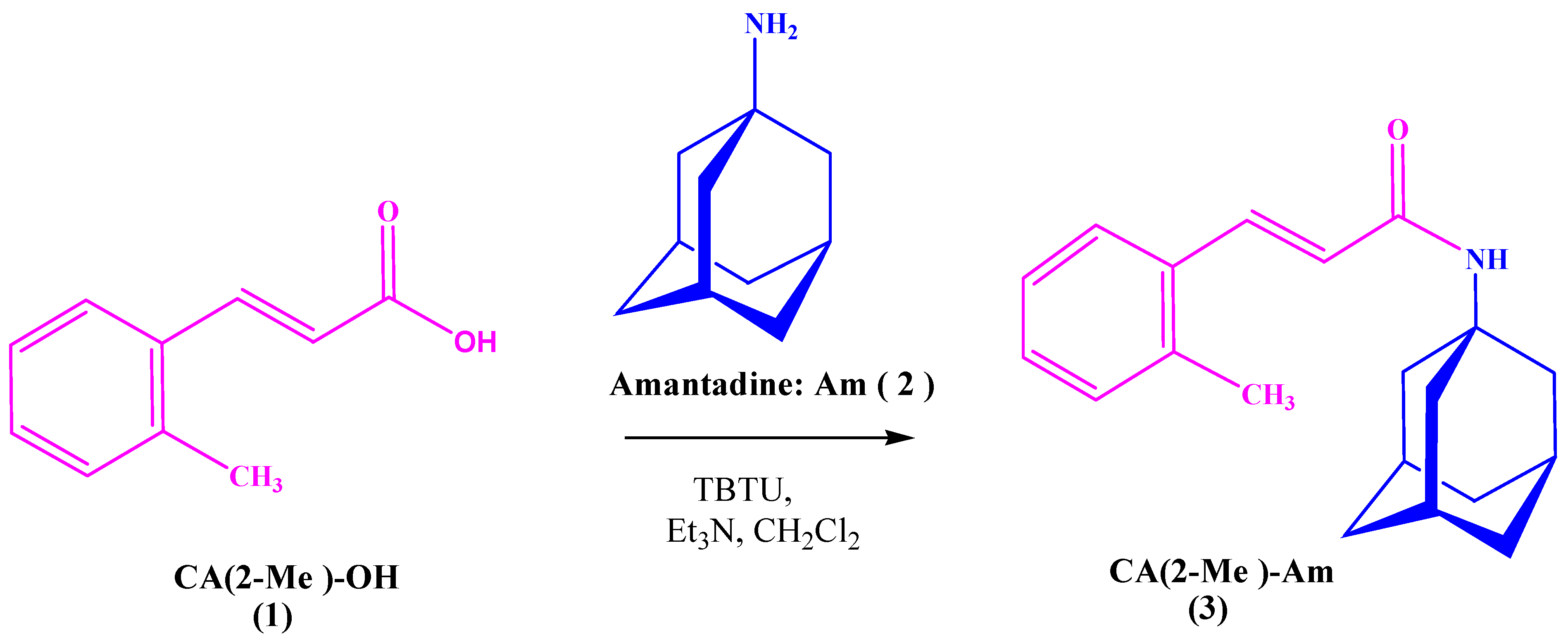
2.2. Synthesis of (E)- N-(2-Methylcinnamoyl)-Amantadine (3)

2-Methylcinnamic acid (1.8 g, 11.4 mmol) was suspended in 30 mL of CH2Cl2, and then, after adding Et3N (1.6 mL, 11.4 mmol), the obtained colorless liquid was treated by solid TBTU (3.7 g, 11.4 mmol). After being stirred for ≈10 min, to the mixture, we added amantadine (2.4 g, 12.6 mmol) and Et3N (1.8 mL, 12.6 mmol), dissolved (under sonication) in 40 mL CH2Cl2. Thus, the reaction mixture was stirred at room temperature for 3 h, and then was diluted with an additional 30 mL CH2Cl2. The organic phase was washed with 5% aqueous NaHCO3 (5 × 50 mL) and brine (3 × 50 mL), dried over Na2SO4, and concentrated in vacuo. Furthermore, after purification, the amide was obtained (3.3 g, 89%) as white crystals.
Compound (3): white crystals (CH3CN); mp 188–189 ⁰C; 1H NMR (DMSO-d6, 600 MHz) δ 7.59 (s, 1H), 7.56 (d, J = 15.6 Hz, 1H), 7.48 (d, J = 7.3 Hz, 1H), 7.27–7.20 (m, 3H), 6.58 (d, J = 15.6 Hz, 1H), 2.35 (s, 3H), 2.03 (bs, 3H), 2.00 (bs, 6H), 1.64 (bs, 6H); 13C NMR (DMSO-d6, 151 MHz) δ 164.5, 137.0, 135.7, 134.4, 131.1, 129.4, 126.8, 126.3, 125.3, 51.4, 41.5, 36.5, 29.3, 19.9; HRMS m/z 318.1830 (calcd for C20H25NNaO, 318.1828).

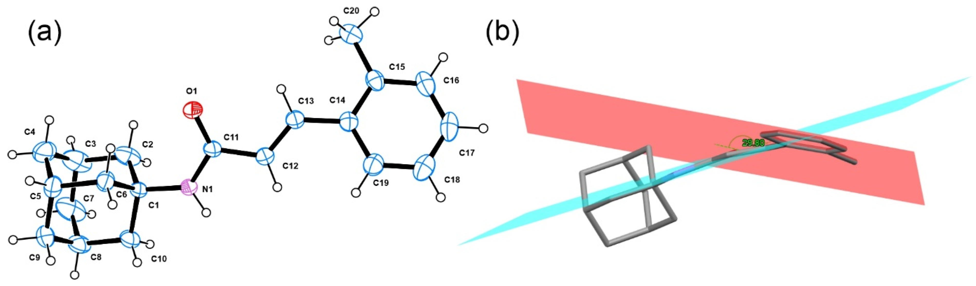
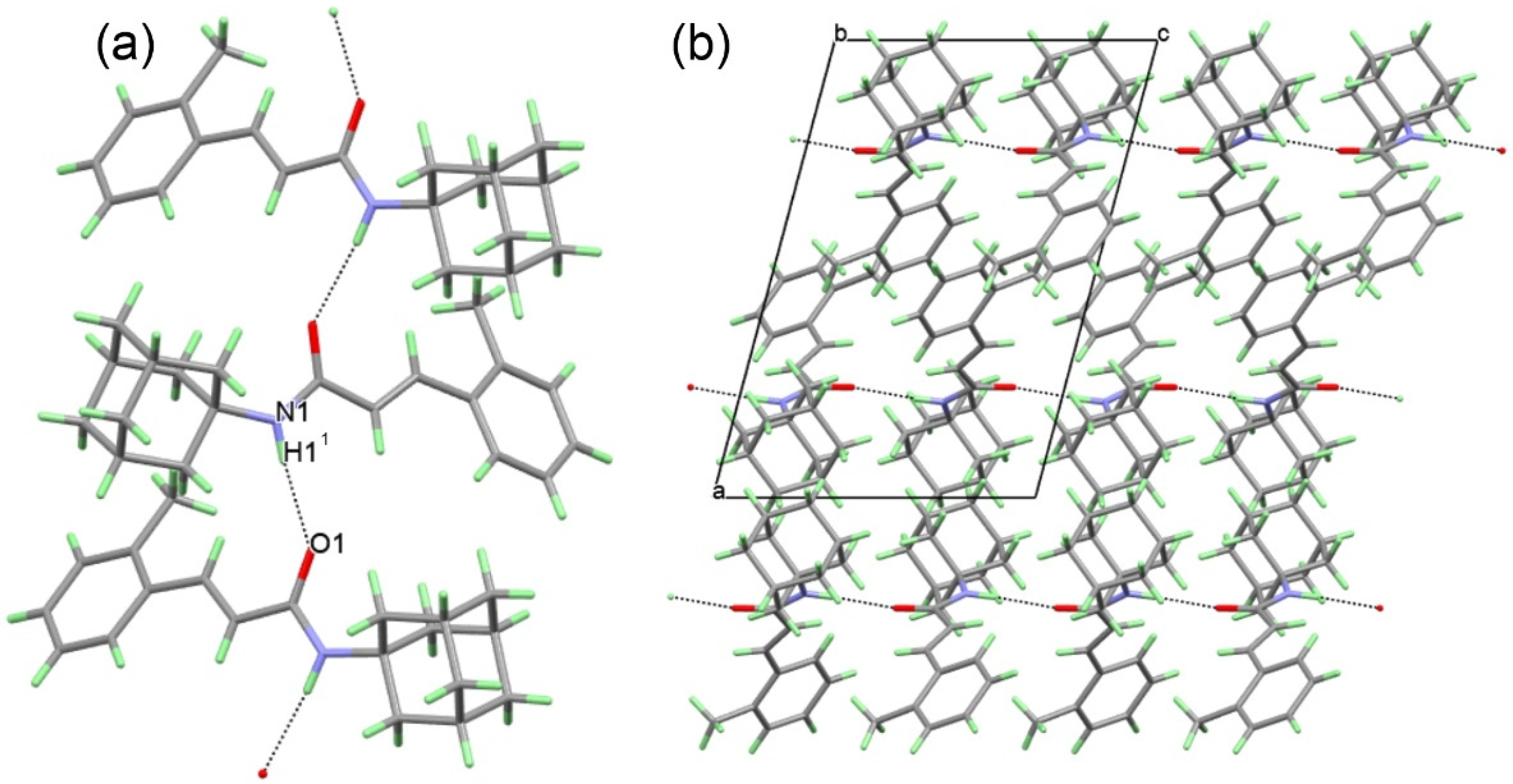
2.3. Single Crystal X-ray Diffraction (SCXRD) of (E)-N-(2-methylcinnamoyl)-amantadine (3)

Single crystals of compound 3 were grown from 1:1 v/v benzene methanol solution. A crystal with suitable size and quality was selected and was mounted on a glass capillary. The diffraction peak intensities and coordinates were collected on Bruker D8 Venture diffractometer (Bruker AXS GmbH, Karlsruhe, Germany) equipped with a PhotonII CMOS detector using micro-focus MoKα radiation (λ = 0.71073 Å). Data were processed with CrysAlisPro software [17]. The structure was solved with intrinsic methods using ShelxT [18] and refined by the full-matrix least-squares method on the F2 with ShelxL program [19]. All non-hydrogen atoms were located successfully from the Fourier map and were refined anisotropically. Hydrogen atoms were placed on calculated positions (C–Haromatic = 0.93, C–Hmethyl = 0.96 Å, and C–Hmethylenic = 0.97 Å, riding on the parent atom (Ueq = 1.2). The H atom near the nitrogen was located from a different Fourier map. Complete crystallographic data for the structure of the title compound reported in this paper were deposited in the CIF format at the Cambridge Crystallographic Data Center as 2205297. These data can be obtained free of charge via http://www.ccdc.cam.ac.uk/conts/retrieving.html, deposited on 5 September 2022 (or from the CCDC, 12 Union Road, Cambridge CB2 1EZ, UK; Fax: +441223336033; e-mail: deposit@ccdc.cam.ac.uk).

2.4. Neurobehavioral Studies

Mice (C57BL/6, male, 8 weeks old) were obtained from Erboj (Animal Breeding Center, Slivniza, Sofia). The animals were housed two per cage under constant laboratory conditions (25 ± 3°C, 12/12 h light/dark cycle) with food and water available ad libitum. The habituation period was 5 days before the start of the experiment. The protocol of all experiments was in accordance with the requirements of the European Communities Council Directive 86/609/EEC and rules of the Bioethics Committee 30/03/2021, Institute of Neurobiology, Bulgarian Academy of Sciences.
CA(2-Me)-Am (3) was dissolved in oleanolic acid (OA). We tested five doses of amide 3 applied per os on 25 male mice C57BL/6 and found the dose of 20 mg/kg to be the most effective.
Mice were divided into four experimental groups (n = 8 in each group) as follows: (1) control, treated with normal saline (i.p.) for 12 consecutive days; (2) MPTP (30 mg/kg/day, i.p.) applied daily for 5 consecutive days in accordance with the work of Shin et al. [20]; (3) MPTP (30 mg/kg, i.p.) + CA(2-Me)-Am (20 mg/kg, per os) applied for 12 consecutive days, 5 days simultaneously with MPTP and 7 days after MPTP; (4) MPTP (30 mg/kg, i.p.) + OA (per os) applied daily for 12 consecutive days.
All mice training was conducted before MPTP administration.

2.4.1. Rotarod Test

Mice from all experimental groups were placed on a gyratory with a fixed speed of 7 rpm/min, and the time on rotarod was determined. The observation period was 5 min. All animals were pre-trained on the rotarod apparatus before treatment in order to reach stable performance. The training consisted of one session per day over 3 consecutive days. The test was made on the 13th day, and the average time per group was calculated after the experiment and repeated four times [21].

2.4.2. Passive Avoidance Test

Learning and memory performance in mice was evaluated using passive avoidance learning test [22]. Acquisition phase: during this phase, each animal was placed in the illuminated compartment. When the rodents innately entered into the dark compartment, they received a mild electrical foot shock (0.5 mA, 3 s). In this trial, the initial latency (IL) of entrance into the dark chamber of each animal was recorded, and mice with ILs > 60 s were excluded from the study. Test phase: on the 13th and 14th days, each mouse was placed in the illuminated chamber, and the entry into the dark chamber was measured as step through latency (STL). The behavioral observations were carried out between 9 a.m. and 12 a.m.

2.4.3. Statistical Analysis

The results were expressed as means ± the standard error of the mean (SEM) or as percentage changes over the mean compared to the control. Statistical analyses of the data were performed by one-way analysis of variance (ANOVA) followed by Dunnett post hoc comparison test. Differences were considered significant at p < 0.05.

3. Results and Discussion

3.1. Chemistry

Herein, the 2-methylcinnamic acid amide (CA(2-Me)-Am; 3) was synthesized as outlined in Scheme 1. Generally, the amidation of 2-methylcinnamic acid (CA(2-Me)-OH; 1) with amantadine (Am; 2) was carried out in the presence of tertiary amine (triethylamine, Et3N) and by one of the preferred coupling reagents for in situ activation, such as TBTU [16] to amide 3.
The structure of the newly obtained compound (3) was confirmed by the 1H NMR, 13C NMR, HRMS, and single crystal analysis powder diffraction. The title compound (CA(2-Me)-Am; 3) crystallized in the monoclinic P21/c space group, with one molecule in the asymmetric unit (Figure 1). The bond distances and angles (Table 1) within the adamantane and 2-methylcinnamic acid were comparable with those observed in other structures [23,24,25,26,27].
The angle between the phenyl and acrylamide moieties was 29.8 °, disclosing that the conjugation was not stringent, e.g., the conjugation could be disrupted. In the molecule of (E)–N-(2-methylcinnamoyl)-amantadine, one hydrogen donor (N–H) and one acceptor (carbonyl oxygen) were present. In the crystal structure, the molecules produced one-dimensional chains with a graph set C 1 1 4 [28,29] (Figure 2a). The three-dimensional packing of the molecules (Figure 2b) did not reveal additional weak interactions, and thus the stabilization of the crystal structure was achieved by the N1-H1…O1 hydrogen bond (N1…O1 of 3.065(5) Å).

3.2. In Vivo Evaluation of Amide 3 in an Experimental Mouse Model of PD

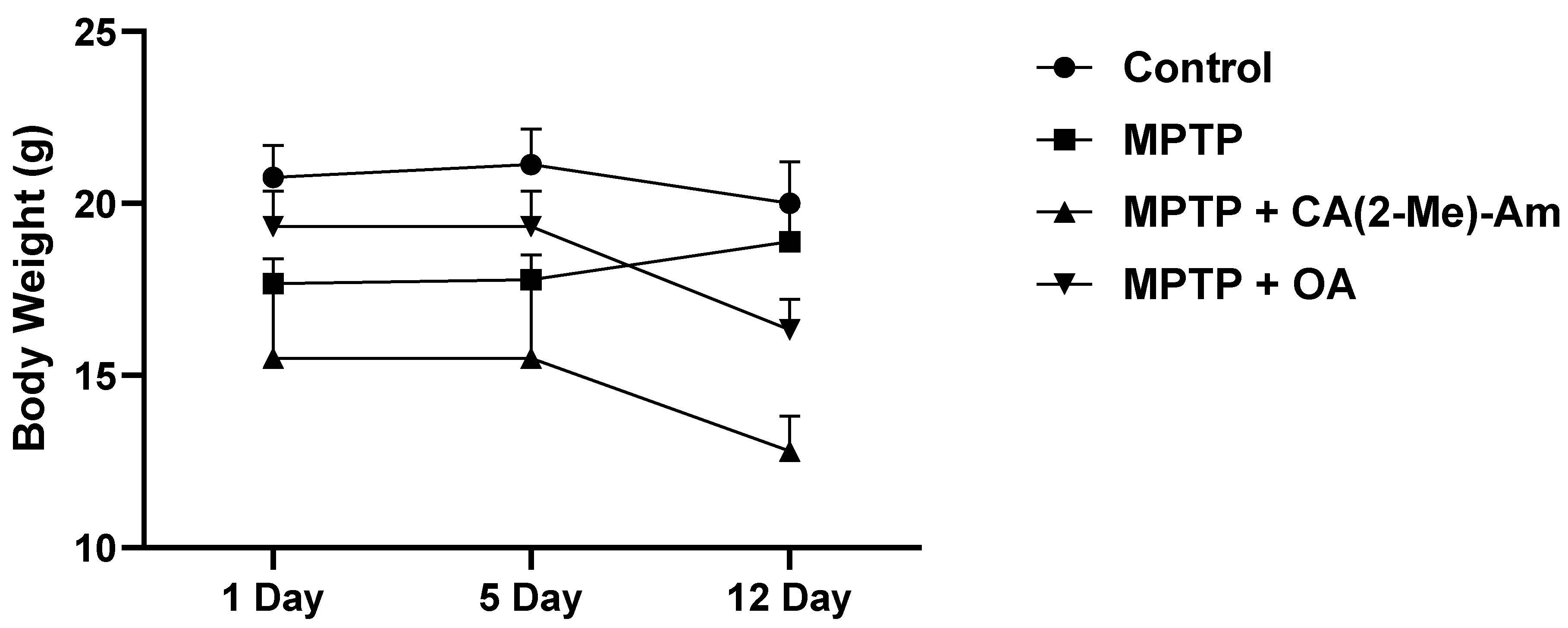
3.2.1. Effect of CA(2-Me)-Am (3) on the Weight of Experimental Animals
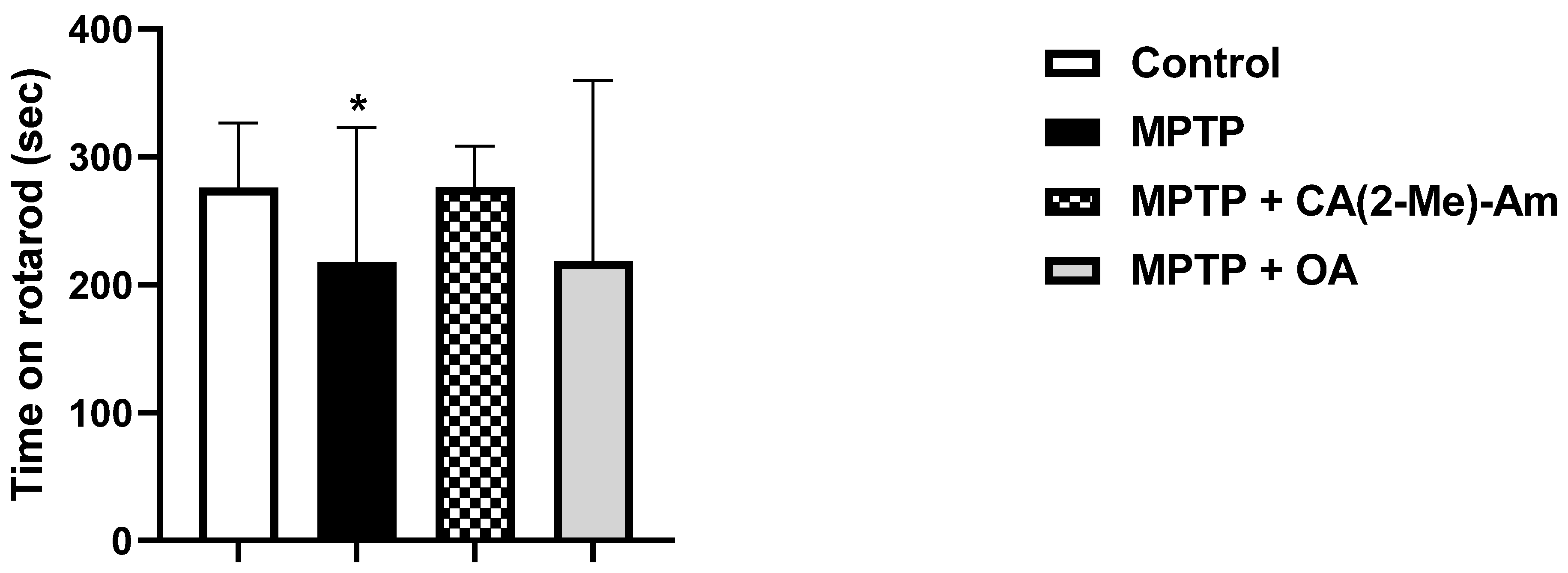
There was no significant change in the weight of the control mice over the 12-day period. In those treated with MPTP, we observed a 6.92% weight gain within the group. In the MPTP + CA(2-Me)-Am and MPTP + OA mice, weight reduction was recorded at the end of the observed period at 17.41% and 15.51%, respectively (Figure 3).

3.2.2. Rotarod Test

The studies performed demonstrated that the group of mice treated either with the MPTP toxin or with MPTP + OA spent less time on the rotating lever of the rotarod apparatus as compared to the controls, which is an indication of a motor-impairing effect. The reduction was by 21.13 % (p < 0.05) for the MPTP group, and by 20.99 % (p < 0.05) for the MPTP + OA group (Figure 4). In the MPTP + CA(2-Me)-Am group, the time that experimental animals spent on the rotary lever was comparable to that of the control group (Figure 4).

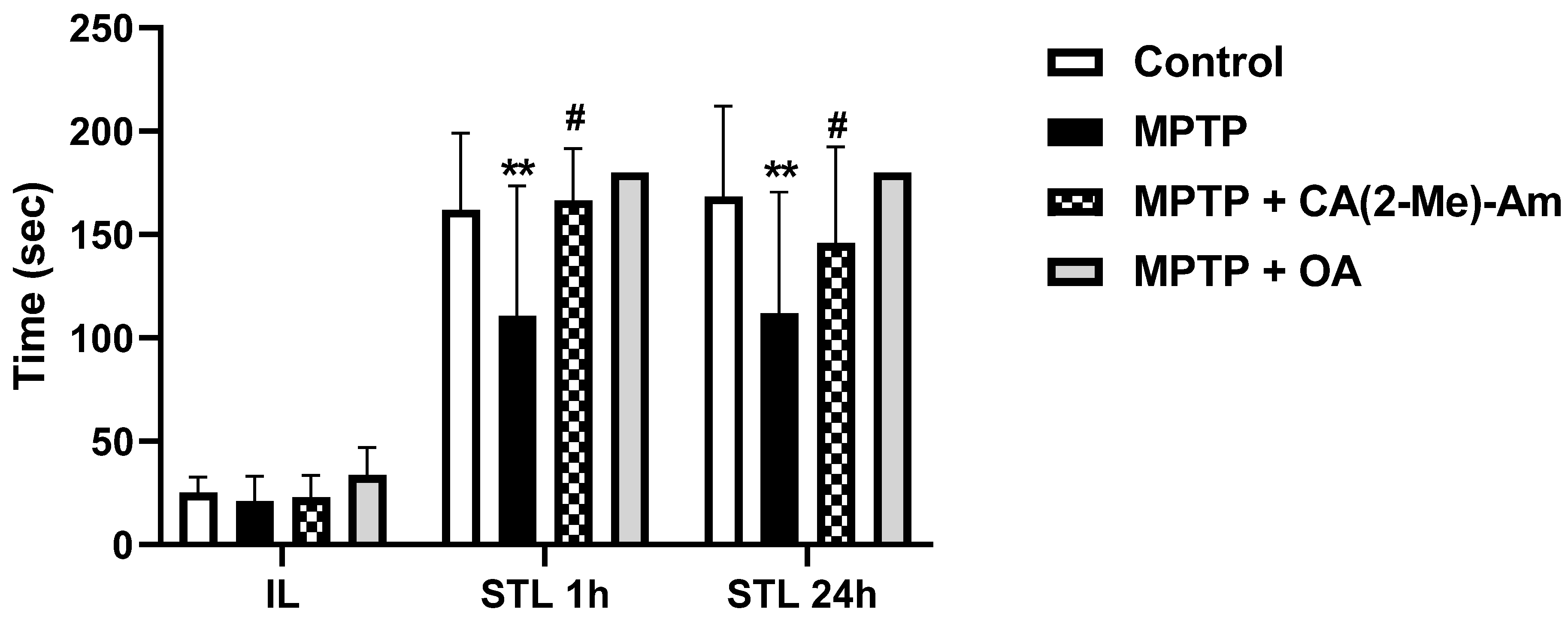
3.2.3. Passive Avoidance Test

The administration of the MPTP toxin caused a decrease in the step-through latency time by 31.56% (p < 0.01) at 1st h and by 33.45% (p < 0.01) at 24th h after the training of mice as compared to controls, which is evidence of memory and learning deficits. Administration of CA(2-Me)-Am increased the latent reaction time by 33.49% (p < 0.05) at the 1st h and by 33.84% (p < 0.05) at 24th h as compared to the MPTP-treated group, an indication of a memory-protective effect of the newly synthesized amantadine derivative (Figure 5).

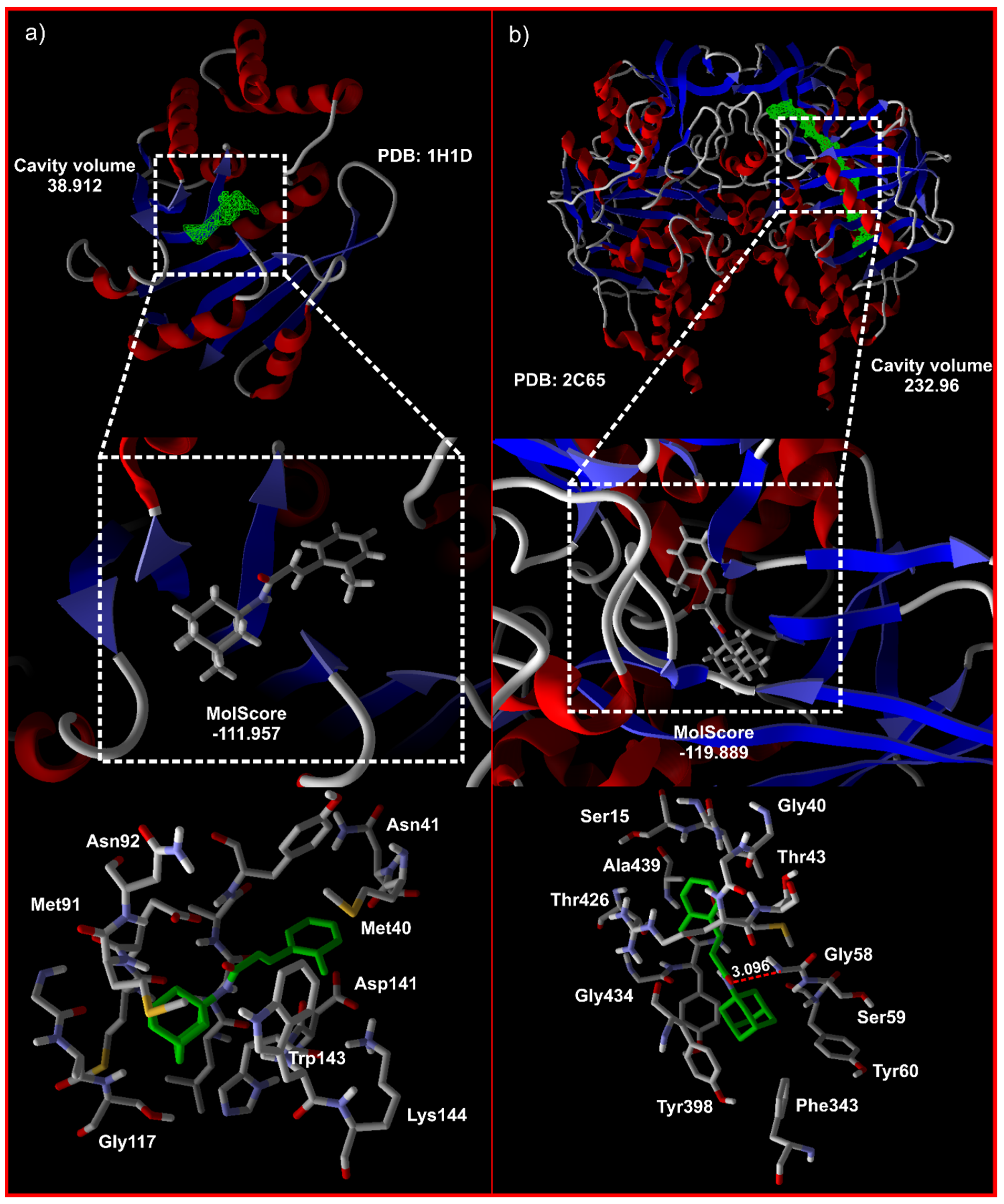
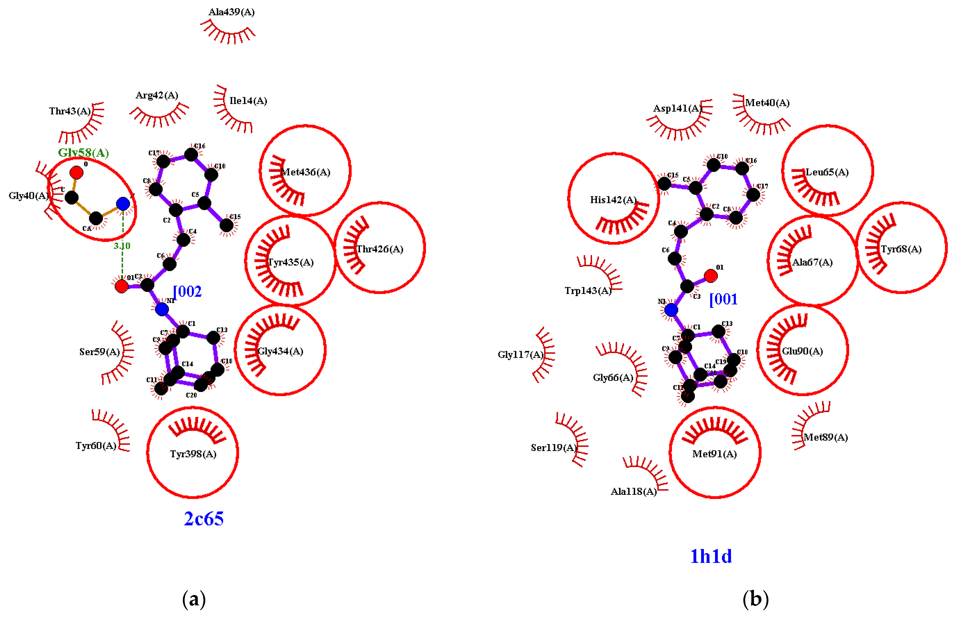
3.3. Molecular Docking

The docking of (E)-N-(2-methylcinnamoyl)-amantadine (3) was performed against four different targets associated with PD: A2aAR (3EML) [31], COMT (1H1D) [32], MAO-B (2C65) [33], and NMDA (7SAD) [34] (Table 2).
The docking approach involved predicting the conformation and orientation of ligands within a targeted binding site. Initially, the reference drug memantine present in 7SAD [34] was employed to adjust the docking parameters. The structures of the target enzymes were obtained from the Protein Data Bank (PDB), the coordinates of the small molecules were generated from the crystal structure of the (E)–N-(2-methylcinnamoyl)-amantadine, and positioning in the active site and docking were conducted using Molegro Virtual Docker (MVD2019.7.0.0-2019-03-18-1B win32). Details regarding the docking validation are provided in Figures S4 and S5. The UCSF Chimera [35] and Ligplot+ 2.2.5 [36] were used for visualization and interactions detection. On the basis of the scores obtained from the docking results, the interaction of (E)–N-(2-methylcinnamoyl)-amantadine with MAO-B was most favorable (score of –119.889, Figure 6). The second possibility not to be excluded is the interaction with COMT (–111.957). However, while for MAO-B, a hydrogen bonding interaction was detected for COMT, no hydrogen bonding interaction “enzyme…ligand” was identified (Figure 7). Interestingly, the active sites of COMT and MAO-B shared a large amount of analogical AA and thus it may be possible to design a ligand that will interact with both enzymes.
Parkinson’s disease has high social significance, resulting from a progressive loss of nigrostriatal dopaminergic neurons. The decrease in striatal dopaminergic innervation due to this loss is responsible for motor disturbances characteristic of the disease, such as akinesia, muscular rigidity, and tremor, and cognitive function impairment later appears. Amantadine is an agent that raises the concentration of dopamine in the synaptic cleft in PD. Currently, levodopa is considered to be the gold standard for symptomatic treatment of PD. However, long-term treatment with levodopa is complicated by motor fluctuations and dyskinesia. Everything stated above requires the search for compounds that can replace levodopa and improve the efficacy of amantadine in the treatment of PD. In this line of thinking, we performed investigations of a newly obtained amantadine derivative CA(2-Me)-Am (3) on neuromuscular coordination, learning, and memory in an experimental mouse model of PD. The obtained data showed that amide 3 restored neuromuscular coordination and memory performance of parkinsonian animals to the control level, giving an indication of its beneficial protective effects.
Moreover, the molecular docking study investigations showed that from the considered four targets, (E)–N-(2-methylcinnamoyl)-amantadine interacted preferably with MAO-B, followed by COMT. The detected hydrogen bonding interaction could be used for development of modified potential antiviral drug candidates.
In conclusion, our results demonstrated ameliorating effects of the newly synthesized compound CA(2-Me)-Am in an experimental model of Parkinson’s disease, which deserves further investigations.

Supplementary Materials

The following supporting information can be downloaded at: https://www.mdpi.com/article/10.3390/cryst12111518/s1, NMR and MS spectra of compound 3 are provided.

Author Contributions

Synthesis, supervision, writing and manuscript conceptualization, M.C.; NMR spectroscopy studies, N.P. and M.Š.; study, M.Š.; single crystal X-ray diffraction experiments, R.R. and H.S.-D.; docking studies, writing, B.S.; neurobehavioral studies, writing, R.K., L.T., M.L., A.P. and K.T. All authors have read and agreed to the published version of the manuscript.

Funding

This work was funded by the Bulgarian National Science Fund (BNSF), grant number KP-06-Russia/7-2019.

Institutional Review Board Statement

The animal study protocol was approved by the Commission of Bioethics (CBE) at the Institute of Neurobiology, Bulgarian Academy of Sciences—BAS/CBE/018/2020.

Informed Consent Statement

Not applicable.

Data Availability Statement

Crystallographic data for the structure of the title compound (E)-N-(2-methylcinnamoyl)-amantadine (CA(2-Me)-Am; 3)) was deposited in the CIF format with the Cambridge Crystallographic Data Center as 2205297. These data can be obtained free of charge via http://www.ccdc.cam.ac.uk/conts/retrieving.html, deposited on 05 September 2022 (or from the CCDC, 12 Union Road, Cambridge CB2 1EZ, UK; Fax: +441223336033; e-mail: deposit@ccdc.cam.ac.uk).

Conflicts of Interest

The authors declare no conflict of interest.

References

  1. He, W.; Goodkind, D.; Kowal, P.R. An Aging World: 2015; International Population Reports; United States Census Bureau: Suitland-Silver Hill, MD, USA, 2016. [Google Scholar]
  2. Parkinson Disease: A Public Health Approach: Technical Brief; World Health Organization: Geneva, Switzerland, 2022.
  3. Mhyre, T.R.; Boyd, J.T.; Hamill, R.W.; Maguire-Zeiss, K.A. Parkinson’s Disease. Subcell. Biochem. 2012, 65, 389–455. [Google Scholar] [CrossRef] [PubMed] [Green Version]
  4. Wyss-Coray, T. Ageing, Neurodegeneration and Brain Rejuvenation. Nature 2016, 539, 180–186. [Google Scholar] [CrossRef] [PubMed] [Green Version]
  5. Liu, J.; Obando, D.; Liao, V.; Lifa, T.; Codd, R. The Many Faces of the Adamantyl Group in Drug Design. Eur. J. Med. Chem. 2011, 46, 1949–1963. [Google Scholar] [CrossRef] [PubMed]
  6. Wanka, L.; Iqbal, K.; Schreiner, P.R. The Lipophilic Bullet Hits the Targets: Medicinal Chemistry of Adamantane Derivatives. Chem. Rev. 2013, 113, 3516–3604. [Google Scholar] [CrossRef] [PubMed] [Green Version]
  7. Schmidtke, M.; Zell, R.; Bauer, K.; Krumbholz, A.; Schrader, C.; Suess, J.; Wutzler, P. Amantadine Resistance among Porcine H1N1, H1N2, and H3N2 Influenza A Viruses Isolated in Germany between 1981 and 2001. Intervirology 2006, 49, 286–293. [Google Scholar] [CrossRef]
  8. Nelson, M.I.; Simonsen, L.; Viboud, C.; Miller, M.A.; Holmes, E.C. The Origin and Global Emergence of Adamantane Resistant A/H3N2 Influenza Viruses. Virology 2009, 388, 270–278. [Google Scholar] [CrossRef] [Green Version]
  9. Weinstock, D.M.; Zuccotti, G. The Evolution of Influenza Resistance and Treatment. JAMA 2009, 301, 1066–1069. [Google Scholar] [CrossRef]
  10. Schwab, R.S.; England, A.C.; Poskanzer, D.C.; Young, R.R. Amantadine in the Treatment of Parkinson’s Disease. JAMA 1969, 208, 1168–1170. [Google Scholar] [CrossRef]
  11. Rascol, O.; Fabbri, M.; Poewe, W. Amantadine in the Treatment of Parkinson’s Disease and Other Movement Disorders. Lancet Neurol. 2021, 20, 1048–1056. [Google Scholar] [CrossRef]
  12. Adisakwattana, S. Cinnamic Acid and Its Derivatives: Mechanisms for Prevention and Management of Diabetes and Its Complications. Nutrients 2017, 9, E163. [Google Scholar] [CrossRef]
  13. Liu, L.; Hudgins, W.R.; Shack, S.; Yin, M.Q.; Samid, D. Cinnamic Acid: A Natural Product with Potential Use in Cancer Intervention. Int. J. Cancer 1995, 62, 345–350. [Google Scholar] [CrossRef]
  14. Prorok, T.; Jana, M.; Patel, D.; Pahan, K. Cinnamic Acid Protects the Nigrostriatum in a Mouse Model of Parkinson’s Disease via Peroxisome Proliferator-Activated Receptorα. Neurochem. Res. 2019, 44, 751–762. [Google Scholar] [CrossRef]
  15. Hemmati, A.A.; Alboghobeish, S.; Ahangarpour, A. Effects of Cinnamic Acid on Memory Deficits and Brain Oxidative Stress in Streptozotocin-Induced Diabetic Mice. Korean J. Physiol. Pharmacol. Off. J. Korean Physiol. Soc. Korean Soc. Pharmacol. 2018, 22, 257–267. [Google Scholar] [CrossRef] [Green Version]
  16. Knorr, R.; Trzeciak, A.; Bannwarth, W.; Gillessen, D. New Coupling Reagents in Peptide Chemistry. Tetrahedron Lett. 1989, 30, 1927–1930. [Google Scholar] [CrossRef]
  17. Rigaku Oxford Diffraction. CrysAlis pro. Rigaku Oxford Diffraction, CrysAlis pro CrysAlis Pro. 2015. Available online: https://www.rigaku.com/products/crystallography/crysalis (accessed on 20 October 2022).
  18. Sheldrick, G.M. SHELXT-Integrated Space-Group and Crystal-Structure Determination. Acta Crystallogr. Sect. Found. Adv. 2015, 71, 3–8. [Google Scholar] [CrossRef] [Green Version]
  19. Sheldrick, G.M. A Short History of SHELX. Acta Crystallogr. A 2008, 64, 112–122. [Google Scholar] [CrossRef] [Green Version]
  20. Shin, K.S.; Zhao, T.T.; Choi, H.S.; Hwang, B.Y.; Lee, C.K.; Lee, M.K. Effects of Gypenosides on Anxiety Disorders in MPTP-Lesioned Mouse Model of Parkinson׳s Disease. Brain Res. 2014, 1567, 57–65. [Google Scholar] [CrossRef]
  21. Manna, S.; Bhattacharyya, D.; Mandal, T.K.; Dey, S. Neuropharmacological Effects of Deltamethrin in Rats. J. Vet. Sci. 2006, 7, 133–136. [Google Scholar] [CrossRef] [Green Version]
  22. Shahidi, S.; Komaki, A.; Mahmoodi, M.; Atrvash, N.; Ghodrati, M. Ascorbic Acid Supplementation Could Affect Passive Avoidance Learning and Memory in Rat. Brain Res. Bull. 2008, 76, 109–113. [Google Scholar] [CrossRef]
  23. Land, M.A.; Robertson, K.N.; Ylijoki, K.E.O.; Clyburne, J.A.C. Reactivity of 1,3-Dichloro-1,3-Bis(Dimethylamino)-Propenium Salts with Primary Amines. New J. Chem. 2021, 45, 13558–13570. [Google Scholar] [CrossRef]
  24. Pereira, A.K.d.S.; Manzano, C.M.; Nakahata, D.H.; Clavijo, J.C.T.; Pereira, D.H.; Lustri, W.R.; Corbi, P.P. Synthesis, Crystal Structures, DFT Studies, Antibacterial Assays and Interaction Assessments with Biomolecules of New Platinum(Ⅱ) Complexes with Adamantane Derivatives. New J. Chem. 2020, 44, 11546–11556. [Google Scholar] [CrossRef]
  25. Perlovich, G.L.; Ryzhakov, A.M.; Tkachev, V.V.; Hansen, L.K.; Raevsky, O.A. Sulfonamide Molecular Crystals: Structure, Sublimation Thermodynamic Characteristics, Molecular Packing, Hydrogen Bonds Networks. Cryst. Growth Des. 2013, 13, 4002–4016. [Google Scholar] [CrossRef]
  26. Sarcevica, I.; Orola, L.; Veidis, M.V.; Podjava, A.; Belyakov, S. Crystal and Molecular Structure and Stability of Isoniazid Cocrystals with Selected Carboxylic Acids. Cryst. Growth Des. 2013, 13, 1082–1090. [Google Scholar] [CrossRef]
  27. Swapna, B.; Maddileti, D.; Nangia, A. Cocrystals of the Tuberculosis Drug Isoniazid: Polymorphism, Isostructurality, and Stability. Cryst. Growth Des. 2014, 14, 5991–6005. [Google Scholar] [CrossRef]
  28. Etter, M.C.; MacDonald, J.C.; Bernstein, J. Graph-Set Analysis of Hydrogen-Bond Patterns in Organic Crystals. Acta Crystallogr. B 1990, 46, 256–262. [Google Scholar] [CrossRef]
  29. Etter, M.C. Encoding and Decoding Hydrogen-Bond Patterns of Organic Compounds. Acc. Chem. Res. 1990, 23, 120–126. [Google Scholar] [CrossRef]
  30. Farrugia, L.J. WinGX and ORTEP for Windows: An Update. J. Appl. Crystallogr. 2012, 45, 849–854. [Google Scholar] [CrossRef]
  31. Jaakola, V.-P.; Griffith, M.T.; Hanson, M.A.; Cherezov, V.; Chien, E.Y.T.; Lane, J.R.; Ijzerman, A.P.; Stevens, R.C. The 2.6 Angstrom Crystal Structure of a Human A2A Adenosine Receptor Bound to an Antagonist. Science 2008, 322, 1211–1217. [Google Scholar] [CrossRef] [Green Version]
  32. Bonifácio, M.J.; Archer, M.; Rodrigues, M.L.; Matias, P.M.; Learmonth, D.A.; Carrondo, M.A.; Soares-Da-Silva, P. Kinetics and Crystal Structure of Catechol-o-Methyltransferase Complex with Co-Substrate and a Novel Inhibitor with Potential Therapeutic Application. Mol. Pharmacol. 2002, 62, 795–805. [Google Scholar] [CrossRef] [Green Version]
  33. Binda, C.; Hubálek, F.; Li, M.; Herzig, Y.; Sterling, J.; Edmondson, D.E.; Mattevi, A. Binding of Rasagiline-Related Inhibitors to Human Monoamine Oxidases: A Kinetic and Crystallographic Analysis. J. Med. Chem. 2005, 48, 8148–8154. [Google Scholar] [CrossRef]
  34. Chou, T.-H.; Epstein, M.; Michalski, K.; Fine, E.; Biggin, P.C.; Furukawa, H. Structural Insights into Binding of Therapeutic Channel Blockers in NMDA Receptors. Nat. Struct. Mol. Biol. 2022, 29, 507–518. [Google Scholar] [CrossRef]
  35. Pettersen, E.F.; Goddard, T.D.; Huang, C.C.; Couch, G.S.; Greenblatt, D.M.; Meng, E.C.; Ferrin, T.E. UCSF Chimera--a Visualization System for Exploratory Research and Analysis. J. Comput. Chem. 2004, 25, 1605–1612. [Google Scholar] [CrossRef] [Green Version]
  36. Wallace, A.C.; Laskowski, R.A.; Thornton, J.M. LIGPLOT: A Program to Generate Schematic Diagrams of Protein-Ligand Interactions. Protein Eng. Des. Sel. 1995, 8, 127–134. [Google Scholar] [CrossRef]
Scheme 1. Synthetic route of amide 3.
Crystals 12 01518 sch001
Figure 1. (a) ORTEP [30] view and numbering scheme of the molecule present in the asymmetric unit of (E)–N-(2-methylcinnamoyl)-amantadine; the thermal ellipsoids were drawn with 50% probability, and hydrogen atoms are shown as small spheres with arbitrary radii. (b) Observed angle between the mean plane of the phenyl (C1/C15/C16/C17/C18/C19) and acrylamide (C13/C12/C11/O1/N1) moieties (C13/C12/C11/O1/N1).
Crystals 12 01518 g001
Figure 2. The observed (a) hydrogen bonding interaction stabilizing the crystal structure of (E)–N-(2-methylcinnamoyl)-amantadine and (b) a view along the b axis of the three-dimensional packing of the molecules and formation of C11(4) chains propagating along the [10] plane.
Crystals 12 01518 g002
Figure 3. Effect of CA(2-Me)-Am on the weight of the experimental animals. Data are presented as means with their respective standard errors (m ± S.E.M; n = 8; * p < 0.05).
Crystals 12 01518 g003
Figure 4. Effect of CA(2-Me)-Am on neuromuscular coordination. The asterisks above bars indicate significant differences in number of falls per minute for each experimental group versus the control at * p < 0.05. Statistical analysis was performed by one-way analysis of variance (ANOVA) followed by Dunnett’s post hoc comparison test.
Crystals 12 01518 g004
Figure 5. The effect of CA(2-Me)-Am on initial latency (IL) and step-through latency (STL) in a single-trial passive avoidance test in a mouse model of PD. Significance vs. control group: **p < 0.01; significance vs. MPTP-treated group: # p < 0.05. Statistical analysis was performed by one-way analysis of variance (ANOVA), followed by Dunnett’s post hoc comparison test.
Crystals 12 01518 g005
Figure 6. Visualization of the docking studies and molecular interaction of (E)-N-(2-methylcinnamoyl)-amantadine with (a) COMT and (b) MAO-B. The hydrogen bonding interaction N-H…O=C is shown in as red dashed line with the A…D distance of 3.096 Å.
Crystals 12 01518 g006
Figure 7. Observed interactions after docking of (E)–N-(2-methylcinnamoyl)-amantadine into the active site of two putative PD targets (a) MAO-B (2C65) and (b) COMT (1H1D). The hydrogen bonding interactions are shown in green; the hydrophobic contacts are shown as Crystals 12 01518 i001 for enzymes and with Crystals 12 01518 i002 for ligands; the similar residues for both enzymes are shown as Crystals 12 01518 i003.
Crystals 12 01518 g007
Table 1. The most important data collection and crystallographic refinement parameters for (E)=N-(2-methylcinnamoyl)-amantadine.
Empirical formulaC20H25NO
Formula weight295.41
Temperature/K290.00
Crystal systemmonoclinic
Space groupP21/c
a/Å14.692(2)
b/Å11.900(2)
c/Å9.9904(18)
α/°90
β/°104.943(5)
γ/°90
Volume/Å31687.6(5)
Z4
ρcalcg/cm31.163
μ/mm-10.071
F(000)640.0
Crystal size/mm30.3 × 0.25 × 0.2
RadiationMoKα (λ = 0.71073)
2Θ range for data collection/°4.466 to 52.842
Index ranges-18 ≤ h ≤ 17, -14 ≤ k ≤ 12, -12 ≤ l ≤ 12
Reflections collected10,910
Independent reflections3443 (Rint = 0.0595, Rsigma = 0.0654)
Data/restraints/parameters3443/0/205
Goodness-of-fit on F21.018
Final R indexes (I > =2σ (I))R1 = 0.0605, wR2 = 0.1171
Final R indexes (all data)R1 = 0.1142, wR2 = 0.1405
Largest diff. peak/hole/e Å−30.17/−0.13
Table 2. Molecular docking score (kcal/mol) of (E)–N-(2-methylcinnamoyl)-amantadine against selected targets associated with PD.
Molegro Virtual
Docker Score
Detected Hydrogen
Bonding Interaction
(E)–N-(2-methylcinnamoyl)-amantadine
NMAD (7SAD) [34]-80.240No
COMT (1H1D) [32]-111.957No
A2aAR (3EML) [31]-83.578C=O…O-H Tyr271
D…A 3.38 Å
MAO-B (2C65) [33]-119.889C=O…N-H Gly58
D…A 3.09 Å
NMAD, N-methyl-D-aspartate receptor; COMT, catechol-O-methyltransferase; A2aAR, A2A adenosine receptor; MAO-B, monoamine oxidase B.
Publisher’s Note: MDPI stays neutral with regard to jurisdictional claims in published maps and institutional affiliations.

