Article

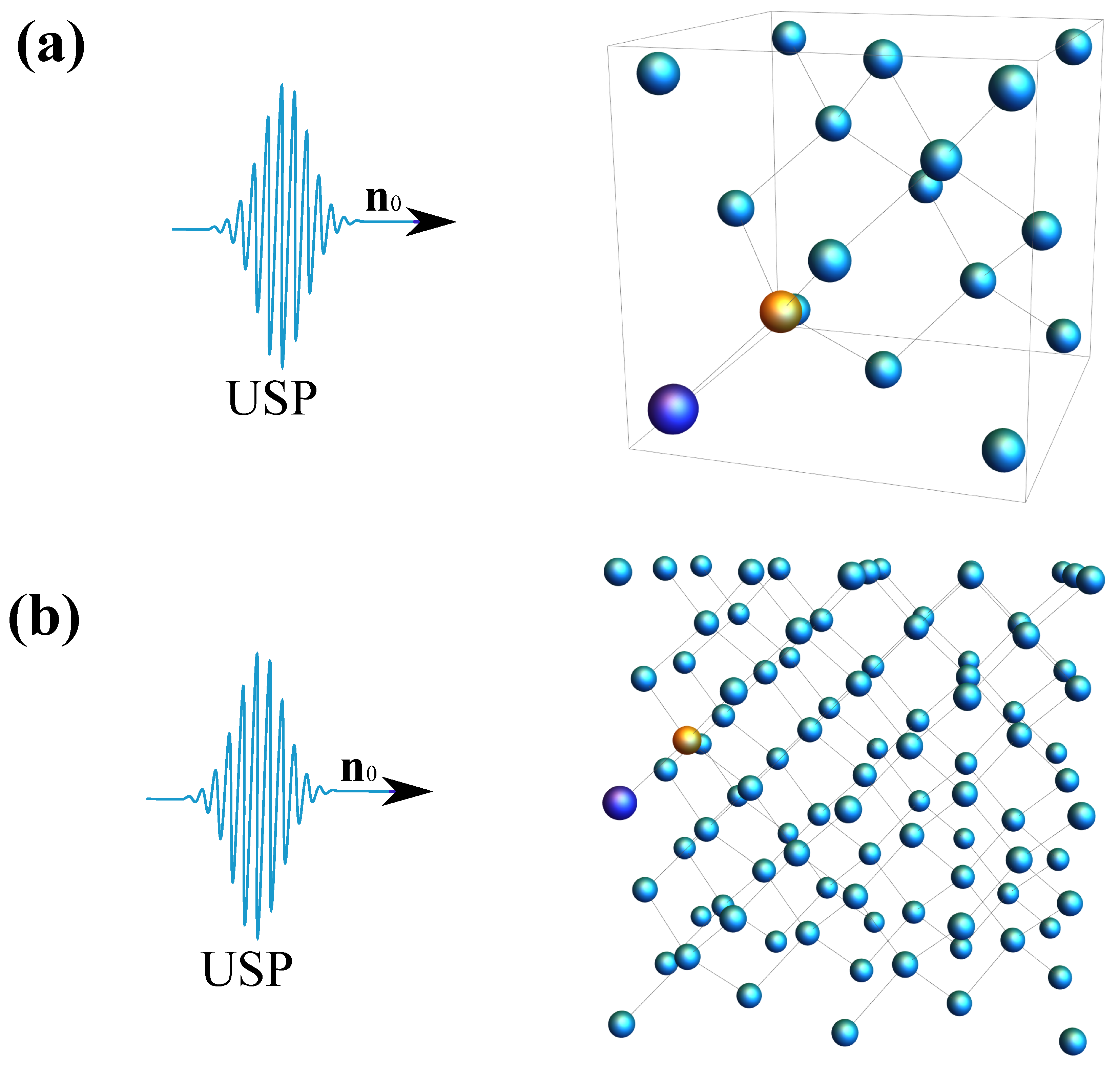
Scattering of Ultrashort X-ray Pulses on Diamonds with NV Centers

Laboratory for Diagnostics of Carbon Materials and Spin-Optical Phenomena in Wide-Bandgap Semiconductors, Northern (Arctic) Federal University, Nab. Severnoi Dviny 17, 163002 Arkhangelsk, Russia
*
Author to whom correspondence should be addressed.
Crystals 2022, 12(10), 1417; https://doi.org/10.3390/cryst12101417
Submission received: 15 September 2022 / Revised: 4 October 2022 / Accepted: 6 October 2022 / Published: 8 October 2022

Abstract

:
The scattering of ultra-short X-ray pulses (USPs) is an important component of diffraction analysis of matter. Usually, the specific scattering of such USPs is not taken into account to determine the structure of a substance. Taking into account the specifics of scattering on complex structures will give more accurate results when deciphering complex structures. In this work, it is shown that when X-ray USPs are scattered on diamond with NV centers, it is necessary to take into account the pulse duration. The results obtained can be very different from the widely used theory of diffraction analysis, which confirms the need to take into account the specifics of USP scattering when diagnosing complex structures. It is shown that the scattering spectra are very sensitive to the concentration of NV-centres in the diamond structure, and this can be used in diffraction analysis.

1. Introduction

X-ray diffraction analysis of matter (XRD) using ultrashort pulses (USP) has now gained great importance [1,2,3]. It is now possible to study the structure of matter with high spatial and temporal resolution using such USPs, as new sources of such pulses are available for research. One of the most promising sources of USP are free-electron XFELs [4]. Investigations at such facilities are currently carried out using femtosecond pulses. At present, attosecond pulse generation is being reported by advances in X-ray free-electron lasers [5,6]. In addition, the so-called sub-femtosecond barrier (pulses close to attosecond) with high peak power has already been reached and it makes it possible to study various excitations in molecular systems, such as the motion of valence electrons with high spatial and temporal resolution [7]. As powerful sources of USPs are created, the challenge is to develop new theoretical approaches that take into account the specific interaction of such USPs with various complex structures [8,9]. Such structures include promising materials for quantum technologies. Such materials, among others, can be to attributed diamond with NV centres. The NV centre (nitrogen-vacancy centre) in diamond is one of the many point defects in diamond, which appear when the carbon atom is removed from the lattice node and the resulting vacancy binds with the nitrogen atom. The defect is unique in that its individual centre electron spins are easily manipulated by light, magnetic, electric and microwave fields, allowing quantum information (qubits) to be written on the centre nucleus spin. Such manipulation is possible even at room temperature [10,11]. Of main interest for quantum technologies are the so called NV centres (commonly denoted as NV), which have an extra electron located at the vacancy site and form a pair with spin S = 1 with one of the vacancy electrons.
Ultra-high resolution diffraction analysis of such structures is a promising trend in modern physics. Such analysis is based on scattering of X-rays by various periodic and complex structures. Usually, in the theoretical analysis of XRD scattering of X-ray ultrashort pulses are described as scattering of plane waves of infinite duration in time [3,12]. In other words, the specificity of USP scattering on such structures is not taken into account, which leads to inaccurate use of well-known approaches in XRD [3,8]. Scattering of femto- and attosecond USPs with high spatial and temporal resolution on these kinds of structures is being actively studied, because old approaches often do not take the pulse duration parameter into account, leading to errors [8,13,14,15,16,17,18,19,20,21,22,23]. It has recently been shown that the scattering of attosecond USPs on DNA nucleotides may differ significantly from the previously known XRD theory [24,25]. Thus, the study of USP scattering on diamond with NV centres, taking into account the specificity of their scattering, is an important task.
In this paper, the specificity of USP scattering on diamond with NV centres will be investigated. The results obtained have rather simple analytical form and can be applied for calculation of scattering spectra not only with NV centres but also for other colour structures in diamond. It is shown that the scattering spectra are sensitive to the number of NV centres in the diamond. Moreover, it is shown that the consideration of the USP durations leads to a marked difference of the theory presented here to the previously known and widely used one.
Further, we will use the atomic system of units: m e = 1; = 1; | e | = 1, where e is the electron charge, is the Dirac constant, m e is the electron mass.

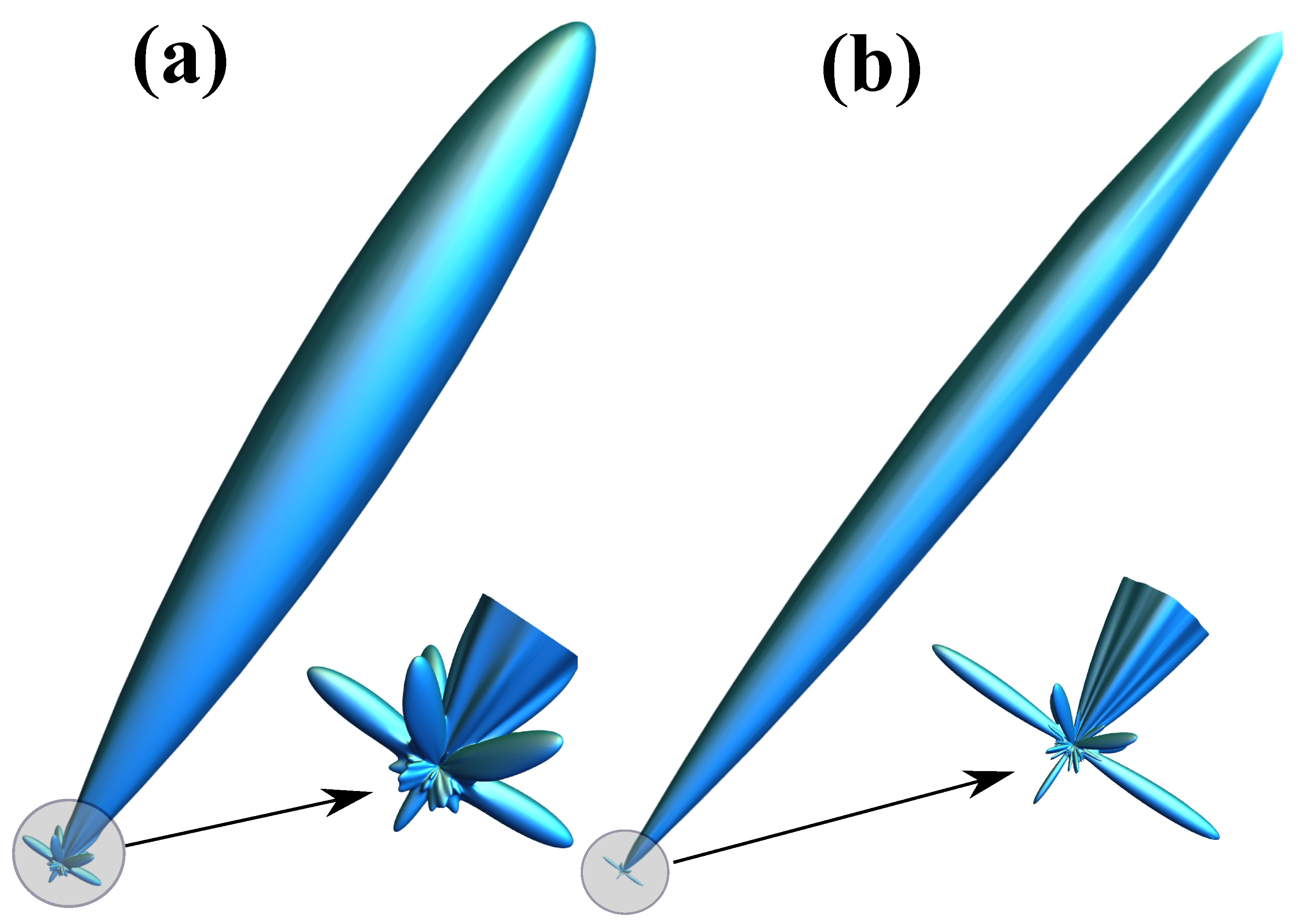
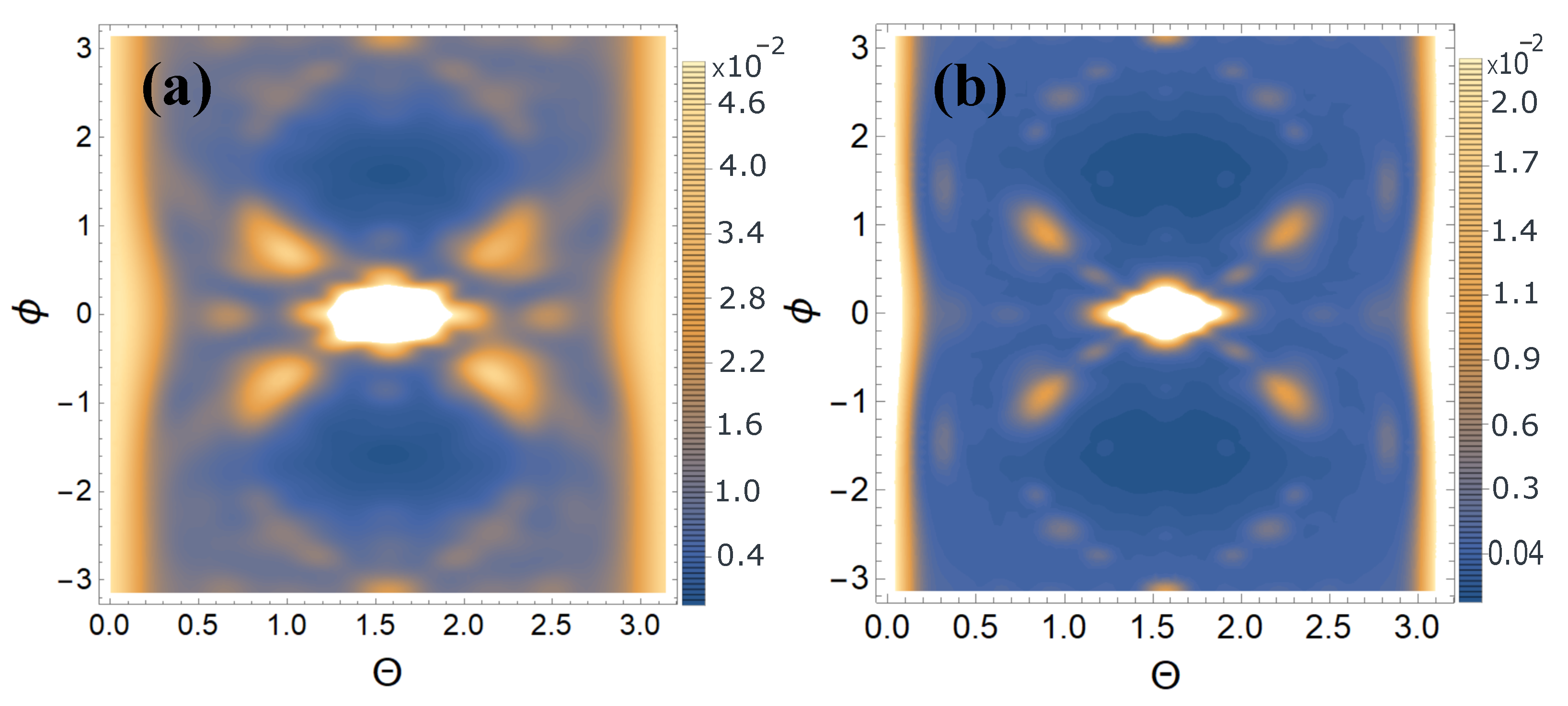
2. USP Scattering on Diamond with NV Centres

Consider a diamond with an NV center on which the USP falls in the direction n 0 . As a computational example, consider the system shown in Figure 1.
The duration of such a pulse τ , which we will consider many times shorter than the characteristic atomic time τ a 1 , i.e., τ τ a . Using this condition one can apply the sudden perturbation approximation. In the sudden perturbation approximation it is assumed that the Hamiltonian of the system before the interaction can be neglected, since the electron in the atom becomes as if “frozen” in the USP field, i.e., has no time to evolve under the action of this field. Indeed, this is so because the momentum interaction with the electron in the atom is too fast [26]. Moreover, the condition of applicability of the sudden interaction approximation τ τ a is not strict and can be extended to the case of X-ray USPs. As shown in [18,26], with X-ray USPs, it is sufficient to assume that ω 0 τ a 1 , where ω 0 is the carrier frequency of incident USPs, to extend the theory of sudden perturbation to the case of X-ray USPs. To consider our problem in general terms, we will use the electromagnetic field strength of the USPs in the form E ( r , t ) = E 0 h ( t n 0 r / c ) , i.e., we will consider the momentum to be spatially inhomogeneous, where E 0 is the field amplitude, h ( t n 0 r / c ) is an arbitrary function defining the USP shape, c is the speed of light (in a.u. c 137 ). In what follows, we will assume USPs fields such that E 0 / c 2 1 or in intensity units I 10 25 W / cm 2 . In other words, we consider the USPs fields not strong enough to account for the magnetic field of the USPs. In this case, as was shown in [24,25], the scattering spectra (scattered energy USP to a unit solid angle in a unit frequency interval) can be represented as
d 2 ε d Ω k d ω = E 0 n 2 ( 2 π ) 2 | h ˜ ( ω ) | 2 c 3 i = 1 s N e , i N A , i ( 1 | F i | 2 ) + i , j = 1 s δ i , j N e , i N e , j F i F j * ,
where N e , i is the number of electrons in the atom i variety; N A , i is the number of atoms i variety; F i = 1 N e , i ρ e , i ( r ) e i p r d 3 r is the form factor of the i atom of the variety with electron density ρ e , i ( r ) ; h ˜ ( ω ) = + h ( η ) e i ω η d η , and p = ω c ( n n 0 ) has the meaning of recoil momentum when a USP is scattered on a bound electron. In Equation (1) factor δ i , j = A i , A j e i p ( R Ai R A j ) is a crucial quantity in USPs scattering since it depends only on the position of atoms in space, i.e., on the coordinates of the atoms i of the variety (with number A i ) whose position in space is determined by the radius vector R A i . It is well known that the main case of interest for XRD is when τ ω 0 1 ( τ is the pulse duration, ω 0 is the USP carrier frequency). If we assume that τ , i.e., the radiation source is continuous, then we get the previously well known XRD theory. It should be added that it is the old theory used in XRD even in the case of ultrashort pulses, i.e., without taking into account the specificity of USP scattering.
Using Equation (1) we can obtain a physically measurable value (scattering energy to a unit solid angle)
d ε d Ω k = E 0 n 2 ( 2 π ) 2 c 3 τ | f ˜ ( x ) | 2 d x [ i = 1 s N e , i N A , i ( 1 | F i ( p 0 ) | 2 ) + i , j = 1 s γ i , j ( p 0 , p τ ) N e , i N e , j F i ( p 0 ) F j * ( p 0 ) ] , γ i , j ( p 0 , p τ ) = A i , A j e i p 0 ( R A i R A j ) | f ˜ ( x ) | 2 e i x p τ ( R A i R A j ) d x | f ˜ ( x ) | 2 d x .
where F i ( p 0 ) , δ i , j ( p 0 ) at p 0 = ω 0 c ( n n 0 ) and p τ = 1 c τ ( n n 0 ) ; h = e i ( ω 0 t k 0 r ) f ( ( t n 0 r ) / τ ) , where the f function defines the USP profile, then we get f ˜ ( ( ω ω 0 ) τ ) = e i ( ω ω 0 ) τ η f ( η ) d η is the Fourier transform of the function f ( η ) . The electron density of such atoms ρ e , i ( r ) = N e , i 4 π r k = 1 3 A k , i α k , i 2 e α k , i r , where A k , i , α k , i are constant coefficients defined in [27]. The result is a simple expression for F i = k = 1 3 A k , i α k , i 2 p 2 + α k , i 2 . Next, we need to determine the form of the incident USP, which we choose as a Gaussian form h ( t ) = e i ( ω 0 t k 0 r ) e α 2 ( t n 0 r / c ) 2 , where α = 1 / τ , k 0 = n 0 ω 0 / c . We have chosen the shape of the pulse as a Gaussian, since such a pulse is one of the best known for describing the shape of USPs. Indeed, for example, in [28] a subcyclic pulse beam (SCPB) was studied and its exact solution was found. If we consider a special case of this solution, i.e., ( ω 0 / α 1 ) then the solution takes the form of just a Gaussian pulse. Performing the calculations with the selected USP we obtain that h ˜ ( ω ) = π α e ( ω ω 0 ) 2 / 4 α 2 . We will be interested in the most interesting case of multi-cycle momentum, i.e., when ω 0 / α 1 , which is mainly used in diffraction analysis of matter. Using Equation (2) we obtain
d ε d Ω k = E 0 n 2 4 c 3 α 2 π i = 1 s N e , i N A , i ( 1 | F i ( p 0 ) | 2 ) + i , j = 1 s γ i , j ( p 0 , p τ ) N e , i N e , j F i ( p 0 ) F j * ( p 0 ) , γ i , j ( p 0 , p τ ) = A i , A j e i p 0 ( R A i R A j ) ) e 1 2 ( p τ ( R A i R A j ) ) 2 .
In our case, s can be considered equal to 2, since the presented system can be represented by two independent atoms: carbon (C) and nitrogen (N). It should be taken into account that the place where there is a vacancy is empty, i.e., there are no atoms there. Equation (3) takes into account the specificity of USP scattering on the system under study. Indeed, in this expression there are characteristics responsible for the duration τ of the USP. If in this expression we take into account only the coherent USP scattering term (the second sum in Equation (3)) and we take τ away, we obtain the well-known expression for the scattering of long duration (monochromatic) X-rays [25]. Indeed, at τ the expressions for p τ = 1 c τ ( n n 0 ) 0 and Equation (3) becomes proportional to τ (Fermi’s golden rule). Thus, the main difference between the Equation (3) from the previously known theory is determined by the factor γ i , j ( p 0 , p τ ) , analysis of which determines the difference of our theory from the previously known one. It turns out that this difference is mainly observed when using attosecond and shorter pulses. Indeed, at such pulses when τ 1 the factor e 1 2 ( p τ ( R A i R A j ) ) 2 is close to unity at quite small R A i R A j (groups of units, tens and hundreds of atoms). In this case on such atoms, where R A i R A j is small enough, the scattering is coherent, otherwise the factor γ i , j ( p 0 , p τ ) quickly tends to zero and the scattering goes in more complicated way. In the case of continuous radiation (when τ ) the coherent radiation comes from all atoms. Qualitatively, this can be explained quite simply. The portion of the atoms in the matter that falls within the spatial region of size of the USP ( c τ ) emits coherently. This is a very important clarification, since if some region with NV centers with characteristic length L is many times larger than ( c τ ), i.e., L c τ , then the results of our theory and the previously known one will be very different. This, in turn, will lead to incorrect use of the previously known theory of diffraction analysis (XRD) of matter. As an example, let’s take the USP scattering calculations on the systems presented in Figure 1. Calculation of scattering spectra using Equation (3) are shown in Figure 2 and Figure 3. The pulse falls as shown in Figure 1 with photon energy ω 0 = 7.46 keV, pulse duration τ = 10 (as). The results in Figure 3 are presented in relative units normalized to the maximum value of the spectrum.
Let us show that using USP scattering is sensitive to finding NV centers in the diamond, even at low concentrations. To do this, let us show the difference δ (normalized to the maximum value of the spectrum, i.e., δ = d ε d Ω k N V d ε d Ω k / d ε d Ω k m a x ) between scattering spectra of diamond lattice with NV center and diamond lattice. The results of the calculations are shown in Figure 4.
From Figure 2, Figure 3 and Figure 4, one can see that the scattering spectra are quite sensitive to the NV centers in the diamond structure, i.e., sensitive to the concentration of NV centers in the diamond. Indeed, comparing the graphs in Figure 3 and Figure 4 one can see that for case (a) the spectral values do not differ by more than an order of magnitude, and in case (b) by more than 2 orders of magnitude.

3. Conclusions

Thus, using Equation (3), we obtained two important results. The first result is responsible for the necessity to use the theory of USP scattering on complex multi-atomic systems developed in this work and in [24,25] in the case of using attosecond pulses. We have shown that when using attosecond pulses, the previously well known XRD theory can carry significant errors and it is necessary to use the theory developed here. The theory developed here should be used in the case of USP scattering on a diamond structure with NV centers when the parameter γ i , j ( p 0 , p τ ) differs from unity. The reason why the parameter γ i , j ( p 0 , p τ ) may differ from unity arises when the spatial pulse duration ( c τ ) is comparable or smaller than the size of the NV centers being studied in the diamond structure. This can be when the NV centers are concentrated in some region in the diamond. It should be added that nowadays more and more attention is paid to single NV centers and their relatively small groups, as well as their orientation in the diamond structure. In order to study such NV centers in the diamond structure using attosecond pulses, the theory developed here should be used. The second important conclusion is that the USP scattering spectra are quite sensitive to the concentration of NV centers in the diamond structure. This leads to the fact that USP scattering can be used to determine the concentration of NV centers. It should be added that determining the concentration of NV centers in a diamond structure is a rather difficult task, which is relevant at present. Each number of NV centers, their orientation and spatial arrangement is reflected in the scattering spectra. Depending on the task, it is necessary to carry out the calculation using Equation (3).

Author Contributions

Conceptualization, D.M. and M.E.; methodology, D.M.; software, D.M. and K.M.; validation, D.M. and K.M.; formal analysis, D.M. and M.E.; writing—original draft preparation, D.M.; writing—review and editing, D.M. and M.E.; project administration, D.M. All authors have read and agreed to the published version of the manuscript.

Funding

The research was supported by Grant of the President of the Russian Federation No. MD-4260.2021.1.2; state assignment of the Russian Federation No. 0793-2020-0005 and No. FSRU-2021-0008.

Institutional Review Board Statement

Not applicable.

Informed Consent Statement

Not applicable.

Data Availability Statement

Request to corresponding author of this article.

Conflicts of Interest

The authors declare no conflict of interest.

References

  1. Jones, N. Crystallography: Atomic secrets. Nature 2018, 505, 602–603. [Google Scholar] [CrossRef] [PubMed] [Green Version]
  2. Benediktovich, A.; Feranchuk, I.; Ulyanenkov, A. Theoretical Concepts of X-Ray Nanoscale Analysis; Springer: Berlin/Heidelberg, Germany, 2014. [Google Scholar]
  3. Eseev, M.K.; Matveev, V.I.; Makarov, D.N. Diagnostics of Nanosystems with the Use of Ultrashort X-Ray Pulses: Theory and Experiment (Brief Review). JETP Lett. 2021, 114, 387–405. [Google Scholar] [CrossRef]
  4. Schoenlein, R.; Elsaesser, T.; Holldack, K.; Huang, Z.; Kapteyn, H.; Murnane, M.; Woerner, M. Recent advances in ultrafast X-ray sources. Philos. Trans. R. Soc. A 2019, 377, 20180384. [Google Scholar] [CrossRef] [PubMed] [Green Version]
  5. Duris, J.; Li, S.; Driver, T.; Champenois, E.G.; MacArthur, J.P.; Lutman, A.A.; Zhang, Z.; Rosenberger, P.; Aldrich, J.W.; Coffee, R.; et al. Tunable isolated attosecond X-ray pulses with gigawatt peak power from a free-electron laser. Nat. Photonics 2020, 14, 30–36. [Google Scholar] [CrossRef] [Green Version]
  6. Maroju, P.K.; Grazioli, C.; Di Fraia, M.; Moioli, M.; Ertel, D.; Ahmadi, H.; Plekan, O.; Finetti, P.; Allaria, E.; Giannessi, L.; et al. Attosecond pulse shaping using a seeded free-electron laser. Nature 2020, 578, 386–391. [Google Scholar] [CrossRef] [PubMed]
  7. Mukamel, S.; Healion, D.; Zhang, Y.; Biggs, J.D. Multidimensional attosecond resonant X-ray spectroscopy of molecules: Lessons from the optical regime. Annu. Rev. Phys. Chem. 2013, 64, 101–127. [Google Scholar] [CrossRef] [Green Version]
  8. Dixit, G.; Vendrell, O.; Santra, R. Imaging electronic quantum motion with light. Proc. Natl. Acad. Sci. USA 2012, 109, 11636–11640. [Google Scholar] [CrossRef] [Green Version]
  9. Leone, S.R.; McCurdy, C.W.; Burgdörfer, J.; Cederbaum, L.S.; Chang, Z.; Dudovich, N.; Feist, J.; Greene, C.H.; Ivanov, M.; Kienberger, R.; et al. What will it take to observe processes in “real time”? Nat. Photonics 2014, 8, 162–166. [Google Scholar] [CrossRef]
  10. Balasubramanian, G.; Chan, I.Y.; Kolesov, R.; Al-Hmoud, M.; Tisler, J.; Shin, C.; Kim, C.; Wojcik, A.; Hemmer, P.R.; Krueger, A.; et al. Nanoscale imaging magnetometry with diamond spins under ambient conditions. Nature 2008, 455, 648–651. [Google Scholar] [CrossRef] [Green Version]
  11. Doherty, M.W.; Manson, N.B.; Delaney, P.; Jelezko, F.; Wrachtrup, J.; Hollenberg, L.C. The nitrogen-vacancy colour centre in diamond. Phys. Rep. 2013, 528, 1–45. [Google Scholar] [CrossRef]
  12. James, R.W. The Optical Principles of the Diffraction of X-rays (Ox Bow); Ox Bow Press: Woodbridge, CT, USA, 1982. [Google Scholar]
  13. Henriksen, N.E.; Moller, K.B. On the Theory of Time-Resolved X-ray Diffraction. J. Phys. Chem. B 2008, 112, 558–567. [Google Scholar] [CrossRef] [PubMed]
  14. Astapenko, V.A.; Sakhno, E.V. Excitation of a quantum oscillator by short laser pulses. Appl. Phys. B 2020, 126, 23. [Google Scholar] [CrossRef]
  15. Rosmej, F.B.; Astapenko, V.A.; Lisitsa, V.S.; Li, X.D.; Khramov, E.S. Scattering of ultrashort laser pulses on “ion-sphere” in dense plasmas. Contrib. Plasma Phys. 2019, 59, 189–196. [Google Scholar] [CrossRef]
  16. Makarov, D.N. Quantum theory of scattering of ultrashort electromagnetic field pulses by polyatomic structures. Opt. Express 2019, 27, 31989–32008. [Google Scholar] [CrossRef] [PubMed]
  17. Eseev, M.K.; Goshev, A.A.; Makarov, D.N. Scattering of Ultrashort X-ray Pulses by Various Nanosystems. Nanomaterials 2020, 10, 1355. [Google Scholar] [CrossRef] [PubMed]
  18. Eseev, M.K.; Goshev, A.A.; Makarova, K.A.; Makarov, D.N. X-ray diffraction analysis of matter taking into account the second harmonic in the scattering of powerful ultrashort pulses of an electromagnetic field. Sci. Rep. 2021, 11, 3571. [Google Scholar] [CrossRef] [PubMed]
  19. Moller, K.B.; Henriksen, N.E. Time-resolved x-ray diffraction: The dynamics of the chemical bond. Struc. Bond. 2012, 142, 185. [Google Scholar]
  20. Tanaka, S.; Chernyak, V.; Mukamel, S. Time-resolved x-ray spectroscopies: Nonlinear response functions and liouville-space pathways. Phys. Rev. A 2001, 63, 63405–63419. [Google Scholar] [CrossRef]
  21. Dixit, G.; Slowik, J.; Santra, R. Proposed Imaging of the Ultrafast Electronic Motion in Samples using X-Ray Phase Contrast. Phys. Rev. Lett. 2013, 110, 137403. [Google Scholar] [CrossRef] [PubMed] [Green Version]
  22. Kraus, P.M.; Zürch, M.; Cushing, S.K.; Neumark, D.M.; Leone, S.R. The ultrafast X-ray spectroscopic revolution in chemical dynamics. Nat. Rev. Chem. 2018, 2, 82–94. [Google Scholar] [CrossRef] [Green Version]
  23. Peng, P.; Marceau, C.; Villeneuve, D.M. Attosecond imaging of molecules using high harmonic spectroscopy. Nat. Rev. Phys. 2019, 1, 144–155. [Google Scholar] [CrossRef]
  24. Makarov, D.; Kharlamova, A. Scattering of X-ray Ultrashort Pulses by Complex Polyatomic Structures. Int. J. Mol. Sci. 2022, 23, 163. [Google Scholar] [CrossRef] [PubMed]
  25. Makarov, D.N.; Makarova, K.A.; Kharlamova, A.A. Specificity of scattering of ultrashort laser pulses by molecules with polyatomic structure. Sci. Rep. 2022, 12, 4976. [Google Scholar] [CrossRef] [PubMed]
  26. Makarov, D.N.; Eseev, M.K.; Makarova, K.A. Analytical wave function of an atomic electron under the action of a powerful ultrashort electromagnetic field pulse. Opt. Lett. 2019, 44, 3042–3045. [Google Scholar] [CrossRef]
  27. Salvat, F.; Martnez, J.D.; Mayol, R.; Parellada, J. Analytical Dirac-Hartree-Fock-Slater screening function for atoms (Z = 1-92). Phys. Rev. A 1987, 36, 467–474. [Google Scholar] [CrossRef] [PubMed] [Green Version]
  28. Lin, Q.; Zheng, J.; Becker, W. Subcycle pulsed focused vector beams. Phys. Rev. Lett. 2006, 97, 253902. [Google Scholar] [CrossRef] [PubMed]
Figure 1. The USP falling on the presented system, in case (a) A diamond lattice unit cell with one NV center, where blue is carbon atoms (C), purple is a nitrogen atom (N), and yellow is a vacancy; (b) a diamond lattice with one NV center for 8 unit cells (2 × 2 × 2).
Crystals 12 01417 g001
Figure 2. The figures show 3D scattering spectra of the USP: (a) the diamond unit cell with NV centre (see Figure 1a) and ( b ) this on the diamond structure shown in Figure 1b.
Crystals 12 01417 g002
Figure 3. The figures show the 2D USP scattering spectra presented as contour plots: (a) the diamond unit cell with NV centre (see Figure 1a), (b) this on the diamond structure presented in Figure 1b. The calculation was done using Equation (3). The angles θ and ϕ are represented in a spherical coordinate system, where the z axis is directed upwards and y is in the direction of the incident USP.
Crystals 12 01417 g003
Figure 4. The figures show the contribution to the scattering spectra of a diamond lattice with one NV centre (the δ parameter above is calculated) as 2D contour plots: (a) the diamond unit cell with NV centre (see Figure 1a), (b) this on the diamond structure presented in Figure 1b. The calculation was done using Equation (3). The angles θ and ϕ are represented in a spherical coordinate system, where the z axis is directed upwards and y is in the direction of the incident USP.
Crystals 12 01417 g004
Publisher’s Note: MDPI stays neutral with regard to jurisdictional claims in published maps and institutional affiliations.

