Article

Characterization of Bioactive Glass Synthesized by Sol-Gel Process in Hot Water

1
Faculty of Chemical Engineering, Ho Chi Minh City University of Food Industry, Ho Chi Minh City 700000, Vietnam
2
Institute of Theoretical and Applied Research, Duy Tan University, Hanoi 100000, Vietnam
3
Faculty of Natural Sciences, Duy Tan University, Da Nang 550000, Vietnam
4
Faculty of Chemical Technologies, Kazan National Research Technological University, ul. K. Marksa 68, 420015 Kazan, Tatarstan, Russia
5
Faculty of Chemistry, Ho Chi Minh City University of Education, Ho Chi Minh City 700000, Vietnam
6
Faculty of Pedagogy in Natural Sciences, Sai Gon University, Ho Chi Minh City 700000, Vietnam
7
Department for Management of Science and Technology Development, Ton Duc Thang University, Ho Chi Minh City 700000, Vietnam
8
Faculty of Applied Sciences, Ton Duc Thang University, Ho Chi Minh City 700000, Vietnam
*
Author to whom correspondence should be addressed.
Crystals 2020, 10(6), 529; https://doi.org/10.3390/cryst10060529
Submission received: 2 June 2020 / Revised: 16 June 2020 / Accepted: 18 June 2020 / Published: 21 June 2020
(This article belongs to the Special Issue Sol-Gel Method Applied to Crystalline Materials)

Abstract

:
Bioactive glass 70SiO2-30CaO (mol.%) was successfully synthesized by modified sol–gel in hot water without using an acid catalyst. TG-DSC analysis showed that the amorphous glass could be synthesized by sintering the sample at 700 °C for three hours. The N2 adsorption/desorption and TEM investigations highlighted that the synthetic glass had a mesoporous structure, consisting of spherical particles with sizes in the range of 11–20 nm. The specific surface area, pore volume, and average pore diameter of synthetic glass were 150.13 m2/g, 0.37 cm3/g, and 11.84 nm, respectively. Moreover, synthetic bioactive glass presented interesting bioactivity and good biocompatibility after in vitro experiments in simulated body fluid (SBF) and in cellular medium.

1. Introduction

The original bioactive glass was first discovered in 1970 by Larry L. Hench. Its composition consists of 45% SiO2, 24.5% CaO, 24.5% Na2O, and 6.0% P2O5 (wt.%, noted 45S5). The bioactivity of this glass is expressed by the formation of a new layer of hydroxyapatite Ca10(PO4)6(OH)2 (HA) on its surface when immersed in a physiological solution or implanted in the human body [1,2]. Hydroxyapatite mineral is the main inorganic component, which presents in bone and teeth of humans (about 70% in weight of bone); therefore, it acts as a cohesive bridge between artificial materials and natural bone to repair and restore bone defects [3,4]. After Hench’s discovery, many bioactive glasses have been synthesized and used as artificial biomaterials for bone substitutes. In the past fifty years, melting and sol–gel have been the two main methods used for the synthesis of bioactive glasses. In the melting method, by heating a mixture of starting precursors following a special high-temperature regime, bioactive glasses can be quickly synthesized in large quantities. Some typical glasses have been synthesized using this technique, such as glass 45S5 (45SiO2-24.5CaO-24.5Na2O-6P2O5, wt.%), 46S6 (46SiO2-24CaO-24Na2O-6P2O5, wt.%) with and without doping other elements (Sr, Zn, Mg), S53P4 (53SiO2-20CaO-23Na2O-4P2O5, wt.%), and 13-93 (53SiO2-20CaO-6Na2O-12K2O-5MgO-4P2O5, wt.%). The bioactivity of these synthetic glasses have been confirmed in both in vitro and in vivo experiments. However, melting bioactive glasses require synthesis processes at high temperatures, where volatile compounds, such as P2O5, can be evaporated, resulting in a difference in glassy compositions; obtained glasses usually have dense structures and low specific surface area values, which result in low bioactivities of synthetic glasses [4,5,6]. The sol–gel method overcomes the drawbacks of the melting technique because glasses are synthesized at lower temperatures through sol- and gel-forming processes. The resulting gel is treated in several steps, such as aging, drying, stabilizing, and finally calcining at about 700 °C to transform them into glass materials [6,7,8]. Sol–gel bioactive glasses normally have mesoporous structures with larger specific surface area values, leading to a higher bioactivity [7]. However, most sol–gel bioactive glasses are prepared using strong inorganic acids as catalysts, which have negative effects on health and the environment in view of green chemistry [9]. Therefore, environmentally friendly methods for the synthesis of bioactive glasses are urgently needed. Some glassy systems have been prepared using weaker acids (organic acids) instead of strong acids (inorganic acids); however, the synthetic processes still use acid catalysts [7,10]. Recently, bioactive glass with a composition of 75SiO2-16CaO-5Na2O-4P2O5 (mol.%, noted as S75C16) was successfully synthesized using the sol–gel method without using acid catalysts [11]. The synthesis was performed by stirring the reaction mixture at a high speed (1100 rpm). Under this condition, hydrolysis of alkoxide precursors occurred to form a clear sol after five hours. The obtained bioactive glass was an amorphous material. In this study, the bioactivity of synthetic glass is not reported. In our recent research, a bioactive glass with a composition of 70SiO2-30CaO (mol.%) was prepared using a hydrothermal method following green synthesis without any acid catalyst [12]. The investigation showed interesting bioactivity of the obtained glass. Following the trend of green chemistry, this work reports the preparation of bioactive glass 70SiO2-30CaO (mol.%) using a modified sol–gel method, in which the reaction mixture was stirred in hot water at a rate of 500 rpm and no acid catalysts were used. The physical–chemical properties and bioactivity of the synthetic glass were investigated.

2. Experimental Section

2.1. Acid-Free Sol-Gel Synthesis

The synthesis of bioactive glass was followed as below. Firstly, 21.3 g of calcium nitrate tetrahydrate Ca(NO3)2·4H2O (CNT, ≥98%, Merck) were dissolved in 38.3 g of hot water (temperature = 60 °C). The resulting solution was continuously stirred at a speed of 500 rpm. After that, 43.8 g of tetraethyl orthosilicate Si(OCH2CH3)4 (TEOS, ≥ 99.0%, Sigma-Aldrich) were added dropwise into the reaction vessel. The clear sol was formed after only 2 h. Next, the sol was placed in an oven at 70 °C to transform into gel for 1 day. Then, the wet gel was aged at 100 °C for 1 day and then dried at 150 °C for 6 h. The resulting powder was sintered at 700, 800, 900, and 1000 °C for 3 h. The used drying and sintering processes are referenced in previous studies, with some modifications [13,14,15]. It is worth noting that no acid catalyst was used in this study.

2.2. In Vitro Experiment in SBF

The in vitro experiment was performed by soaking the glass samples in simulated body fluid (SBF) for 1, 3, and 7 days. SBF is a solution with an ionic concentration similar to that of human blood plasma. It was synthesized according to Kokubo’s method [16]. The powder samples were soaked in SBF and stirred at a rate of 100 rpm at 37 °C. The ratio of powder to solution was 1:2 (mg/mL). After soaking, the powder samples were collected and then investigated using physical–chemical methods.

2.3. In Vitro Experiment In Cellular Medium

The experiments were performed according to the protocols reported in a previous study [17]. The cellular environment was standard medium DMEM (Sigma Chemical Co., St. Louis, MO) which contained 15 mM HEPES [(4-(2-hydroxyethyl)-1-piperazineethanesulfonic acid)], 2 mM L-glutamine, 10% FBS (fetal bovine serum), 100 UI/mL penicillin, and 100 µg/mL streptomycin. Two cell lines, osteoblast SaOS2 and endothelial Eahy926, were cultivated in standard medium at 37 °C in a humidified environment with 5% CO2 and 95% humidity. The cellular viability was determined by using the colorimetric MTT assay. The MTT (3-(4,5-Dimethylthiazol-2-yl)-2,5-diphenyltetrazolium bromide, a yellow tetrazole) is transformed into purple formazan in the mitochondria of living cells. The absorbance can be identified by measuring at a wavelength between 500 and 570 nm using a spectrophotometer. This reduction takes place only when mitochondrial reductase enzymes are active, and therefore the conversion can be directly related to the number of living cells.

2.4. Physico–Chemical Characterization

The sintering behavior of synthetic bioactive glass was verified using a thermogravimetry-differential scanning calorimetry (TG-DSC, SETERAM, LABSYS Evo). Glass powder was put in a platinum crucible, followed by heating from 30 to 1000 °C at a ratio of 10 °C.min−1. The phase composition of synthetic glass was analyzed using X-ray diffraction (XRD, D8-Advance) with Cu-Kα radiation (λ = 1.5406Å). The samples were measured in the range from 5 to 100° (2θ), with a step of 0.02°. The elemental composition of synthetic glass was identified by energy dispersive X-ray spectroscopy (EDX, Horiba H-7593). The structural textures consist of a specific surface area, pore size and pore volume were verified by N2 adsorption/desorption using a micromeritic porosimeter (Quantachrome Instruments). The specific surface area was determined using the Brunauer–Emmett–Teller (BET) method. The pore size and pore volume were calculated from the desorption curve using the Barrett–Joyner–Halanda (BJH) technique. The morphology was observed by transmission electron microscopy (TEM, JEOL-1400 microscope, Japan) and field emission scanning electron microscopy (FE-SEM, S-4800, Japan).

3. Results and Discussion

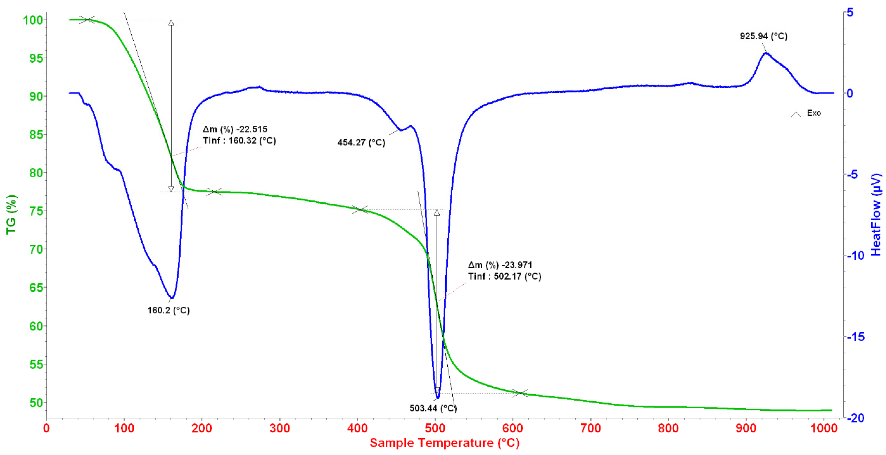
3.1. Thermal Analysis

TG-DSC analysis of the as-sintered sample is shown in Figure 1. Two mass-loss regions were determined in the ranges of 30–210 °C and 400–610 °C. The first one with an endothermic peak at 160.2 °C, is characteristic of the removal of water [13,14]. The second one with two endothermic peaks at 454.27 and 503.44 °C, is attributed to the decomposition of NO3- groups [13,14]. An exothermic peak without any mass loss at 925.94 °C corresponds to the crystallization of CaSiO3 phase [15]. From the sintering behavior, the selected temperature for glass synthesis was determined at 700 °C, where water and nitrate groups were completely removed.

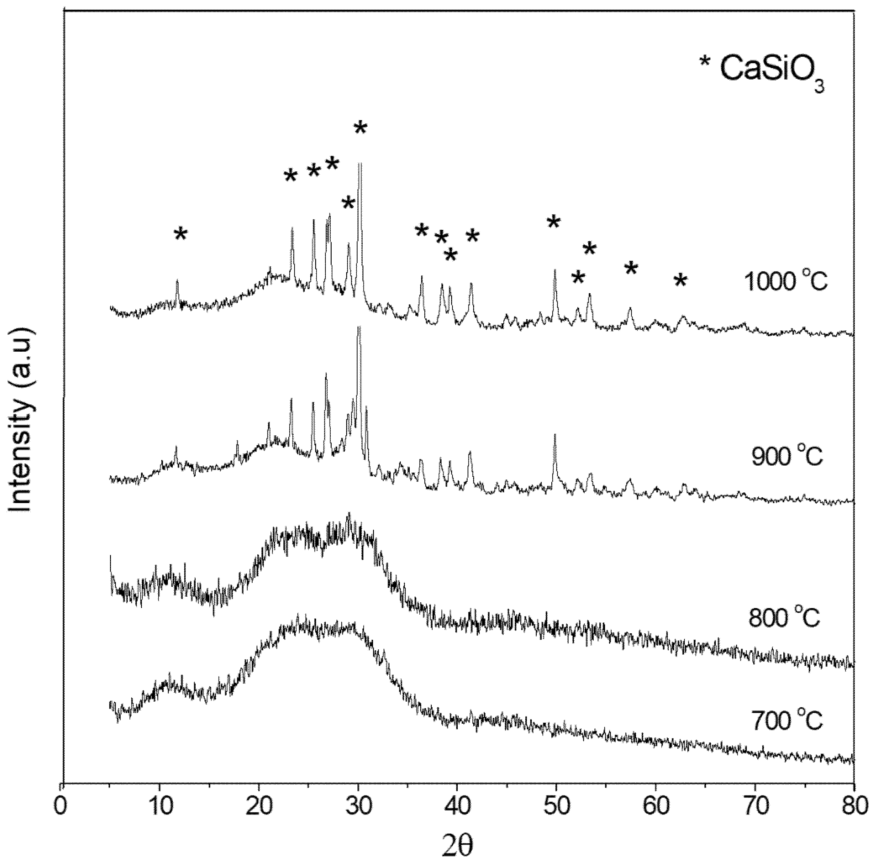
3.2. Phase Composition

Figure 2 shows XRD diagrams of glass samples treated at 700, 800, 900 and 1000 °C. The bioactive glass synthesized at 700 °C showed broad diffraction halos, characteristic of the amorphous state. Therefore, the bioactive glass 70SiO2-30CaO, synthesized using a modified sol–gel method without any acid catalyst, kept the nature of amorphous glass as it was prepared the conventional sol–gel method and the hydrothermal method [12,13,14]. Therefore, the suitable temperature for synthesis of amorphous bioactive glass was selected to be 700 °C. The elemental composition of synthetic glass was identified using the EDX technique. The final values were derived from an average of five different positions. The analytical result showed that the CaO content in synthetic glass is lower than the theoretical value (Table 1). Specifically, as the temperatures increased from 700 °C to 1000 °C, the bioactive glass exhibited a crystallization of the CaSiO3 phase. This result is consistent with the TG-DSC analysis, where the phase transformation was noted at a temperature of 925.94 °C.

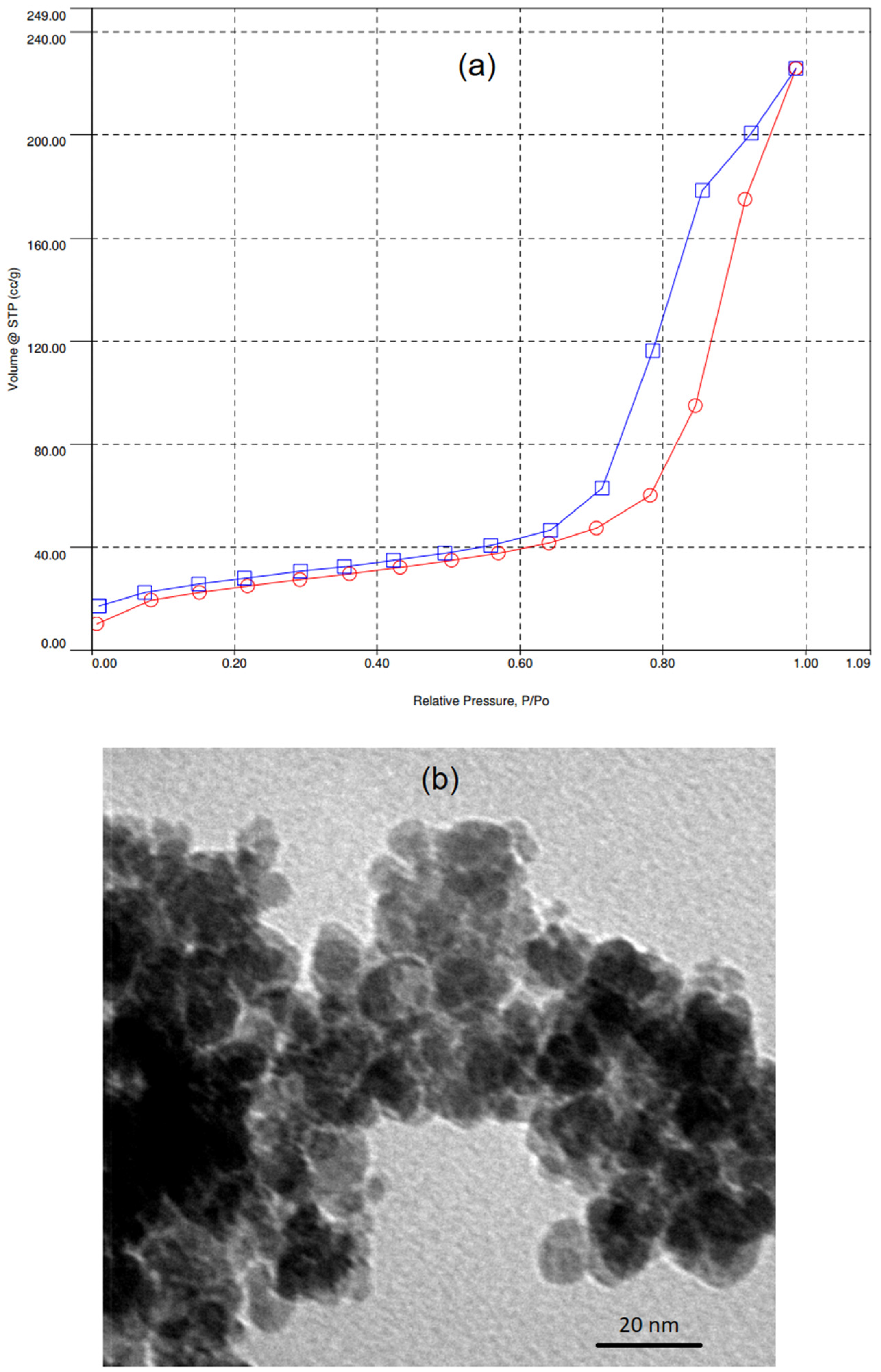
3.3. Structural Characterization

Textural and morphology of synthetic glass were evaluated by N2 adsorption/desorption and TEM analyses (Figure 3). The isotherm for synthetic glass is a typical type IV, following to IUPAC classification, attributing to mesoporous structure of synthetic material (Figure 3a) [18]. The hysteresis loop is type H2, characteristic of complex pore textures [18]. In addition, the pore size distribution and pore volume were derived using the BJH model. The textural characteristics are given in Table 2. The synthetic glass in this study exhibited a high specific surface area value and small pore size compared to previous studies [12,15]. The morphology was examined using TEM. The observation shows spherical particles with the sizes in range of 11–20 nm (Figure 3b). These particles seem to be aggregated to form the mesoporous structure of synthetic glass. This phenomenon is consistent with the previous studies [19,20].

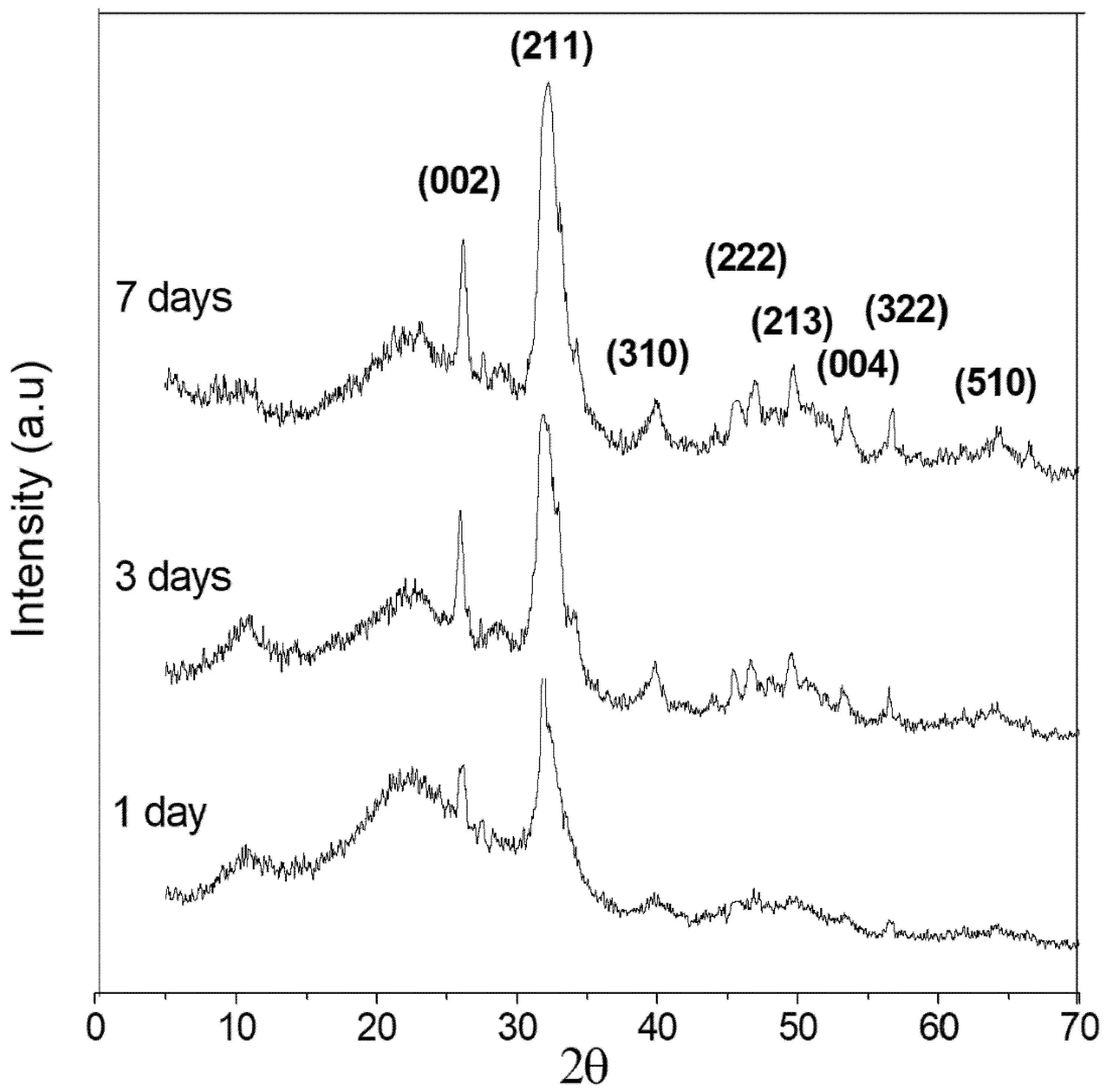
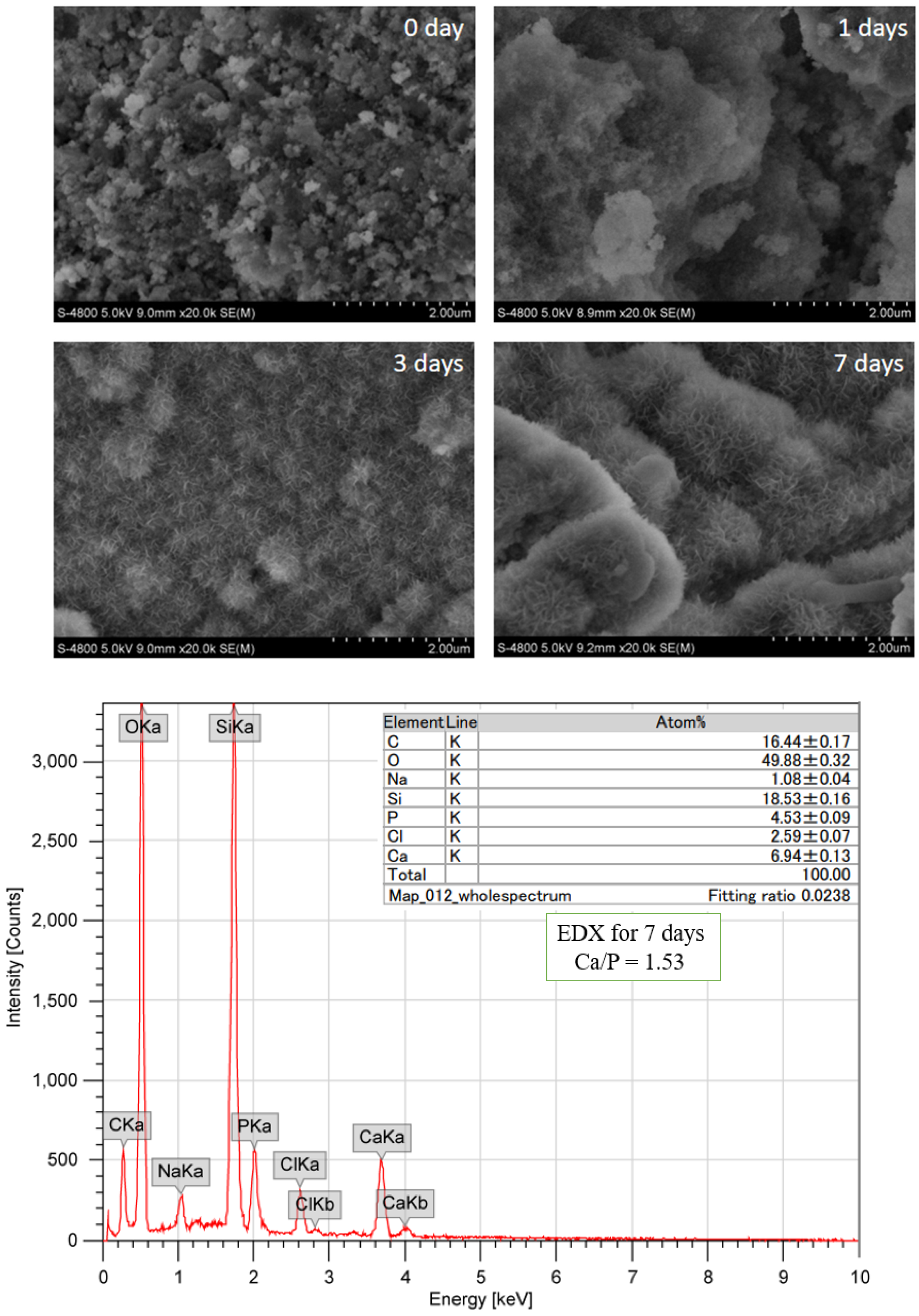
3.4. Apatite Formation after in Vitro Experiment

The XRD patterns of bioactive glass after immersion in SBF for 1, 3 and 7 days are shown in Figure 4. The apatite formation was evaluated by comparing with the standard XRD diagram of HA (File JCPDS. No. 09432) [21,22]. After being soaked for just 1 day in SBF, two characteristic peaks of HA phase at about 26° (002) and 32° (211) were revealed. As the immersed time increased, these two peaks became more visible in terms of shape and intensity. Moreover, other peaks also appeared, as seen in the XRD pattern of bioactive glass, after seven days in SBF. The obtained results confirmed the bioactivity of bioactive glass in this study.
Figure 5 presents FE-SEM micrographs of glass after soaking in SBF solution for different amounts of days. Bio-mineralization was confirmed by the formation of new HA crystals, covering the surfaces of the glass samples after immersion, compared to the sample of initial glass. The HA crystals grow up as a function of immersed times. After 7 days, the HA layer was uniform and well-defined. This layer entirely covered the surface of glass. Furthermore, the EDX analysis for the sample at 7 days in SBF indicated that the Ca/P molar ratio was equal to 1.53, which is quite close to the theoretical value of HA (1.67). The Ca/P crystallization may continue to form an HA crystal layer.

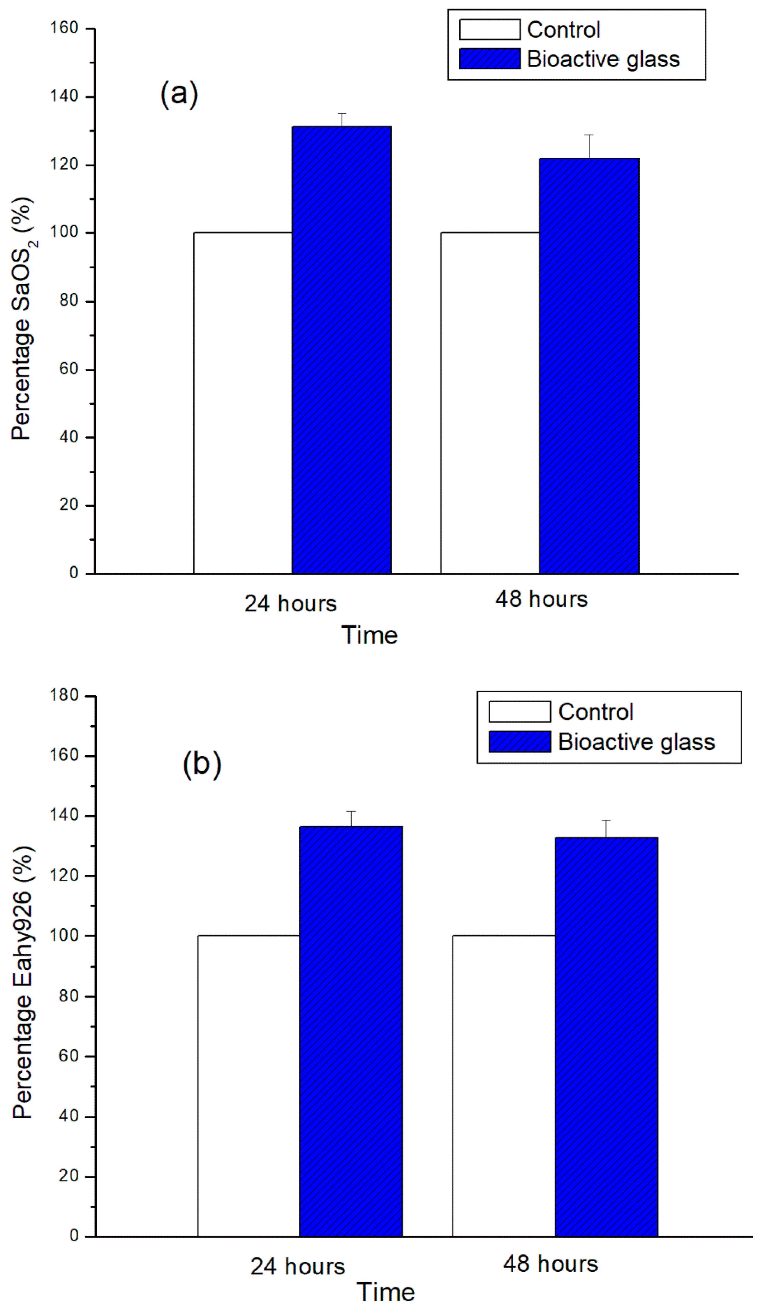
3.5. Cellular Biocompatibility

The cellular viabilities in the cultural environment are showed in Figure 6. After 24 h, the cellular viabilities were 131.2 and 136.5 % for SaOS2 and Eahy926 cells, respectively. The viability of cells without contact with glass powder was chosen as control (100%). After 48 h, a slight decrease in cell viability was noted for both SaOS2 and Eahy926 lines, compared to 24 h. This may need the renewal of culturing media. According to ISO standard 10993-5 (test for cytotoxicity, in vitro methods), the material is toxic when its cellular viability is lower than 70% [20]. Therefore, the bioactive glass in this study shows a good compatibility after culture with SaOS2 and Eahy926 cells for 24 and 48 h.

4. Conclusions

The bioactive glass with the composition of 70SiO2-30CaO (mol.%) was successfully synthesized by a modified sol–gel process in hot water without using acid catalysts. The obtained glass is a totally amorphous material with a mesoporous structure. The in vitro experiment in SBF confirmed the bioactivity of glass by formation of apatite after one day. In vitro assay in cellular medium confirmed good biocompatibility of synthetic glass. Therefore, the bioactive glass synthesized by an environmentally friendly method in this study can find potential application as artificial bone substitutes.

Author Contributions

T.H.D.: Writing, methodology, editing; T.H.B.: Writing and analysis; E.V.G.: review and editing; A.T.T.: experimental and analysis; A.T.N.: writing and editing; T.T.H.H.: methodology and analysis and X.V.B.: writing, analysis and editing. All authors have read and agreed to the published version of the manuscript.

Funding

This research received no external funding.

Conflicts of Interest

The authors confirm that there are no conflict of interest.

References

  1. Hench, L.L. The story of Bioglass®. J. Mater. Sci. Mater. Electron. 2006, 17, 967–978. [Google Scholar] [CrossRef] [PubMed]
  2. De Grado, G.F.; Keller, L.; Idoux-Gillet, Y.; Wagner, Q.; Musset, A.-M.; Benkirane-Jessel, N.; Bornert, F.; Offner, D. Bone substitutes: A review of their characteristics, clinical use, and perspectives for large bone defects management. J. Tissue Eng. 2018, 9, 1–18. [Google Scholar] [CrossRef] [Green Version]
  3. Ferraz, M.P.; Monteiro, F.J.; Manuel, C.M. Hydroxyapatite nanoparticles: A review of preparation methodologies. J. Appl. Biomater. Biomech. 2010, 2, 74–80. [Google Scholar]
  4. Oudadesse, H.; Dietrich, E.; Bui, X.; Le Gal, Y.; Pellen-Mussi, P.P.O.; Cathelineau, G. Enhancement of cells proliferation and control of bioactivity of strontium doped glass. Appl. Surf. Sci. 2011, 257, 8587–8593. [Google Scholar] [CrossRef]
  5. Jones, J. Review of bioactive glass: From Hench to hybrids. Acta Biomater. 2013, 9, 4457–4486. [Google Scholar] [CrossRef]
  6. Sepulveda, P.; Jones, J.; Hench, L.L. Characterization of melt-derived 45S5 and sol-gel-derived 58S bioactive glasses. J. Biomed. Mater. Res. 2001, 58, 734–740. [Google Scholar] [CrossRef]
  7. Owens, G.J.; Singh, R.; Foroutan, F.; Alqaysi, M.; Han, C.-M.; Mahapatra, C.; Kim, H.-W.; Knowles, J.C. Sol–gel based materials for biomedical applications. Prog. Mater. Sci. 2016, 77, 1–79. [Google Scholar] [CrossRef] [Green Version]
  8. Lukowiak, A.; Lao, J.; Lacroix, J.; Nedelec, J.-M. Bioactive glass nanoparticles obtained through sol-gel chemistry. Chem. Commun. 2013, 49, 6620. [Google Scholar] [CrossRef]
  9. Poliakoff, M.; Fitzpatrick, J.M.; Farren, T.R.; Anastas, P. Green Chemistry: Science and Politics of Change. Science 2002, 297, 807–810. [Google Scholar] [CrossRef] [Green Version]
  10. Lei, B.; Chen, X.; Wang, Y.; Zhao, N.; Du, C.; Fang, L. Fabrication, structure and biological properties of organic acid-derived sol–gel bioactive glasses. Biomed. Mater. 2010, 5, 54103. [Google Scholar] [CrossRef]
  11. Ben-Arfa, B.A.E.; Fernandes, H.R.; Salvado, I.M.M.; Ferreira, J.M.F.; Pullar, R.C. Effects of catalysts on polymerisation and microstructure of sol-gel derived bioglasses. J. Am. Ceram. Soc. 2018, 101, 2831–2839. [Google Scholar] [CrossRef]
  12. Hoa, B.T.; Hoa, H.T.T.; Tien, N.A.; Khang, N.H.D.; Viktorovna, G.E.; Tuan, T.A.; Vuong, B.X. Green synthesis of bioactive glass 70SiO2-30CaO by hydrothermal method. Mater. Lett. 2020, 274, 128–132. [Google Scholar] [CrossRef]
  13. Martinez, A.; Izquierdo-Barba, I.; Vallet-Regí, M. Bioactivity of a CaO−SiO2Binary Glasses System. Chem. Mater. 2000, 12, 3080–3088. [Google Scholar] [CrossRef]
  14. Roman, J.; Padilla, S.; Vallet-Regi, M. Sol−Gel Glasses as Precursors of Bioactive Glass Ceramics. Chem. Mater. 2003, 15, 798–806. [Google Scholar] [CrossRef]
  15. Saravanapavan, P.; Hench, L.L. Mesoporous calcium silicate glasses. J. Non. Cryst. Sol. 2003, 318, 1–13. [Google Scholar] [CrossRef]
  16. Kokubo, T.; Takadama, H. How useful is SBF in predicting in vivo bone bioactivity? Biomaterials 2006, 27, 2907–2915. [Google Scholar] [CrossRef]
  17. Standard ISO 10993-5, Biological evaluation of medical devices Part 5: Test for in vitro cytotoxicity (2009). Available online: https://www.iso.org/standard/36406.html (accessed on 21 June 2020).
  18. Thommes, M.; Kaneko, K.; Neimark, A.V.; Olivier, J.P.; Rodriguez-Reinoso, F.; Rouquerol, J.; Sing, K.S. Physisorption of gases, with special reference to the evaluation of surface area and pore size distribution (IUPAC Technical Report). Pure Appl. Chem. 2015, 87, 1051–1069. [Google Scholar] [CrossRef] [Green Version]
  19. Zheng, K.; Boccaccini, A.R. Sol-gel processing of bioactive glass nanoparticles: A review. Adv. Colloid Interface Sci. 2017, 249, 363–373. [Google Scholar] [CrossRef]
  20. Xavier, K.; Charlotte, V.; Jean-Marie, N. Deeper insights into a bioactive glass nanoparticle synthesis protocol to control its morphology, dispersibility, and composition. ACS Omega 2019, 4, 5768–5775. [Google Scholar] [CrossRef]
  21. Fihri, A.; Len, C.; Varma, R.S.; Solhy, A. Hydroxyapatite: A review of syntheses, structure and applications in heterogeneous catalysis. Co-ord. Chem. Rev. 2017, 347, 48–76. [Google Scholar] [CrossRef]
  22. Tranquillo, E.; Barrino, F.; Poggetto, G.D.; Blanco, I. Sol–Gel Synthesis of Silica-Based Materials with Different Percentages of PEG or PCL and High Chlorogenic Acid Content. Mater. 2019, 12, 155. [Google Scholar] [CrossRef] [PubMed] [Green Version]
Figure 1. TG-DSC curves of the as-sintered glass sample.
Crystals 10 00529 g001
Figure 2. XRD patterns of glass samples heated at 700, 800, 900 and 1000 °C.
Crystals 10 00529 g002
Figure 3. (a) N2 adsorption/desorption isotherm and (b) TEM image of synthetic bioactive glass.
Crystals 10 00529 g003
Figure 4. XRD patterns of bioactive glass after immersion in SBF for 1, 3 and 7 days.
Crystals 10 00529 g004
Figure 5. FE-SEM and EDX analyses of bioactive glass samples after immersion in simulated body fluid (SBF).
Crystals 10 00529 g005
Figure 6. Cellular viabilities on bioactive glass after 24 h and 48 h: (a) for osteoblast cells SaOS2 and (b) for endothelial cells Eahy926.
Crystals 10 00529 g006
Table 1. The composition of bioactive glass 70SiO2-30CaO.
Composition (mol.%)SiO2CaO
Theoretical7030
Experimental74.6 ± 0.0525.4 ± 0.09
Table 2. Nitrogen adsorption/desorption characterization of bioactive glass 70SiO2-30CaO.
SamplesSpecific Surface Area (m2/g)Total Pore Volume (cm3/g)Average Pore Diameter (nm)References
70SiO2-30CaO150.130.3711.84This study
Modified sol-gel method
70SiO2-30CaO140.40.6720.9Reference [12]
Hydrothermal method
70SiO2-30CaO1260.4715Reference [15]
Conventional sol-gel method

