
Mixed-Phase Fe2O3 Derived from Natural Hematite Ores/C3N4 Z-Scheme Photocatalyst for Ofloxacin Removal
 , , and
Abstract
:1. Introduction
2. Results and Discussion
3. Materials and Methods
3.1. Chemicals
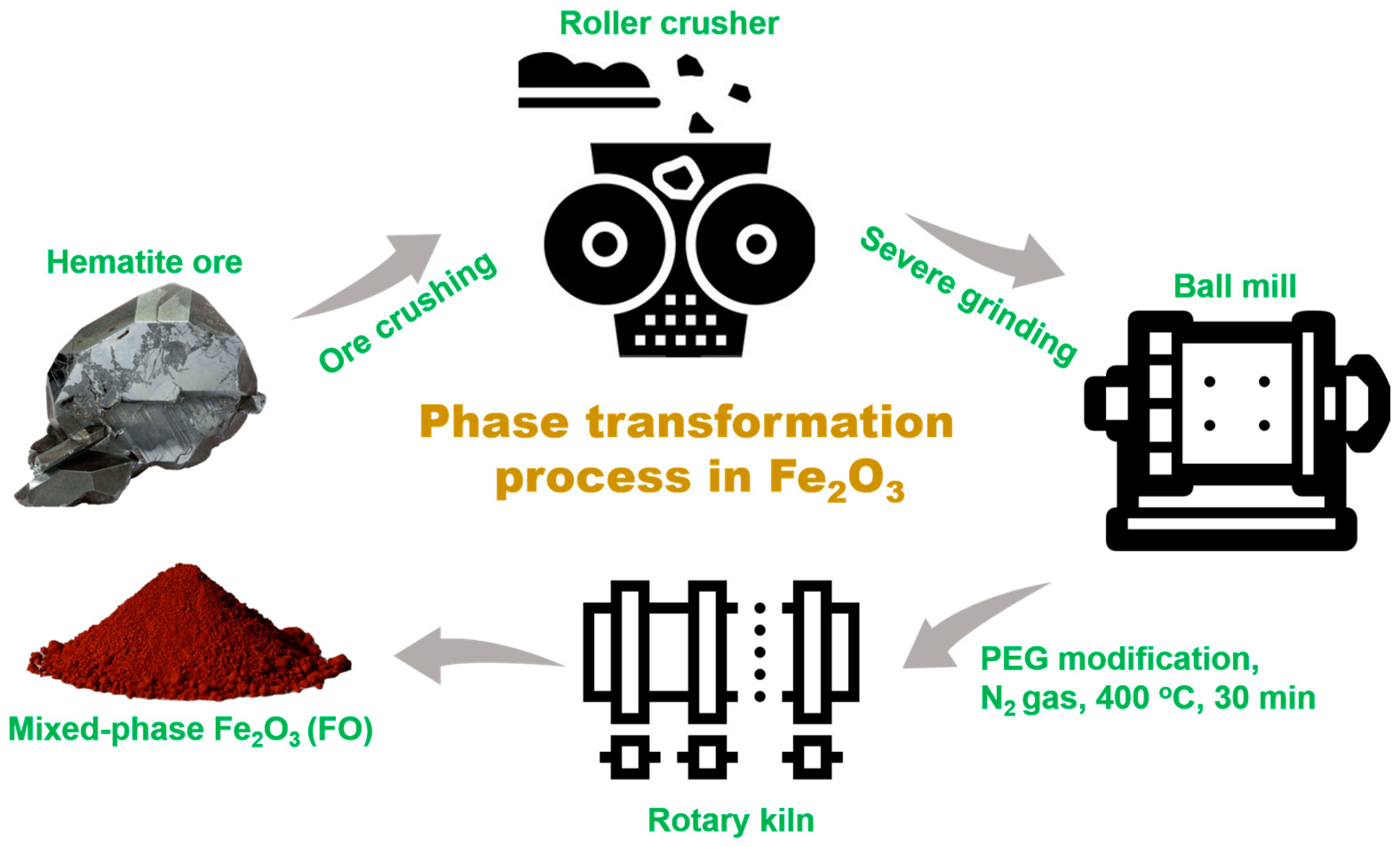
3.2. Partial Phase Transformation of Natural Hematite
3.3. Synthesis of CNFO-X% Composites and Pristine g-C3N4
3.4. Characterization
3.5. Photocatalytic Degradation of Ofloxacin
4. Conclusions
Supplementary Materials
Author Contributions
Funding
Data Availability Statement
Acknowledgments
Conflicts of Interest
References
- de Souza, H.O.; dos Costa, R.S.; Quadra, G.R.; dos Fernandez, M.A.S. Pharmaceutical Pollution and Sustainable Development Goals: Going the Right Way? Sustain. Chem. Pharm. 2021, 21, 100428. [Google Scholar] [CrossRef]
- Wilkinson, J.L.; Boxall, A.B.A.; Kolpin, D.W.; Leung, K.M.Y.; Lai, R.W.S.; Galbán-Malagón, C.; Adell, A.D.; Mondon, J.; Metian, M.; Marchant, R.A.; et al. Pharmaceutical Pollution of the World’s Rivers. Proc. Natl. Acad. Sci. USA 2022, 119, e2113947119. [Google Scholar] [CrossRef] [PubMed]
- Ta, Q.T.H.; Cho, E.; Sreedhar, A.; Noh, J.-S. Mixed-Dimensional, Three-Level Hierarchical Nanostructures of Silver and Zinc Oxide for Fast Photocatalytic Degradation of Multiple Dyes. J. Catal. 2019, 371, 1–9. [Google Scholar] [CrossRef]
- Peres, M.S.; Maniero, M.G.; Guimarães, J.R. Photocatalytic Degradation of Ofloxacin and Evaluation of the Residual Antimicrobial Activity. Photochem. Photobiol. Sci. 2015, 14, 556–562. [Google Scholar] [CrossRef] [PubMed]
- Gao, P.; Tian, X.; Nie, Y.; Yang, C.; Zhou, Z.; Wang, Y. Promoted Peroxymonosulfate Activation into Singlet Oxygen over Perovskite for Ofloxacin Degradation by Controlling the Oxygen Defect Concentration. Chem. Eng. J. 2019, 359, 828–839. [Google Scholar] [CrossRef]
- Mahlaule-Glory, L.M.; Mathobela, S.; Hintsho-Mbita, N.C. Biosynthesized Bimetallic (ZnOSnO2) Nanoparticles for Photocatalytic Degradation of Organic Dyes and Pharmaceutical Pollutants. Catalysts 2022, 12, 334. [Google Scholar] [CrossRef]
- Fedorov, K.; Dinesh, K.; Sun, X.; Soltani, R.D.; Wang, Z.; Sonawane, S.; Boczkaj, G. Synergistic Effects of Hybrid Advanced Oxidation Processes (AOPs) Based on Hydrodynamic Cavitation Phenomenon—A Review. Chem. Eng. J. 2022, 432, 134191. [Google Scholar] [CrossRef]
- Ge, J.; Zhang, Y.; Heo, Y.-J.; Park, S.-J. Advanced Design and Synthesis of Composite Photocatalysts for the Remediation of Wastewater: A Review. Catalysts 2019, 9, 122. [Google Scholar] [CrossRef]
- Zhou, P.; Navid, I.A.; Ma, Y.; Xiao, Y.; Wang, P.; Ye, Z.; Zhou, B.; Sun, K.; Mi, Z. Solar-to-Hydrogen Efficiency of More than 9% in Photocatalytic Water Splitting. Nature 2023, 613, 66–70. [Google Scholar] [CrossRef]
- Liu, C.; Zhang, Q.; Zou, Z. Recent Advances in Designing ZnIn2S4-Based Heterostructured Photocatalysts for Hydrogen Evolution. J. Mater. Sci. Technol. 2023, 139, 167–188. [Google Scholar] [CrossRef]
- Antonopoulou, M.; Papadaki, M.; Rapti, I.; Konstantinou, I. Photocatalytic Degradation of Pharmaceutical Amisulpride Using G-C3N4 Catalyst and UV-A Irradiation. Catalysts 2023, 13, 226. [Google Scholar] [CrossRef]
- Thi, L.N.; Phan, T.T.T.; Ngoc, T.N.; Viswanath, N.S.M.; Le, H.T.T.; Tran Thi, L.; Tien-Trung, N.; Nguyen, L.T.; Nhiem, D.N.; Tran Huu, H.; et al. Prussian Blue Decorated G-C3N4—From Novel Synthesis to Insight Study on Charge Transfer Strategy for Improving Visible-Light Driven Photo-Fenton Catalytic Activity. J. Alloys Compd. 2022, 916, 165331. [Google Scholar] [CrossRef]
- Wen, J.; Xie, J.; Chen, X.; Li, X. A Review on G-C3N4-Based Photocatalysts. Appl. Surf. Sci. 2017, 391, 72–123. [Google Scholar] [CrossRef]
- Wang, H.; Zhang, L.; Chen, Z.; Hu, J.; Li, S.; Wang, Z.; Liu, J.; Wang, X. Semiconductor Heterojunction Photocatalysts: Design, Construction, and Photocatalytic Performances. Chem. Soc. Rev. 2014, 43, 5234–5244. [Google Scholar] [CrossRef]
- Konstas, P.-S.; Konstantinou, I.; Petrakis, D.; Albanis, T. Synthesis, Characterization of g-C3N4/SrTiO3 Heterojunctions and Photocatalytic Activity for Organic Pollutants Degradation. Catalysts 2018, 8, 554. [Google Scholar] [CrossRef]
- Hanif, M.A.; Akter, J.; Kim, Y.S.; Kim, H.G.; Hahn, J.R.; Kwac, L.K. Highly Efficient and Sustainable ZnO/CuO/g-C3N4 Photocatalyst for Wastewater Treatment under Visible Light through Heterojunction Development. Catalysts 2022, 12, 151. [Google Scholar] [CrossRef]
- Wang, Z.; Lin, Z.; Shen, S.; Zhong, W.; Cao, S. Advances in Designing Heterojunction Photocatalytic Materials. Chin. J. Catal. 2021, 42, 710–730. [Google Scholar] [CrossRef]
- Ong, W.-J.; Tan, L.-L.; Ng, Y.H.; Yong, S.-T.; Chai, S.-P. Graphitic Carbon Nitride (g-C3N4)-Based Photocatalysts for Artificial Photosynthesis and Environmental Remediation: Are We a Step Closer To Achieving Sustainability? Chem. Rev. 2016, 116, 7159–7329. [Google Scholar] [CrossRef]
- Li, Y.; Xia, Z.; Yang, Q.; Wang, L.; Xing, Y. Review on G-C3N4-Based S-Scheme Heterojunction Photocatalysts. J. Mater. Sci. Technol. 2022, 125, 128–144. [Google Scholar] [CrossRef]
- Ziaur Rahman, M.; Golam Kibria, M.; Buddie Mullins, C. Metal-Free Photocatalysts for Hydrogen Evolution. Chem. Soc. Rev. 2020, 49, 1887–1931. [Google Scholar] [CrossRef]
- Védrine, J.C. Heterogeneous Catalysis on Metal Oxides. Catalysts 2017, 7, 341. [Google Scholar] [CrossRef]
- Wu, W.; Jiang, C.; Roy, V.A. Recent Progress in Magnetic Iron Oxide–Semiconductor Composite Nanomaterials as Promising Photocatalysts. Nanoscale 2015, 7, 38–58. [Google Scholar] [CrossRef]
- Singh, P.; Sharma, K.; Hasija, V.; Sharma, V.; Sharma, S.; Raizada, P.; Singh, M.; Saini, A.K.; Hosseini-Bandegharaei, A.; Thakur, V.K. Systematic Review on Applicability of Magnetic Iron Oxides–Integrated Photocatalysts for Degradation of Organic Pollutants in Water. Mater. Today Chem. 2019, 14, 100186. [Google Scholar] [CrossRef]
- Mishra, M.; Chun, D.-M. α-Fe2O3 as a Photocatalytic Material: A Review. Appl. Catal. A Gen. 2015, 498, 126–141. [Google Scholar] [CrossRef]
- Thakur, D.; Ta, Q.T.H.; Noh, J.-S. Photon-Induced Superior Antibacterial Activity of Palladium-Decorated, Magnetically Separable Fe3O4/Pd/Mpg-C3N4 Nanocomposites. Molecules 2019, 24, 3888. [Google Scholar] [CrossRef]
- Li, X.; Qiu, Y.; Zhu, Z.; Chen, T.; Zhang, H.; Yin, D. Construction of Magnetically Separable Dual Z-Scheme g-C3N4/α-Fe2O3/Bi3TaO7 Photocatalyst for Effective Degradation of Ciprofloxacin under Visible Light. Chem. Eng. J. 2022, 440, 135840. [Google Scholar] [CrossRef]
- Lin, Y.-F.; Chang, C.-Y. Design of Composite Maghemite/Hematite/Carbon Aerogel Nanostructures with High Performance for Organic Dye Removal. Sep. Purif. Technol. 2015, 149, 74–81. [Google Scholar] [CrossRef]
- Baig, U.; Gondal, M.A.; Dastageer, M.A.; Sajid, M. Maghemite Nanoparticles Decorated Semiconducting Graphitic Carbon Nitride Hetero-Structured Nanocomposite: Facile Synthesis, Characterizations and Its Visible Light Active Photocatalytic System for Removal of Hazardous Organic Pollutants from Aqueous Solutions. Colloids Surf. A Physicochem. Eng. Asp. 2022, 641, 128427. [Google Scholar] [CrossRef]
- Ghasemifard, M.; Heidari, G.; Ghamari, M.; Fathi, E.; Izi, M. Synthesis of Porous Network-Like α-Fe2O3 and α/γ-Fe2O3 Nanoparticles and Investigation of Their Photocatalytic Properties. Nanotechnol. Russ. 2019, 14, 353–361. [Google Scholar] [CrossRef]
- Li, F.; Liu, Y.; Sun, Z.; Zhao, Y.; Liu, R.; Chen, L.; Zhao, D. Photocatalytic Oxidative Desulfurization of Dibenzothiophene under Simulated Sunlight Irradiation with Mixed-Phase Fe2O3 Prepared by Solution Combustion. Catal. Sci. Technol. 2012, 2, 1455–1462. [Google Scholar] [CrossRef]
- Sridharan, K.; Kuriakose, T.; Philip, R.; Park, T.J. Transition Metal (Fe, Co and Ni) Oxide Nanoparticles Grafted Graphitic Carbon Nitrides as Efficient Optical Limiters and Recyclable Photocatalysts. Appl. Surf. Sci. 2014, 308, 139–147. [Google Scholar] [CrossRef]
- Shenoy, S.; Chuaicham, C.; Okumura, T.; Sekar, K.; Sasaki, K. Simple Tactic Polycondensation Synthesis of Z-Scheme Quasi-Polymeric g-C3N4/CaFe2O4 Composite for Enhanced Photocatalytic Water Depollution via p-n Heterojunction. Chem. Eng. J. 2023, 453, 139758. [Google Scholar] [CrossRef]
- Deraz, N.M.; Alarifi, A. Novel Processing and Magnetic Properties of Hematite/Maghemite Nano-Particles. Ceram. Int. 2012, 38, 4049–4055. [Google Scholar] [CrossRef]
- Shenoy, S.; Tarafder, K.; Sridharan, K. Graphitic C3N4/CdS Composite Photocatalyst: Synthesis, Characterization and Photodegradation of Methylene Blue under Visible Light. Phys. B Condens. Matter 2020, 595, 412367. [Google Scholar] [CrossRef]
- Shenoy, S.; Jang, E.; Park, T.J.; Gopinath, C.S.; Sridharan, K. Cadmium Sulfide Nanostructures: Influence of Morphology on the Photocatalytic Degradation of Erioglaucine and Hydrogen Generation. Appl. Surf. Sci. 2019, 483, 696–705. [Google Scholar] [CrossRef]
- Chuaicham, C.; Noguchi, Y.; Shenoy, S.; Shu, K.; Trakulmututa, J.; Srikhaow, A.; Sekar, K.; Sasaki, K. Simultaneous Photocatalytic Sugar Conversion and Hydrogen Production Using Pd Nanoparticles Decorated on Iron-Doped Hydroxyapatite. Catalysts 2023, 13, 675. [Google Scholar] [CrossRef]
- Xu, Q.; Zhu, B.; Jiang, C.; Cheng, B.; Yu, J. Constructing 2D/2D Fe2O3/g-C3N4 Direct Z-Scheme Photocatalysts with Enhanced H2 Generation Performance. Sol. RRL 2018, 2, 1800006. [Google Scholar] [CrossRef]
- Sanad, M.M.S.; Farahat, M.M.; El-Hout, S.I.; El-Sheikh, S.M. Preparation and Characterization of Magnetic Photocatalyst from the Banded Iron Formation for Effective Photodegradation of Methylene Blue under UV and Visible Illumination. J. Environ. Chem. Eng. 2021, 9, 105127. [Google Scholar] [CrossRef]
- Jiang, Z.; Wan, W.; Li, H.; Yuan, S.; Zhao, H.; Wong, P.K. A Hierarchical Z-Scheme α-Fe2O3/g-C3N4 Hybrid for Enhanced Photocatalytic CO2 Reduction. Adv. Mater. 2018, 30, 1706108. [Google Scholar] [CrossRef] [PubMed]
- Zhao, G.; Ding, J.; Zhou, F.; Chen, X.; Wei, L.; Gao, Q.; Wang, K.; Zhao, Q. Construction of a Visible-Light-Driven Magnetic Dual Z-Scheme BiVO4/g-C3N4/NiFe2O4 Photocatalyst for Effective Removal of Ofloxacin: Mechanisms and Degradation Pathway. Chem. Eng. J. 2021, 405, 126704. [Google Scholar] [CrossRef]
- Su, Q.; Li, J.; Yuan, H.; Wang, B.; Wang, Y.; Li, Y.; Xing, Y. Visible-Light-Driven Photocatalytic Degradation of Ofloxacin by g-C3N4/NH2-MIL-88B(Fe) Heterostructure: Mechanisms, DFT Calculation, Degradation Pathway and Toxicity Evolution. Chem. Eng. J. 2022, 427, 131594. [Google Scholar] [CrossRef]
- Chen, R.; Ding, S.; Fu, N.; Ren, X. Preparation of a G-C3N4/Ag3PO4 Composite Z-Type Photocatalyst and Photocatalytic Degradation of Ofloxacin: Degradation Performance, Reaction Mechanism, Degradation Pathway and Toxicity Evaluation. J. Environ. Chem. Eng. 2023, 11, 109440. [Google Scholar] [CrossRef]
- Adhikari, S.; Lee, H.H.; Kim, D.-H. Efficient Visible-Light Induced Electron-Transfer in z-Scheme MoO3/Ag/C3N4 for Excellent Photocatalytic Removal of Antibiotics of Both Ofloxacin and Tetracycline. Chem. Eng. J. 2020, 391, 123504. [Google Scholar] [CrossRef]
- Zhang, D.; Qi, J.; Ji, H.; Li, S.; Chen, L.; Huang, T.; Xu, C.; Chen, X.; Liu, W. Photocatalytic Degradation of Ofloxacin by Perovskite-Type NaNbO3 Nanorods Modified g-C3N4 Heterojunction under Simulated Solar Light: Theoretical Calculation, Ofloxacin Degradation Pathways and Toxicity Evolution. Chem. Eng. J. 2020, 400, 125918. [Google Scholar] [CrossRef]
- Saravanakumar, K.; Mamba, G.; Muthuraj, V. 1D/2D MnWO4 Nanorods Anchored on g-C3N4 Nanosheets for Enhanced Photocatalytic Degradation Ofloxacin under Visible Light Irradiation. Colloids Surf. A Physicochem. Eng. Asp. 2019, 581, 123845. [Google Scholar] [CrossRef]
- Chen, D.; Xie, Z.; Zeng, Y.; Lv, W.; Zhang, Q.; Wang, F.; Liu, G.; Liu, H. Accelerated Photocatalytic Degradation of Quinolone Antibiotics over Z-Scheme MoO3/g-C3N4 Heterostructure by Peroxydisulfate under Visible Light Irradiation: Mechanism; Kinetic; and Products. J. Taiwan Inst. Chem. Eng. 2019, 104, 250–259. [Google Scholar] [CrossRef]
- Dao, D.Q.; Nguyen, T.K.A.; Pham, T.-T.; Shin, E.W. Synergistic Effect on Photocatalytic Activity of Co-Doped NiTiO3/g-C3N4 Composites under Visible Light Irradiation. Catalysts 2020, 10, 1332. [Google Scholar] [CrossRef]
- Lee, G.-J.; Lee, X.-Y.; Lyu, C.; Liu, N.; Andandan, S.; Wu, J.J. Sonochemical Synthesis of Copper-Doped BiVO4/g-C3N4 Nanocomposite Materials for Photocatalytic Degradation of Bisphenol A under Simulated Sunlight Irradiation. Nanomaterials 2020, 10, 498. [Google Scholar] [CrossRef]
- Zhao, Y.; Shi, H.; Yang, D.; Fan, J.; Hu, X.; Liu, E. Fabrication of a Sb2MoO6/g-C3N4 Photocatalyst for Enhanced RhB Degradation and H2 Generation. J. Phys. Chem. C 2020, 124, 13771–13778. [Google Scholar] [CrossRef]


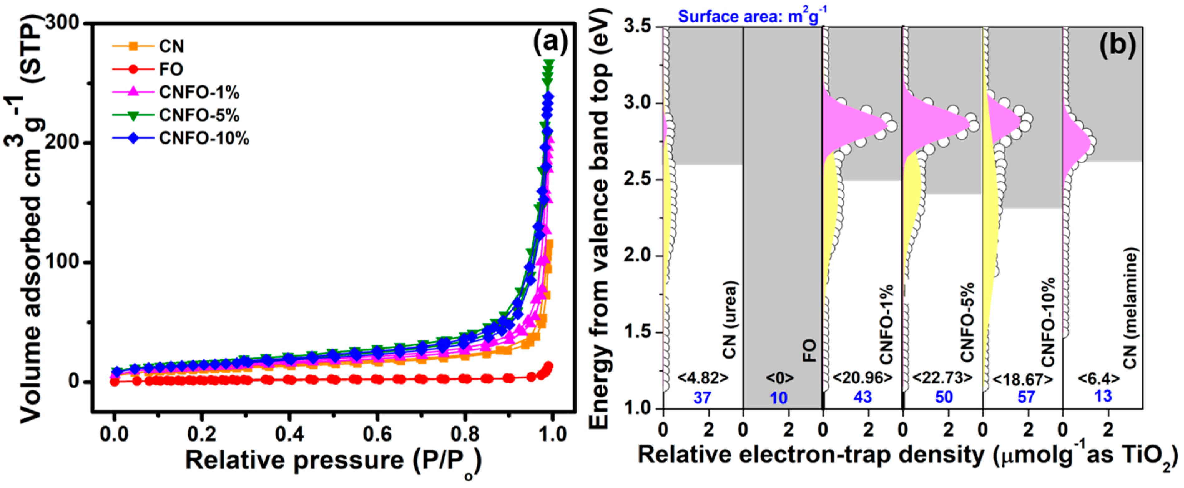
| Catalyst | Bandgap (eV) | Surface Area (m2g−1) | OFX Degradation Efficiency (%) | Reaction Rate Constant (min−1) |
|---|---|---|---|---|
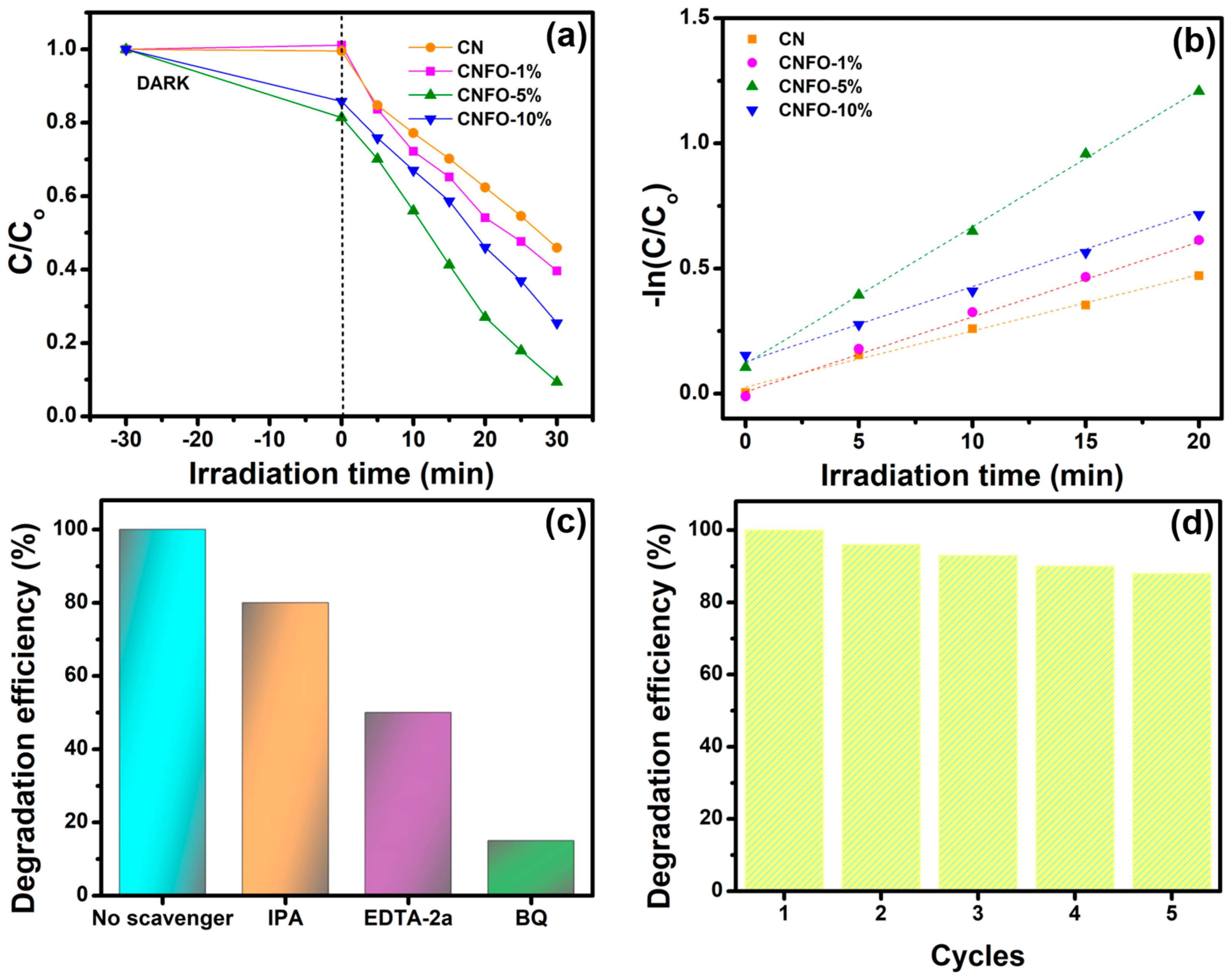
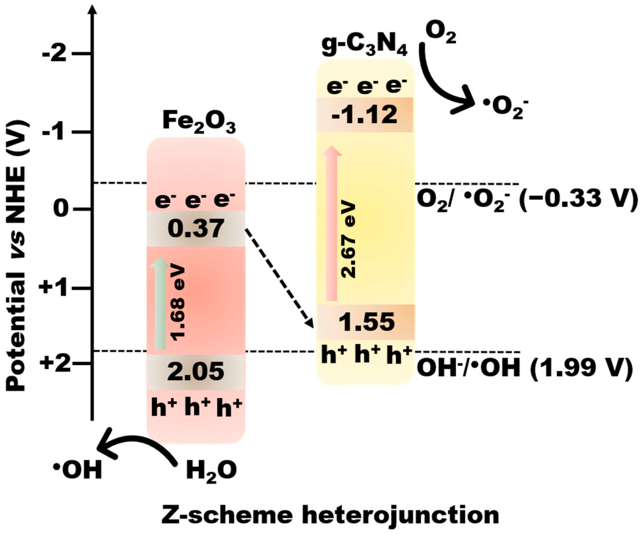
| CN | 2.67 | 37 | 44 | 0.0169 |
| FO | 1.68 | 10 | - | - |
| CNFO-1% | 2.49 | 43 | 60 | 0.0264 |
| CNFO-5% | 2.48 | 50 | 93 | 0.1271 |
| CNFO-10% | 2.46 | 57 | 75 | 0.1194 |
| Catalyst | Light Source | Degradation Efficiency (%), Time (min) | Type of Heterojunction | Ref. |
|---|---|---|---|---|
| BiVO4/g-C3N4/NiFe2O4 | 300 W Xe lamp (λ > 420 nm) | 93.8, 20 | Dual Z-scheme | [40] |
| g-C3N4/NH2-MIL88B(Fe) | 300 W Xe lamp (λ > 420 nm) | 96.5, 150 | Type-II | [41] |
| g-C3N4/Ag3PO4 | 500 W Xe lamp | 71.9, 10 | Z-scheme | [42] |
| MoO3/Ag/C3N4 | 150 W Xe lamp | 96, 100 | Z-scheme | [43] |
| NaNbO3/g-C3N4 | 300 W Xe lamp | 99.5, 30 | Type-II | [44] |
| MnWO4/g-C3N4 | 150 mW/cm2 W lamp | 90.4, 70 | Type-II | [45] |
| MoO3/g-C3N4 | 350 W Xe lamp (λ > 420 nm) | 94.4, 120 | Z-scheme | [46] |
| α/γ-Fe2O3/g-C3N4 | 300 W Xe lamp (λ > 420 nm) | 93, 30 | Z-scheme | Present work |
Disclaimer/Publisher’s Note: The statements, opinions and data contained in all publications are solely those of the individual author(s) and contributor(s) and not of MDPI and/or the editor(s). MDPI and/or the editor(s) disclaim responsibility for any injury to people or property resulting from any ideas, methods, instructions or products referred to in the content. |
© 2023 by the authors. Licensee MDPI, Basel, Switzerland. This article is an open access article distributed under the terms and conditions of the Creative Commons Attribution (CC BY) license (https://creativecommons.org/licenses/by/4.0/).
Share and Cite
Shenoy, S.; Farahat, M.M.; Chuaicham, C.; Sekar, K.; Ramasamy, B.; Sasaki, K. Mixed-Phase Fe2O3 Derived from Natural Hematite Ores/C3N4 Z-Scheme Photocatalyst for Ofloxacin Removal. Catalysts 2023, 13, 792. https://doi.org/10.3390/catal13050792
Shenoy S, Farahat MM, Chuaicham C, Sekar K, Ramasamy B, Sasaki K. Mixed-Phase Fe2O3 Derived from Natural Hematite Ores/C3N4 Z-Scheme Photocatalyst for Ofloxacin Removal. Catalysts. 2023; 13(5):792. https://doi.org/10.3390/catal13050792
Chicago/Turabian StyleShenoy, Sulakshana, Mohsen M. Farahat, Chitiphon Chuaicham, Karthikeyan Sekar, Boopathy Ramasamy, and Keiko Sasaki. 2023. "Mixed-Phase Fe2O3 Derived from Natural Hematite Ores/C3N4 Z-Scheme Photocatalyst for Ofloxacin Removal" Catalysts 13, no. 5: 792. https://doi.org/10.3390/catal13050792