Article

Metabolic Biodegradation Pathway of Fluoranthene by Indigenous Trichoderma lixii and Talaromyces pinophilus spp.

by
Samson O. Egbewale
1,
Ajit Kumar
1,
Mduduzi P. Mokoena
1,2 and
Ademola O. Olaniran
1,*
1
Discipline of Microbiology, Westville Campus, University of KwaZulu-Natal, Durban 4001, South Africa
2
Department of Pathology, School of Medicine, University of Limpopo, Private Bag X1106, Sovenga 0727, South Africa
*
Author to whom correspondence should be addressed.
Catalysts 2023, 13(5), 791; https://doi.org/10.3390/catal13050791
Submission received: 27 February 2023 / Revised: 6 April 2023 / Accepted: 20 April 2023 / Published: 23 April 2023
(This article belongs to the Special Issue New Trends in Industrial Biocatalysis)

Abstract

:
Two indigenous ascomycetes fungi, Trichoderma lixii strain FLU1 (TlFLU1) and Talaromyces pinophilus strain FLU12 (TpFLU12), were isolated from benzo(b)fluoranthene-enriched activated sludge and tested for bio-catalytically degrade fluoranthene as a sole carbon source. TlFLU1 and TpFLU12 degraded 98 and 99% of 400 mg/L of fluoranthene after 16 and 12 d incubation period, respectively. Degradation correlated with the upregulation of expression of ligninolytic enzymes. The GC-MS and FTIR analysis of the degradation products suggest that the degradation is initiated at the C1-C2 position of the compound ring via oxygenation and ring cleavage to form 9-oxo-9H-fluorene-1-carboxylic acid before undergoing ring cleavage to yield fluorenone, which then proceeds through the ß-Ketoadipate pathway via benzene-1,2,3-tricarboxylic acid. The degradation rate is better fitted in the first-order and zero-order kinetic model for TlFLU1 and TpFLU12, respectively. The metabolites from the TlFLU1 degradation media are shown to be toxic in Vibryo parahaemolyticus after 6 h of exposure with effective concentration (EC50) and toxicity unit (TU) values of 14.25 mg/L and 7.018%, respectively, while also being observed as non-toxic from TpFLU12 degradation media with an EC50 and TU values of 197.1 mg/L and 0.507%, respectively. Results from this study show efficient metabolism of fluoranthene into an innocuous state by TlFLU1 and TpFLU12.

Graphical Abstract

1. Introduction

Among the ubiquitous group of organic xenobiotics found in the various matrices of the ecosystem, polycyclic aromatic hydrocarbons (PAHs) have become a major environmental priority owing to the carcinogenicity, size-dependent genotoxicity of graphene nanoplatelets in human stem cells, and toxicity of graphene and graphene oxide nano-walls against bacteria and mutagenic effects [1], which are characterized by low aqueous solubility and high lipophilicity to biological functions [2]. Due to the increase in bioaccumulation, biomagnification, and persistent toxicity of this pollutant, the United State Environmental Protection Agency (USEPA) and the United Nations Environmental Program (UNEP) created a list of 16 priority PAH compounds that needed to be eradicated from the environment [3,4]. Sources of PAHs include the incomplete combustion of fossil fuel, ship traffic, oil spilling/leakages from corroded tanks at petrol stations, urban runoff, and industrial activities [5,6]. Some PAHs exhibit carbocatalytic activity as they can act as catalysts to break down hazardous aromatic molecules due to their unique electronic structure and reactivity [7]. PAHs have multiple fused aromatic rings with pi-electrons that make them highly reactive and able to act as electron donors or acceptors when exposed to hazardous aromatic molecules, they undergo redox reactions, breaking them into less toxic compounds [8].
Fluoranthene is a model compound for a PAH degradation study due to its relative abundance in the environment, resistance to degradation, and the fact that it can accumulate in the soil, water, and air [9]. In plants, it causes some serious biochemical and physiological changes, such as photosynthetic pigments alteration, metabolic activity reduction, and cell membranes disruption leading to defoliation, chlorosis, necrosis, and oxidative stress defense compromise [10,11]. Acute and chronic toxicity of fluoranthene with varied orders of magnitude has been well documented among a diverse group of freshwater and saltwater organisms [12,13]. Additionally, its acute toxicity in animals such as rats includes dysfunction processes, including ataxia, decreased grip strengths, increased landing foot splay, loss of aerial righting, increased urination and defecation, and decreased responses to sensory stimuli in both sexes with server neurological deficits [14]. Its long exposure in humans through pathways, such as inhalation, dermal touch, and ingestion, has been linked to an increased risk of lung cancer, as well as cardiovascular disease (CVD), including atherosclerosis, thrombosis, hypertension, and myocardial infarction (MI) [15]. Apart from its carcinogenicity, mutagenicity, and teratogenicity, preclinical studies have shown a direct link between PAH exposure, oxidative stress, CVD illness, mortality development, and endocrine disruption [16,17].
Although PAHs can naturally disintegrate from the environment via chemical oxidation, photolysis, volatilization, and adsorption, the chemical stabilities of PAHs make these methods inefficient for their total removal coupled with many toxic derivatives which are more toxic than their parent compounds [18]. In addition, techniques such as physical, chemical, and biological techniques have been established, optimized, and utilized in the amelioration of PAH contaminated sites, but significant problems, such as technological complexity, high cost, and a general lack of acceptance are the drawbacks encountered with the physical and chemical treatment techniques [19]. Biological treatment techniques (Biodegradation) using biocatalysts have recently attracted a great deal of interest in the PAHs contaminated site clean-up owing to its safe clean-up means, least disruptive nature, complete degradation, and cost-effective method compared with the conventional physicochemical method, which often leaves behind toxic derivatives which are more toxic than the parent compounds [20]. The role of microbes in PAH degradation has been documented, but degradation rates in some microbes (bacteria) are often not proportional to the contaminant released into an environment and, thus, leading to the accumulation of PAHs in the contaminated sites that can remain for decades [5].
Fungi have been appraised as one of the pre-dominant biodegrading agents found in the environment, which are free-living and ubiquitous [21,22]. They employ their metabolic versatility and resilience to utilize PAHs as a carbon and energy source, and thereby mineralize them to simpler compounds, carbon dioxide, and water, or transformed them into other non-toxic molecules under harsh environmental conditions, such as low moisture and high concentrations of PAHs [23,24]. However, fungi belonging to the Ascomycota family, such as Aspergillus sp., Aspergillus terricola, Trichoderma reesei, Aspergillus ochraceus, Cunninghamella elegans, and Cunninghamella echinulata, indigenous to contaminated sites have been reported to have efficiently degraded PAH compounds, such as Anthracene, Benzo (k) fluoranthene, Benzo(a)anthracene, and Fluoranthene [25,26,27]. In addition, ligninolytic fungi, including Nematoloma frowardii, Coriolopsis byrsina, Pleurotus eryngii, and Ganoderma lucidum, have been well documented for their effective degradation of PAHs [28,29,30]. Although the biodegradation pathways of two- and three-ring PAHs by fungi are well documented while information regarding the routes and reactions by which the oxidization of higher molecular weight especially four-ring PAHs (fluoranthene) is still limited. Based on the available literature and based on metabolites identified over the last 10 years during fluoranthene degradation, only two biodegradation pathways routes have been reported in fungi belonging to the mushroom-forming fungi (basidiomycetes). The first route is initiated by deoxygenation at C-7 and C-8 position via extra and intra-diol ring cleavage to form acenaphthylene-type metabolites before entering into the ß-ketoadipate pathway via benzene-1,2,3-tricarboxylic acid while the second route involves deoxygenation at C-2 and C-3 position via ortho-cleavage to form a product known as 9-fluorenone-1-carboxylic acid, which is further degraded to yield benzene-1,2,3-tricarboxylic acid and phthalic acid [31].
Thus, in the present study, two indigenous fungal isolates, Trichoderma lixii strain FLU1 and Talaromyces pinophilus strain FLU12, were investigated for their ability to bio-catalytically degrade fluoranthene. The biodegradation kinetics and metabolic pathways were also established via profiling of metabolites produced and enzymes involved in the degradation process. Furthermore, empirical data on the acute toxicity of fluoranthene and its transient metabolic products were elucidated.

2. Results

2.1. Fungal Cultural Morphological Characteristics and Molecular Analyses

The fungal cultural morphological characteristics and the sequence analysis of the ITS rDNA gene were used to identify the strains. Isolate one, grown on a PDA plate, exhibited concentric green conidial production rings. The center of the plate had denser conidiation, while an irregular yellow zone devoid of conidia surrounded the inoculum. Green conidia were distributed throughout the bit granular plate, with white pustules on the green mat. White mycelia without conidial formation were also present. A single green and yellowish concentric ring with a cluster of yellow conidia was observed around the point of inoculation. Light green and yellowish conidia were present throughout the plate, with more conidia towards the margins and the inoculum, except for the white mycelia covering the Petri dish. A single cottony concentric ring with dark green conidiation was found around the inoculum (Figure S1A). Additionally, its microscopic morphology reveals that it has septate hyphae, its conidiophores and conidia are green to yellow green in colour and are produced in chains that measure around 2.5 × 1.5 µm, ellipsoidal to oblong in shape, and typically measure 3 × 2 µm (Figure S1B). ITS gene sequence (submitted as NCBI accession number: MK208694) analysis of the isolate showed that it has 100% genetic similarity with Trichoderma lixii and is named as Trichoderma lixii strain FLU1 (TlFLU1). Isolate two, grown on PDA plate, forms concentric rings with whitish yellow conidial production, and the conidia production is denser in the center than towards the margins. An irregular white pustule zone without conidia is also present around the inoculum. The density of conidia production is higher towards the center than the margins, and some white pustules are present (Figure S2A). Additionally, its microscopic morphology reveals that the mycelium consists of long, smooth branching hyphae that are septate, and around 2 µm in diameter, it has unbranched conidiophores around 4 µm in diameter with a spherical to oval shape. It has smooth-walled with a greenish grey colour. It has spherical vesicles containing phialides at the tip. These phialides look like flask-shaped acerose phialides that produce asexual spores, along with elongated sausage-shaped cells (Figure S2B). ITS rDNA sequence (submitted as NCBI accession number: MK208704) analysis of the isolate showed that it has 100% similarity with Talaromyces pinophilus and named as Talaromyces pinophilus strain FLU12 (TpFLU12). FLU is abbreviated for fluoranthene for both strains. Additionally, the phylogenetic typing of these fungal strains with other fungal nucleotide sequences obtained from the NCBI database through the neighbor-joining tree method revealed that TlFLU1 and Hypocrea lixii with NCBI accession number JF923807 fall within the same clade and shared a 32% evolutionary relationship (Figure S3A) while TpFLU12 and Talaromyces pinophilus with NCBI accession number MN006617 fall within the same clade and shared a 70% evolutionary relationship (Figure S3B).

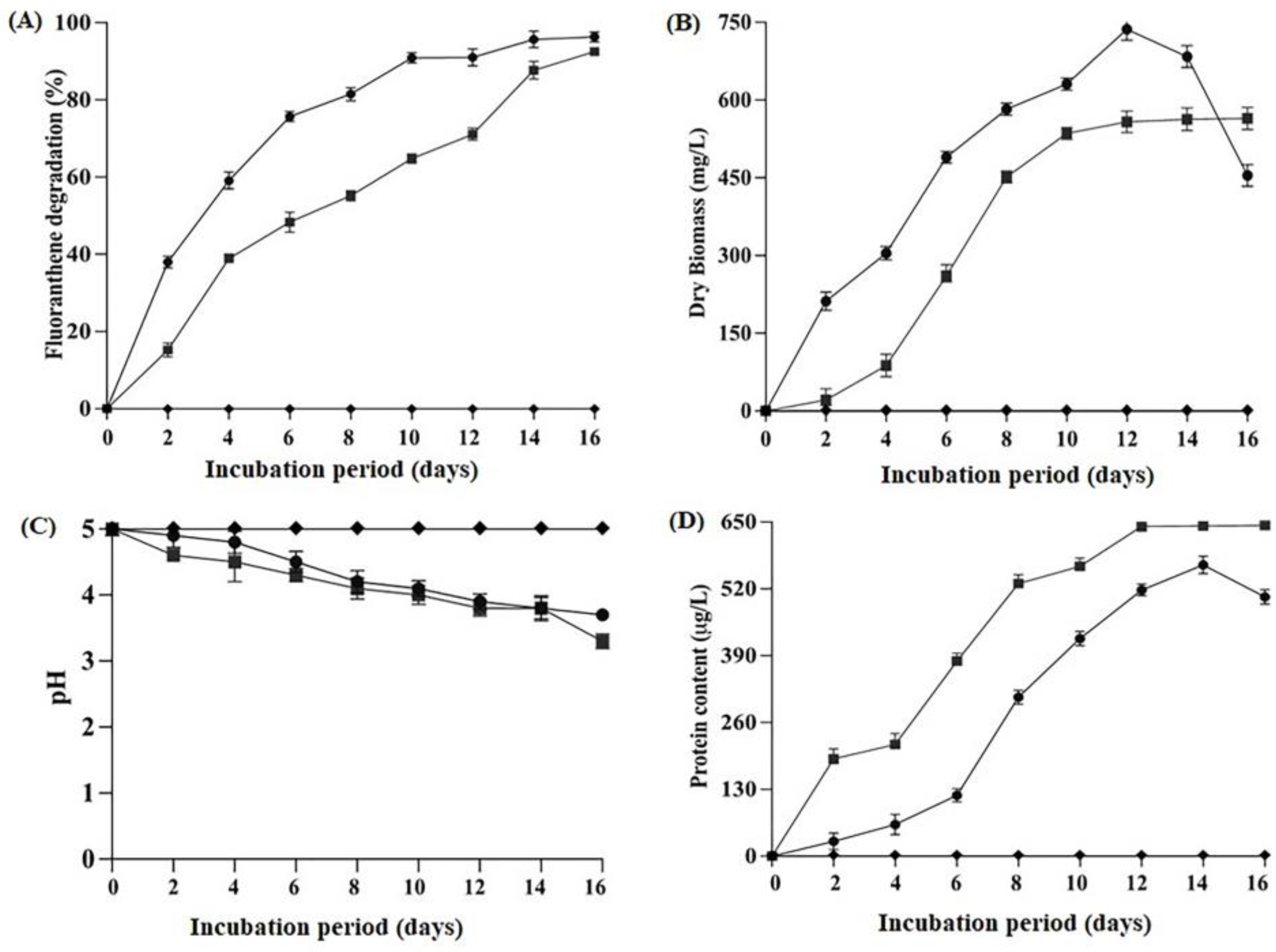
2.2. Degradation Efficiency, Biomass Production, Protein Content and Change in pH Condition

As shown in Figure 1A, degradation of fluoranthene increases with an increased incubation period in the flask inoculated with strains TlFLU1 and TpFLU12. After 10 d incubation period, TlFLU1 degraded 90.9% of the fluoranthene while TpFLU12 could degrade 92.5% after 16 d incubation period. The dry biomass production and protein content of these fungal strains also increased with an increase in the incubation period, except for some slight decrease observed after reaching their maximum production days. The maximum dry biomass production of 736.8 mg/L was observed at 12 d after the incubation period for TlFLU1, while 564.4 mg/L was recorded for TpFLU12 at 16 d after the incubation period (Figure 1B). The pH of the inoculation media decreases, from pH 5 to 3.87 (TlFLU1) and 3.66 (TpFLU12) with an increased incubation period. The pH of the control (no inoculum) remains unchanged at the end of the incubation period (Figure 1C); however, the total protein content of about 643.4 μg/mL at 16 d and 566.5 μg/mL at 14 d was recorded for TlFLU1 and TpFLU12, respectively (Figure 1D).

2.3. Degradation Kinetics Study

The kinetic model for the degradation of fluoranthene by TlFLU1 and TpFLU12 is shown in Table 1 and Figure S4. Overall, the degradation kinetics best fitted first-order with the regression coefficient (R2) values of 0.987 and 0.919 for TlFLU1 and TpFLU12, respectively. TpFLU12 was also shown to degrade the compound following zero-order degradation kinetics with a regression coefficient (R2) values of 0.968. The first-order degradation kinetics was characterized by a degradation rate constant (k) and half-life (DT50) values of 0.210 d−1 and 2.254 d−1 for TlFLU1, while 0.15 d−1 and 2.588 d−1 are for TpFLU12, respectively. In addition, a degradation rate constant (k = 22.280 d−1) and half-life (D1/2 = 2.254 d−1) was obtained for TpFLU12 while following the zero-order degradation kinetics.

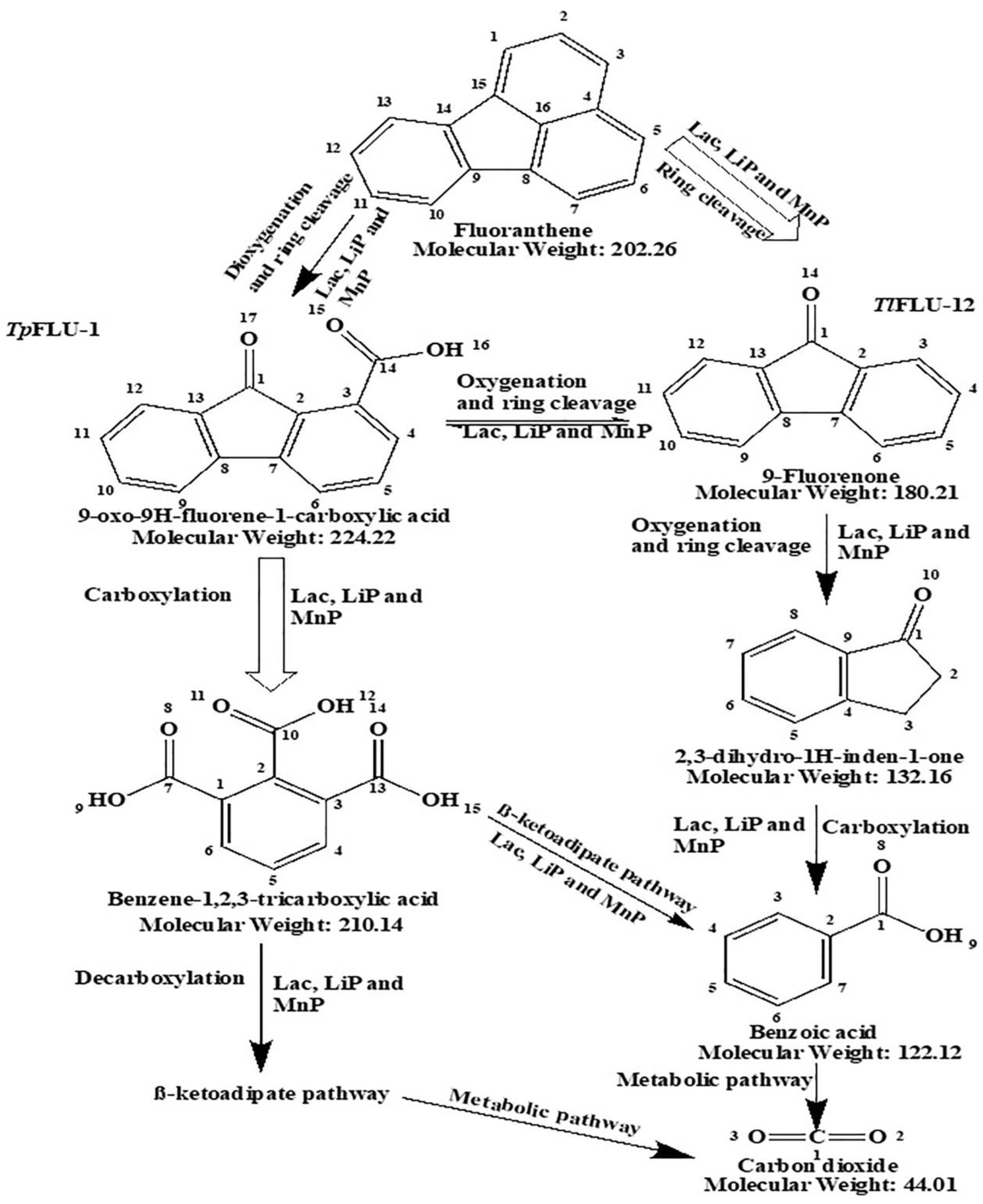
2.4. Fluoranthene Degradation Metabolites with Its Pathway

The different metabolites formed during the degradation of fluoranthene by TlFLU1 and TpFLU12 are presented in Figure 2 and Table S1, respectively. Two metabolites, namely 9-oxo-9H-fluorene-1-carboxylic acid, with a retention time (RT) of 14.2 min at molecular ion (M+) m/z: 224, and benzene-1,2,3-tricarboxylic acid (RT: 12.3 and m/z: 148), were observed in the TlFLU1 inoculated flasks while three metabolites, namely fluorenone (RT: 23.9, molecular ion (M+) at m/z 180), was observed in the TpFLU12 inoculated flask at a varying retention times (RT), such as 18.4 min, 12.3 min, and 7.9 min, respectively, for 2,3-dihydro-1H-inden-1-one with molecular ion (M+) at m/z 104, benzene-1,2,3-tricarboxylic acid with molecular ion (M+) at m/z 148, and benzoic acid with molecular ion (M+) at m/z 105. However, the parent compound (control: fluoranthene) was detected at 28.4 min. The theoretical arrangement of these degradation products pathways indicates that parent compound fluoranthene degradation is initiated at the C1–C2 position via oxygenation and ring cleavage to form 9-oxo-9H-fluorene-1-carboxylic acid before undergoing ring cleavage to yield fluorenone, which then proceeds through oxygenation and side-chain removal into ß-ketoadipate pathway via benzene-1,2,3-tricarboxylic acid and ring cleavage to form 2,3-dihydro-1H-inden-1-one before undergoing side-chain removal to yield benzoic acid and CO2 release, respectively, as a dead-end product.

2.5. Analysis of the Transformed Fluoranthene Functional Groups

The FTIR spectra of the changes in the functional group of fluoranthene metabolites are presented in Table 2 and Figure S5. The parent compound (control, fluoranthene) showed an intense peak at 3202 cm−1 due to O-H stretching of the alcohol rings, an adsorption peak at 1610 cm−1 is an attribution of –C=C aromatic ring stretch, while an adsorption peak at 622 cm−1 with attribution of –C-H bending of the substituted benzene ring. However, in the TlFLU1 degradation product spectra, a significant shift in the control sample functional groups was observed with four intense adsorption peaks. An observed adsorption peak at 3284 cm−1, with the attribution of broad O–H stretching of the alcohol rings, was followed by an adsorption peak at 1650 cm−1, which has an attribution of –C=O stretching of the carbonyl/carboxylic. Additionally, a peak at 1385 cm−1 was linkable to the bending of aliphatic CH and CH2 groups was observed to be followed by the intense peak at 600 cm−1, which has an attribution of the –C≡C–H: C–H bend in alkynes. In the TpFLU12 inoculated flask sample, apart from the shift in absorption peaks of control samples which was an attribute of O–H stretching of the alcohol rings observed, seven more adsorption peaks were observed, namely peak at 1730 cm−1, linkable to the presence of C=O rings of the quinone compounds, followed by high adsorption intensity peak at 1421 cm−1 with an attribute of aromatic ring vibrations, and plane bending of C–O–H. Additionally, the observed adsorption intensity at 1385 cm−1 is linkable to the bending of aliphatic CH and CH2 groups, followed by a high peak intensity at 1280 cm−1, which corresponds with the C-O functional group due to carboxylic acid stretch. Furthermore, the observed high-intensity peak at 1030 cm−1 corresponds with the C-OH and O-CH3 stretch, while the adsorption intensity at 610 cm−1 could be linked with the –C≡C–H: C–H bend in alkynes.

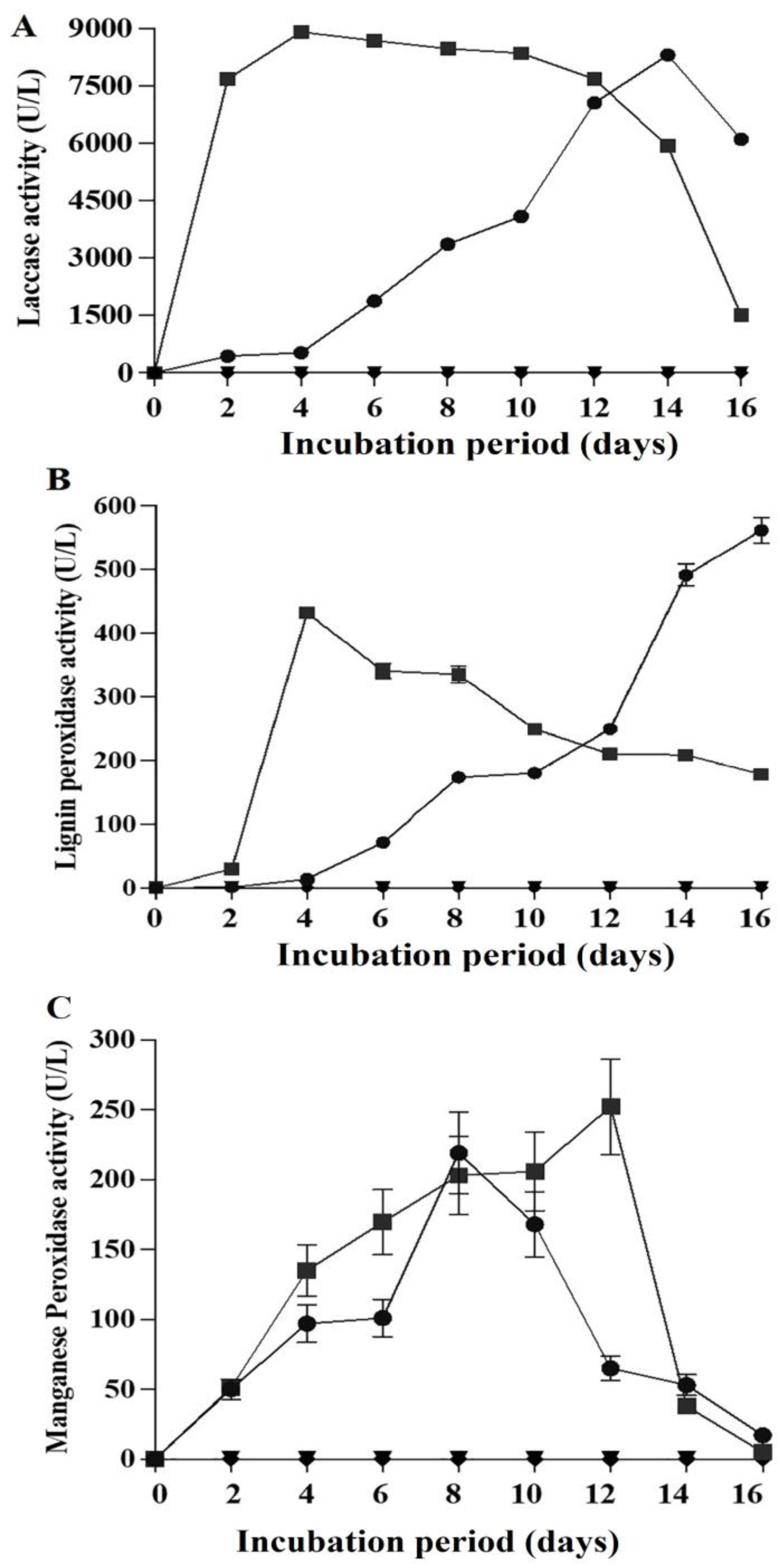
2.6. Ligninolytic Activities in the Presence of Fluoranthene

The ligninolytic activities of TlFLU1 and TpFLU12 during fluoranthene degradation are presented in Figure 3. Overall, the production of ligninolytic enzyme activities was dependent on the incubation period. Laccase enzyme showed superior activity in the flask inoculated with TpFLU12 in comparison to TlFLU1. The maximum laccase activity of 8310.4 U/L at 14 d after the incubation period was observed in TlFLU1 inoculated flask, while a maximum laccase activity of 8910.5 U/L was recorded for TpFLU12 at 2 d after the incubation period (Figure 3A). Conversely, Lignin peroxidase activity was observed to be a maximum with 561.2 U/L at 16 d after the incubation period in TlFLU1 inoculated flask while a maximum activity of 431.7 U/L was documented at 4 d after the incubation period in TpFLU12 inoculated flask (Figure 3B). In addition, Manganese peroxidase activity was observed to be maximum with 219.2 U/L at 8 d after the incubation period in the TlFLU1 inoculated flask, while Manganese peroxidase activity of 252.3 U/L was recorded at 12 d after the incubation period (Figure 3C). However, no significant production of laccase, Lignin peroxidase, and Manganese peroxidase was recorded in BSM + TlFLU1 and BSM + TpFLU12 inoculated flask.

2.7. Ecotoxicity Test

The time-course changes in toxicity of fluoranthene degradation products were assessed by survival, EC50 and TU (toxicity unit) of Vibrio parahaemolyticus after 6 h of exposure time (Figure 4 and Table 3). Toxicity was at its peak at 0 d (parent compound) with 0 Log (CFU/mL) of the marine bacterium. The bacterium survival (Log CFU/mL) on each degradation product of TlFLU1 (Figure 4A) and TpFLU12 (Figure 4B) was dependent on degradation product concentration. Additionally, the bacterium survival was observed to be increasing, with an increase in the degradation product per day for TlFLU1 and TpFLU12. Maximum survival of 7.68 Log (CFU/mL) was observed in the 16 d degradation product of TlFLU1, followed by 14 d product with 6.50 while the least survival of 5.25 Log (CFU/mL) which remains constant on degradation products of 2 to 12 d. However, the 16 d degradation product and the CN (No degradation product/PAHs) with 7.90 Log (CFU/mL), are statistically not significant (p ≤ 0.05) (Figure 4A). Conversely, the marine bacterium survival was significantly high in every degradation product of TpFLU12 with the maximum survival of 7.85 Log (CFU/mL) at 16 d and least survival of 7.32 Log (CFU/mL). Additionally, the degradation products of 10 d, 12 d, 14 d, 16 d, and the CN (No degradation product/PAHs) with 7.90 Log (CFU/mL) were statistically not significant (p ≤ 0.05) (Figure 4B). However, the effective concentration (EC50) and toxicity unit (TU) of 14.25 mg/L and 7.018% with toxicity class of harmful was obtained for TlFLU1 degradation products, while that of TpFLU12 was classified as non-toxic with effective concentration (EC50) and toxicity unit (TU) of 197.1 mg/L and 0.507%. It is noteworthy that significant differences are observed in EC50 and TU in the degradation products of TlFLU1 and TpFLU12 (Table 3).

3. Discussion

The use of high PAHs tolerant fungi indigenous to a heavily PAHs contaminated site should be the preliminary attribute for selecting desirable PAH-degrading fungal species during any biodegradation procedures [33]. Although few studies have shown the ability of two of the most arguably diverse groups of fungi (Trichoderma and Talaromyces) in the degradation of different classes of hydrocarbons, such as alicyclic, heterocyclics, and some PAH compounds [34,35]. There are no available reports that account for the degradation of fluoranthene by these bio-catalytically fungi either as an individual strain or in a consortium.
In this study, Trichoderma lixii strain FLU1 (TlFLU1) and Talaromyces pinophilus strain FLU12 (TpFLU12), with a remarkably high degree of physiological and genetic PAHs tolerance were identified. This observation is not surprising because high evolutionary relationships are common characteristics of fungi with diverse substrate ranges with high tolerance to them [36]. Additionally, fungi from the genus Trichoderma and Talaromyces have been linked to high PAHs tolerance capability, consistent with their phylogenetic relationship [35,37]. Similarly, the high evolutionary relationships and high PAHs tolerance can be attributed to the extreme metabolic diversity of these fungi [35].
The capability of TlFLU1 and TpFLU12 to effectively metabolize fluoranthene as a sole carbon source could be linked to their extensive substrate affinity for different classes of PAHs when used as a carbon source [38]. The increment in fluoranthene degradation rate by the fungi with an increase in incubation period is comparable to other reports where PAH compounds degradation is proportional to the incubation period [27,39]. Similarly, the ability of the tested fungi to exhibit higher and faster fluoranthene degradation (99%) at 12 d of incubation period compared to earlier reported white-rot fungus (Pleurotus eryngii), where 80% of fluoranthene was degraded at 30 d after incubation period under agitated conditions [31]. The efficiency of PAHs degradation can decrease at higher concentrations, possibly due to mass transfer limitations or adsorbent material saturation. Other factors, such as pH, temperature, and the type and concentration of enzymes, can also affect the efficiency of PAH removal. For example, during the biodegradation of anthracene by Aspergillus sydowii strain bpo1, high concentration of anthracene, extracellular enzymes activities (laccase, lignin, and manganese peroxidase), pH, and temperature individually have significant impact on the biodegradation efficiency [40]. The significant increase in the dry weight biomass observed is directly correlated with fluoranthene degradation [41]. Furthermore, the pH reduction observed could be explained by the accumulation of certain acidic metabolites formed during PAH metabolic processes [24]. Additionally, the reduction in pH during degradation suggests the production of CO2 during the aromatic ring decarboxylation [3].
Despite the existence of copious studies on the biodegradation of PAHs by fungi belonging to the family Ascomycota, little is known about fluoranthene persistence through degradation kinetics. It was evident from the study that the high degradation rate constant K (0.150–0.210 d−1) is directly proportional to low half-life time (2.254–2.588 T50), which implies less persistence of fluoranthene due to the fast degradation rate by TlFLU1 and TpFLU12 (Table 1). This result agrees with the previous report [42], where the high degradation constant rate (0.164–0.204 d−1) was, consequently, proportional to low half-life (3.397–4.218 T50) leading to the fast degradation of benzo(ghi)perylene and perylene by yeast consortium YC02 and YC04. Similarly, previous study [40] demonstrated the less persistence of a PAH compound (anthracene) with a low half-life time (t1/2) = 3.2 and 5.5 days for concentrations of 400 and 500 mg/L, respectively, which corroborate the result in this study. Additionally, the perceived high biodegradation kinetic constant rates could also be linked to the production of ligninolytic enzymes which non-specifically oxidize fluoranthene through the removal of an electron or a hydrogen atom [43]. In addition, the degradation kinetics model which best fitted first-order kinetics for TlFLU1 and better fitted first-order kinetics for TpFLU12 are not unusual observations but significant in gaining an insight into the rate at which fluoranthene concentration is reduced by half and the most popular model for assessing any biodegradation kinetics [44]. Contrary to the ideal case of first-order kinetics, which is often employed in evaluating degradation kinetics, the ability of TpFLU12 to best fit zero-order kinetics with the highest R2 = 0.968 is an indication of a linear rate of degradation with a finite persistence half-life of fluoranthene. The most probable explanation for such an observation could be attributed to the PAH compound ring number since the persistence of four four-ring PAHs (fluoranthene and pyrene) in the environment often obey zero-order biodegradation kinetics, which is a time-dependent process with a degradation rate constant directly proportional to PAHs concentration compared to the first-order kinetic model [45].
The result of the GC-MS analysis suggests that the fluoranthene degradation pathway by fungi belonging to the Ascomycota family (TlFLU1 and TpFLU12) is principally initiated at the C1–C2 position of the compound ring via oxygenation and ring cleavage to form 9-oxo-9H-fluorene-1-carboxylic acid before undergoing ring cleavage to yield fluorenone, which then proceeds through oxygenation and side-chain removal into ß-Ketoadipate via benzene-1,2,3-tricarboxylic acid (Table 2 and Figure 2). This same metabolic pathway has been reported previously [46], as one of the routes by which a white rot-fungi, Armillaria sp. F022 degrades fluoranthene into major products, such as 9-fluorenone-1-carboxylic acid, benzene-1,2,3-tricarboxylic acid, and phthalic acid. Similarly, this same route has been documented as a major route through which fluoranthene metabolism proceeds via dioxygenation and meta cleavage at C-1 and C-2 positions to form 9-fluorenone-1-carboxylic acid which later undergoes decarboxylation to form 9-fluorenone in Pasteurella sp. IFA and Mycobacterium sp. PYR-1 [12]. Additionally, another study [47] attributed the conversion of 9-fluorenone-1-carboxylic acid to 9-fluorenone as a result of decarboxylation. Unfortunately, phthalic acid was not picked up during the GC-MS analysis of both TlFLU1 and TpFLU12 inoculated flask which could be attributed to the instability of this product or probably it was used by the fungi for its metabolism. Additionally, the oxidation of 9-fluorenone aromatic ring to 2,3-dihydro-1H-Inden-1-ol, which was later converted to benzoic acid via ring cleavage, could be linked to the production of laccase, lignin peroxidase, and manganese peroxidase. This observation aligned with another findings [48], where laccase and peroxidase enzymes mediated the oxidation of fluorene to benzoic acid by a marine-derived fungus, Mucor irregularis strain bpo1. In addition, the formation of 2,3-dihydro-1H-Inden-1-ol was a result of carboxyl ring oxidation in 9-fluorenone, which then yielded in the loss of aldehyde substituent before undergoing decarboxylation of its resulting β-keto acid [49]. It is noteworthy that 9-oxo-9H-fluorene-1-carboxylic acid and 9-fluorenone are not stable products due to their absence in chronological order in both the TlFLU1 and TpFLU12 inoculated flasks.
The use of FTIR as a sensitive, high throughput and non-destructive mechanism for monitoring the vibrational frequency changes in functional groups of PAHs during biodegradation pathway mapping has been well documented [28]. The observed spectral absorption peaks at 3202 cm−1–3340 cm−1 in both the control sample and fungal inoculated flasks could attribute to the O–H broad stretching of the alcohol rings due to the presence of ethyl-acetate used in during the extraction process [50,51]. The shift in adsorption peak of the control sample at wavelength 1610 cm−1 to 1650–1730 cm−1 is linkable to the presence of C=O rings of the quinone compounds [52]. Similarly, a previous study opined that a peak at 1650 cm−1 is attributable to C=C and C=O asymmetric vibration stretching of the aromatic and carbonyl compound [53]. The high adsorption intensity peak at 1421 cm−1 in the TpFLU12 sample, is an attribute of aromatic ring vibrations and plane bending of C–O–H [54]. Additionally, the adsorption intensity at 1385 cm−1 in the fungi inoculated flasks could be a result of the bending of aliphatic CH and CH2 groups [52]. A previous report [55], analogously attributed a close peek at 1380 cm−1 to a planar bending of the CH ring. Furthermore, the high peak intensity observed at 1280 cm−1 of the TpFLU12 degradation sample corresponds with C–O functional group due to the carboxylic acid stretch [56]. The observed high-intensity peak at 1030 cm−1 of the TpFLU12 sample corresponds with the C–OH and O–CH3 stretch [54]. Likewise, the peak intensity could be linked with the vibration of C–C, C–OH, C–H ring and side group [55]. The adsorption intensity of the fungi inoculated flasks at 600–610 cm−1 could be linked with –C≡C–H: C–H bend in alkynes [28].
The high production of extracellular enzymes (Lac, LiP, and MnP) in the presence of fluoranthene suggests that they are PAH inducible and played a crucial role in the oxidation of the aromatic rings [57,58]. Similarly, ligninolytic enzymes have been implicated in the mechanistic and oxidative activities involving oxygenation, ring cleavage, carboxylation, and decarboxylation in the metabolic pathway of fluorene to various intermediate products [48]. In addition, the high laccase observed in strain TpFLU12, in comparison to strain TlFLU1, is not an unusual occurrence, since previous studies have shown that these extracellular enzymes are not always produced at the same rate during the oxidation of fluoranthene, anthracene, naphthalene, and phenanthrene [31]. A preliminary study for a tolerance test (Table S3, Figure S6A–C) using a solid plate method revealed that the fungi could produce only the extracellular enzyme in the presence of PAHs (low molecular weight and high molecular weight). Additionally, in the first 8 days of the degradation assay, intracellular enzymatic activities were not detectable.
Knowledge about the mechanism and metabolic pathways for fluoranthene degradation by fungi belonging to the Ascomycota cannot be completed without assessing the toxicity of degradation intermediate products formed for a proper environmental impact assessment. It was evident that the formation of one or more metabolites could induce acute toxicity, despite the increase in bacterium survival (log CFU/mL) on degradation product after day 0 in comparison to the non-degradation product treated assay (Figure 4). A similar observation was reported previously [59], where biodegradation of bisphenol A by some thermos-tolerant ascomycetes produces metabolites of higher toxicity than the parent compound. Additionally, the low bacterium survival (log CFU/mL) in the degradation product of the TlFLU1 treated assay in comparison to TpFLU12 could be linked to the occurrence of carboxylation on the C1- position of PAHs and its metabolic products which have been established in lowered defensing mechanism and altered the normal biological functions of the test organism [60]. The presence of aromatic structures with ortho-substituted carbonyl and hydroxyl groups metabolic product could have accounted for the low bacterium survival (log CFU/mL) [61]. Additionally, the low EC50 and high percentage TU recorded for TlFLU1 treated assay further corroborates the observed low bacterium survival (Log CFU/mL) and the general notion that PAHs metabolites could be more toxic than the parent PAHs. A review [62] further attests that PAHs biodegradation often yielded toxic metabolites which covalently bound to the fish cellular macromolecules (proteins, DNA and RNA) which later resulted in cell damage, mutagenesis, teratogenesis, and carcinogenesis. Similarly, a report [63] demonstrated how high molecular weight PAHs with their derivatives, such as NOx, O3, and OH radicals, significantly invoke more developmentally toxic and CYP1A expression in five distinct tissues, including vasculature, liver, skin, neuromasts, and yolk of zebrafish. Additionally, PAHs toxicity increases due to an increase in solubility and/or inherent toxicity of metabolites [63]. Conversely, the high EC50 and low TU values of the degradation process by TpFLU12 confirm the efficiency of the fungal strain in biodegradation and detoxifying toxic metabolic products into an innocuous state, especially with compounds with the dimer OH auras as the dead-end degradation product [64]. Additionally, high EC50 and low TU values further confirmed the use of marine bacterium as an efficient and cheap bioassay protocol in the environmental risk assessment of organo-pollutants, such as fluoranthene and its metabolites, due to their sensitivity to chemical toxicants [65].

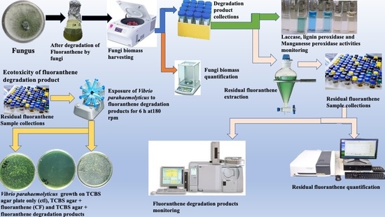
4. Materials and Methods

4.1. Chemicals and Media

Fluoranthene, ethyl acetate, acetone, n-hexane, 2,2- azino-bis-3-ethyl-benzthiozoline-6-sulphonic acid (ABTS), 2, 6-dimethoxy phenol (DMP), azure B, (NH4)2SO4, K2HPO4, NaH2PO4.2H2O, NaCl, MgSO4.7H2O, nitrilotriacetic acid, CaCl2.2H2O, MnCl2.2H2O, FeSO4.7H2O, Co(NO3).6H2O, ZnSO4, CuSO4, H3BO3, Na2MoO4, Al2(SO4)3 H2O, potato dextrose agar, and thiosulphate-citrate-bile salts-sucrose (TCBS) agar were procured from Merck (Burlington, MA, USA). All solvents and reagents are of HPLC grade with ≥98% purity.

4.2. Preparation of Standard Solutions

Fluoranthene stock solution was prepared by dissolving 500 mg of fluoranthene in 10 mL acetone, sterilized by membrane filtration (0.22 µm, Millipore, Merck, Burlington, MA, USA) and stored at 4 °C. Basal salt medium (BSM) was prepared, as described previously [41], and contain (in g/L): (NH4)2SO4 2.4; K2HPO4 1.55; NaH2PO4.2H2O 0.85; NaCl 0.5; MgSO4.7H2O 0.26 and 1mL of Trace Elements (mg/L): Nitrilotriacetic acid 15; CaCl2.2H2O 15; MnCl2.2H2O 6; FeSO4.7H2O 1; Co(NO3).6H2O 1; ZnSO4 1; CuSO4 0.1; H3BO3 0.1; Na2MoO4 0.1; Al2(SO4)3.H2O 0.1. The pH of the medium was adjusted to 5 with 1 N hydrochloric acid (HCl).

4.3. Fungal Strains and Molecular Identification

Two indigenous fungal strains namely, Trichoderma lixii strain FLU1(TlFLU1) and Talaromyces pinophilus FLU12 (TpFLU12) previously isolated from a benzo (b) fluoranthene enriched wastewater-activated sludge with tolerance to 3, 4, and 5-ring PAH compounds and with ability to use them as sole carbon sources (Unpublished data). TlFLU1 and TpFLU12 cultures were grown separately on potato dextrose agar plates and incubated at 30 °C for 96 h. DNA was extracted from each fungal strain using the ZR Fungal/Bacterial DNA MiniPrepTM kit (Zymo Research) according to the manufacturer’s instructions and used in amplifying, sequencing, and analyzing the fungi intergenic ITS1-5.8S-ITS2 region of the ribosomal DNA (rDNA). The ITS region of the fungal strains were amplified from the extracted DNA using the primers, ITS 4 (5′-TCCTCCGCTTATTGATATGC-3′) and ITS 5 (5′-GGAAGTAAAAGTCGTAACAAGG-3′), in a PCR assay. The PCR mixtures consisted of 10 ng/μL DNA, 5 μL of 10 X reaction buffer, 2 μL 25 mM MgCl2, 2.5 μL of each primer (0.5 μM), 0.25 μL of Taq DNA polymerase, 1 μL of 10 mM dNTPs and volume made up to 50 μL with ultrapure water (MilliQ H2O). A T100 Thermal Cycler (BioRad, Hercules, CA, USA) was used for amplification with the initial denaturation at 95 °C for 2 min followed by 30 cycles of denaturation at 95 °C for 30s, then annealing at 53 °C for 45 s and elongation at 72 °C for 45 s, with a final extension at 75 °C for 10 min. The PCR products were resolved on 1.0% (w/v) agarose gels (Seakem, Thermo Fisher Scientific, Arendalsvägen, Göteborg, Sweden) and visualized after staining with ethidium bromide (0.5 μg/mL) using the Chemigenius Bio-imaging System (Syngene, Cambridge, UK). Positive amplicons (~600 bp) were excised and sequenced at Inqaba Biotechnogical Industries, Pretoria, South Africa, and the sequences were compared against the GenBank database. Homologs were identified using the BLASTn program at the National Center for Biotechnology Information (NCBI) (https://blast.ncbi.nlm.nih.gov/Blast.cgi (accessed on: 21 April 2023)). A phylogenetic approach was used for alignment. The evolutionary relationship between the fungi nucleotide sequence and other sequences was matched on NCBI database using muscle (multiple sequence comparison by UMPGA) implanted in MEGA 11. Alignments were examined manually, and a phylogenetic tree was constructed with a neighbor-joining likelihood approach and bootstrap resampling of 1000 replicates using MEGA 11 [66].

4.4. Degradation of Fluoranthene

The potential of the fungal strains to utilize fluoranthene (400 mg/L) as the sole carbon and energy source was carried out in a 250 mL Erlenmeyer flask containing 100 mL BSM broth. BSM broth added with fluoranthene was separately inoculated with two mycelium discs of 10 mm diameter from the edge of an active growing 5 days old culture of each fungal strain from potato dextrose agar plate. The flasks were incubated in the dark at 30 °C on a rotary shaker at 180 rpm for 16 days. Sterile BSM broth containing only fluoranthene (400 mg/L) was set up as a negative control sample to monitor the effect of abiotic factors. Samples were periodically drawn at 2 day intervals for the quantification of residual fluoranthene, metabolite formation, ligninolytic enzyme activity determination, and ecotoxicity test. The initial concentration of 400 mg/L was used based on its zero fungal growth inhibition on both selected strains (Table S2). The pH of the culture broth was also monitored periodically using a pH/temperature bench meter fitted with a digital probe 230 VAC (HANNA instruments, Romania).

4.5. Fungal Strains Biomass Estimation

The fungal biomass was estimated as described previously [31]. The periodically collected culture broth (4 mL) was centrifuged at 10,000 rpm for 15 min. The supernatant was collected for residual fluoranthene quantifications while the pellets were removed and washed thrice with ethyl acetate to recover any absorbed fluoranthene before passing through a pre-dried and pre-weighed Whatman filter paper no. 1. Thereafter, the Whatman filter paper was dried to a constant weight at 80 °C, and the dry weight of the biomass was determined. It is worthwhile to note that washing the pellet with ethyl acetate (1:10 w/v, 5 times) was necessitated so as not to underestimate the residual fluoranthene concentration since fluoranthene is known to be hydrophobic and to limit biosorption of the PAH by the fungi mycelia pellets.

4.6. Extraction, Quantification of Residual Fluoranthene and Kinetics of Degradation

The residual fluoranthene in the culture medium was extracted and quantified using a UV-Vis spectrometer, as described previously [57], with slight modifications. For brevity, the culture broth (supernatant) obtained during the biomass estimation was mixed with ethyl-acetate (1:4 v/v) in a separating funnel. The mixture was then vigorously shaken for 5 min and allowed to stand for 20 min to enhance the separation of aqueous and organic phases using the standard extraction method. The organic phase was collected, dried over 10 g anhydrous Na2SO4, and evaporated to dryness at 40 °C under reduced pressure. The dried fractions were then dissolved with the same extraction solvent and diluted 10-fold with ethyl acetate before quantifying the residual fluoranthene in a scanning spectrum mode of Agilent Cary 60 UV-Vis spectrophotometer (Agilent, Santa Clara, CA, USA) from a wavelength of 400 to 200 nm. The absorption spectrum of fluoranthene showed peak maxima at a wavelength of 265 nm using a quartz cuvette with an optical path length of 10 mm. The fluoranthene residual concentration of the medium was determined by using a standard curve derived from known concentrations of fluoranthene (R2 value of 0.958). Additionally, a recovery study was carried out in a sterile BSM broth (uninoculated with fungi) with an appropriate volume of known fluoranthene concentrations under a static condition before analytical quantification. This set-up was replicated with an average fluoranthene recovery of 97.9 ± 7.17%. The Kinetic models of the fluoranthene degradation by the fungi were plotted following the methods, as described previously [67].

4.7. Determination of Intermediates Compounds by GC-MS Analysis

The periodically extracted residual fluoranthene fractions were pooled together and dried at 40 °C under reduced pressure. The dried fractions were then re-dissolved to a final volume of 1 mL in ethyl acetate before analyzing the intermediate compounds using GC–MS as follows, one microliter of the extracted fractions was injected into an Elite-5MS capillary GC column with a length of 30.0 m; ID 0.25 mm; film thickness 250 µm, split injection (injector temperature 280 °C, split 1/8 for samples and 1/20 for standard samples); oven temperature programmed from 60 °C (held for 2 min) to 300 °C (15 min) at 10 °C/min. The carrier gas used was helium at a flow rate of 1 mL/min. The ion trap detector and NIST (National Institute of standard technology) and USA library was used for intermediate compound analysis at a full scan at 10–220 m/z [30].

4.8. Fourier-Transform Infrared Spectroscopy (FTIR) Analysis

The structural changes in the functional group of intermediate compounds were monitored with FTIR spectroscopy in a scanning absorbance mode of PerkinElmer spectrum 100 fitted with a universal attenuated total reflectance (ATR) accessory (PerkinElmer, Waltham, MA, USA). Hundred microliters of sample used for the intermediate compound detection were scanned between wavenumber 4000–400 cm−1. All obtained data were corrected for the background spectrum.

4.9. Quantitative Estimation of Ligninolytic Enzymes Activities

4.9.1. Laccase Assay

Laccase activity was carried out by measuring the increase in absorbance during the oxidation of 2,2- Azino-bis-3-ethyl-benzthiozoline-6-sulphonic acid (ABTS) [68]. The reaction mixture contained 100 μL of 50 mM ABTS and 800 μL of 20 mM Na-Acetate buffer (pH 4.5), and 100 μL of appropriately diluted enzyme extract. The reaction mixture was incubated at 30 °C for 15 min and thereafter stopped by adding 40 μL of 20% trichloroacetic acid. The absorbance was measured at 420 nm using an Agilent Cary 60 UV-Vis spectrophotometer (Agilent, Santa Clara, CA, USA). The enzyme activity was expressed in unit L−1 of the culture filtrate. A unit of laccase activity was defined as 1 µmol of ABTS oxidized per min.

4.9.2. Lignin Peroxidase Assay

Lignin peroxidase assay was performed by measuring the reduction in the substrate (Azure B), as described previously [28]. The reaction mixture contained 749.5 μL of 125 mM sodium tartrate buffer (pH 3.0), 83.5 μL of enzyme extract, and 0.16 mM of substrate Azure B. The enzymatic reaction was also catalyzed with pure 83.5 μL of 2 mM Hydrogen peroxide (pH 3.0) before incubating at 30 °C for 15 min. The color change was measured using Agilent Cary 60 UV-Vis spectrophotometer at 651 nm and expressed in unit L−1 of the culture filtrate. One unit of the lignin peroxidase activity was defined as 1 µmol of Azure B reduced per min.

4.9.3. Manganese Peroxidase Assay

Manganese peroxidase activity was determined by monitoring the oxidation of 2, 6-DMP at 469 nm [68]. Briefly, the reaction mixture contained 749.5 μL of sodium tartrate buffer (50 mM, pH 4.0), 83.5 μL of enzyme extract and 2 mM of 2, 6-DMP. The enzymatic reaction was also catalyzed with pure 83.5 μL of 0.4 mM hydrogen peroxide (pH 4.0) before incubating at 30 °C for 15 min. The enzyme activities were expressed as unit L−1 of the culture filtrate. One unit of the manganese peroxidase activity was defined as 1 µmol of the substrate oxidized per min.

4.10. Ecotoxicity Test

The ecotoxicity test of the culture medium (undergoing degradation) was evaluated at different incubation periods based on bacterial survival using a modified liquid-to-plate micro-counting method [69]. Briefly, 100 μL of diluted standardized overnight culture broth of Vibrio parahaemolyticus (ATCC 17802) with an absorbance value of 0.14 at λmax 600 nm (corresponding to a final inoculum of 1.0 × 10⁸ CFU/mL) was exposed to each degradation product in marine broth (9:1 v/v) to a final volume of 1 mL in 15 mL centrifuge tube. To avoid sedimentation, each toxicant was incubated at 30 °C in a 15 mL centrifuge tube and mixed on MRC Lab suspension mixer SM-3600 (MRC laboratory equipment, Essex, UK) at 180 rpm for 6 h. The suspensions from each exposed toxicant at the initial and final exposure time were serially diluted before spread plating 10 μL on 45 mm TCBS agar plates and incubated at 30 °C for 96 h in triplicates. Bacterium survival for each degradation product was calculated by log transformation of the colony-forming unit (CFU/mL) using GraphPad Prism software (v. 7.05). Additionally, a control (CN) and no degradation product/PAHs was also set up to compare the bacterium survival without stress. The toxicity unit (TU) of the 50% effect endpoint of the toxicant was calculated as the reciprocal of EC50 multiplied by 100 [70]. It is noteworthy to state that the EC50 toxicity classification was interpreted based on Directive 79/831/EEC [32]. Briefly, the metabolites were classified as very toxic (EC50 ≤ 1 mg/L), toxic (1 mg/L ≤ EC50 ≤ 10 mg/L), harmful (10 mg/L ≤ EC50 ≤ 100 mg/L), and non-toxic (EC50 ≥ 100 mg/L).

4.11. Data Analysis

The experiments were set up in a completely randomized design (CRD). Data obtained were presented graphically as mean values and error bars using GraphPad prism statistical analysis package version 8.4.3 (471) (San Diego, CA, USA). The effective concentration (EC50) and toxicity unit (TU) of transformation product resulting in 50% reduction in active bacterial replicating/forming colonies after 6 h exposure was evaluated using non-linear regression (log (agonist) vs. response model) of colony-forming unit (CFU/mL). Additionally, the bacterium survival data (log CFU/mL) was subjected to analysis of variance (ANOVA) while its means were ranked with Bonferroni’s multiple comparisons test (BMCT) at p ≤ 0.05.

5. Conclusions

This study reveals that two indigenous fungal strains, TlFLU1 and TpFLU12, can effectively degrade fluoranthene. The fungi can metabolize fluoranthene as a sole source of carbon and energy through the ß-Ketoadipate pathway via benzene-1,2,3-tricarboxylic acid. The ability of these strains to completely metabolize fluoranthene is linked to their production of ligninolytic enzymes (Lac, LiP, and MnP) and their growth. The study also found that the time-course changes in toxicity of degradation products accumulated during the metabolism process by TlFLU1 are toxic to marine bacterium survival and classified as harmful due to the recorded EC50 (14.25 mg/L) and toxicity unit (7.000%) while that of TpFLU12 were non-toxic with EC50 (197.1 mg/L) and toxicity unit (0.507%), suggesting it as a proficient strain in metabolizing fluoranthene into an innocuous state and the enzymes reported may be used as biocatalysts in the biotechnology industries.

Supplementary Materials

The following supporting information can be downloaded at: https://www.mdpi.com/article/10.3390/catal13050791/s1, Figure S1: morphological characteristics of TlFLU1 grown on a PDA plate; Figure S2: morphological characteristics of TpFLU12 grown on a PDA plate; Figure S3: neighbor-joining phylogenetic tree for TlFLU1 and TpFLU12 with other fungal strains. Figure S4: Fluoranthene degradation kinetic modelling (A) Zero Order, (B) First Order, and (C) Second Order. TlFLU1 (●), TpFLU12 (■), d = days; Figure S5: FTIR spectra of fluoranthene biodegradation metabolites; Figure S6: Preliminary assay for Ligninolytic enzyme production on solid media. Keys: - flouranthene + no-fungus; + flouranthene + fungus; laccase (oxidation of ABTs to Green colour zones); LiP (Oxi-dation of Azure B to Green colour zones); MnP (oxidation of phenol red to form yellow colour zones); Table S1: GC-MS profile of the metabolite formed during fluoranthene degradation; Table S2: The average fungal growth and growth inhibition after 10 days of exposure to varying fluoranthene concentrations; Table S3: Preliminary assay for extracellular enzyme production on solid media.

Author Contributions

Conceptualization, S.O.E. and A.O.O.; methodology, S.O.E. and A.K.; validation, S.O.E., A.K. and A.O.O.; formal analysis, S.O.E. and A.K.; investigation, S.O.E. and A.O.O.; resources, M.P.M. and A.O.O.; data curation, S.O.E. and A.K.; writing—S.O.E.; writing—A.K. and A.O.O.; visualization, S.O.E.; supervision, M.P.M. and A.O.O.; project administration, A.O.O.; funding acquisition, A.O.O. and M.P.M. All authors have read and agreed to the published version of the manuscript.

Funding

National Research Foundation, South Africa (Grant No: 94036 and 92803).

Data Availability Statement

Data are contained within the article or Supplementary Material.

Acknowledgments

Authors thank University of KwaZulu Natal for fellowship to Samson O. Egbewale.

Conflicts of Interest

The authors declare no conflict of interest.

References

  1. Masiol, M.; Hofer, A.; Squizzato, S.; Piazza, R.; Rampazzo, G.; Pavoni, B. Carcinogenic and mutagenic risk associated to airborne particle-phase polycyclic aromatic hydrocarbons: A source apportionment. Atmos. Environ. 2012, 60, 375–382. [Google Scholar] [CrossRef]
  2. Zafra, G.; Cortés-Espinosa, D.V. Biodegradation of polycyclic aromatic hydrocarbons by Trichoderma species: A mini review. Environ. Sci. Pollut. Res. 2015, 22, 19426–19433. [Google Scholar] [CrossRef] [PubMed]
  3. Ping, L.; Zhang, C.; Zhang, C.; Zhu, Y.; He, H.; Wu, M.; Tang, T.; Li, Z.; Zhao, H. Isolation and characterization of pyrene and benzo[a]pyrene-degrading Klebsiella pneumonia PL1 and its potential use in bioremediation. Appl. Microbiol. Biotechnol. 2014, 98, 3819–3828. [Google Scholar] [CrossRef]
  4. Haneef, T.; Ul Mustafa, M.R.; Yusof, K.W.; Isa, M.H.; Bashir, M.J.K.; Ahmad, M.; Zafar, M. Removal of polycyclic aromatic hydrocarbons (PAHs) from produced water by Ferrate (VI) oxidation. Water 2020, 12, 3132. [Google Scholar] [CrossRef]
  5. Aranda, E.; Godoy, P.; Reina, R.; Badia-Fabregat, M.; Rosell, M.; Marco-Urrea, E.; García-Romera, I. Isolation of Ascomycota fungi with capability to transform PAHs: Insights into the biodegradation mechanisms of Penicillium oxalicum. Int. Biodeterior. Biodegrad. 2017, 122, 141–150. [Google Scholar] [CrossRef]
  6. Moghadam, S.; Ebrahimipour, M.; Abtahi, G.; Ghassempour, B.; Hashtroudi, A.; Seyed, M. Biodegradation of polycyclic aromatic hydrocarbons by a bacterial consortium enriched from mangrove sediments. J. Environ. Health Sci. Eng. 2014, 12, 114. [Google Scholar] [CrossRef] [PubMed]
  7. Hung, C.-M.; Chen, C.-W.; Huang, C.-P.; Tsai, M.-L.; Dong, C.-D. Metal-free carbocatalysts derived from macroalga biomass (Ulva lactuca) for the activation of peroxymonosulfate toward the remediation of polycyclic aromatic hydrocarbons laden marine sediments and its impacts on microbial community. Environ. Res. 2022, 208, 112782. [Google Scholar] [CrossRef] [PubMed]
  8. Maharaj Kumari, K.; Lakhani, A. PAHs in gas and particulate phases: Measurement and control. In Environmental Contaminants: Measurement, Modelling and Control; Gupta, T., Agarwal, A.K., Agarwal, R.A., Labhsetwar, N.K., Eds.; Springer: Singapore, 2018; pp. 43–75. [Google Scholar] [CrossRef]
  9. Kumar, S.; Upadhayay, S.K.; Kumari, B.; Tiwari, S.; Singh, S.N.; Singh, P.K. In vitro degradation of fluoranthene by bacteria isolated from petroleum sludge. Bioresour. Technol. 2011, 102, 3709–3715. [Google Scholar] [CrossRef]
  10. Bhushan, K.; Chouhan, S.; Tandey, R.; Mandal, V. Central effect of polycyclic aromatic hydrocarbons liberated from thermal power units on the metabolic and oxidative status of Raphanus sativus. JSM Environ. Sci. Ecol. 2020, 8, 1071. [Google Scholar]
  11. Tomar, R.S.; Jajoo, A. PSI becomes more tolerant to fluoranthene through the initiation of cyclic electron flow. Funct. Plant Biol. 2017, 44, 978–984. [Google Scholar] [CrossRef]
  12. Šepič, E.; Bricelj, M.; Leskovšek, H. Degradation of fluoranthene by Pasteurella sp. IFA and Mycobacterium sp. PYR-1: Isolation and identification of metabolites. J. Appl. Microbiol. 1998, 85, 746–754. [Google Scholar] [CrossRef] [PubMed]
  13. Spehar, R.L.; Poucher, S.; Brooke, L.T.; Hansen, D.J.; Champlin, D.; Cox, D.A. Comparative toxicity of fluoranthene to freshwater and saltwater species under fluorescent and ultraviolet light. Arch. Environ. Contam. Toxicol. 1999, 37, 496–502. [Google Scholar] [CrossRef]
  14. Saunders, C.R.; Shockley, D.C.; Knuckles, M.E. Fluoranthene-induced neurobehavioral toxicity in F-344 rats. Int. J. Toxicol. 2003, 22, 263–276. [Google Scholar] [CrossRef]
  15. Mallah, M.A.; Changxing, L.; Mallah, M.A.; Noreen, S.; Liu, Y.; Saeed, M.; Xi, H.; Ahmed, B.; Feng, F.; Mirjat, A.A.; et al. Polycyclic aromatic hydrocarbon and its effects on human health: An overeview. Chemosphere 2022, 296, 133948. [Google Scholar] [CrossRef] [PubMed]
  16. Hao, G.; Liu, F.; Zhang, Q.; Ali Mallah, M.; Ali Mallah, M.; Liu, Y.; Xi, H.; Wang, W.; Feng, F. Relationship between polycyclic aromatic hydrocarbons and cardiovascular diseases: A systematic review. Front. Public Health 2021, 9, 763706. [Google Scholar] [CrossRef]
  17. Kubincová, P.; Sychrová, E.; Raška, J.; Basu, A.; Yawer, A.; Dydowiczová, A.; Babica, P.; Sovadinová, I. Polycyclic aromatic hydrocarbons and endocrine disruption: Role of testicular gap junctional intercellular communication and connexins. Toxicol. Sci. 2019, 169, 70–83. [Google Scholar] [CrossRef]
  18. Nur-Aainaa-Syafini, M.R.; Tay, K.S.; Abu Bakar, N.K.; Uche Emenike, C.; Krishnan, S.; Shahul Hamid, F.; Abas, M.R. Degradation of polycyclic aromatic hydrocarbons (pyrene and fluoranthene) by bacterial consortium isolated from contaminated road side soil and soil termite fungal comb. Environ. Earth Sci. 2015, 74, 5383–5391. [Google Scholar] [CrossRef]
  19. Alegbeleye, O.O.; Opeolu, B.O.; Jackson, V. Bioremediation of polycyclic aromatic hydrocarbon (PAH) compounds: (acenaphthene and fluorene) in water using indigenous bacterial species isolated from the Diep and Plankenburg rivers, Western Cape, South Africa. Braz. J. Microbiol. 2017, 48, 314–325. [Google Scholar] [CrossRef]
  20. Jain, P.K.; Gupta, V.K.; Gaur, R.K.; Lowry, M.; Jaroli, D.P.; Chauhan, U.K. Bioremediation of petroleum oil contaminated soil and water. Res. J. Environ. Toxicol. 2011, 5, 1–26. [Google Scholar] [CrossRef]
  21. Ipeaiyeda, A.R.; Nwauzor, G.O.; Akporido, S.O. Biodegradation of polycyclic aromatic hydrocarbons in agricultural soil contaminated with crude oil from Nigeria refinery using Pleurotus sajor-caju. J. Bioremediat. Biodegrad. 2015, 6, 1000301. [Google Scholar] [CrossRef]
  22. Nasrawi, H.A. Biodegradation of crude oil by fungi isolated from Gulf of Mexico. J. Bioremediat. Biodegrad. 2012, 3, 1000147. [Google Scholar] [CrossRef]
  23. Deshmukh, R.; Khardenavis, A.A.; Purohit, H.J. Diverse metabolic capacities of fungi for bioremediation. Indian J. Microbiol. 2016, 56, 247–264. [Google Scholar] [CrossRef] [PubMed]
  24. Ye, J.-S.; Yin, H.; Qiang, J.; Peng, H.; Qin, H.-M. Biodegradation of anthracene by Aspergillus fumigatus. J. Hazard. Mater. 2011, 185, 174–181. [Google Scholar] [CrossRef] [PubMed]
  25. Lahkar, J.; Deka, H. Isolation of polycyclic aromatic hydrocarbons (PAHs) degrading fungal candidate from oil-contaminated soil and degradation potentiality study on anthracene. Polycycl. Aromat. Comp. 2017, 37, 141–147. [Google Scholar] [CrossRef]
  26. Sharma, A.; Singh, S.B.; Sharma, R.; Chaudhary, P.; Pandey, A.K.; Ansari, R.; Vasudevan, V.; Arora, A.; Singh, S.; Saha, S.; et al. Enhanced biodegradation of PAHs by microbial consortium with different amendment and their fate in in-situ condition. J. Environ. Manag. 2016, 181, 728–736. [Google Scholar] [CrossRef] [PubMed]
  27. Yao, L.; Teng, Y.; Luo, Y.; Christie, P.; Ma, W.; Liu, F.; Wu, Y.; Luo, Y.; Li, Z. Biodegradation of polycyclic aromatic hydrocarbons (PAHs) by Trichoderma reesei FS10-C and effect of bioaugmentation on an aged PAH-contaminated soil. Bioremediat. J. 2015, 19, 9–17. [Google Scholar] [CrossRef]
  28. Agrawal, N.; Shahi, S.K. Degradation of polycyclic aromatic hydrocarbon (pyrene) using novel fungal strain Coriolopsis byrsina strain APC5. Int. Biodeterior. Biodegrad. 2017, 122, 69–81. [Google Scholar] [CrossRef]
  29. Sack, U.; Hofrichter, M.; Fritsche, W. Degradation of polycyclic aromatic hydrocarbons by Manganese peroxidase of Nematoloma frowardii. FEMS Microbiol. Lett. 1997, 152, 227–234. [Google Scholar] [CrossRef] [PubMed]
  30. Teh, Z.C.; Hadibarata, T. Enhanced degradation of pyrene and metabolite identification by Pleurotus eryngii F032. Water Air Soil Pollut. 2014, 225, 1–8. [Google Scholar] [CrossRef]
  31. Hadibarata, T.; Kristanti, R.A.; Hamdzah, M. Biosorption and biotransformation of fluoranthene by the white-rot fungus Pleurotus eryngii F032. Biotechnol. Appl. Biochem. 2014, 61, 126–133. [Google Scholar] [CrossRef]
  32. Lin, Z.; Du, J.; Yin, K.; Wang, L.; Yu, H. Mechanism of concentration addition toxicity: They are different for nonpolar narcotic chemicals, polar narcotic chemicals and reactive chemicals. Chemosphere 2004, 54, 1691–1701. [Google Scholar] [CrossRef] [PubMed]
  33. Hesham, A.E.-L.; Mohamed, E.A.; Mawad, A.M.M.; Elfarash, A.; Abd El-Fattah, B.S.; El-Rawy, M.A. Molecular characterization of Fusarium Solani degrades a mixture of low and high molecular weight polycyclic aromatic hydrocarbons. Open Biotechnol. J. 2017, 11, 27–35. [Google Scholar] [CrossRef]
  34. Govarthanan, M.; Fuzisawa, S. Biodegradation of aliphatic and aromatic hydrocarbons using the filamentous fungus Penicillium sp. CHY-2 and characterization of its manganese peroxidase activity. RSC Adv. 2017, 7, 20716–20723. [Google Scholar] [CrossRef]
  35. Zafra, G.; Absalon, A.E.; Cuevas, M.D.C.; Cortes-Espinosa, D.V. Isolation and selection of a highly tolerant microbial consortium with potential for PAH biodegradation from heavy crude oil-contaminated soils. Water Air Soil Pollut. 2014, 225, 1–18. [Google Scholar] [CrossRef]
  36. Asemoloye, M.D.; Ahmad, R.; Jonathan, S.G. Transcriptomic responses of catalase, peroxidase and laccase encoding genes and enzymatic activities of oil spill inhabiting rhizospheric fungal strains. Environ. Pollut. 2018, 235, 55–64. [Google Scholar] [CrossRef]
  37. Álvarez-Barragán, J.; Cravo-Laureau, C.; Wick, L.Y.; Duran, R. Fungi in PAH-contaminated marine sediments: Cultivable diversity and tolerance capacity towards PAH. Mar. Pollut. Bull. 2021, 164, 112082. [Google Scholar] [CrossRef]
  38. Reyes-César, A.; Absalón, Á.E.; Fernández, F.J.; González, J.M.; Cortés-Espinosa, D.V. Biodegradation of a mixture of PAHs by non-ligninolytic fungal strains isolated from crude oil-contaminated soil. World J. Microbiol. Biotechnol. 2014, 30, 999–1009. [Google Scholar] [CrossRef] [PubMed]
  39. Memić, M.; Vrtačnik, M.; Boh, B.; Pohleven, F.; Mahmutović, O. Biodegradation of PAHs by ligninolytic fungi Hypoxylon Fragiforme and Coniophora Puteana. Polycycl. Aromat. Compd. 2017, 6638, 1–8. [Google Scholar] [CrossRef]
  40. Bankole, P.O.; Semple, K.T.; Jeon, B.H.; Govindwar, S.P. Enhanced enzymatic removal of anthracene by the mangrove soil-derived fungus, Aspergillus sydowii BPOI. Front. Environ. Sci. Eng. 2020, 14, 113. [Google Scholar] [CrossRef]
  41. Saraswathy, A.; Hallberg, R. Degradation of pyrene by indigenous fungi from a former gasworks site. FEMS Microbiol. Lett. 2002, 210, 227–232. [Google Scholar] [CrossRef]
  42. Mandal, S.K.; Das, N. Biodegradation of perylene and benzo [ghi] perylene (5-6 rings) using yeast consortium: Kinetic study, enzyme analysis and degradation pathway. J. Environ. Biol. 2018, 39, 5–15. [Google Scholar] [CrossRef]
  43. Lee, H.; Jang, Y.; Lee, Y.M.; Lee, H.; Kim, G.H.; Kim, J.J. Enhanced removal of PAHs by Peniophora incarnata and ascertainment of its novel ligninolytic enzyme genes. J. Environ. Manag. 2015, 164, 10–18. [Google Scholar] [CrossRef] [PubMed]
  44. Asemoloye, M.D.; Ahmad, R.; Jonathan, S.G. Synergistic action of rhizospheric fungi with Megathyrsus maximus root speeds up hydrocarbon degradation kinetics in oil polluted soil. Chemosphere 2017, 187, 1–10. [Google Scholar] [CrossRef]
  45. Lu, X.Y.; Li, B.; Zhang, T.; Fang, H.H.P. Enhanced anoxic bioremediation of PAHs-contaminated sediment. Bioresour. Technol. 2012, 104, 51–58. [Google Scholar] [CrossRef] [PubMed]
  46. Hadibarata, T.; Kristanti, R.A. Biotransformation studies on fluoranthene, a four-ring polycylic aromatic hydrocarbon, by white-rot fungus Armillaria sp. F022. Agric. Agric. Sci. Procedia 2015, 3, 45–50. [Google Scholar] [CrossRef]
  47. Jin, J.; Yao, J.; Liu, W.; Zhang, Q.; Liu, J. Fluoranthene degradation and binding mechanism study based on the active-site structure of ring-hydroxylating dioxygenase in Microbacterium paraoxydans JPM1. Environ. Sci. Pollut. Res. 2017, 24, 363–371. [Google Scholar] [CrossRef]
  48. Bankole, P.O.; Semple, K.T.; Jeon, B.H.; Govindwar, S.P. Biodegradation of fluorene by the newly isolated marine-derived fungus, Mucor irregularis strain bpo1 using response surface methodology. Ecotoxicol. Environ. Saf. 2021, 208, 111619. [Google Scholar] [CrossRef]
  49. Casellas, M.; Grifoll, M.; Bayona, J.M.; Solanas, A.M. New metabolites in the degradation of fluorene by Arthrobacter sp. strain F101. Appl. Environ. Microbiol. 1997, 63, 819–826. [Google Scholar] [CrossRef]
  50. Oliveira, R.N.; Mancini, M.C.; de Oliveira, F.C.S.; Passos, T.M.; Quilty, B.; Thiré, R.M.d.S.M.; McGuinness, G.B. FTIR analysis and quantification of phenols and flavonoids of five commercially available plants extracts used in wound healing. Rev. Mater. 2016, 21, 767–779. [Google Scholar] [CrossRef]
  51. Sharma, M.; Kumar, S.; Pandey, O.P. Study of energy transfer from capping agents to intrinsic vacancies/defects in passivated ZnS nanoparticles. J. Nanopart. Res. 2010, 12, 2655–2666. [Google Scholar] [CrossRef]
  52. Mecozzi, M.; Sturchio, E. Computer assisted examination of infrared and near infrared spectra to assess structural and molecular changes in biological samples exposed to pollutants: A case of study. J. Imaging 2017, 3, 11. [Google Scholar] [CrossRef]
  53. Boguta, P.; Sokołowska, Z.; Skic, K. Use of thermal analysis coupled with differential scanning calorimetry, quadrupole mass spectrometry and infrared spectroscopy (TG-DSC-QMS-FTIR) to monitor chemical properties and thermal stability of fulvic and humic acids. PLoS ONE 2017, 12, e0189653. [Google Scholar] [CrossRef] [PubMed]
  54. Balogun, A.O.; Lasode, O.A.; Li, H.; McDonald, A.G. Fourier transform infrared (FTIR) study and thermal decomposition kinetics of Sorghum bicolour glume and Albizia pedicellaris residues. Waste Biomass Valorization 2015, 6, 109–116. [Google Scholar] [CrossRef]
  55. Auta, R.; Adamus, G.; Kwiecien, M.; Radecka, I.; Hooley, P. Production and characterization of bacterial cellulose before and after enzymatic hydrolysis. Afr. J. Biotechnol. 2017, 16, 470–482. [Google Scholar]
  56. Mostafa, Y.S.; Alrumman, S.A.; Alamri, S.A.; Otaif, K.A.; Mostafa, M.S.; Alfaify, A.M. Bioplastic (poly-3-hydroxybutyrate) production by the marine bacterium Pseudodonghicola xiamenensis through date syrup valorization and structural assessment of the biopolymer. Sci. Rep. 2020, 10, 1–14. [Google Scholar] [CrossRef] [PubMed]
  57. Teerapatsakul, C.; Pothiratana, C.; Chitradon, L.; Thachepan, S. Biodegradation of polycyclic aromatic hydrocarbons by a thermotolerant white rot fungus Trametes polyzona RYNF13. J. Gen. Appl. Microbiol. 2016, 62, 303–312. [Google Scholar] [CrossRef] [PubMed]
  58. Zafra, G.; Moreno-montaño, A.; Absalón, Á.E.; Cortés-espinosa, D.V. Degradation of polycyclic aromatic hydrocarbons in soil by a tolerant strain of Trichoderma asperellum. Environ. Sci. Pollut. Res. 2015, 22, 1034–1042. [Google Scholar] [CrossRef]
  59. Mtibaà, R.; Olicón-Hernández, D.R.; Pozo, C.; Nasri, M.; Mechichi, T.; González, J.; Aranda, E. Degradation of bisphenol A and acute toxicity reduction by different thermo-tolerant ascomycete strains isolated from arid soils. Ecotoxicol. Environ. Saf. 2018, 156, 87–96. [Google Scholar] [CrossRef]
  60. Kim, Y.C.; Lee, S.R.; Jeon, H.J.; Kim, K.; Kim, M.J.; Choi, S.D.; Lee, S.E. Acute toxicities of fluorene, fluorene-1-carboxylic acid, and fluorene-9-carboxylic acid on zebrafish embryos (Danio rerio): Molecular mechanisms of developmental toxicities of fluorene-1-carboxylic acid. Chemosphere 2020, 260, 127622. [Google Scholar] [CrossRef]
  61. Kamaya, Y.; Fukaya, Y.; Suzuki, K. Acute toxicity of benzoic acids to the crustacean Daphnia magna. Chemosphere 2005, 59, 255–261. [Google Scholar] [CrossRef] [PubMed]
  62. Tuvikene, A. Aquatic ecotoxicology-study of freshwater systems stressed by toxic chemical. Ann. Zool. Fenn. 1995, 32, 295–309. [Google Scholar]
  63. So-Young, L.; Kwon, J.-H. Enhancement of toxic efficacy of alkylated polycyclic aromatic hydrocarbons transformed by Sphingobium quisquiliarum. Int. J. Environ. Res. Public Health 2020, 17, 6416. [Google Scholar] [CrossRef]
  64. Arunprasath, T.; Sudalai, S.; Meenatchi, R.; Jeyavishnu, K.; Arumugam, A. Biodegradation of triphenylmethane dye malachite green by a newly isolated fungus strain. Biocatal. Agric. Biotechnol. 2019, 17, 672–679. [Google Scholar] [CrossRef]
  65. Strotmann, U.; Flores, D.P.; Konrad, O.; Gendig, C. Bacterial toxicity testing: Modification and evaluation of the luminescent bacteria test and the respiration inhibition test. Processes 2020, 8, 1349. [Google Scholar] [CrossRef]
  66. Tamura, K.; Stecher, G.; Kumar, S. MEGA11: Molecular Evolutionary Genetics Analysis Version 11. Mol. Bio. Evol. 2021, 38, 3022–3027. [Google Scholar] [CrossRef]
  67. Ortega-González, D.K.; Cristiani-Urbina, E.; Flores-Ortíz, C.M.; Cruz-Maya, J.A.; Cancino-Díaz, J.C.; Jan-Roblero, J. Evaluation of the removal of pyrene and fluoranthene by Ochrobactrum anthropi, fusarium sp. and their coculture. Appl. Biochem. Biotechnol. 2015, 175, 1123–1138. [Google Scholar] [CrossRef]
  68. Patel, H.; Gupte, A.; Gupte, S. Effect of different culture conditions and inducers on production of laccase by a basidiomycete fungal isolate Pleurotus ostreatus HP-1 under solid state fermentation. BioResour 2009, 4, 268–284. [Google Scholar]
  69. Rotini, A.; Tornambè, A.; Cossi, R.; Iamunno, F.; Benvenuto, G.; Berducci, M.T.; Maggi, C.; Thaller, M.C.; Cicero, A.M.; Manfra, L.; et al. Salinity-based toxicity of CuO nanoparticles, CuO-bulk and Cu ion to Vibrio anguillarum. Front. Microbiol. 2017, 8, 2076. [Google Scholar] [CrossRef]
  70. Czech, B.; Jośko, I.; Oleszczuk, P. Ecotoxicological evaluation of selected pharmaceuticals to Vibrio fischeri and Daphnia magna before and after photooxidation process. Ecotoxicol. Environ. Saf. 2014, 104, 247–253. [Google Scholar] [CrossRef]
Figure 1. (A) Percentage degradation of fluoranthene in BSM by TlFLU1 and TpFLU12, (B) dry biomass of individual fungi strains, (C) change in pH medium during fluoranthene degradation, and (D) total protein content of each fungus. Control (♦), TlFLU1 (●), TpFLU12 (■).
Catalysts 13 00791 g001
Figure 2. The proposed fluoranthene metabolic pathway by TlFLU1 and TpFLU12. The un-bold arrows denote an unknown transient product which were not detected by GCMS analysis.
Catalysts 13 00791 g002
Figure 3. Extracellular ligninolytic enzyme activities response to fluoranthene degradation. (A) Laccase activity, (B) lignin peroxidase activity, and (C) manganese peroxidase activity. Control (♦), Fluoranthene + TlFLU1 (●), Fluoranthene + TpFLU12 (■), BSM + TlFLU1 (▼), BSM + TpFLU12 (▲).
Catalysts 13 00791 g003
Figure 4. V. parahaemolyticus survival after 6 h exposure to fluoranthene degradation products of (A) TlFLU1 and (B) TpFLU12. CN (Negative Control (no fluoranthene in the growth medium)) and ****: significant difference at p < 0.001.
Catalysts 13 00791 g004
Table 1. Kinetic parameters for the degradation of fluoranthene by TlFLU1 and TpFLU12.
Kinetic ModelParameterStrains
TlFLU1TpFLU12
Zero orderRegression EquationCd = −21.24 d + 290.7Cd = −22.28 d + 367.7
Cd − C0 = KdK (d−1)21.2422.28
D1/2 = C0/2K0D1/29.4128.969
R20.8020.968
First orderRegression EquationlnCd = −0.2100 d + 5.924lnCd = −0.1503 d + 6.184
lnCd= K1d + lnCd K (d−1)0.2100.1503
D1/2 = ln2/KdDT502.2542.588
R20.9870.919
Second orderRegression Equation1/Cd = 0.0040 d − 0.0081/Cd = 0.0109 d − 0.03229
1/Ct = 1/C0 + K2dK (d−1)0.00400.0109
D1/2 =1/C0K2DT500.6250.229
R20.8490.749
R2: Regression coefficient; K: Degradation rate constant; D1/2 and DT50: Half-life.
Table 2. Assignment of the infrared spectra absorbance bands of fluoranthene degradation metabolites.
Functional GroupFrequency (cm−1)Assignment
ControlTlFLU1TpFLU12
i3202–3340O–H stretch of the alcohol rings
ii1610–1730C=C and C=O asymmetric vibration stretching of
the aromatic, carbonyl and quinone compounds
iiiNDND1421Plane bending of C–O–H and aromatic ring
vibrations
ivNDND1385Aliphatic bending of CH and CH2
vND1030NDC–C, C–OH and O–CH3 stretching
vi600–610–C≡C–H: C–H bend in alkynes
ND: Not detected.
Table 3. Acute toxicity (EC50) of daily fluoranthene biodegradation products of TlFLU1 and TpFLU12.
Acute ToxicityStrains
ProfileTlFLU1TpFLU12
EC50 (mg/L)14.25 b197.1 a
TU (%)7.018 a0.05 b
ClassHarmfulnon-toxic
Key: Very toxic (EC50 < 1 mg/L); Toxic (1 mg/L < EC50 ≤ 10 mg/L); Harmful (10 mg/L < EC50 ≤ 100 mg/L); Non-toxic (≥100 mg/L) [32]. Values with the same alphabet as superscripts are not statistically different (p ≤ 0.05).
Disclaimer/Publisher’s Note: The statements, opinions and data contained in all publications are solely those of the individual author(s) and contributor(s) and not of MDPI and/or the editor(s). MDPI and/or the editor(s) disclaim responsibility for any injury to people or property resulting from any ideas, methods, instructions or products referred to in the content.

