Review

Multifunctional Enzymes in Microbial Secondary Metabolic Processes

State Key Laboratory for Conservation and Utilization of Bio-Resources in Yunnan, School of Life Sciences, Yunnan University, Kunming 650091, China
*
Author to whom correspondence should be addressed.
Catalysts 2023, 13(3), 581; https://doi.org/10.3390/catal13030581
Submission received: 31 December 2022 / Revised: 8 March 2023 / Accepted: 9 March 2023 / Published: 14 March 2023
(This article belongs to the Special Issue Microbial Biocatalysis)

Abstract

:
Microorganisms possess a strong capacity for secondary metabolite synthesis, which is represented by tightly controlled networks. The absence of any enzymes leads to a change in the original metabolic pathway, with a decrease in or even elimination of a synthetic product, which is not permissible under conditions of normal life activities of microorganisms. In order to improve the efficiency of secondary metabolism, organisms have evolved multifunctional enzymes (MFEs) that can catalyze two or more kinds of reactions via multiple active sites. However, instead of interfering, the multifunctional catalytic properties of MFEs facilitate the biosynthetic process. Among the numerous MFEs considered of vital importance in the life activities of living organisms are the synthases involved in assembling the backbone of compounds using different substrates and modifying enzymes that confer the final activity of compounds. In this paper, we review MFEs in terms of both synthetic and post-modifying enzymes involved in secondary metabolic biosynthesis, focusing on polyketides, non-ribosomal peptides, terpenoids, and a wide range of cytochrome P450s(CYP450s), and provide an overview and describe the recent progress in the research on MFEs.

1. Introduction

The life activities of organisms involve a large number of functionally rich enzymes, among which multifunctional enzymes (MFEs) are a class of enzymes with two or more catalytic functions. The polypeptide chains of MFEs are linked together by covalent linkages, which help maintain the homeostasis of MFEs. In addition, the function of polypeptide chains in MFEs is not affected by the separation of polypeptide chains, which is an important way to distinguish them from multi-enzyme complexes [1]. There are also a large number of MFEs in the biosynthesis pathway of microbial secondary metabolites, especially synthetases and modifying enzymes. To assemble the basic skeleton of the products, synthases use specific substrates, which are the basis upon which the products are distinguished [2,3]. The most representative synthetic enzymes are polyketide synthases (PKSs), non-ribosomal peptide synthases (NRPSs), and terpene synthases (TPSs). The modification enzymes, which are the main contributors to the abundance of secondary metabolites and the key to their ultimate activity, modify the product skeleton, resulting in products with different functions. As an important modifying enzyme, cytochrome P450s (CYP450s) participate in the post-modification process, and the abundant number and function of CYP450s indicates that they are multifunctional [4]. Hence, it is reasonable to speculate that MFEs can make full use of energy and substances in vivo and in vitro, enabling a faster and more accurate response when regulation occurs and facilitating the vital activities of the organism. Therefore, exploring MFEs in microbial secondary metabolic processes is a meaningful task to more clearly demonstrate the process of certain important biosynthetic products (Figure 1) and to provide precise data for application.

2. Multifunctional Synthases in the Synthesis of Secondary Metabolic Processes

Polyketides, non-ribosomal peptides (NRP), and terpenoids are secondary metabolites widely found in microorganisms [5,6] and have been widely applied in antibiotic, anti-infective, antineoplastic, and immunization applications due to their excellent activity [3,7,8]. In this section, we mainly introduce the MFEs that are typically representative secondary metabolites (Figure 1). Interestingly, PKSs and NRPSs are analogous in many aspects concerning the function of domains and the biosynthesis process. On the one hand, the basic extension domains of PKSs are the acyltransferase (AT), acyl carrier protein (ACP), and condensation (KS) domains, which play the role of substrate loading, substrate transport, and condensation, respectively [9,10,11,12], while in NRPSs, these roles are performed by the adenylation domain A (A), peptidyl carrier protein (PCP) domain, and condensation domain C (C), respectively [13,14,15,16,17]. In the context of the biosynthesis process, the ACP (PCP) of PKSs (NRPSs) transports the substrate that is loaded and activated by AT (A), and finally KS (C) catalyzes the attachment of the substrate to the intermediate until the completion of all catalytic processes, after which the compound skeleton is formed. The backbone is extended via catalysis by the structural domain, which is eventually released by the terminating domain (TE) and modified by a post-modifying enzyme, resulting in the final active compound [13,14,16,17,18,19,20,21,22,23,24,25,26,27,28,29,30,31]. MFEs are ubiquitous, especially according to the available classification, and type I PKSs and type I NRPS are all MFEs. They are further divided into two categories, modular and iterative, where we focus more on the elaboration of iterative PKSs (IPKSs) and iterative NRPSs (INRPSs). Compared with modular enzymes, iterative enzymes contain structural domains that are reused in multiple rounds to complete product extension, although they have only one functional module. Interestingly, despite the smaller size and fewer structural domains of iterative enzymes, their synthesis capacity is not weaker than that of modular enzymes and, moreover, has the potential to be developed. Regarding multifunctional TPSs, their multifunctionality is more often seen in bifunctional enzymes with both isoprenoid transferase (PT) and TPS activities—for example, bifunctional terpene synthases (BTSs), which are bifunctional enzymes that continuously catalyze the proceeding key steps in terpene biosynthesis and have excellent terpenoid synthesis capabilities. In addition, there exist some TPSs with modifying roles that are directly involved in the post-modification process in addition to cyclization, all of which will be elaborated in this section.

2.1. Multifunctional Polyketide Synthases

6-Methylsalicylic acid synthase (6-MSAS), first isolated from Penicillium patulum, is a representative of IPKS [32]. 6-MSAS was found to be involved in the biosynthesis of 6-methylsalicylic acid (6-MSA), which is one of the simplest polypeptides produced [33]. The single protein of 6-MSAS contains all the catalytically active domains required for the synthesis of 6-MSA [34]. 6-MSAS catalyzes the biosynthesis of 6-MSA from one acetyl coenzyme A and three malonyl coenzyme A units by successive decarboxylation condensation [35,36,37]. 6-MSAS initiates the reaction by loading an acetyl starting unit and a malonyl extension unit, and the growing polyketide midbody is bound to the ACP until the end of synthesis. Usually, PKSs releases products through the termination domain (TE), which is absent in 6-MSAS. Thus, the question of what 6-MSAS releases its products through has persisted for some time. There are specialized domains for product release in other IPKSs, such as the Claisen cyclase domain, as well as the R domain [38,39]. The results of Tomomi et al. suggest that the product release of 6-MSAS is inextricably linked to its dehydratase domain (DH), called the thioester hydrolase domain (TH), with which 6-MSAS hydrolyzes the thioester bond of the tetraketone intermediate linked to the ACP for product release (Figure 2) [40].
In addition, IPKSs responsible for the biosynthesis of aromatic polyketides also occur in bacteria, where they may even be more prevalent. As the first bacterial 6-MSAS identified for 6-MSA biosynthesis, ChlB1, first isolated from Streptomyces antibioticus Tü99, is involved in the biosynthesis of the antibiotic chlorothricin (CHL) [41]. ChlB1 is responsible for the synthesis 6-MSA, which is methylated and chlorinated to give the 2-methoxy-5-chloro-6-methylsalicylic acid fraction of CHL [42,43]. In the biosynthesis of maduropeptin (MDP), an enediene from actinomyces, MdpB, similar to ChlB1, is involved in the biosynthesis of part of 6-MSA, namely the part that is modified and used as a component of MDP [44]. PtmQ from Streptomyces pactum ATCC 27456, which is highly similar to ChlB1, is thought to be involved in the biosynthesis of 6-MSA in this organism [45]. As the first discovered bacterial 6-MSAS, ChlB1 is homologous to the fungal 6-MSAS in organization and sequence, and phylogenetic analysis shows that ChlB1 is genetically closer to AviM, CalO5, and NNS, which are bacterial IPKSs [43].
The representative IPKS LovB is responsible for the biosynthesis of lovastatin, a cholesterol-lowering drug formed from dihydromonacolin L (DML) [46] found in Aspergillus oryzae. LovB contains an enoyl reductase (ER)-like structural domain that lacks the corresponding activity and therefore requires the assistance of a separate enoyl reductase, LovC, for the biosynthesis of lovastatin [47,48]. The entire process of lovastatin biosynthesis involves eight cycles of synthesis, and throughout the biosynthesis of lovastatin, the individual structural domains of LovB are used in selective combinations: the MT–KR–DH–ER domains combination are used in the third cycle; the KR–DH–ER domains combination are used in the fourth and sixth cycles; the KR–DH domains combination are used in the first, second, and fifth cycles; and only the KR domain is used in the seventh and eighth cycles (Figure 3). The study by Suzanne et al. demonstrated that the C-terminus of LovB contains neither the TE nor other common unloading domains. When using LovB and LovC to produce polyketides in vitro, the polyketides remain attached to LovB and, therefore, the other structural domains release DML [49]. A study by Xu et al. identified that the gene lovG, located between lovB and lovC in the biosynthetic gene cluster of lovastatin, belongs to the esterase–lipase family of serine hydrolases and is involved in the release of LovB products, which is in agreement with the results that the presence of a LovB-independent structure is involved in DML release [50].
IPKSs play an important role in the formation of polyketone structures during the biosynthesis of a noteworthy class of antibiotics, the enediynes. Calicheamicin, a non-pigmented enediyne from Micromonospora that can be used as an antitumor agent, possesses two specific polyketide structural elements, PKS E and PKS O, of which PKS E is known as the calicheamicin warhead and is an important functional structure of calicheamicin. Two genes in the biosynthetic gene cluster of calicheamicin, calE8 and calO5, encode two independent IPKSs, CalE8 and CalO5, respectively. CalO5 shares a high degree of similarity to the IPKS-AviM responsible for avilamycin synthesis. Joachim found that the CalE8 of a Micromonospora disruption mutant produced neither calicheamicin nor enediene activity nor compounds that would be produced by disruption of the PKS O synthesis machinery, consistent with disruption of PKS E, suggesting a role for CalE8 on PKS E [51]. Another study showed that an IPKS-SgcE from Streptomyces globisporus is involved in the biosynthesis of the slug portion of the enediene antibiotic C-1027, which, unlike calicheamicin, is a class of pigment protein enediene [52]. By comparing the slug structures of C-1027, calicheamicin, and the fellow nonchromoprotein dynemicin, it was hypothesized that a highly conserved PKS is responsible for the biosynthesis of the slug structures of both the pigmented and nonchromoprotein enediynes [51]. In addition, it was shown that the naphthalene fraction of enediyne, another enediyne antitumor antibiotic, is synthesized via condensation of six intact acetate units by naphthalene synthase, an IPKS that, similarly to CalO5 above, shares a high degree of sequence homology with AviM [53].

2.2. Multifunctional Non-Ribosomal Peptide Synthase

INRPSs are often found to be involved in the biosynthesis of NRPs with repetitive sequence composition, and these NRPSs often reuse a domain, especially the TE. Many microbial siderophores are synthesized by NRPSs [54], such as coelichelin, fuscachelin C, etc. Hai et al. proposed the INRPS involved in fuscachelin C biosynthesis and demonstrated a mechanism for fungal NRPS in assembling iron carriers [55]. Enterobactin NRPS, a typical representative INRPS, is a siderophore found in Gram-positive bacteria that promote cellular iron transport. Enterobactin is a circular trimer consisting of a dihydroxybenzoyl group and serine. Prior to biosynthesis, conversion of dihydroxybenzoyl from an aromatic amino acid precursor branching acid to an isobranching acid is first required, then to 2,3-dihydro-2,3-dihydroxybenzoate, and finally to 2,3-dihydroxybenzoic acid (DHB) [56]. Amide linkage of DHB and L-serine is catalyzed by EntD, EntE, EntF, and the C-terminal aryl carrier of EntB [57], in which EntF consists of four domains, C, A, PCP, and TE. L-Serine is activated by domain A and subsequently bound to PCP in the form of an acyl-S-pantothiamine intermediate awaiting binding to DHB [58], which is activated by EntE and binds to EntB [59]. The formation of a peptide bond is catalyzed by domain C of EntF, which binds DHB and L-serine, forming a dihydroxybenzoyl–serine unit. This is followed by the most interesting process in the biosynthesis of enterobactin, wherein the dihydroxybenzoyl–serine unit is transferred to the TE of EntF, leaving the site open for assembly of the next unit (Figure 4). This process is repeated three times and, finally, the cyclization on TE forms a cyclic trimer of dihydroxybenzoyl–serine units, which are finally released by hydrolysis [60]. Even more interesting is that there are countless examples of such iterative use of the TE in such modules as 2,3-dihydroxybenzoate (DHB)-glycine, an iron carrier produced by Bacillus subtilis during iron deficiency that is very similar to enterobactin and is deduced to be a DHB–Gly–Thr trimer with the same DHB portion as enterobactin. Similarly, 2,3-dihydroxybenzoate (DHB)–glycine uses the TE at the end of the C-terminus of INRPS for iterative repeated triple condensation to form the final product. The proteins DhbE, DhbB, and DhbF are involved in the whole biosynthesis process, among which DhbE and DhbB function similarly to EntE and EntB in enterobactin for loading and activating DHB; DhbF is different from EntF, being divided into DhbF–Gly and DhbF–Thr, which are correspondingly responsible for loading Gly and Thr to form the DHB–Gly–Thr monomer, after which TE plays an iterative role to complete biosynthesis of the product [58]. The biosynthetic processes of enedimycin, valinomycin, echinocandin, and gramicin, compounds of bacterial origin, are similar in that they are all formed from repetitive condensation of TE monomers [61,62,63,64,65], which seems to foreshadow the similarity in the biosynthesis of this class of compounds in bacteria.
In 2012, Oikawa et al. performed in vitro expression and biochemical testing of the NRPS for saframycin A biosynthesis and proposed that NRPS SfmC plays an iterative role in completing the final assembly of saframycin A, which is a potent antitumor antibiotic. SfmC plays an iterative role by add two tyrosine derivative units to its skeleton. Unlike the biosynthesis process of NRP, such as for enterobactin described above, the C-terminal structural domain R of SfmC, according to the in vitro reaction results, catalyzes multiple steps of other reduction reactions during this biosynthesis in addition to playing a role in catalyzing the reductive cleavage and release of the mature polypeptide chain from the last PCP structural domain adjacent to the R structural domain [66].

2.3. Multifunctional Terpene Synthases

Two key enzymes play a role in terpenoid biosynthesis: isoprenoid transferases (PTs), which are responsible for the production of C5 precursors, and terpene synthases (TPSs), which catalyze the formation of the basic terpene backbone by cyclizing these chain precursors synthesized by PTs. Generally speaking, PTs and TPSs are two separate enzymes; however, PTs and TPSs have increasingly been found to coexist in the same protein as separate active sites involved in catalytic sequential reactions, and these bifunctional enzymes are called bifunctional terpene synthases (BTSs) [67,68]. Type I BTSs contain αα structures, such as the fusicoccadiene synthase PaFS from Phomopsis amygdali, while type II BTSs have αβγ structures, such as the copalyl diphosphate synthase (CDS) from Penicillium verruculosum [69,70,71]. In recent years, both PaFS and CDS have been researched relatively in-depth by David’s group [72]. PaFS is the first BTS identified to catalyze the first two steps of diterpene glycoside fusicoccadiene biosynthesis [68,73]. Its C-terminal PT structural domain generates geranylgeranyl diphosphate (GGPP), which is used by the N-terminal class I cyclase structural domain to generate the tricyclic hydrocarbon backbone of fusicoccadiene. CDS was the first BTS found to possess both PT and class II TPS activity [74]. The α structural domain of PT catalyzes the condensation of C5 dimethylallyl diphosphate with three successive additions of C-5 isopentenyl diphosphate (IPP) to form C-20 GGPP, which is then cyclized in the N-terminal class II cyclase structural domain to form copalyl diphosphate [75]. Based on the attraction of the efficient biological activities of terpenoids, various techniques are being actively attempted in the expectation of improving product yields or obtaining structurally novel structures. The yields achieved using ophiobolin synthase, a BTS that is highly homologous to PaFS, have been increased more than 100-fold by constructing a high-yield chassis, a method that can efficiently increase synthetic yields [76]. It was demonstrated that the depth of the PT active site determines the final length of the product [77], and fixed-point mutagenesis experiments showed that the length of the product chain can be changed by altering the depth of the active site pocket [78]. Thus, David et al. demonstrated that CDS can be modified to produce sesquiterpenes and that PT partially produces farnesyl diphosphate (FPP) instead of GGPP [75]. Extending the biosynthetic potential of BTS through combinatorial biology is a good option, but this aspect remains to be explored in relation to PKSs and NRPSs. Some studies have made some progress in demonstrating the applicability of combinatorial biology approaches to the development of TPS [79].
In another study, Tiangang Liu selected two TPSs with a substrate admixture by constructing a phylogenetic tree, and their broad substrate specificity was verified by modifying the mevalonate pathway such that it could provide sufficient amounts of C5 precursors. Six BTSs were constructed by combining the screened TPSs with PT for efficient synthesis of FPPS, GGPPS, and GFPPS, resulting in seven new terpenoids with three new backbones [80]. This modular-like combination seems to be a feasible way to exploit the potential of TPSs and rapidly expand the number of terpenoids. However, because of limited substrate specificity, there is still a need to discover new TPSs. New compounds can be obtained by constructing multifunctional TPS using combinatorial biology methods [81].
Genome-based mining for BTS is a fast and efficient route, and new enzymes are constantly being discovered [80,82,83,84,85,86]. Two BTSs, PbSS and PvPS, were presumably obtained by analyzing the genomes of Penicillium brasilianum NBRC 6234 and Penicillium verruculosum TPU1311, and their cyclization mechanisms were investigated using isotope labeling experiments [87]. Genome mining of Aspergillus ustus 094102 led to identification of a BTS, AuAS, whose heterologous expression resulted in five new diester terpenoids, while coexpression of AuAS and the upstream CYP450 (AuAP450) yielded four new diester terpene alcohols [88]. Subsequently, AcAS from Aspergillus calidoustus, a homologue of AuAS, was heterologously expressed and found to be involved in the biosynthesis of six ester terpenoids, one of which is the new skeleton ester terpenoid calidoustene, and the other five are the five diester terpenoids found after the aforementioned heterologous expression of AuAS (Figure 5) [89]. Liu et al. made remarkable progress by systematically analyzing the genomes of 477 fungal species and found a total of 227 homologous BTS genes, which includes 20 PTTS genes whose functions are known. In order to obtain more information on the catalytic functions of these BTSs, 74 were selected based on the results of functional identification. Yeast engineered strains of 74 BTSs were constructed using a high dipterpene producing yeast chassis, of which 34 new functional BTS were successfully identified. This study presents some new evidence on the origin of BTSs and the consistency of their functions, as well as the convergent evolution of gene functions in fungi, plants, and bacteria [90].
In addition to the BTSs mentioned above, there are also some multifunctional TPSs that tend to have some additional modifying effects, and these additional modification activities are often conducive to the formation of the final products. The volatile sesquiterpene derivative geosmin from Streptomyces coelicolor is synthesized by geosmin synthase (GS), the first bifunctional TPS found in bacteria, and is present in various Gram-positive soil bacteria. GS is a bifunctional TPS with two α structural domains: the N-terminal α structural domain of type I TPS catalyzes the FPP reaction to form cyclized sesquiterpenes, which are precursors of geosmin, and the C-terminal α structural domain catalyzes the cyclization break reaction to produce hyoscyamine and acetone [91,92,93]. A bifunctional synthase encoded by the gene crtYB was found to synthesize octahydro lycopene and cyclize lycopene to β-carotene. First, the octahydro lycopene-β-carotene synthase active domain condenses two GGPP molecules to produce octahydro lycopene, and four desaturation reactions subsequently take place to produce lycopene. Finally, lycopene cyclase activity results in cyclization of both ends of lycopene to produce β-carotene [94].

3. Multifunctional Post-Modifying Enzymes in the Synthesis of Secondary Metabolic Processes

After the synthases complete the assembly of the compound backbone, modifying enzymes, such as oxidases, methyltransferases, glycosyltransferases, and halogenases, catalyze highly diverse and complex structural modifications, a critical step in conferring biological activity to the compounds [2,3]. Among them, oxidases are extensively involved in structural modifications to obtain complex and highly oxidized structures, which are essential for the products to acquire biological activity. The main oxidases commonly found in microbial secondary metabolism are CYP450s, flavin-dependent monooxygenases (FMO), and dioxygenases represented by non-heme α-ketoglutarate-dependent dioxygenases, which present multifunctional activity.

3.1. Multifunctional Cytochrome P450

As an essential modifier enzyme in many secondary metabolic processes, the CYP450 gene is typically integrated in biosynthetic clusters [95] and functions to hydroxylate hydrocarbon bonds through insertion of oxygen atoms to produce hydroxylated metabolites and H2O [96], which can catalyze region-specific and stereospecific oxidation of precursors, allowing for structural diversity and enhanced biological activity. More and more MFEs are being discovered, and many CYP450s from fungi and bacteria are found to be multifunctional.
The same CYP450 enzymes can catalyze different biosynthetic processes or different stages of the same biosynthetic process. Most of the multifunctional CYP450s function in the same biosynthetic process. In contrast to pure hydroxylation and epoxidation reactions, these multifunctional CYP450s are usually responsible for sequential reactions, catalyzing sequential hydroxylation or epoxidation combinatorial reactions, directly using the products of the previous reaction as substrates [97,98]. The most typical example of this is the synthesis of gibberellins (GAs) in fungi, for example, a gibberellic acid called GA3, a tetracyclic dihydroxy gamma-lactone acid containing two ethylene bonds and a free carboxylic acid group. In fungi, such as Fusarium fujikuroi, the main final product of the gibberellin synthesis pathway is gibberellic acid, which is synthesized by seven genes located in a gene cluster. During subsequent modification, four CYP450 genes encode four multifunctional P450s, namely P450-1, P450-2, P450-3, and P450-4 [99]. Five oxidases participate in the GA biosynthetic pathway, including P450-1, P450-2, P450-3, P450-4, and a 2-oxoglutarate-dependent dioxygenase DES. The role played by oxidases in the biosynthesis of GA3 is well elucidated: P450-4 is responsible for the oxidation of ent-kaurene to ent-kaurenoic acid. P450-1 oxidizes ent-kaurenoic acid to GA14 and catalyzes four sequential steps in the GA biosynthetic pathway: 7β-hydroxylation, oxidation of C-6 to give a contractile ring B, 3β-hydroxylation, and C-7 oxidation [100]; P450-2 functions as a GA20-oxidase, oxidizing GA14 to GA4, and is able to oxidize GA12 to GA9 [101]; P450-3 encodes a 13-hydroxylase responsible for the conversion of GA7 to GA3 and also catalyzes the 13-hydroxylation of GA4 to GA1 [102]; and 2-oxoglutarate-dependent dioxygenase DES is responsible for desaturation and conversion of GA4 to GA7 (Figure 6) [102,103].
A multifunctional CYP450 is involved in the biosynthesis of a well-known compound, lovastatin, and its backbone synthesis is described in the IPKS section above. However, there is no clear explanation of how the cyclic ninhydrin DML is converted to monacolin J and of how the double bond and hydroxylation are formed. Both in vivo and in vitro characterizations by Jorge Barriuso and Don T. Nguyen showed that LovA can catalyze the regio- and stereospecific hydroxylation of monacolin L-acid to produce monacolin J acid. MycG, a multifunctional CYP450 found in Micromonospora griseorubida A11725, which is involved in the biosynthesis of mycinamycin II, a 16-membered macrolide antibiotic. Mycinamycin II contains two types of sugars, deglycosamine and mycinamycin, at C5 and C21, respectively. Genetic complementation analysis and in vitro characterization of MycG confirmed that it is a multifunctional CYP450 that catalyzes sequential hydroxylation and epoxidation of substrates [104]. During the biosynthesis of mycinamycin II, MycG can continuously catalyze the hydroxylation of C14 and the epoxidation of C12 and C13 of macrolides on the substrate mycinamicin IV to obtain mycinamycin II [105,106]. AurH, a CYP450 from Streptomyces thioluteus, catalyzes two sequential oxidation reactions in the biosynthesis of the polyketide antibiotic aureothin, in which AurH first catalyzes hydroxylation of the allyl groups and then oxidation of the methyl groups to generate tetrahydrofuran rings, which are important active structures of aureothin [107,108]. The researchers who discovered this multifunctional CYP450 subsequently revealed the structural and biochemical basis of AurH-catalyzed tetrahydrofuran ring formation [109]. In addition, the multifunctional CYP450 Fma-P450 is involved in three successive oxidation steps of the antiparasitic and antiangiogenic drug, namely the terpenoid fumagillin from Aspergillus fumigatus [110]. CYP450 RosC is responsible for the formation of hydroxymethyl, formyl, and carboxyl groups during the biosynthesis of rosamicin (16-membered macrolide) produced by Micromonospora rosaria IFO 13697 [111,112,113]. Similar examples have been described elsewhere [114,115,116,117]. Aflatoxin is a class of toxic and carcinogenic microbial secondary metabolites, among which the most famous and toxic is aflatoxin B1. To obtain the final chemical structure in the biosynthesis of aflatoxin B1, two successive oxidation reactions are required. These two successive oxidation reactions have been shown to be catalyzed by a CYP450 OrdA encoded by a single gene. The synthesis intermediate o-methyl-sterimatocystin is first catalyzed by OrdA, which adds a hydroxyl group to the A-ring of O-methyl-sterimatocystin and then continues to produce an unstable seven-membered lactone ring under OrdA catalysis. After deacidification and rearrangement, it is converted to aflatoxin B1. ordA-encoded proteins have previously been found to be involved in the biosynthesis of aflatoxins B1, G1, B2, and G2 by functioning as a CYP450s, and they have shown the ability to convert OMST to aflatoxin B1 independently (Figure 7) [118,119].
A fraction of multifunctional CYP450 can function in different synthetic processes due to their wide range of substrate properties. For example, the first reported CYP450 hydroxylase, PikC, can accept macrolide substrates of different ring sizes, catalyzing the addition of hydroxyl groups at two different positions on the macrolide. In Streptomyces venezuelae, it is the enzyme responsible for hydroxylation during the biosynthesis of the 12-membered ring compounds methymycin and neomethymycin and the 14-membered ring ketolide pikromycin [120]. CYP170A is a bifunctional enzyme that catalyzes the last two consecutive allylic oxidations in the biosynthesis of albaflavenone [121]. CYP170A1 first catalyzes the hydroxylation reaction to generate a heteromeric mixture of albaflavenols and then converts these heteromeric albaflavenols to albaflavenone [122]. Crystal structure and mutagenesis analysis revealed that CYP170A1 also has a separate non-CYP450 active site, a novel TPS active site that converts FPP into a mixture of farnesene isomers [123].

3.2. Other Oxygenases

FMO is a common enzyme for biochemical Baeyer–Villiger oxidation and also accomplishes a variety of oxidation reactions, including epoxidation, hydroxylation, oxidative decarboxylation, halogenation, and sulfonation oxidation, and MFEs exist for this class of oxidases [124,125,126]. XanO4 is an MFE responsible for catalyzing the oxidative substitution of carbonyl and methoxy in anthraquinone compounds during the biosynthesis of yellow phospholipids, leading to the formation of xanthenone rings and the replacement of methoxy with hydroxyl groups. Through isotopic labeling experiments, it has been shown that the reaction involves the sequential insertion of two oxygen atoms and is accompanied by demethoxylation [127]. Cytochalasin, an angiogenesis inhibitor produced by Aspergillus oryzae, contains a vinyl carbonate fraction, and CcsB is an FMO responsible for the introduction of the carbonate fraction via two oxidations of the ketomacrocyclic precursor [128]. Bradley and colleagues have shown that the FMO EncM catalyzes two sequential oxidations of the polyketide midbody, followed by an unusual Favorskii-type rearrangement to produce enteromycin [129,130].
Non-heme α-ketoglutarate-dependent dioxygenases are representatives of dioxygenases that are widely distributed in nature and usually use α-ketoglutarate as a co-substrate and ferrous iron as a cofactor to oxidize the substrate through decarboxylation of α-ketoglutarate into succinic acid with subsequent oxidative coupling of the substrate. Hydroxylation is probably the most common reaction performed by these enzymes, but they can also catalyze other oxidation reactions [131,132,133]. Austinol is a fungal diterpene derived from 3,5-dimethylmustardic acid with a unique chemical structure and a remarkable and unusual spirolactone ring system, whose formation requires the α-ketoglutarate-dependent dioxygenase AusE, along with two FMOs for catalysis. AusE is responsible for iterative oxidation steps, including oxidation of the spirocyclic ring formation reactions, to produce the austinol scaffold [134]. This is the first example of an α-ketoglutarate-dependent dioxygenase that catalyzes the formation of spirolactone rings. PrhA from Penicillium brasilianum has high homology with AusE, and the two enzymes can catalyze the same substrate via a similar catalysis process, except that AusE first desaturates at C1-C2 to form preaustinoid A2 and then rearranges, leading to the formation of spirolactone in preaustinoid A3, whereas PrhA first desaturates at C5-C6 to form berkeleyone B and then rearranges the A/B ring to form the cycloheptadiene portion of berkeleydione [135,136]. Interchanging the non-conserved residues on AusE and PrhA by targeted mutagenesis was used to demonstrate the possibility of functional conversion. After interchanging residues Val150 and Ala232 in PrhA and the corresponding residues Leu150 and Ser232 in AusE, both mutants lost their original function. The results demonstrate that the catalytic function of non-heme iron α-ketoglutarate-dependent oxygenases can be altered by a certain degree of substitution with non-conserved residues, and that subtle differences in the active site structure can lead to dramatic changes in the reaction results, serving as an example for the modification of such enzymes [137].
Other multifunctional non-heme iron α-ketoglutarate-dependent oxygenases, such as CAS, catalyze three oxidation reactions in the production of clavulanic acid, an important β-lactamase inhibitor [138,139]. In cephalosporin C biosynthesis, DAOC, a bifunctional enzyme present in eukaryotes, such as Cephalosporium acremonium, is responsible for the sequential oxidative ring expansion of penicillin N, to produce deacetoxycephalosporin C, and its hydroxylation to form deacetylcephalosporin C [140,141]. A bifunctional α-KG oxygenase, carbapenem synthase (CarC), catalyzes differential isomerization and desaturation reactions in the formation of carbapenem-3-carboxylate, a carbapenem antibiotic [142].

3.3. Post-Modifying Enzymes in the Synthesis of Lanthipeptides

Peptide secondary metabolites of microorganisms are a class of natural products with a wide range of biological activities, typically represented by NRP, and another class represented by ribosomally synthesized post-translationally modified peptides (RiPPs) [143,144], which result from the ribosomal synthesis of polypeptide chains followed by post-modification. The biosynthesis of RiPPs is usually accompanied by a critical hydrolysis step that separates the N-terminal lead peptide from the C-terminal core peptide [145], of which the core peptide is the biologically active RiPPs. Lanthipeptides comprise one of the largest subfamilies of RiPPs and are widely utilized as an antibiotic [145]. According to the biosynthesis pathway, lanthipeptides are classified into four categories [146], and with the exception of type I lanthipeptides, the post-modification enzymes of the other three types of lanthipeptides are MFEs. Although these enzymes are involved in post-modification, they are, in most cases, referred to as lanthipeptide synthases.
Type I lanthipeptides are post-modified by two enzymes: LanB, a dehydratase, and LanC, a cyclase. Type II lanthipeptides are synthesized by LanM, a bifunctional enzyme with a dehydratase domain at the N-terminal end and a cyclase domain at the C-terminal end. Type III and IV lanthipeptides are post-modified by the trifunctional enzymes LanKC and LanL, both of which contain an N-terminal cleavage domain and a central kinase domain with a LanC-like domain at the C-terminal end [147]. However, different structures exist at the end of their C-termini, with the C-terminal domain of LanL containing a conserved zinc-binding motif, and such motifs are not found in the C-terminal cyclase domain of LanKC [148,149]. Notably, new lanthipeptides are still being discovered, and the first V-type lanthipeptide cacaoidin, a glycosylated lanthipeptide, was discovered in 2020 [150,151].
All lanthipeptides contain either the cyclic amino acid lanoline (Lan) or the methyl lanoline (MeLan), the formation of which requires the dehydration of Ser and Thr residues into dehydroalanine (Dha) and dehydrobutyrine (Dhb), followed by a subsequent cyclization reaction in which the Cys residues undergo conjugate addition to Dha and Dhb, corresponding to the formation of thioether cross-linked lanoline and methyllanoline, respectively [152]. In addition, the dehydration mechanism for Ser and Thr residues is the main way to distinguish lanthipeptides [153]. Phylogenomic studies have shown that although these enzymes have very similar cyclase structural domains, the three classes of LanM, LanKC, and LanL have evolved independently. At present, there are a few studies on the structures of these post-modifying enzymes. Only the crystal structure of CylM, which is involved in type II lanthipeptides synthesis, has been revealed and shows the expected structure [154].
The discovery of type III lanthipeptides was relatively late. The first type III lanthipeptide synthase identified was RamC, which is involved in the biosynthesis of the lanthipeptide SapB. A sequence analysis showed that this enzyme contains a Ser/Thr protein kinase structural domain and a C-terminal structural domain [155]. Shortly afterward, a new lanthipeptide synthase with homology to the N-terminus of RamC was discovered, which was shown to contain three catalytic structural domains, including a kinase-like domain responsible for phosphorylation of Ser/Thr residues, an N-terminal lyase domain responsible for phosphate elimination, and a C-terminal cyclase domain. The truncated protein without the C-terminal cyclase domain can also independently catalyze the dehydration reaction. The C-terminal cyclase domain has the same cyclization strategy as LanM and LanC proteins, except that this enzyme contains a conserved zinc-binding motif, which shows that its C-terminal cyclase domain is zinc ion-dependent, whereas RamC-type enzymes do not have a zinc-binding motif [156]. Type III and IV lanthipeptides synthases differ in that the C-terminal cyclization sequence of type III lanthipeptides synthase has no zinc-binding motif and is known as LanKC, whereas the C-terminal cyclization sequence of type IV lanthipeptide synthase contains a zinc-binding motif and is known as LanL [157]. The mechanism of LanL cyclization is similar to that of cyclase LanC due to the similarity of the cyclization structural domain. The cyclization mechanism of LanKC is the most puzzling because their cyclization structural domains do not have a conserved zinc-binding motif. In addition, the presence of a lanthionin or labionin carbon ring in type III lanthipeptides indicates the presence of another type of cyclization, and there are no clear studies showing how this occurs.

3.4. Other Multifunctional Modifying Enzymes

The introduction of fluorine, chlorine, bromine, and iodine substituents into compounds by halogenases can lead to organohalides with high biological activity, due to the significant electronic and spatial properties conferred by the halogen part [158,159]. Of the nearly 5000 or so halogenated natural products identified, chlorinated and brominated metabolites predominate, with iodinated and fluorinated metabolites being fairly rare [160]. A new halogenase, AoiQ, has been identified in Aspergillus oryzae and is representative of multifunctional halogenases. It is involved in the synthesis of the halogenated polyketide compound dichlorodiaporthin and is responsible for catalyzing the highly regiospecific sequential aliphatic dichloride of independent polyketide substrates. Prior to this, only a few aliphatic halogenases have been biochemically characterized compared with the large number of well-studied aromatic halogenases, and all are Fe II ketoglutarate-dependent enzymes. This is the first flavin-dependent halogenase (FDH) for fungal aliphatic halogenation. Bioinformatic analysis and functional genetics indicate that AoiQ is a bifunctional enzyme. Successful recombination of AoiQ in vivo and in vitro demonstrates its ability to progressively add gem-dichloride inactive carbon atoms onto independent substrates. cDNA sequencing confirmed that AoiQ has 1014 amino acids and contains two distinct functional domains: one with FDH activity for catalytic halogenation and the other with a conserved S-adenosylmethionine (SAM) binding domain with methyltransferase activity, suggesting it is an unidentified halogenase–methyltransferase heterodimer. Furthermore, the halogenase site catalyzes an unactivated methyl group, whereas almost all other in vitro reconstituted FDHs are involved in aromatic substitution reactions using Cl+ equivalents, and achieving regioselective functionalization of inactive carbon atoms, such as aliphatic halogenation, is a major synthetic challenge. The discovery of the first characterized fungal aliphatic halogenase, AoiQ, provided a solution for aliphatic halogenation [161,162]. XanH is a bifunctional protein with both reducing and chlorinating functions, but the reduced products cannot be fully and efficiently utilized. By constructing the fusion enzyme FDR-XanH, additional flavin reductase (FDR) can be added to facilitate the halogenation reaction, and its activity has been verified [163,164].
Carbamoylation is ubiquitous in secondary metabolism, causing oxygen and nitrogen atoms to undergo O-carbamoylation and N-carbamoylation, and this post-modification often imparts antibiotic activity and cytotoxicity to its products. The biosynthesis of ansamitocin, an antitumor agent similar to maytansinoid [165], involves the addition of seven polyketide units that undergo a series of post-modifications, including carbamoylation [166]. After some genes were shown to be involved in ansamycin modification [167,168,169], carbamoylated asm21 was presumed to be involved, and the experimental results also indicate that asm21 encodes a carbamoyltransferase. 4″-O-Aminoformylansamycin was isolated and identified as a novel ansamitocin with an aminoformylated polyketoskeleton and glycosylated moiety. As the only carbamoyltransferase gene present in the ansamiline biosynthetic gene cluster, Asm21 is thought to be involved in carbamylation of the glycosyl fraction, a conjecture that was confirmed. The results indicate that ASM21 is a MFE with dual carbamoylation activity on both the polyketide backbone and the glycosyl fraction (Figure 8). Avermectin, a macrolide natural product containing a 6,6 helix ketone group, is an important antiparasitic drug, and a bifunctional post-modifying enzyme was also found to be involved in its biosynthesis. A specific protein, AveC, has no sequence homology to any enzyme of known function and was long thought to only be involved in dehydration. This protein was found to catalyze stereospecific spironoketosis and dehydration of the dihydroxyketone intermediate during the biosynthesis of avermectin, and the dehydration precedes the formation of helical ketones [170]. To some extent, it facilitates the progress of biosynthesis of spironoketone-containing compounds.

4. Future Perspective

MFEs in secondary metabolic processes are highly efficient and versatile. It can be said that catalytic approaches based on MFEs can be used to increase the efficiency of synthesis and save space and substrate loss, as evidenced by their conservation in microorganisms. MFEs have been continuously and systematically studied, which allows us to keep abreast of their progress. In addition to the continuous search for microbial resources with production potential and fermentation isolation, various techniques have been actively applied to explore novel compounds, including genome mining, heterologous expression, and combinatorial biology, and these approaches are used in conjunction. The application of combinatorial biology in the modification of multifunctional synthetases can yield some unnatural products. It is of great value to design a perfect biosynthetic pathway by combining various methods and to exploit the functions of multifunctional enzymes using suitable biomaterials. In any case, the emergence of MFEs has improved the use of energy, matter, and space by organisms, and this advantage can be exploited by humans.

Author Contributions

J.-T.W., T.-T.S. and P.-J.Z. proposed the concept; J.-T.W. and P.-J.Z. drafted the manuscript, L.D. and J.X. participated in the data collection. All authors have read and agreed to the published version of the manuscript.

Funding

This research was supported by the National Natural Science Foundation of China (Grant No. 31970060 (P.-J.Z.) and 32270132 (P.-J.Z.)) and the Yunnan Science and Technology Special Project (202102AA100013 and 202001BB050061).

Data Availability Statement

Not applicable.

Conflicts of Interest

The authors declare no conflict of interest.

References

  1. Stark, G.R. Multifunctional proteins: One gene—More than one enzyme. Trends Biochem. Sci. 1977, 2, 64–66. [Google Scholar] [CrossRef]
  2. Hopwood, D.A. Genetic contributions to understanding polyketide synthases. Chem. Rev. 1997, 97, 2465–2498. [Google Scholar] [CrossRef]
  3. Fischbach, M.A.; Walsh, C.T. Assembly-line enzymology for polyketide and nonribosomal Peptide antibiotics: Logic, machinery, and mechanisms. Chem. Rev. 2006, 106, 3468–3496. [Google Scholar] [CrossRef] [PubMed]
  4. Guengerich, F.P.; Macdonald, T.L. Chemical mechanisms of catalysis by cytochromes-P-450-a unified view. Acc. Chem. Res. 1984, 17, 9–16. [Google Scholar] [CrossRef]
  5. Grunewald, J.; Marahiel, M.A. Chemoenzymatic and template-directed synthesis of bioactive macrocyclic peptides. Microbiol. Mol. Biol. Rev. 2006, 70, 121–146. [Google Scholar] [CrossRef] [Green Version]
  6. Velkov, T.; Lawen, A. Non-ribosomal peptide synthetases as technological platforms for the synthesis of highly modified peptide bioeffectors--Cyclosporin synthetase as a complex example. Biotechnol. Annu Rev. 2003, 9, 151–197. [Google Scholar] [CrossRef]
  7. Koglin, A.; Walsh, C.T. Structural insights into nonribosomal peptide enzymatic assembly lines. Nat. Prod. Rep. 2009, 26, 987–1000. [Google Scholar] [CrossRef] [Green Version]
  8. Christianson, D.W. Structural and chemical biology of terpenoid cyclases. Chem. Rev. 2017, 117, 11570–11648. [Google Scholar] [CrossRef] [Green Version]
  9. Shen, B. Polyketide biosynthesis beyond the type I, II and III polyketide synthase paradigms. Curr. Opin. Chem. Biol. 2003, 7, 285–295. [Google Scholar] [CrossRef]
  10. Sultana, A.; Kallio, P.; Jansson, A.; Wang, J.S.; Niemi, J.; Mantsala, P.; Schneider, G. Structure of the polyketide cyclase SnoaL reveals a novel mechanism for enzymatic aldol condensation. EMBO J. 2004, 23, 1911–1921. [Google Scholar] [CrossRef] [Green Version]
  11. Chooi, Y.H.; Tang, Y. Navigating the fungal polyketide chemical space: From genes to molecules. J. Org. Chem. 2012, 77, 9933–9953. [Google Scholar] [CrossRef] [PubMed] [Green Version]
  12. Piel, J. Biosynthesis of polyketides by trans-AT polyketide synthases. Nat. Prod. Rep. 2010, 27, 996–1047. [Google Scholar] [CrossRef] [PubMed]
  13. Conti, E.; Stachelhaus, T.; Marahiel, M.A.; Brick, P. Structural basis for the activation of phenylalanine in the non-ribosomal biosynthesis of gramicidin S. EMBO J. 1997, 16, 4174–4183. [Google Scholar] [CrossRef] [PubMed] [Green Version]
  14. May, J.J.; Kessler, N.; Marahiel, M.A.; Stubbs, M.T. Crystal structure of DhbE, an archetype for aryl acid activating domains of modular nonribosomal peptide synthetases. Proc. Natl. Acad. Sci. USA 2002, 99, 12120–12125. [Google Scholar] [CrossRef] [PubMed] [Green Version]
  15. Stachelhaus, T.; Mootz, H.D.; Marahiel, M.A. The specificity-conferring code of adenylation domains in nonribosomal peptide synthetases. CHEM Biol. 1999, 6, 493–505. [Google Scholar] [CrossRef] [PubMed] [Green Version]
  16. Stachelhaus, T.; Marahiel, M.A. Modular structure of peptide synthetases revealed by dissection of the multifunctional enzyme GrsA. J. Biol. Chem. 1995, 270, 6163–6169. [Google Scholar] [CrossRef] [Green Version]
  17. Weissman, K.J. The structural biology of biosynthetic megaenzymes. Nat. Chem. Biol. 2015, 11, 660–670. [Google Scholar] [CrossRef]
  18. Bevitt, D.J.; Cortes, J.; Haydock, S.F.; Leadlay, P.F. 6-deoxyerythronolide-b synthase-2 from Saccharopolyspora-erythraea-cloning of the structural gene, sequence-analysis and inferred domain-structure of the multifunctional enzyme. Eur. J. Biochem. 1992, 204, 39–49. [Google Scholar] [CrossRef]
  19. Cortes, J.; Haydock, S.F.; Roberts, G.A.; Bevitt, D.J.; Leadlay, P.F. An unusually large multifunctional polypeptide in the erythromycin-producing polyketide synthase of Saccharopolyspora-erythraea. Nature 1990, 348, 176–178. [Google Scholar] [CrossRef]
  20. Donadio, S.; Katz, L. Organization of the enzymatic domains in the multifunctional polyketide synthase involved in erythromycin formation in Saccharopolyspora-erythraea. Gene 1992, 111, 51–60. [Google Scholar] [CrossRef]
  21. Donadio, S.; Staver, M.J.; McAlpine, J.B.; Swanson, S.J.; Katz, L. Modular organization of genes required for complex polyketide biosynthesis. Science 1991, 252, 675–679. [Google Scholar] [CrossRef] [PubMed]
  22. Chen, A.Y.; Cane, D.E.; Khosla, C. Structure-based dissociation of a type I polyketide synthase module. CHEM Biol. 2007, 14, 784–792. [Google Scholar] [CrossRef] [PubMed] [Green Version]
  23. Nivina, A.; Yuet, K.P.; Hsu, J.; Khosla, C. Evolution and diversity of assembly-line polyketide synthases. Chem. Rev. 2019, 119, 12524–12547. [Google Scholar] [CrossRef] [PubMed]
  24. Stachelhaus, T.; Huser, A.; Marahiel, M.A. Biochemical characterization of peptides carrier protein (PCP), the thiolation domain of multifunctional peptide synthetases. CHEM Biol. 1996, 3, 913–921. [Google Scholar] [CrossRef] [PubMed] [Green Version]
  25. vonDohren, H.; Keller, U.; Vater, J.; Zocher, R. Multifunctional peptide synthetases. Chem. Rev. 1997, 97, 2675–2705. [Google Scholar] [CrossRef]
  26. Stachelhaus, T.; Mootz, H.D.; Bergendahl, V.; Marahiel, M.A. Peptide bond formation in nonribosomal peptide biosynthesis-Catalytic role of the condensation domain. J. Biol. Chem. 1998, 273, 22773–22781. [Google Scholar] [CrossRef] [Green Version]
  27. Finking, R.; Marahiel, M.A. Biosynthesis of nonribosomal peptides. Annu. Rev. Microbiol. 2004, 58, 453–488. [Google Scholar] [CrossRef]
  28. Takeda, K.; Kemmoku, K.; Satoh, Y.; Ogasawara, Y.; Shin-ya, K.; Dairi, T. N-phenylacetylation and nonribosomal peptide synthetases with substrate promiscuity for biosynthesis of heptapeptide variants, JBIR-78 and JBIR-95. ACS Chem. Biol. 2017, 12, 1813–1819. [Google Scholar] [CrossRef]
  29. Miller, B.R.; Gulick, A.M. Structural biology of nonribosomal peptide synthetases. Methods Mol. Biol. 2016, 1401, 3–29. [Google Scholar] [CrossRef] [Green Version]
  30. Walsh, C.T. Insights into the chemical logic and enzymatic machinery of NRPS assembly lines. Nat. Prod. Rep. 2016, 33, 127–135. [Google Scholar] [CrossRef]
  31. Marahiel, M.A. A structural model for multimodular NRPS assembly lines. Nat. Prod. Rep. 2016, 33, 136–140. [Google Scholar] [CrossRef] [PubMed]
  32. Spencer, J.B.; Jordan, P.M. Purification and properties of 6-methylsalicylic acid synthase from Penicillium patulum. Biochem. J. 1992, 288, 839–846. [Google Scholar] [CrossRef] [PubMed] [Green Version]
  33. Beck, J.; Ripka, S.; Siegner, A.; Schiltz, E.; Schweizer, E. The multifunctional 6-methylsalicylic acid synthase gene of Penicillium-patulum-its gene structure relative to that of other polyketide synthases. Eur. J. Biochem. 1990, 192, 487–498. [Google Scholar] [CrossRef] [PubMed]
  34. Dimroth, P.; Walter, H.; Lynen, F. Biosynthesis of 6-methylsalicylic acid. Eur. J. Biochem. 1970, 13, 98–110. [Google Scholar] [CrossRef]
  35. Parascandolo, J.S.; Havemann, J.; Potter, H.K.; Huang, F.; Riva, E.; Connolly, J.; Wilkening, I.; Song, L.; Leadlay, P.F.; Tosin, M. Insights into 6-methylsalicylic acid bio-assembly by using chemical probes. Angew. Chem. 2016, 55, 3463–3467. [Google Scholar] [CrossRef] [Green Version]
  36. Child, C.J.; Spencer, J.B.; Bhogal, P.; ShoolinginJordan, P.M. Structural similarities between 6-methylsalicylic acid synthase from Penicillium patulum and vertebrate type I fatty acid synthase: Evidence from thiol modification studies. Biochemistry 1996, 35, 12267–12274. [Google Scholar] [CrossRef]
  37. Nicholson, T.P.; Rudd, B.A.M.; Dawson, M.; Lazarus, C.M.; Simpson, T.J.; Cox, R.J. Design and utility of oligonucleotide gene probes for fungal polyketide synthases. CHEM Biol. 2001, 8, 157–178. [Google Scholar] [CrossRef] [Green Version]
  38. Fujii, I.; Watanabe, A.; Sankawa, U.; Ebizuka, Y. Identification of Claisen cyclase domain in fungal polyketide synthase WA, a naphthopyrone synthase of Aspergillus nidulans. CHEM Biol. 2001, 8, 189–197. [Google Scholar] [CrossRef] [Green Version]
  39. Bailey, A.M.; Cox, R.J.; Harley, K.; Lazarus, C.M.; Simpson, T.J.; Skellam, E. Characterisation of 3-methylorcinaldehyde synthase (MOS) in Acremonium strictum: First observation of a reductive release mechanism during polyketide biosynthesis. Chem. Commun. 2007, 39, 4053–4055. [Google Scholar] [CrossRef]
  40. Moriguchi, T.; Kezuka, Y.; Nonaka, T.; Ebizuka, Y.; Fujii, I. Hidden function of catalytic domain in 6-methylsalicylic acid synthase for product release. J. Biol. Chem. 2010, 285, 15637–15643. [Google Scholar] [CrossRef] [Green Version]
  41. Gerhard, A.; Muntwyler, R.; Kellerschierlein, W. Metabolic products of microorganisms.147. unexpected transformation in chlorothricin series. Helv. Chim. Acta. 1975, 58, 1323–1338. [Google Scholar] [CrossRef] [PubMed]
  42. Jia, X.Y.; Tian, Z.H.; Shao, L.; Qu, X.D.; Zhao, Q.F.; Tang, J.; Tang, G.L.; Liu, W. Genetic characterization of the chlorothricin gene cluster as a model for spirotetronate antibiotic biosynthesis. Chem. Biol. 2006, 13, 575–585. [Google Scholar] [CrossRef] [PubMed] [Green Version]
  43. Shao, L.; Qu, X.D.; Jia, X.Y.; Zhao, Q.F.; Tian, Z.H.; Wang, M.; Tang, G.L.; Liu, W. Cloning and characterization of a bacterial iterative type I polyketide synthase gene encoding the 6-methylsalicyclic acid synthase. Biochem. Biophys. Res. Commun. 2006, 345, 133–139. [Google Scholar] [CrossRef] [PubMed]
  44. Van Lanen, S.G.; Oh, T.J.; Liu, W.; Wendt-Pienkowski, E.; Shen, B. Characterization of the maduropeptin biosynthetic gene cluster from Actinomadura madurae ATCC 39144 supporting a unifying paradigm for enediyne biosynthesis. J. Am. Chem. Soc. 2007, 129, 13082–13094. [Google Scholar] [CrossRef] [Green Version]
  45. Ito, T.; Roongsawang, N.; Shirasaka, N.; Lu, W.L.; Flatt, P.M.; Kasanah, N.; Miranda, C.; Mahmud, T. Deciphering pactamycin biosynthesis and engineered production of new pactamycin analogues. ChemBioChem 2009, 10, 2253–2265. [Google Scholar] [CrossRef]
  46. Wang, J.L.; Liang, J.D.; Chen, L.; Zhang, W.; Kong, L.L.; Peng, C.; Su, C.; Tang, Y.; Deng, Z.X.; Wang, Z.J. Structural basis for the biosynthesis of lovastatin. Nat. Commun. 2021, 12, 867. [Google Scholar] [CrossRef] [PubMed]
  47. Kennedy, J.; Auclair, K.; Kendrew, S.G.; Park, C.; Vederas, J.C.; Hutchinson, C.R. Modulation of polyketide synthase activity by accessory proteins during lovastatin biosynthesis. Science 1999, 284, 1368–1372. [Google Scholar] [CrossRef]
  48. Ma, S.M.; Li, J.W.H.; Choi, J.W.; Zhou, H.; Lee, K.K.M.; Moorthie, V.A.; Xie, X.K.; Kealey, J.T.; Da Silva, N.A.; Vederas, J.C.; et al. Complete reconstitution of a highly reducing iterative polyketide synthase. Science 2009, 326, 589–592. [Google Scholar] [CrossRef] [Green Version]
  49. Hendrickson, L.; Davis, C.R.; Roach, C.; Nguyen, D.K.; Aldrich, T.; McAda, P.C.; Reeves, C.D. Lavastatin biosynthesis in Aspergillus terreus: Characterization of blocked mutants, enzyme activities and a multifunctional polyketide synthase gene. CHEM Biol. 1999, 6, 429–439. [Google Scholar] [CrossRef] [Green Version]
  50. Xu, W.; Chooi, Y.H.; Choi, J.W.; Li, S.; Vederas, J.C.; Da Silva, N.A.; Tang, Y. LovG: The thioesterase required for dihydromonacolinl release and lovastatin nonaketide synthase turnover in lovastatin biosynthesis. Angew. Chem. 2013, 52, 6472–6475. [Google Scholar] [CrossRef] [Green Version]
  51. Ahlert, J.; Shepard, E.; Lomovskaya, N.; Zazopoulos, E.; Staffa, A.; Bachmann, B.O.; Huang, K.X.; Fonstein, L.; Czisny, A.; Whitwam, R.E.; et al. The calicheamicin gene cluster and its iterative type I enediyne PKS. Science 2002, 297, 1173–1176. [Google Scholar] [CrossRef] [PubMed]
  52. Liu, W.; Christenson, S.D.; Standage, S.; Shen, B. Biosynthesis of the enediyne antitumor antibiotic C-1027. Science 2002, 297, 1170–1173. [Google Scholar] [CrossRef] [PubMed]
  53. Sthapit, B.; Oh, T.J.; Lamichhane, R.; Liou, K.; Lee, H.C.; Kim, C.G.; Sohng, J.K. Neocarzinostatin naphthoate synthase: An unique iterative type I PKS from neocarzinostatin producer Streptomyces carzinostaticus. FEBS Lett. 2004, 566, 201–206. [Google Scholar] [CrossRef] [PubMed]
  54. Barry, S.M.; Challis, G.L. Recent advances in siderophore biosynthesis. Curr. Opin. Chem. Biol. 2009, 13, 205–215. [Google Scholar] [CrossRef]
  55. Hai, Y.; Jenner, M.; Tang, Y. Fungal siderophore biosynthesis catalysed by an iterative nonribosomal peptide synthetase. Chem. Sci. 2020, 11, 11525–11530. [Google Scholar] [CrossRef] [PubMed]
  56. Raymond, K.N.; Dertz, E.A.; Kim, S.S. Enterobactin: An archetype for microbial iron transport. Proc. Natl. Acad. Sci. USA 2003, 100, 3584–3588. [Google Scholar] [CrossRef] [Green Version]
  57. Gehring, A.M.; Bradley, K.A.; Walsh, C.T. Enterobactin biosynthesis in Escherichia coli: Isochorismate lyase (EntB) is a bifunctional enzyme that is phosphopantetheinylated by EntD and then acylated by EntE using ATP and 2,3-dihydroxybenzoate. Biochemistry 1997, 36, 8495–8503. [Google Scholar] [CrossRef]
  58. Reichert, J.; Sakaitani, M.; Walsh, C.T. Characterization of EntF as a serine-activating enzyme. Protein Sci. 1992, 1, 549–556. [Google Scholar] [CrossRef] [Green Version]
  59. Drake, E.J.; Nicolai, D.A.; Gulick, A.M. Structure of the EntB multidomain nonribosomal peptide synthetase and functional analysis of its interaction with the EntE adenylation domain. CHEM Biol. 2006, 13, 409–419. [Google Scholar] [CrossRef] [Green Version]
  60. Shaw-Reid, C.A.; Kelleher, N.L.; Losey, H.C.; Gehring, A.M.; Berg, C.; Walsh, C.T. Assembly line enzymology by multimodular nonribosomal peptide synthetases: The thioesterase domain of E-coli EntF catalyzes both elongation and cyclolactonization. CHEM Biol. 1999, 6, 385–400. [Google Scholar] [CrossRef] [Green Version]
  61. Haese, A.; Schubert, M.; Herrmann, M.; Zocher, R. Molecular characterization of the enniatin synthetase gene encoding a multifunctional enzyme catalyzing n-methyldepsipeptide formation in Fusarium-scirpi. Mol. Microbiol. 1993, 7, 905–914. [Google Scholar] [CrossRef] [PubMed]
  62. Cheng, Y.Q. Deciphering the biosynthetic codes for the potent anti-SARS-CoV cyclodepsipeptide valinomycin in Streptomyces tsusimaensis ATCC 15141. ChemBioChem 2006, 7, 471–477. [Google Scholar] [CrossRef] [PubMed]
  63. Watanabe, K.; Hotta, K.; Praseuth, A.P.; Koketsu, K.; Migita, A.; Boddy, C.N.; Wang, C.C.C.; Oguri, H.; Oikawa, H. Total biosynthesis of antitumor nonribosomal peptides in Escherichia coli. Nat. Chem. Biol. 2006, 2, 423–428. [Google Scholar] [CrossRef]
  64. Kohli, R.M.; Trauger, J.W.; Schwarzer, D.; Marahiel, M.A.; Walsh, C.T. Generality of peptide cyclization catalyzed by isolated thioesterase domains of nonribosomal peptide synthetases. Biochemistry 2001, 40, 7099–7108. [Google Scholar] [CrossRef]
  65. Hoyer, K.M.; Mahlert, C.; Marahiel, M.A. The iterative gramicidin S thioesterase catalyzes peptide ligation and cyclization. CHEM Biol. 2007, 14, 13–22. [Google Scholar] [CrossRef] [PubMed]
  66. Koketsu, K.; Minami, A.; Watanabe, K.; Oguri, H.; Oikawa, H. Pictet-Spenglerase involved in tetrahydroisoquinoline antibiotic biosynthesis. Curr. Opin. Chem. Biol. 2012, 16, 142–149. [Google Scholar] [CrossRef]
  67. Mitsuhashi, T.; Abe, I. Chimeric terpene synthases possessing both terpene cyclization and prenyltransfer activities. ChemBioChem 2018, 19, 1106–1114. [Google Scholar] [CrossRef]
  68. Toyomasu, T.; Tsukahara, M.; Kaneko, A.; Niida, R.; Mitsuhashi, W.; Dairi, T.; Kato, N.; Sassa, T. Fusicoccins are biosynthesized by an unusual chimera diterpene synthase in fungi. Proc. Natl. Acad. Sci. USA 2007, 104, 3084–3088. [Google Scholar] [CrossRef] [Green Version]
  69. Srere, P.A. Complexes of sequential metabolic enzymes. Annu. Rev. Biochem. 1987, 56, 89–124. [Google Scholar] [CrossRef]
  70. Miles, E.W.; Rhee, S.; Davies, D.R. The molecular basis of substrate channeling. J. Biol. Chem. 1999, 274, 12193–12196. [Google Scholar] [CrossRef] [Green Version]
  71. Huang, X.Y.; Holden, H.M.; Raushel, F.M. Channeling of substrates and intermediates in enzyme-catalyzed reactions. Annu. Rev. Biochem. 2001, 70, 149–180. [Google Scholar] [CrossRef] [PubMed] [Green Version]
  72. Faylo, J.L.; Ronnebaum, T.A.; Christianson, D.W. Assembly-line catalysis in bifunctional terpene synthases. Acc. Chem. Res. 2021, 54, 3780–3791. [Google Scholar] [CrossRef] [PubMed]
  73. Chen, M.B.; Chou, W.K.W.; Toyomasu, T.; Cane, D.E.; Christianson, D.W. Structure and function of fusicoccadiene synthase, a hexameric bifunctional diterpene synthase. ACS Chem. Biol. 2016, 11, 889–899. [Google Scholar] [CrossRef] [PubMed] [Green Version]
  74. Mitsuhashi, T.; Okada, M.; Abe, I. Identification of chimeric alpha beta gamma diterpene synthases possessing both Type II terpene cyclase and prenyltransferase activities. ChemBioChem 2017, 18, 2104–2109. [Google Scholar] [CrossRef] [PubMed]
  75. Ronnebaum, T.A.; Eaton, S.A.; Brackhahn, E.A.E.; Christianson, D.W. Engineering the prenyltransferase domain of a bifunctional assembly-line terpene synthase. Biochemistry 2021, 60, 3162–3172. [Google Scholar] [CrossRef]
  76. Yuan, W.; Lv, S.; Chen, L.Y.; Zhao, Y.; Deng, Z.X.; Hong, K. Production of sesterterpene ophiobolin by a bifunctional terpene synthase in Escherichia coli. Appl. Microbiol. Biotechnol. 2019, 103, 8785–8797. [Google Scholar] [CrossRef]
  77. Ronnebaum, T.A.; Gupta, K.; Christianson, D.W. Higher-order oligomerization of a chimeric alpha beta gamma bifunctional diterpene synthase with prenyltransferase and class II cyclase activities is concentration-dependent. J. Struct. Biol. 2020, 210, 107463. [Google Scholar] [CrossRef]
  78. Chang, T.H.; Guo, R.T.; Ko, T.P.; Wang, A.H.J.; Liang, P.H. Crystal structure of type-III geranylgeranyl pyrophosphate synthase from Saccharomyces cerevisiae and the mechanism of product chain length determination. J. Biol. Chem. 2006, 281, 14991–15000. [Google Scholar] [CrossRef] [Green Version]
  79. Andersen-Ranberg, J.; Kongstad, K.T.; Nielsen, M.T.; Jensen, N.B.; Pateraki, I.; Bach, S.S.; Hamberger, B.; Zerbe, P.; Staerk, D.; Bohlmann, J.; et al. Expanding the landscape of diterpene structural diversity through stereochemically controlled combinatorial biosynthesis. Angew. Chem. 2016, 55, 2142–2146. [Google Scholar] [CrossRef] [Green Version]
  80. Bian, G.K.; Han, Y.C.; Hou, A.W.; Yuan, Y.J.; Liu, X.H.; Deng, Z.X.; Liu, T.G. Releasing the potential power of terpene synthases by a robust precursor supply platform. Metab. Eng. 2017, 42, 1–8. [Google Scholar] [CrossRef]
  81. Yuan, Y.J.; Litzenburger, M.; Cheng, S.; Bian, G.K.; Hu, B.; Yan, P.; Cai, Y.S.; Deng, Z.X.; Bernhardt, R.; Liu, T.G. Sesquiterpenoids produced by combining two sesquiterpene cyclases with promiscuous myxobacterial CYP260B1. ChemBioChem 2019, 20, 677–682. [Google Scholar] [CrossRef] [PubMed]
  82. Ye, Y.; Minami, A.; Mandi, A.; Liu, C.W.; Taniguchi, T.; Kuzuyama, T.; Monde, K.; Gomi, K.; Oikawa, H. Genome mining for sesterterpenes using bifunctional terpene synthases reveals a unified intermediate of di/sesterterpenes. J. Am. Chem. Soc. 2015, 137, 11846–11853. [Google Scholar] [CrossRef] [PubMed]
  83. Narita, K.; Sato, H.; Minami, A.; Kudo, K.; Gao, L.; Liu, C.W.; Ozaki, T.; Kodama, M.; Lei, X.G.; Taniguchi, T.; et al. Focused genome mining of structurally related sesterterpenes: Enzymatic formation of enantiomeric and diastereomeric products. Org. Lett. 2017, 19, 6696–6699. [Google Scholar] [CrossRef] [PubMed]
  84. Chiba, R.; Minami, A.; Gomi, K.; Oikawa, H. Identification of ophiobolin F synthase by a genome mining approach: A sesterterpene synthase from Aspergillus clavatus. Org. Lett. 2013, 15, 594–597. [Google Scholar] [CrossRef]
  85. Matsuda, Y.; Mitsuhashi, T.; Quan, Z.Y.; Abe, I. Molecular basis for stellatic acid biosynthesis: A genome mining approach for discovery of sesterterpene synthases. Org. Lett. 2015, 17, 4644–4647. [Google Scholar] [CrossRef]
  86. Jiang, L.; Zhang, X.; Sato, Y.; Zhu, G.L.; Minami, A.; Zhang, W.Y.; Ozaki, T.; Zhu, B.; Wang, Z.X.; Wang, X.Y.; et al. Genome-based discovery of enantiomeric pentacyclic sesterterpenes catalyzed by fungal bifunctional terpene synthases. Org. Lett. 2021, 23, 4645–4650. [Google Scholar] [CrossRef]
  87. Mitsuhashi, T.; Rinkel, J.; Okada, M.; Abe, I.; Dickschat, J.S. Mechanistic characterization of two chimeric sesterterpene synthases from Penicillium. Chemistry 2017, 23, 10053–10057. [Google Scholar] [CrossRef] [PubMed]
  88. Guo, J.J.; Cai, Y.S.; Cheng, F.C.; Yang, C.J.; Zhang, W.Q.; Yu, W.L.; Yan, J.J.; Deng, Z.X.; Hong, K. Genome mining reveals a multiproduct sesterterpenoid biosynthetic gene cluster in Aspergillus ustus. Org. Lett. 2021, 23, 1525–1529. [Google Scholar] [CrossRef]
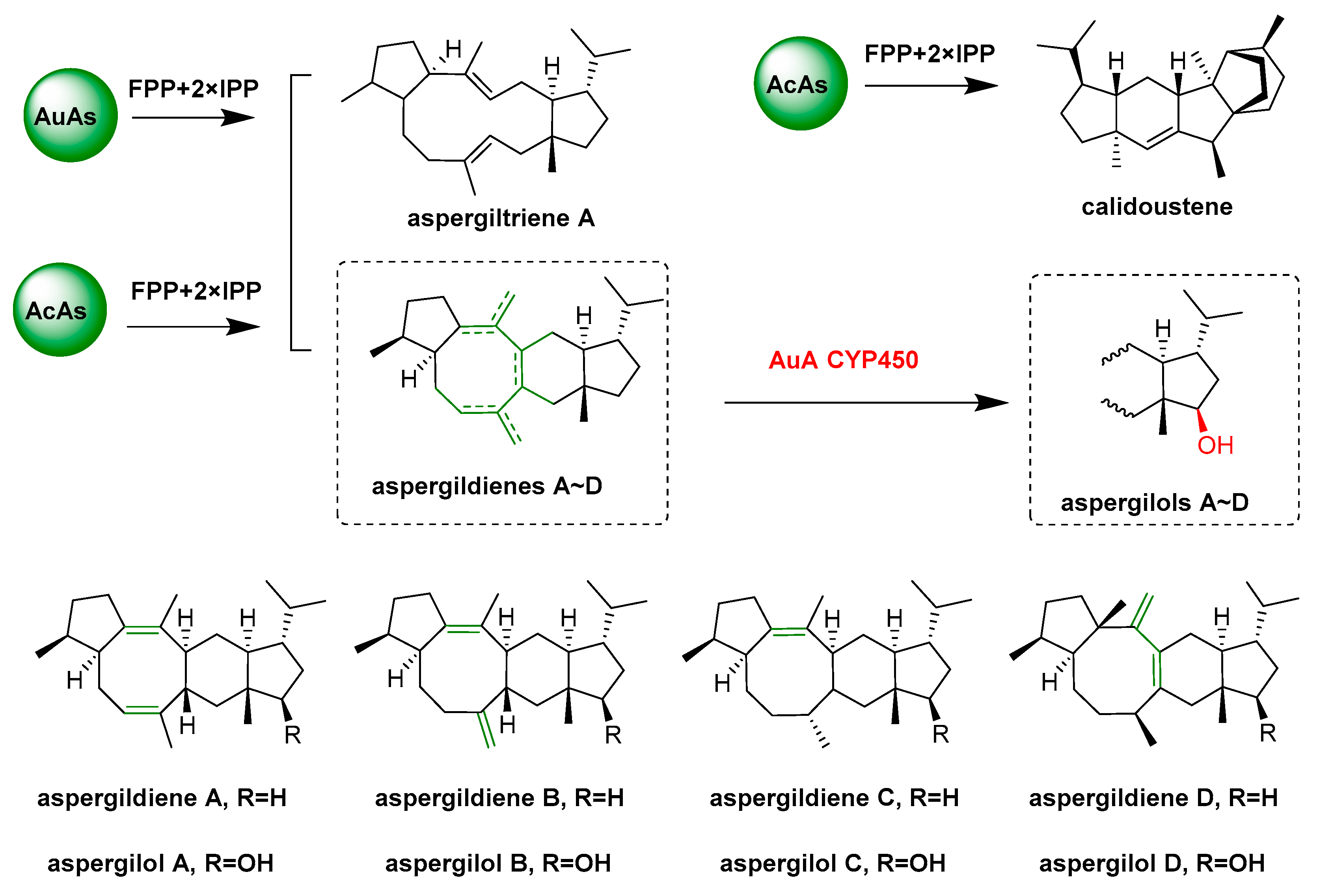
  89. Quan, Z.Y.; Hou, A.W.; Goldfuss, B.; Dickschat, J.S. Mechanism of the bifunctional multiple product sesterterpene synthase AcAS from Aspergillus calidoustus. Angew. Chem. Int. Ed. 2022, 61, e202117273. [Google Scholar] [CrossRef]
  90. Chen, R.; Jia, Q.D.; Mu, X.; Hu, B.; Sun, X.; Deng, Z.X.; Chen, F.; Bian, G.K.; Liu, T.G. Systematic mining of fungal chimeric terpene synthases using an efficient precursor-providing yeast chassis. Proc. Natl. Acad. Sci. USA 2021, 118, e2023247118. [Google Scholar] [CrossRef]
  91. Cane, D.E.; Watt, R.M. Expression and mechanistic analysis of a germacradienol synthase from Streptomyces coelicolor implicated in geosmin biosynthesis. Proc. Natl. Acad. Sci. USA 2003, 100, 1547–1551. [Google Scholar] [CrossRef] [PubMed] [Green Version]
  92. Harris, G.G.; Lombardi, P.M.; Pemberton, T.A.; Matsui, T.; Weiss, T.M.; Cole, K.E.; Koksal, M.; Murphy, F.V.; Vedula, L.S.; Chou, W.K.W.; et al. Structural studies of geosmin synthase, a bifunctional sesquiterpene synthase with alpha alpha domain architecture that catalyzes a unique cyclization-fragmentation reaction sequence. Biochemistry 2015, 54, 7142–7155. [Google Scholar] [CrossRef] [PubMed] [Green Version]
  93. Liato, V.; Aider, M. Geosmin as a source of the earthy-musty smell in fruits, vegetables and water: Origins, impact on foods and water, and review of the removing techniques. Chemosphere 2017, 181, 9–18. [Google Scholar] [CrossRef]
  94. Verdoes, J.C.; Krubasik, P.; Sandmann, G.; van Ooyen, A.J.J. Isolation and functional characterisation of a novel type of carotenoid biosynthetic gene from Xanthophyllomyces dendrorhous. Mol. Gen. Genet. 1999, 262, 453–461. [Google Scholar] [CrossRef]
  95. Lamb, D.C.; Ikeda, H.; Nelson, D.R.; Ishikawa, J.; Skaug, T.; Jackson, C.; Omura, S.; Waterman, M.R.; Kelly, S.L. Cytochrome P450 complement (CYPome) of the avermectin-producer Streptomyces avermitilis and comparison to that of Streptomyces coelicolor A3(2). Biochem. Biophys. Res. Commun. 2003, 307, 610–619. [Google Scholar] [CrossRef] [PubMed]
  96. Whitehouse, C.J.C.; Bell, S.G.; Wong, L.L. P450(BM3) (CYP102A1): Connecting the dots. Chem. Soc. Rev. 2012, 41, 1218–1260. [Google Scholar] [CrossRef]
  97. Podust, L.M.; Sherman, D.H. Diversity of P450 enzymes in the biosynthesis of natural products. Nat. Prod. Rep. 2012, 29, 1251–1266. [Google Scholar] [CrossRef] [Green Version]
  98. Li, S.Y.; Tietz, D.R.; Rutaganira, F.U.; Kells, P.M.; Anzai, Y.; Kato, F.; Pochapsky, T.C.; Sherman, D.H.; Podust, L.M. Substrate recognition by the multifunctional cytochrome P450 MycG in mycinamicin hydroxylation and epoxidation reactions. J. Biol. Chem. 2012, 287, 37880–37890. [Google Scholar] [CrossRef] [Green Version]
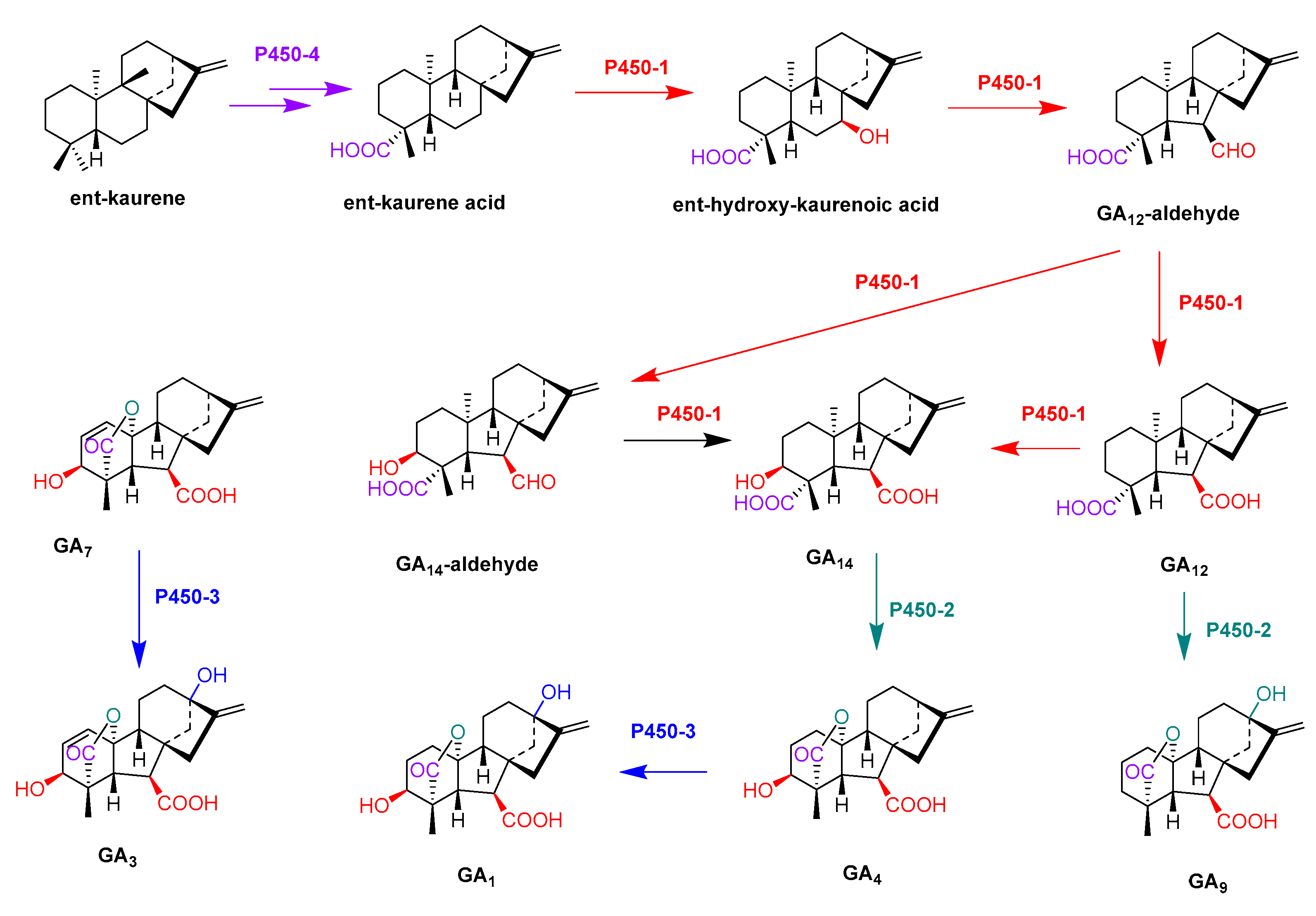
  99. Tudzynski, B.; Kawaide, H.; Kamiya, Y. Gibberellin biosynthesis in Gibberella fujikuroi: Cloning and characterization of the copalyl diphosphate synthase gene. Curr. Genet. 1998, 34, 234–240. [Google Scholar] [CrossRef]
  100. Rojas, M.C.; Hedden, P.; Gaskin, P.; Tudzynki, B. The P450-1 gene of gibberella fujikuroi encodes a multifunctional enzyme in gibberellin biosynthesis. Proc. Natl. Acad. Sci. USA 2001, 98, 5838–5843. [Google Scholar] [CrossRef] [Green Version]
  101. Tudzynski, B.; Rojas, M.C.; Gaskin, P.; Hedden, P. The gibberellin 20-oxidase of Gibberella fujikuroi is a multifunctional monooxygenase. J. Biol. Chem. 2002, 277, 21246–21253. [Google Scholar] [CrossRef] [PubMed] [Green Version]
  102. Tudzynski, B.; Mihlan, M.; Rojas, M.C.; Linnemannstons, P.; Gaskin, P.; Hedden, P. Characterization of the final two genes of the gibberellin biosynthesis gene cluster of Gibberella fujikuroi-des and P450-3 encode GA(4) desaturase and the 13-hydroxylase, respectively. J. Biol. Chem. 2003, 278, 28635–28643. [Google Scholar] [CrossRef] [PubMed] [Green Version]
  103. Bhattacharya, A.; Kourmpetli, S.; Ward, D.A.; Thomas, S.G.; Gong, F.; Powers, S.J.; Carrera, E.; Taylor, B.; Gonzalez, F.N.D.; Tudzynski, B.; et al. Characterization of the fungal gibberellin desaturase as a 2-Oxoglutarate-dependent dioxygenase and its utilization for enhancing plant growth. Plant Physiol. 2012, 160, 837–845. [Google Scholar] [CrossRef] [PubMed] [Green Version]
  104. Inouye, M.; Takada, Y.; Muto, N.; Beppu, T.; Horinouchi, S. Characterization and expression of a P-450-like mycinamicin biosynthesis gene using a novel Micromonospora escherichia-coli shuttle cosmid vector. Mol. Gen. Genet. 1994, 245, 456–464. [Google Scholar] [CrossRef] [PubMed]
  105. Anzai, Y.; Li, S.Y.; Chaulagain, M.R.; Kinoshita, K.; Kato, F.; Montgomery, J.; Sherman, D.H. Functional analysis of MycCI and MycG, cytochrome P450 enzymes involved in biosynthesis of mycinamicin macrolide antibiotics. CHEM Biol. 2008, 15, 950–959. [Google Scholar] [CrossRef] [Green Version]
  106. Anzai, Y.; Tsukada, S.; Sakai, A.; Masuda, R.; Harada, C.; Domeki, A.; Li, S.Y.; Kinoshita, K.; Sherman, D.H.; Kato, F. Function of cytochrome P450 enzymes MycCI and MycG in Micromonospora griseorubida, a producer of the macrolide antibiotic mycinamicin. Antimicrob. Agents Chemother. 2012, 56, 3648–3656. [Google Scholar] [CrossRef] [PubMed] [Green Version]
  107. Richter, M.E.A.; Traitcheva, N.; Knupfer, U.; Hertweck, C. Sequential asymmetric polyketide heterocyclization catalyzed by a single cytochrome P450 monooxygenase (AurH). Angew. Chem. Int. Ed. 2008, 47, 8872–8875. [Google Scholar] [CrossRef]
  108. Muller, M.; He, J.; Hertweck, C. Dissection of the late steps in aureothin biosynthesis. ChemBioChem 2006, 7, 37–39. [Google Scholar] [CrossRef]
  109. Zocher, G.; Richter, M.E.A.; Mueller, U.; Hertweck, C. Structural fine-tuning of a multifunctional cytochrome P450 monooxygenase. J. Am. Chem. Soc. 2011, 133, 2292–2302. [Google Scholar] [CrossRef]
  110. Lin, H.C.; Tsunematsu, Y.; Dhingra, S.; Xu, W.; Fukutomi, M.; Chooi, Y.H.; Cane, D.E.; Calvo, A.M.; Watanabe, K.; Tang, Y. Generation of complexity in fungal terpene biosynthesis: Discovery of a multifunctional cytochrome P450 in the fumagillin pathway. J. Am. Chem. Soc. 2014, 136, 4426–4436. [Google Scholar] [CrossRef]
  111. Iizaka, Y.; Takeda, R.; Senzaki, Y.; Fukumoto, A.; Anzai, Y. Cytochrome P450 enzyme RosC catalyzes a multistep oxidation reaction to form the non-active compound 20-carboxyrosamicin. FEMS Microbiol. Lett. 2017, 364, fnx110. [Google Scholar] [CrossRef] [PubMed] [Green Version]
  112. Iizaka, Y.; Higashi, N.; Ishida, M.; Oiwa, R.; Ichikawa, Y.; Takeda, M.; Anzai, Y.; Kato, F. Function of cytochrome P450 enzymes RosC and RosD in the biosynthesis of rosamicin macrolide antibiotic produced by Micromonospora rosaria. Antimicrob. Agents Chemother. 2013, 57, 1529–1531. [Google Scholar] [CrossRef] [PubMed] [Green Version]
  113. Qi, Z.; Kang, Q.J.; Jiang, C.Y.; Han, M.; Bai, L.Q. Engineered biosynthesis of pimaricin derivatives with improved antifungal activity and reduced cytotoxicity. Appl. Microbiol. Biotechnol. 2015, 99, 6745–6752. [Google Scholar] [CrossRef] [PubMed]
  114. Proctor, R.H.; Plattner, R.D.; Desjardins, A.E.; Busman, M.; Butchko, R.A.E. Fumonisin production in the maize pathogen Fusarium verticillioides: Genetic basis of naturally occurring chemical variation. J. Agric. Food Chem. 2006, 54, 2424–2430. [Google Scholar] [CrossRef] [Green Version]
  115. Matsuda, Y.; Iwabuchi, T.; Wakimoto, T.; Awakawa, T.; Abe, I. Uncovering the unusual D-ring construction in terretonin biosynthesis by collaboration of a multifunctional cytochrome P450 and a unique isomerase. J. Am. Chem. Soc. 2015, 137, 3393–3401. [Google Scholar] [CrossRef]
  116. Barriuso, J.; Nguyen, D.T.; Li, J.W.H.; Roberts, J.N.; MacNevin, G.; Chaytor, J.L.; Marcus, S.L.; Vederas, J.C.; Ro, D.K. Double oxidation of the cyclic nonaketide dihydromonacolin l to Monacolin j by a single cytochrome P450 monooxygenase, LovA. J. Am. Chem. Soc. 2011, 133, 8078–8081. [Google Scholar] [CrossRef]
  117. Cochrane, R.V.K.; Vederas, J.C. Highly selective but multifunctional oxygenases in secondary metabolism. Acc. Chem. Res. 2014, 47, 3148–3161. [Google Scholar] [CrossRef]
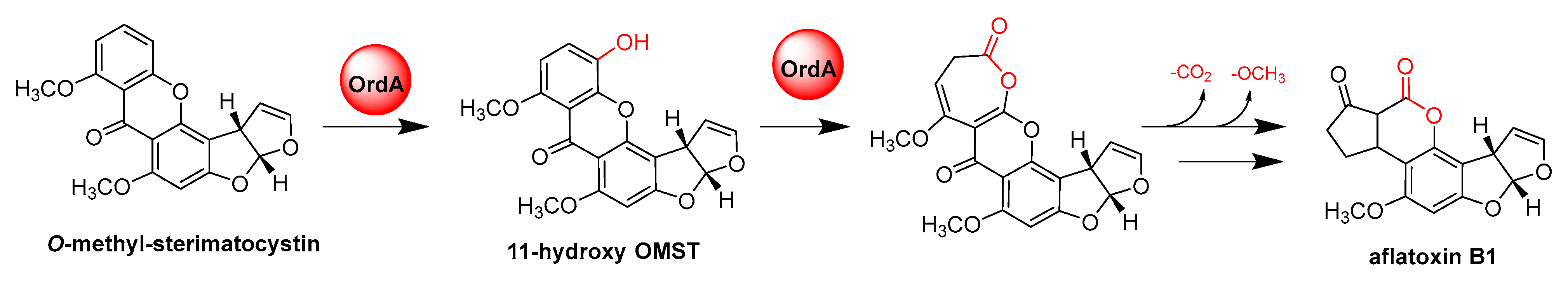
  118. Udwary, D.W.; Casillas, L.K.; Townsend, C.A. Synthesis of 11-hydroxyl O-methylsterigmatocystin and the role of a cytochrome P-450 in the final step of aflatoxin biosynthesis. J. Am. Chem. Soc. 2002, 124, 5294–5303. [Google Scholar] [CrossRef]
  119. Yu, J.; Chang, P.K.; Ehrlich, K.C.; Cary, J.W.; Montalbano, B.; Dyer, J.M.; Bhatnagar, D.; Cleveland, T.E. Characterization of the critical amino acids of an Aspergillus parasiticus cytochrome P-450 monooxygenase encoded by ordA that is involved in the biosynthesis of aflatoxins B1, G1, B2, and G2. Appl. Environ. Microbiol. 1998, 64, 4834–4841. [Google Scholar] [CrossRef] [Green Version]
  120. Xue, Y.Q.; Wilson, D.; Zhao, L.S.; Liu, H.W.; Sherman, D.H. Hydroxylation of macrolactones YC-17 and narbomycin is mediated by the pikC-encoded cytochrome P450 in Streptomyces venezuelae. CHEM Biol. 1998, 5, 661–667. [Google Scholar] [CrossRef] [Green Version]
  121. Zhao, B.; Lin, X.; Lei, L.; Lamb, D.C.; Kelly, S.L.; Waterman, M.R.; Cane, D.E. Biosynthesis of the sesquiterpene antibiotic albaflavenone in Streptomyces coelicolor A3(2). J. Biol. Chem. 2008, 283, 8183–8189. [Google Scholar] [CrossRef] [PubMed] [Green Version]
  122. Zhao, B.; Lei, L.; Vassylyev, D.G.; Lin, X.; Cane, D.E.; Kelly, S.L.; Yuan, H.; Lamb, D.C.; Waterman, M.R. Crystal structure of albaflavenone monooxygenase containing a moonlighting terpene synthase active site. J. Biol. Chem. 2009, 284, 36711–36719. [Google Scholar] [CrossRef] [Green Version]
  123. Zhao, B.; Waterman, M.R. Moonlighting cytochrome P450 monooxygenases. IUBMB Life 2011, 63, 473–477. [Google Scholar] [CrossRef] [Green Version]
  124. Huijbers, M.M.E.; Montersino, S.; Westphal, A.H.; Tischler, D.; van Berkel, W.J.H. Flavin dependent monooxygenases. Arch. Biochem. Biophys. 2014, 544, 2–17. [Google Scholar] [CrossRef]
  125. Walsh, C.T.; Wencewicz, T.A. Flavoenzymes: Versatile catalysts in biosynthetic pathways. Nat. Prod. Rep. 2013, 30, 175–200. [Google Scholar] [CrossRef] [PubMed] [Green Version]
  126. Rebehmed, J.; Alphand, V.; de Berardinis, V.; de Brevern, A.G. Evolution study of the Baeyer-Villiger monooxygenases enzyme family: Functional importance of the highly conserved residues. Biochimie 2013, 95, 1394–1402. [Google Scholar] [CrossRef] [PubMed] [Green Version]
  127. Kong, L.X.; Zhang, W.K.; Chooi, Y.H.; Wang, L.; Cao, B.; Deng, Z.X.; Chu, Y.W.; You, D.L. A multifunctional monooxygenase XanO4 catalyzes xanthone formation in xantholipin biosynthesis via a cryptic demethoxylation. Cell Chem. Biol. 2016, 23, 508–516. [Google Scholar] [CrossRef] [PubMed] [Green Version]
  128. Hu, Y.C.; Dietrich, D.; Xu, W.; Patel, A.; Thuss, J.A.J.; Wang, J.J.; Yin, W.B.; Qiao, K.J.; Houk, K.N.; Vederas, J.C.; et al. A carbonate-forming Baeyer-Villiger monooxygenase. Nat. Chem. Biol. 2014, 10, 552–554. [Google Scholar] [CrossRef] [PubMed] [Green Version]
  129. Cheng, Q.; Xiang, L.; Izumikawa, M.; Meluzzi, D.; Moore, B.S. Enzymatic total synthesis of enterocin polyketides. Nat. Chem. Biol. 2007, 3, 557–558. [Google Scholar] [CrossRef] [PubMed]
  130. Xiang, L.; Kalaitzis, J.A.; Moore, B.S. EncM, a versatile enterocin biosynthetic enzyme involved in Favorskii oxidative rearrangement, aldol condensation, and heterocycle-forming reactions. Proc. Natl. Acad. Sci. USA 2004, 101, 15609–15614. [Google Scholar] [CrossRef] [Green Version]
  131. Aik, W.; McDonough, M.A.; Thalhammer, A.; Chowdhury, R.; Schofield, C.J. Role of the jelly-roll fold in substrate binding by 2-oxoglutarate oxygenases. Curr. Opin. Struct. Biol. 2012, 22, 691–700. [Google Scholar] [CrossRef] [PubMed]
  132. Clifton, I.J.; McDonough, M.A.; Ehrismann, D.; Kershaw, N.J.; Granatino, N.; Schofield, C.J. Structural studies on 2-oxoglutarate oxygenases and related double-stranded beta-helix fold proteins. J. Inorg. Biochem. 2006, 100, 644–669. [Google Scholar] [CrossRef] [PubMed]
  133. Hewitson, K.S.; Granatino, N.; Welford, R.W.D.; McDonough, M.A.; Schofield, C.J. Oxidation by 2-oxoglutarate oxygenases: Non-haem iron systems in catalysis and signalling. Philos. Trans. R. Soc. A Math. Phys. Eng. Sci. 2005, 363, 807–828. [Google Scholar] [CrossRef] [PubMed]
  134. Matsuda, Y.; Awakawa, T.; Wakimoto, T.; Abe, I. Spiro-ring formation is catalyzed by a multifunctional dioxygenase in austinol biosynthesis. J. Am. Chem. Soc. 2013, 135, 10962–10965. [Google Scholar] [CrossRef]
  135. Lo, H.C.; Entwistle, R.; Guo, C.J.; Ahuja, M.; Szewczyk, E.; Hung, J.H.; Chiang, Y.M.; Oakley, B.R.; Wang, C.C.C. Two separate gene clusters encode the biosynthetic pathway for the meroterpenoids austinol and dehydroaustinol in Aspergillus nidulans. J. Am. Chem. Soc. 2012, 134, 4709–4720. [Google Scholar] [CrossRef] [Green Version]
  136. Matsuda, Y.; Iwabuchi, T.; Fujimoto, T.; Awakawa, T.; Nakashima, Y.; Mori, T.; Zhang, H.P.; Hayash, F.; Abe, I. Discovery of key dioxygenases that diverged the paraherquonin and acetoxydehydroaustin pathways in Penicillium brasilianum. J. Am. Chem. Soc. 2016, 138, 12671–12677. [Google Scholar] [CrossRef] [PubMed]
  137. Nakashima, Y.; Mori, T.; Nakamura, H.; Awakawa, T.; Hoshino, S.; Senda, M.; Senda, T.; Abe, I. Structure function and engineering of multifunctional non-heme iron dependent oxygenases in fungal meroterpenoid biosynthesis. Nat. Commun. 2018, 9, 104. [Google Scholar] [CrossRef] [Green Version]
  138. Hamed, R.B.; Gomez-Castellanos, J.R.; Henry, L.; Ducho, C.; McDonough, M.A.; Schofield, C.J. The enzymes of beta-lactam biosynthesis. Nat. Prod. Rep. 2013, 30, 21–107. [Google Scholar] [CrossRef]
  139. Townsend, C.A. New reactions in clavulanic acid biosynthesis. Curr. Opin. Chem. Biol. 2002, 6, 583–589. [Google Scholar] [CrossRef]
  140. Lloyd, M.D.; Lipscomb, S.J.; Hewitson, K.S.; Hensgens, C.M.H.; Baldwin, J.E.; Schofield, C.J. Controlling the substrate selectivity of deacetoxycephalosporin/deacetylcephalosporin C synthase. J. Biol. Chem. 2004, 279, 15420–15426. [Google Scholar] [CrossRef] [Green Version]
  141. Tarhonskaya, H.; Szollossi, A.; Leung, I.K.H.; Bush, J.T.; Henry, L.; Chowdhury, R.; Iqbal, A.; Claridge, T.D.W.; Schofield, C.J.; Flashman, E. Studies on deacetoxycephalosporin c synthase support a consensus mechanism for 2-oxoglutarate dependent oxygenases. Biochemistry 2014, 53, 2483–2493. [Google Scholar] [CrossRef] [PubMed]
  142. Phelan, R.M.; Townsend, C.A. Mechanistic insights into the bifunctional non-heme iron oxygenase carbapenem synthase by active site saturation mutagenesis. J. Am. Chem. Soc. 2013, 135, 7496–7502. [Google Scholar] [CrossRef] [PubMed] [Green Version]
  143. Velasquez, J.E.; van der Donk, W.A. Genome mining for ribosomally synthesized natural products. Curr. Opin. Chem. Biol. 2011, 15, 11–21. [Google Scholar] [CrossRef] [PubMed] [Green Version]
  144. Arnison, P.G.; Bibb, M.J.; Bierbaum, G.; Bowers, A.A.; Bugni, T.S.; Bulaj, G.; Camarero, J.A.; Campopiano, D.J.; Challis, G.L.; Clardy, J.; et al. Ribosomally synthesized and post-translationally modified peptide natural products: Overview and recommendations for a universal nomenclature. Nat. Prod. Rep. 2013, 30, 108–160. [Google Scholar] [CrossRef] [PubMed]
  145. Oman, T.J.; van der Donk, W.A. Follow the leader: The use of leader peptides to guide natural product biosynthesis. Nat. Chem. Biol. 2010, 6, 9–18. [Google Scholar] [CrossRef] [Green Version]
  146. Knerr, P.J.; van der Donk, W.A. Discovery, biosynthesis, and engineering of lantipeptides. Annu. Rev. Biochem. 2012, 81, 479–505. [Google Scholar] [CrossRef]
  147. Krawczyk, B.; Voller, G.H.; Voller, J.; Ensle, P.; Sussmuth, R.D. Curvopeptin: A new lanthionine-containing class III lantibiotic and its co-substrate promiscuous synthetase. ChemBioChem 2012, 13, 2065–2071. [Google Scholar] [CrossRef]
  148. Zhang, Q.; Yu, Y.; Velasquez, J.E.; van der Donk, W.A. Evolution of lanthipeptide synthetases. Proc. Natl. Acad. Sci. USA 2012, 109, 18361–18366. [Google Scholar] [CrossRef] [Green Version]
  149. Wang, H.; van der Donk, W.A. Biosynthesis of the class III lantipeptide catenulipeptin. ACS Chem. Biol. 2012, 7, 1529–1535. [Google Scholar] [CrossRef]
  150. van der Donk, W.A.; Nair, S.K. Structure and mechanism of lanthipeptide biosynthetic enzymes. Curr. Opin. Struct. Biol. 2014, 29, 58–66. [Google Scholar] [CrossRef] [Green Version]
  151. Ortiz-Lopez, F.J.; Carretero-Molina, D.; Sanchez-Hidalgo, M.; Martin, J.; Gonzalez, I.; Roman-Hurtado, F.; de la Cruz, M.; Garcia-Fernandez, S.; Reyes, F.; Deisinger, J.P.; et al. Cacaoidin, first member of the new lanthidin RiPP family. Angew. Chem. 2020, 59, 12654–12658. [Google Scholar] [CrossRef] [PubMed]
  152. Xu, M.; Zhang, F.; Cheng, Z.; Bashiri, G.; Wang, J.; Hong, J.L.; Wang, Y.M.; Xu, L.J.; Chen, X.F.; Huang, S.X.; et al. Functional genome mining reveals a class v lanthipeptide containing ad-amino acid introduced by an F420H2-dependent reductase. Angew. Chem. 2020, 59, 18029–18035. [Google Scholar] [CrossRef] [PubMed]
  153. Repka, L.M.; Chekan, J.R.; Nair, S.K.; van der Donka, W.A. Mechanistic understanding of lanthipeptide biosynthetic enzymes. Chem. Rev. 2017, 117, 5457–5520. [Google Scholar] [CrossRef] [PubMed]
  154. Lagedroste, M.; Reiners, J.; Knospe, C.V.; Smits, S.H.J.; Schmitt, L. A structural view on the maturation of lanthipeptides. Front. Microbiol. 2020, 11, 1183. [Google Scholar] [CrossRef]
  155. Kodani, S.; Hudson, M.E.; Durrant, M.C.; Buttner, M.J.; Nodwell, J.R.; Willey, J.M. The SapB morphogen is a lantibiotic-like peptide derived from the product of the developmental gene ramS in Streptomyces coelicolor. Proc. Natl. Acad. Sci. USA 2004, 101, 11448–11453. [Google Scholar] [CrossRef] [Green Version]
  156. Muller, W.M.; Schmiederer, T.; Ensle, P.; Sussmuth, R.D. In vitro biosynthesis of the prepeptide of Type-III lantibiotic labyrinthopeptin A2 including formation of a c-c bond as a post-translational modification. Angew. Chem. 2010, 49, 2436–2440. [Google Scholar] [CrossRef]
  157. Goto, Y.; Li, B.; Claesen, J.; Shi, Y.X.; Bibb, M.J.; van der Donk, W.A. Discovery of unique lanthionine synthetases reveals new mechanistic and evolutionary Insights. PLoS Biol. 2010, 8, e1000339. [Google Scholar] [CrossRef] [Green Version]
  158. Ruiz-Castillo, P.; Buchwald, S.L. Applications of palladium-catalyzed c-n cross-coupling reactions. Chem. Rev. 2016, 116, 12564–12649. [Google Scholar] [CrossRef]
  159. Gkotsi, D.S.; Dhaliwal, J.; McLachlan, M.M.W.; Mulholand, K.R.; Goss, R.J.M. Halogenases: Powerful tools for biocatalysis (mechanisms applications and scope). Curr. Opin. Chem. Biol. 2018, 43, 119–126. [Google Scholar] [CrossRef]
  160. Gribble, G.W. Naturally occurring organohalogen compounds. Acc. Chem. Res. 1998, 31, 141–152. [Google Scholar] [CrossRef]
  161. Chankhamjon, P.; Tsunematsu, Y.; Ishida-Ito, M.; Sasa, Y.; Meyer, F.; Boettger-Schmidt, D.; Urbansky, B.; Menzel, K.D.; Scherlach, K.; Watanabe, K.; et al. Regioselective dichlorination of a non-activated aliphatic carbon atom and phenolic bismethylation by a multifunctional fungal flavoenzyme. Angew. Chem. 2016, 55, 11955–11959. [Google Scholar] [CrossRef] [PubMed]
  162. Liu, M.; Ohashi, M.; Hung, Y.-S.; Scherlach, K.; Watanabe, K.; Hertweck, C.; Tang, Y. AoiQ catalyzes geminal dichlorination of 1,3-diketone natural products. J. Am. Chem. Soc. 2021, 143, 7267–7271. [Google Scholar] [CrossRef] [PubMed]
  163. Kong, L.X.; Wang, Q.; Deng, Z.X.; You, D.L. Flavin adenine dinucleotide-dependent halogenase XanH and engineering of multifunctional fusion halogenases. Appl. Environ. Microbiol. 2020, 86, e01225-20. [Google Scholar] [CrossRef]
  164. Andorfer, M.C.; Belsare, K.D.; Girlich, A.M.; Lewis, J.C. Aromatic halogenation by using bifunctional flavin reductase-halogenase fusion enzymes. ChemBioChem 2017, 18, 2099–2103. [Google Scholar] [CrossRef] [PubMed]
  165. Higashide, E.; Asai, M.; Ootsu, K.; Tanida, S.; Kozai, Y.; Hasegawa, T.; Kishi, T.; Sugino, Y.; Yoneda, M. Ansamitocin, a group of novel maytansinoid antibiotics with antitumour properties from Nocardia. Nature 1977, 270, 721–722. [Google Scholar] [CrossRef] [PubMed]
  166. Yu, T.W.; Bai, L.Q.; Clade, D.; Hoffmann, D.; Toelzer, S.; Trinh, K.Q.; Xu, J.; Moss, S.J.; Leistner, E.; Floss, H.G. The biosynthetic gene cluster of the maytansinoid antitumor agent ansamitocin from Actinosynnema pretiosum. Proc. Natl. Acad. Sci. USA 2002, 99, 7968–7973. [Google Scholar] [CrossRef] [Green Version]
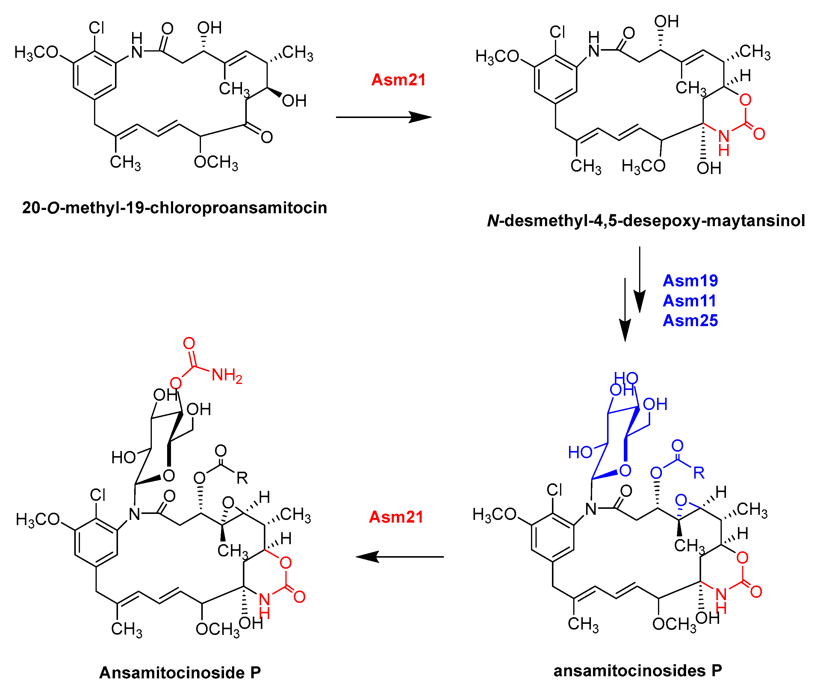
  167. Moss, S.J.; Bai, L.Q.; Toelzer, S.; Carroll, B.J.; Mahmud, T.; Yu, T.W.; Floss, H.G. Identification of Asm19 as an acyltransferase attaching the biologically essential ester side chain of ansamitocins using N-desmethyl-4,5-desepoxymaytansinol, not maytansinol, as its substrate. J. Am. Chem. Soc. 2002, 124, 6544–6545. [Google Scholar] [CrossRef]
  168. Spiteller, P.; Bai, L.Q.; Shang, G.D.; Carroll, B.J.; Yu, T.W.; Floss, H.G. The post-polyketide synthase modification steps in the biosynthesis of the antitumor agent ansamitocin by Actinosynnema pretiosum. J. Am. Chem. Soc. 2003, 125, 14236–14237. [Google Scholar] [CrossRef]
  169. Zhao, P.J.; Bai, L.Q.; Ma, J.; Zeng, Y.; Li, L.; Zhang, Y.R.; Lu, C.H.; Dai, H.Q.; Wu, Z.X.; Li, Y.Y.; et al. Amide N-glycosylation by Asm25, an N-glycosyltransferase of ansamitocins. CHEM Biol. 2008, 15, 863–874. [Google Scholar] [CrossRef]
  170. Sun, P.; Zhao, Q.F.; Yu, F.T.; Zhang, H.; Wu, Z.H.; Wang, Y.Y.; Wang, Y.; Zhang, Q.L.; Liu, W. Spiroketal Formation and modification in avermectin biosynthesis involves a dual activity of AveC. J. Am. Chem. Soc. 2013, 135, 1540–1548. [Google Scholar] [CrossRef]
Figure 1. The main structures mentioned in this paper; their partial biosynthetic processes are catalyzed by MFEs.
Catalysts 13 00581 g001
Figure 2. The representative IPKS 6-MSAS is involved in the biosynthesis of 6-MSA. 6-MSAS repeatedly uses a module to complete the biosynthesis, and each domain is reused in this process. This is a typical way for IPKSs to synthesize products.
Catalysts 13 00581 g002
Figure 3. LovB, as an IPKS, is involved in the biosynthesis of lovastatin. LovB reuses its domain combinations to participate in different stages of lovastatin synthesis, and different domain combinations give lovastatin a complex structure and excellent activity. The methyltransferase domain (MT) is active only during the fourth cycle, producing the intermediate α-methyl 3-ACP, a reaction that follows chain elongation catalyzed by ketone synthase (KS) and precedes the function of the keto reductase (KR), dehydratase (DH), and alkenyl reductase (ER) domains.
Catalysts 13 00581 g003
Figure 4. DHB is synthesized under the action of EntC, EntB, EntA, and EntE, in which EntE (a bifunctional enzyme) repeatedly plays a role. EntF is an INRPS that binds L-serine and catalyzes its binding to DHB. This process occurs three times, during which the intermediate product binds to the TE domain, and the final product, enterobactin, is released under the catalysis of TE.
Catalysts 13 00581 g004
Figure 5. AuAs and AcAs are homologous BTSs containing PT and TPS active domains, and they can use the same substrates to synthesize five different terpenoids: aspergitriene A and aspergidiene A, B, C, and D. AuAs and CYP450 can use aspergidiene A, B, C, and D to produce the corresponding oxidation products aspergilol A, B, C, and D. In addition, AcAs were found to synthesize calidoustens, another compound with a new skeleton.
Catalysts 13 00581 g005
Figure 6. Four CYP450s, P450-1, P450-2, P450-3, and P450-4, are involved in post-modification in the process of GA biosynthesis. All are MFEs and play a variety of oxidative functions, resulting in a variety of products. Among them, P450-1 is the most typical, which can catalyze four oxidation reactions: 7β-hydroxylation, oxidation of C-6 to obtain a contractile ring B, 3β-hydroxylation, and C-7 oxidation.
Catalysts 13 00581 g006
Figure 7. OrdA functions as a multifunctional CYP450 in the modification of aflatoxin B1. OrdA catalyzes a two-step oxidation reaction in the post-modification of aflatoxin B1. Firstly, a hydroxyl group is added to the A-ring of O-methyl-sterimatocystin, and the unstable seven-membered lactone is then generated under OrdA catalysis. Subsequent deacidification and rearrangement are also thought to involve CYP450, a putative formation step shown in the figure.
Catalysts 13 00581 g007
Figure 8. Asm21, as an MFE, participates in post-modification during ansamitocin biosynthesis. Asm21 catalyzes the carbamoylation of polyketoskeleton and the glycosylates part of a special ansamitocin (4″-O-aminoformylansamycin), indicating that it has double carbamoylated activity on the polyketoskeleton and glycosylated part, which sufficiently demonstrates that Asm21 is a multifunctional post-modifying enzyme.
Catalysts 13 00581 g008
Disclaimer/Publisher’s Note: The statements, opinions and data contained in all publications are solely those of the individual author(s) and contributor(s) and not of MDPI and/or the editor(s). MDPI and/or the editor(s) disclaim responsibility for any injury to people or property resulting from any ideas, methods, instructions or products referred to in the content.

