Review

Diclofenac Biodegradation by Microorganisms and with Immobilised Systems—A Review

Institute of Biology, Biotechnology and Environmental Protection, Faculty of Natural Science, University of Silesia in Katowice, Jagiellońska 28, 40-032 Katowice, Poland
*
Author to whom correspondence should be addressed.
Catalysts 2023, 13(2), 412; https://doi.org/10.3390/catal13020412
Submission received: 22 January 2023 / Revised: 11 February 2023 / Accepted: 13 February 2023 / Published: 15 February 2023
(This article belongs to the Special Issue Microbial Biocatalysis)

Abstract

:
Diclofenac is one of the most popular non-steroidal anti-inflammatory drugs. Due to its over-the-counter availability and high consumption along with municipal and hospital wastewater, it enters the sewage treatment plant, where it is not completely degraded. This results in the appearance of diclofenac in the effluents from the treatment plant, and with them, it enters the surface waters. Due to its structure, it is characterised by its high resistance to degradation in the environment. At the same time, it shows documented acute and chronic toxicity to non-target organisms. For this reason, it is necessary to look for cheap solutions that enhance the degradation of diclofenac. The paper discusses both the pathways of microbiological degradation of this drug described so far, as well as modern systems of biocatalyst immobilisation, with a particular emphasis on laccases involved in the biotransformation of diclofenac.

1. Introduction

Pain is a significant public health problem worldwide, with chronic pain affecting approximately 27% of the adult population in Europe and over 100 million adults in the United States. Non-steroidal anti-inflammatory drugs (NSAIDs) were discovered over 100 years ago, and the mechanism of action is based on the inhibition of cyclooxygenase (COX) isoenzymes. To this day, they remain a crucial element in the pharmacological treatment of inflammation and acute and chronic pain. They are essential for treating acute pain in the perioperative period and the cornerstone of treating osteoarthritis and other chronic pain conditions [1]. Due to their low addictive potential, good efficacy and long history of clinical use are often preferred by physicians [2]. In recent years, there has been a steady increase in the production and consumption of these drugs. Currently, over 50 types of NSAIDs are available on the world market, and diclofenac is one of this group’s best-known and popular drugs. It is difficult to calculate the exact global intake of NSAIDs as they are sold under different trade names and are often available over the counter. However, it has been estimated that worldwide consumption of diclofenac is 940 tons per year in capsules, suppositories, tablets, intravenous solutions and ointments, not including veterinary consumption [3].
Due to the constantly growing problem related to detecting active pharmaceutical compounds (PhACs) in groundwater, surface water and drinking water, modern and environmentally friendly methods of wastewater bioremediation with greater efficiency and effectiveness are sought. The threat is exacerbated by the natural processes of water circulation in nature, hydrological connections between ecosystems and the accumulation of various pollutants introduced into the environment for decades. Recently, the microbial degradation of PhACs has been the subject of many studies due to the possibility of complete or partial degradation of harmful compounds or their transformation into less toxic compounds. Due to the unfavourable environmental conditions in which the biodegradation process is carried out, its efficiency drops significantly. Therefore, appropriate immobilisation methods are sought, allowing the immobilisation of biocatalysts. It is conducive to increasing the degradability of pollutants, extending life, and increasing the catalytic activity of biocatalysts. Moreover, it increases the chances of survival and adaptation of microbial cells to the changing environment, including the concentration of toxic compounds [4,5].
The study aims to assess the toxicity of diclofenac to non-target organisms and to analyse the possibility of using immobilised preparations in the biodegradation processes of diclofenac, particularly those based on immobilised laccase and microorganisms with an increased potential for decomposition of this drug.

2. Diclofenac—Characteristics and Distribution in the Environment

Diclofenac [2- (2,6-dichloroanilino)phenylacetic acid] is one of the most widely used non-steroidal anti-inflammatory drugs (NSAIDs), acting as an inhibitor of cyclooxygenase responsible for prostanoid synthesis. After oral administration, diclofenac is rapidly and completely absorbed in the intestines and is detoxified by hydroxylation and glucuronidation. CYP2C9 and CYP3A4 (cytochrome P450 family of proteins) catalyse its oxidation to 4′- and 5’-hydroxylated derivatives, and UDP-glucuronosyltransferase-2B7 (UGT2B70) catalyses glucuronidation. The kidneys excrete 65% of the oxidised metabolites. The rest, as acyl glucuronide, is excreted in the bile. Diclofenac acyl glucuronides are chemically unstable compounds that can be epimerised by acyl migration to 2-, 3- or 4-O-glucuronide, especially in the alkaline environment of bile [6]. Part of diclofenac is not metabolised after ingestion, and the sewage system discharges it to the sewage treatment plant in an unchanged or slightly changed form. It is estimated that the maximum concentrations of this drug in wastewater range from 0.01 to 510 µg/L of diclofenac. Even though the efficiency of removing diclofenac by advanced oxidation processes is as high as 80%, the limitations of physic-chemical methods often preclude their use [7,8].
Alternatives to chemical methods using aggressive chemicals are ecologically safe biological methods. However, diclofenac, a hydrophobic chlorinated derivative with electron-withdrawing and donor groups having log D < 3.2 at pH 8.0, is not susceptible to biological degradation [9]. The treatment efficiency in biological treatment plants is 0–80%, but most often, it is 21–40% [8,10]. Since sewage treatment plants are characterised by a low degradation efficiency of this drug, diclofenac and its derivatives enter the waters [7,8]. Diclofenac appears in soil, surface waters, groundwater and even in drinking water in various parts of the world (Table 1) [11,12,13,14].
The average world concentration in rivers is estimated at 0.021+/−0.722 µg/L, and its concentration in fresh water in extreme cases was recorded even in the range of μg/L [28,29]. Moreover, the presence of diclofenac was observed both in sewage sludge, at concentrations up to 87 ng/g, and in soils, where the observed concentrations reach 5.6 ng/g soil, depending on changing climatic conditions [11]. Such a situation forces the development of new, efficient and, at the same time, cost-effective methods, including those based on immobilised organisms, of removing this compound from water.

3. Toxicity of Diclofenac to Non-Target Organisms

The most tragic effect of diclofenac’s influence on non-target organisms was the almost complete extinction of three species of vultures in the Indian subcontinent [28,30,31]. In the 1990s, in India and Pakistan, this drug was widely used to reduce inflammation caused by trauma and infectious diseases in cattle and buffaloes. When vultures ate the corpse of diclofenac-treated animals, the drug accumulated in the bird’s bodies, causing kidney failure and death [7]. The cause of nephrotoxicity in vultures was the accumulation of uric acid crystals in the visceral organs (visceral fundus). A detailed study found that uric acid and alanine aminotransferase (ALT) levels were significantly elevated, and renal architecture was disturbed [30].
However, effects such as those described above are infrequent due to the low drug concentration observed in the environment. Nowadays, research is underway to analyse the risk of adverse effects on organisms inhabiting the aquatic environment and exposed long-term to low concentrations of diclofenac [32] (Table 2).
Diclofenac physicochemical properties, mainly the n-octanol/water partition coefficient (log Kow, 4.51), are responsible for bioaccumulation in living organisms, primarily aquatic microorganisms [9]. The toxic impact of diclofenac, even at low concentrations of μg/L, has been demonstrated in studies on such species as common carp (Cyprinus carpio), brown trout (Salmo trutta fario), rainbow trout (Oncorhynchus mykiss) and stickleback (Gasterosteus aculeatus) [28]. Exposure of brown trout embryos to diclofenac did not show any evidence of embryotoxicity of this drug up to concentrations of 100 µg/L. NOEC is administered at a level of 500 µg/L for mortality, hatching, development and teratogenicity. Similar results were obtained for the embryonic and larval stages of Danio rerio, Oncorhynchus mykiss and Cyprinus carpio, where significant effects were found only at concentrations higher than 1 mg/L. Juvenile brown trout responded much more sensitively to diclofenac exposure than the larvae. The increase in mortality is alarming, occurring at diclofenac concentrations in the low µg/L range. There was a concentration-dependent increase in mortality in sticklebacks exposed to diclofenac, reaching a significance at 320 µg/L. A similar effect was observed by Näslund et al. [35] for the tricuspid stickleback at 271 μg/L of diclofenac. Acute toxicity tests of diclofenac in adult fish showed that the EC50 for carp was 71 mg/L [36]. Exposure to increasing concentrations of diclofenac from 2 to 32 mg/L also changed the growth curves of the populations of rotifers Plationus patulous and Moina macrocopa, leading to a decrease in the density of organisms with increasing drug concentration and a reduction in the daily population growth [34]. Mortality (LC50 480 ± 50 µg/L) and teratogenicity (EC50 90 ± 20 µg/L) have been demonstrated in Danio rerio after 96–h exposure to diclofenac. Chronic toxicity bioassays on the viability of Danio rerio embryos exposed for ten days to diclofenac allowed for the determination of NOEC and LOEC values at the levels of 4000 and 8000 µg/L, respectively [33]. The chronic toxicity of diclofenac has also been studied at the molecular and biochemical levels in Daphnia magna. Mortality of individuals increased after 24 h of exposure to high concentrations of diclofenac (486 mg/L). Exposure to 2 mg/L of diclofenac resulted in 50% mortality of D. magna after 21-day exposure and a significant reduction in egg production at a concentration of 0.50 mg/L. 96–h exposure to 50 µg/L diclofenac induced substantial changes in the expression of some genes related to detoxification, growth, development and reproduction. Their expression was inhibited after 24 h, and overexpression was observed after 48 h of exposure [34].
Joachim et al. [29] conducted studies on the harmfulness of diclofenac concerning primary producers and consumers in a long-term freshwater mesocosm experiment. The effective concentrations were 0.041, 0.44 and 3.82 µg/L, and the experimental time was six months. In such a constructed experiment simulating natural conditions, it was shown that the toxicity of diclofenac towards non-target organisms was higher than previously observed in laboratory conditions. During the six-month exposure period, the bio-volume of macrophytes (Nasturtium officinale and Callitriche platycarpa) decreased significantly. In Dreissena polymorpha, high mortality, reduced immunity and high genotoxicity were observed at all examined concentrations. Moreover, the highest concentration used changed the structure of the Gasterosteus aculeatus population. After one month of exposure, the total fish stock and the percentage of juveniles decreased while the percentage of adults increased. It led to a general change in the F1 generation length and frequency distribution compared with the control [29].
In the context of the negative impact of diclofenac on non-target organisms, the toxicity of the intermediates of diclofenac biotransformation is very important. Fu et al. [28] studied this phenomenon in two key aquatic invertebrates: Gammarus pulex and Hyalella azteca. In both of these species, diclofenac was converted into several oxidation products and conjugates, including the taurine-diclofenac conjugate and the diclofenac methyl ester. A significant increase in the bioconcentration factor for these intermediates relative to the parent drug has been demonstrated. Moreover, diclofenac methyl ester was also characterised by higher acute toxicity than diclofenac for both species, which correlated well with the increased potential for bioconcentration. The LC50 of diclofenac for H. azteca was 216 mg/L, while the LC50 diclofenac methyl ester was only 0.53 mg/L, which is a 430-fold increase in acute toxicity compared to diclofenac. The diclofenac-taurine conjugate was less toxic to H. azteca than its parent compound, which may be due to its slightly lower hydrophobicity [28]. In addition, it was observed that the two most frequently detected hydroxylated derivatives of diclofenac: 4′-OH-diclofenac and 5-OH-diclofenac can be further oxidised to reactive benzoquinone imines that interact with the protein nucleophilic groups, resulting in the formation of adducts [9]. These studies clearly show that when researching the toxicity of drugs toward non-target organisms, it is also crucial to look at the toxicity of their biotransformation products more broadly [28].
Histological evaluation helps to better understand the mechanism of diclofenac toxicity. In fish exposed to this drug, severe tissue reactions and lesions, especially in the liver, were more frequently observed. Näslund et al. [35] showed renal hematopoietic hyperplasia, and jaw lesions of the tricuspid stickleback at the lowest concentration tested at 4.6 μg/L. Importantly, it is an identified concentration in the environment. Ultrastructural studies of rainbow trout liver revealed glycogen level reduction and macrophage infiltration [36]. Moreover, it has been shown that even low concentrations of diclofenac found in the environment (1 µg/L) lead to cellular changes in the liver, kidneys and gills of rainbow trout, which reduces the functionality of the kidneys and gills [41]. In another in vitro experiment, the toxicity of diclofenac was tested in a range of concentrations of 0.001, 0.01, 0.1, 1 and 10 mg/L on three different cell types of zebra mussels: haemocytes, gill cells and digestive gland cells. After 96 h of exposure, a significantly reduced viability of diclofenac-treated gill cells was observed already in the presence of the lowest concentration applied. Moreover, the viability of diclofenac-treated digestive cells was significantly reduced after 48 h of exposure to 0.01 mg/L, while the haematocyte viability was decreased at a concentration of 0.001 mg/L [34].
The apparent effect of diclofenac on inter-individual relationships in exposed fish is surprising. The percentage of individuals showing signs of aggressive behaviour increased significantly with increasing diclofenac concentration, with a LOEC of 10 µg/L. Behavioural changes of African catfish (Rhamdia quelen) at 25 mg/L of diclofenac were also observed. These included respiratory failure, loss of balance, and irregular swimming, but no signs of aggression were seen. It may be the effect of reduced ability or propensity to act in defence, resulting from the weakened condition of the animals. For example, stickleback was leaving food behind and having a higher percentage of skin ulceration after exposure to diclofenac. Open wounds can cause infections with pathogens, making animals vulnerable to potentially lethal consequences. On the other hand, diclofenac-induced mortality may increase aggressive behaviour through changes in fish density. Another possible explanation for the increased aggressiveness observed in juvenile fish relates to the finding that diclofenac leads to corneal perforation in rainbow trout. The behavioural abnormalities of juvenile brown trout can be attributed to panic reactions due to visual impairment [36,37].
Studies on carp showed a significant increase in hydroperoxide and lipid peroxidation content at a diclofenac concentration of 7.1 mg/L [36]. Reduced lipid peroxidation (LPO) levels were observed in zebrafish embryos after exposure to 30 ng/L diclofenac for 90 min. At the environmental concentration (ng/L), diclofenac showed toxicity to Perna perna mussels, leading to decreased lysosomal membrane stability and increased cyclooxygenase activity, higher levels of oxidative stress and DNA damage. An increased level of lipid peroxidation was also observed in zebra mussels treated with diclofenac at a concentration of 1 μg/L. However, oxidative stress was induced in the mussel Mytilus galloprovincialis even after exposition to 0.25 μg/L of diclofenac [32]. Moreover, in zebrafish, in response to oxidative stress induced by diclofenac and its photolysis products, elevated levels of enzymes such as catalase, superoxide dismutase, and glutathione transferase and lipid peroxidation were observed [44]. The presence of diclofenac results in oxidative stress also in the cultures of microorganisms. Among other things, increased activity has been shown in superoxide dismutase and catalase and the formation of lipid peroxidation products. Changes accompanied this phenomenon on the cell surface and within the biological membrane. Multivariate analysis showed that exposure of the Pseudomonas moorei KB4 strain to diclofenac caused a decrease in the zeta potential with a simultaneous increase in the hydrophobicity of the cell wall. In addition, significant stiffening of the membrane was observed as a result of changes in the fatty acid composition of the membrane (including the appearance of a branched fatty acid-19:0 anteiso and cyclopropane-17:0 cyclo) [9,45]. Not only the parent form of diclofenac but also its intermediates can cause oxidative stress in cells of non-target organisms. Aissaoui et al. [46] assessed the toxicity of diclofenac intermediates obtained during the degradation of this drug by Enterobacter cloacae isolated from compost toward mouse liver cells. It was shown that diclofenac at therapeutic concentrations and its metabolites affected oxidative stress parameters, including a decrease in glutathione reserve, lipid peroxidation and disorders of the liver detoxifying enzymes, including superoxide dismutase, catalase, and glutathione S-transferase. However, these researchers emphasise the lack of a negative influence of diclofenac and its metabolites on the oxidative stress parameters in mice cells after applying environmental concentrations of this drug [46].
Lymnaea stagnalis snails were used to assess the immunotoxicity of diclofenac. They were exposed to diclofenac for three days at environmental concentrations (1–10 µg/L) and therapeutic concentrations (100–1000 µg/L). Diclofenac significantly influenced the immune capacity and the performance of the cochlear haemocytes. This effect is typical of the inflammatory response, confirmed by an increase in NADPH oxidase activity, mainly after using the drug at a concentration of 1000 µg/L [34]. The expression of hepatic c7 genes was also shown to be dependent on the concentration of diclofenac. The c7 protein is a complement of the system part of the innate immune system. It forms a membrane attack complex with other complement component proteins that lead to the lysis of foreign cells. Complement components are linked via the arachidonic acid pathway, which explains the effect of NSAID exposure on c7 [35]. In turn, the cytogenotoxicity of diclofenac was tested in vitro through the 1-h exposition of haemocytes collected from Dreissena polymorpha to 60, 126 and 250 µg/L of the drug. A significant cytotoxic effect in the destabilisation of lysosomal membranes was noted only after exposure to 250 µg/L of diclofenac, while both primary genetic changes (e.g., DNA fragmentation) and permanent DNA damage occurred after exposure to all tested concentrations [34]. Increased DNA fragmentation was also observed in the mussel Mytilus galloprovincialis after exposure to the environmental concentration (2.5 μg/L) [32]. On the other hand, Oryzias latipes showed induction of p53 gene expression after 4-day exposure to 1 μg/L of diclofenac. The p53 gene is an important biomarker in analysing environmental toxin carcinogenicity, and DNA damage, as its product plays a crucial role in cell cycle arrest, apoptosis and DNA repair [41]. The studies on the toxicity of diclofenac do not give conclusive results, and some reports show that in environmental concentrations, diclofenac does not threaten aquatic animals. Memmert et al. [47] indicated very low bioconcentration of diclofenac in fish, corresponding to the chemical properties of diclofenac, including the dissociation constant of pKa 3.99–4.16. Moreover, these authors indicate that although the NOEC is estimated at 10 µg/L, only a concentration above 320 µg/L causes a reduction in growth in zebrafish [46]. However, considering the number of reports of adverse effects of diclofenac on non-target organisms, the conclusions of Näslund et al. [35] seem to be justified. These authors postulate the reduction in the environmental risk associated with diclofenac toxicity through the substitution of diclofenac, where possible from a therapeutic point of view, with naproxen-a drug with a similar effect in the treatment of pain. Although naproxen and diclofenac have similar effects in fish, environmental hazards and risks are lower with naproxen than diclofenac because the toxic effects appear at higher naproxen concentrations than diclofenac [35].
Due to the increasing concentration of NSAIDs in soils, plants are also exposed to the toxic effects of diclofenac. Studies of the stress response to diclofenac of two crops, maize and tomato, showed that the sensitivity to diclofenac is species-dependent. The tomato was more sensitive, with growth inhibition, a decrease in the content of photosynthetic pigments and a decrease in the maximum PSII quantum efficiency and PSII activity. However, no effect of diclofenac in maize was observed in the content of photosynthetic pigments or growth. However, an effect of diclofenac on the quantum efficiency of the PSII photosystem was observed. In both plants, oxidative stress was also observed, manifested by an increased concentration of hydrogen peroxide. In response, the plants triggered a defence mechanism in the form of the synthesis of phenolic compounds [48]. Similarly, Copolovici et al. [49] showed in beans the effect of diclofenac on the reduction in assimilation coefficients and stomatal conductivity for water vapor. In addition, an increase in the concentration of monoterpenes was also found: a-pinene, camphene and 3-carene). The authors postulate that diclofenac may also interfere with the methylerythritol phosphate pathway in plastids [49].

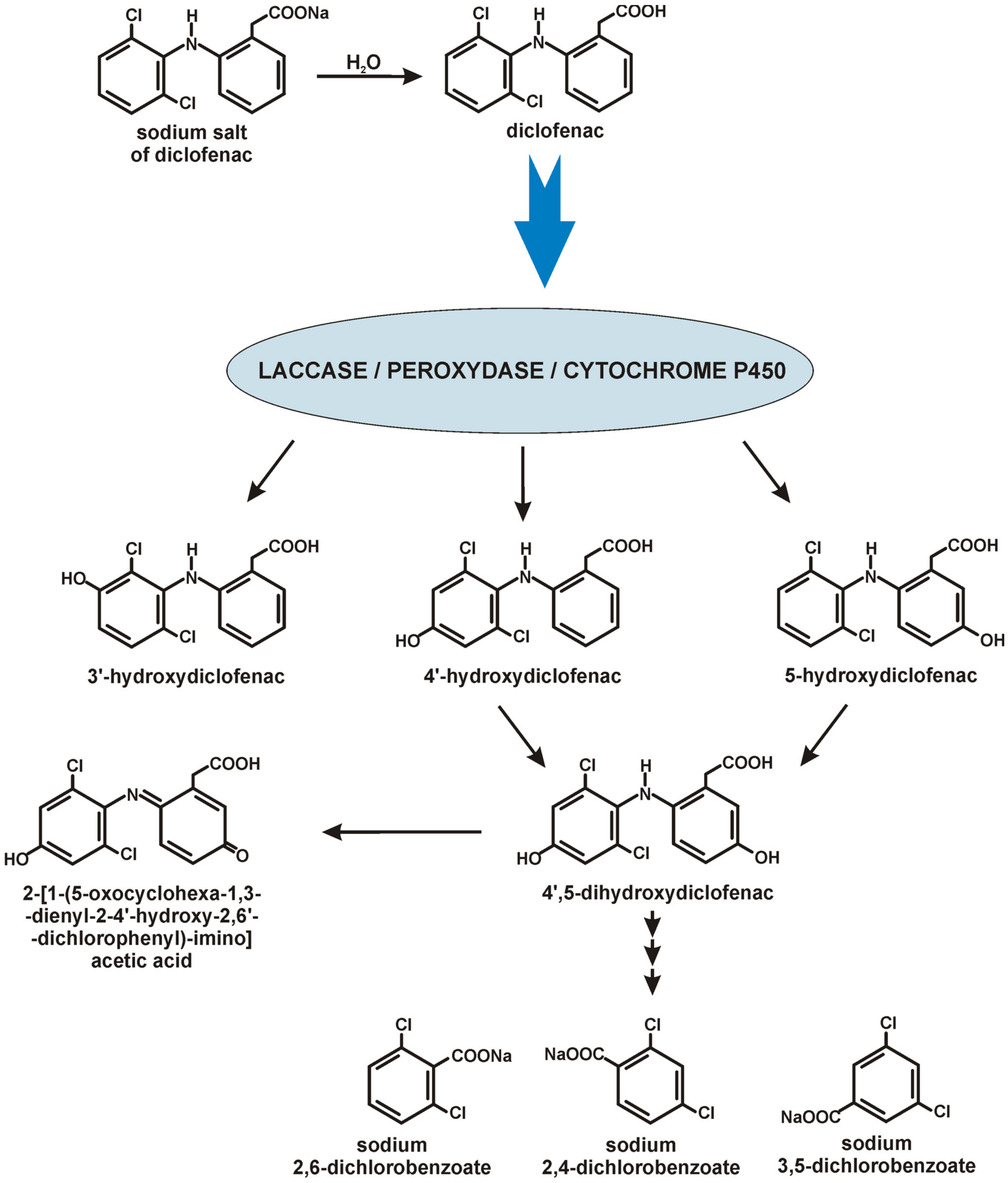
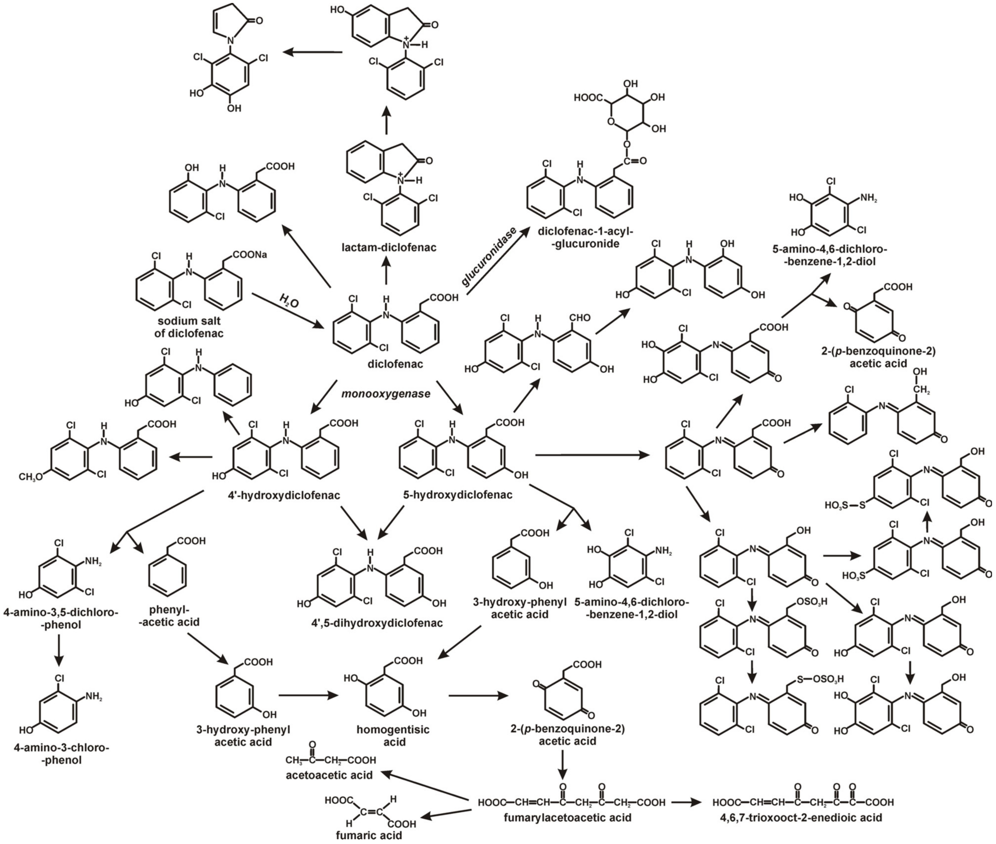
4. Diclofenac Biodegradation by Bacteria and Fungi

Few fungi and bacteria capable of degrading diclofenac have been described so far. Moreover, only a partial decomposition of this drug is most often described in the literature due to the emergence of difficult-to-degradable intermediates. The fungi that can degrade diclofenac include Trametes trogii, T. polyzona, Yarrowia lipolytica, Aspergillus niger, Phanerochaete chrysosporium, Mucor circinelloides, Trichoderma longibrachiatum, Rhizopus microspore [50,51]. Among the bacteria that degrade diclofenac, strains of Raoultella sp. DD4, Bacillus subtilis, Brevibacillus laterosporus, Rhodococcus ruber, Labrys portucalensis F11, Alcaligenes faecalis, Staphylococcus aureus, Staphylococcus haemolyticus, Pseudomonas moorei KB4, Klebsiella sp. KSC and Proteus mirabilis have been described [9,10,45,52,53,54,55,56].
The most frequently observed transformation of diclofenac by fungi is hydroxylation by laccase, manganese peroxidase and the cytochrome P450 enzyme system to intermediates such as 4-hydroxydiclofenac, 5-hydroxydiclofenac, 3-hydroxydiclofenac and 4,5-dihydroxydiclofenac [50,51] (Figure 1).
Moreover, ligninolytic fungi: T. polyzona, M. circinelloides, and T. longibrachiatum degraded diclofenac into the intermediates: adduct of 2,6-dicholorobenzoic, 2,4-dichlorobenzoic and 3,5-dichlorobenzoic acid, which proved the breaking of the C-N bond in the drug structure. After ten days of incubation, these intermediates disappeared from the culture, indicating their further degradation with ring cleavage. A positive correlation was observed between the activity of manganese peroxidase and the drug tolerance of the fungi. In contrast, no such correlation was observed concerning the drug degradation efficiency, which shows that these strains’ mechanism of diclofenac degradation is more complex [51].
Metabolites of diclofenac, commonly observed in fungi, were also shown during bacterial degradation. Among others, strains of Bacillus and Brevibacillus decomposed diclofenac into 4’-hydroxydiclofenac [53].
The first described bacterial strains capable of diclofenac degradation were Raoultella sp. DD4 and Rhodococcus ruber IEGM 346 [52,54]. Raoultella sp. DD4 degraded 0.6 mg/L of diclofenac within 28 days and was highly resistant to the toxic effects of this drug [52]. Rhodococcus ruber strain IEGM 346 can degrade high diclofenac concentrations (50 mg/L). It was confirmed that the C-N bond is broken during degradation, and the aromatic ring opens in the structure of diclofenac. 16 intermediates of the decomposition of this drug by the IEGM 346 strain have been identified. The described pathway leads through a series of oxidation reactions to homogentisic acid. The further oxidation of this acid through a quinone derivative leads to the end products: acetoacetic acid, fumaric acid and 4,6,7-trioxooct-2-enedioic acid (Figure 2).
The adaptation mechanisms of Rhodococcus ruber IEGM 346 to high concentrations of this drug are altered ζ potential of bacterial cells, increased cell hydrophobicity and total cell lipid content, formation of multicellular conglomerates, and altered surface-to-volume ratio [54]. In addition, Moreira et al. [10] showed that the degradation of diclofenac by the Labrys portucalensis F11 strain took place through hydroxylation and the formation of benzoquinone imine as a key metabolite. The resulting product was further decarboxylated and hydroxylated. The stoichiometric release of chlorine and the lack of detected metabolites at the end of the experiments indicated complete degradation of the drug by the F11 strain. It was also the first time that a sulphation reaction was described during bacterial diclofenac decomposition, indicating the similarity of metabolites during bacterial diclofenac degradation to the conjugates which appeared during the Phase II detoxification diclofenac in mammals [8]. For another example, the similarities between the detoxification pathways of drugs in mammals and bacterial metabolism have also been shown by Murshid et al. [55]. They described Staphylococcus sp. and Alcaligenes sp. with the glucuronidase activity responsible for the conjugation of diclofenac with glucuronic acid to diclofenac 1-acyl-glucuronide [55].
Żur et al. [9,45] described the Pseudomonas moorei KB4 strain as capable of degrading 0.5 mg/L of diclofenac in a mono-substrate culture. In contrast, in a culture supplemented with glucose and sodium acetate, this strain degraded 1 mg/L of diclofenac within 12 days. 4′-OH-diclofenac and diclofenac-lactam were identified as intermediates. Gene expression analysis revealed up-regulation of selected genes encoding biotransformation enzymes in the presence of diclofenac such as monooxygenase, dihydroxylating and aromatic ring cleaving dioxygenases, and cytochrome p450 system [9,45]. In turn, the bacterial strain Klebsiella sp. KSC, isolated from the livestock soil, has been described as a strain capable of biodegradation of diclofenac high concentrations. Klebsiella sp. KSC exposition to 70 mg/L of diclofenac caused the mineralisation of diclofenac after 72 h. In this case, 12 biotransformation products of diclofenac have been identified, indicating that hydroxylation, dehydroxylation, decarboxylation and dechlorination are critical steps in the degradation of this compound. As a result of these mechanisms, alcohols and ketones compounds are formed. Both mono-, di-, tri- and tetrahydroxylated derivatives were observed. The generation of such products resulted from removing the carboxyl group and two hydrogens from diclofenac with the simultaneous addition of hydroxyl groups to the parent compound. In addition, hydroxylation products formed after the cleavage of the acetate group from the structure of the parent substance were observed. In addition, the cyclisation product between the carboxyl group and the nitrogen atom was also identified [56].

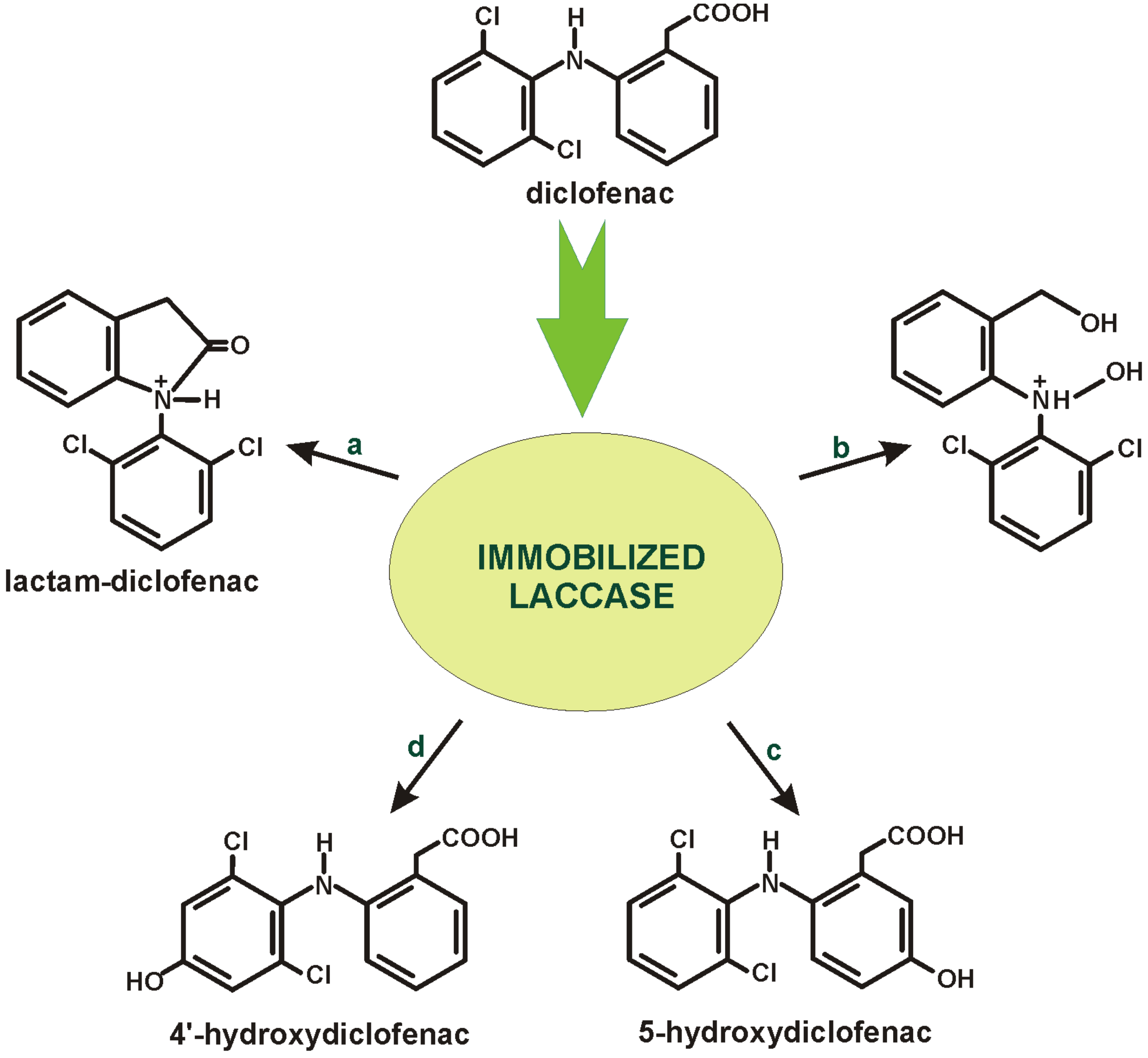
5. Diclofenac Biodegradation in Immobilised Systems

Conventional diclofenac wastewater treatment methods, such as physical and chemical procedures, have severe limitations, such as poor treatment, low efficiency, high cost, generation of hazardous by-products and application to a narrow range of concentrations of organic compounds in the wastewater. Therefore, the challenge for environmental engineers and biotechnologists is to develop an efficient, economical and environmentally safe bioremediation technique to provide outstanding remediation solutions instead of current treatment technologies [57]. Due to the confirmed toxicity of diclofenac towards microorganisms, including microorganisms capable of biodegradation, more and more attention is paid to immobilised biopreparations usage (Table 3).
Currently, the most commonly used methods in immobilising live microorganisms are the so-called self-immobilisation in pellets and adhesion to a fixed or porous surface. The first method is based on the natural tendency of some species of microorganisms in submerged cultures for pellet development. The second method is based on the adhesion of cells to the support material with a secreted exopolysaccharide acting as an adhesive material. Immobilisation in the support material can also occur by encapsulating in pores or through physical or chemical traps in porous solids or matrices. Recently, the use of mushroom pellets has attracted attention due to the appropriate ability to self-immobilise and the possibility of connecting another microorganism to such an aggregate or material, resulting in the formation of self-immobilised biomixes [65].
Both whole cells of microorganisms and selected oxidising enzymes have been used in biodegradation and biotransformation processes [45,52,59]. An example of the use of whole immobilised microorganisms in the degradation process of diclofenac was the designed bioreactor based on a biofilter with immobilised activated sludge microorganisms. After a two-month adaptation period, the removal efficiency of diclofenac in the designed system reached 97.63%. Based on Illumina sequencing, the major bacterial taxa in the biofilter were identified, which included Granulicella pectinivorans, Rhodanobacter terrae, Castellaniella denitrificans, Parvibaculum lavamentivorans, Bordetella petrii, Bryocella elongata and Rhodopseudomonas palustris. Wickerhamiella was the dominant fungal taxa in the immobilised cell biofilter, indicating its leading share in diclofenac degradation in activated sludge systems. Such enormous biodiversity of microorganisms allowed for the efficient operation of the reactor. However, a decrease in pH was observed during its process, which did not affect the degradation efficiency. Moreover, this arrangement proved successful during the implementation of a wide range of carbon sources [8]. Pereira et al. [65] indicate the potential of using the so-called biomixes to enhance microorganisms’ ability to degrade organochlorine derivatives, including diclofenac. In the construction of biopreparation, the ability of fungi and bacteria to autoaggregate is used. These authors, however, point out that this requires research to understand interspecies interactions between fungal granules and bacteria, especially as co-immobilisation affects pellet fixation and the possibility of their use in continuous operation. Such research can contribute to developing cost-effective and efficient biodegradation techniques for diclofenac and other substances resistant to degradation [65].
The literature data show that not only microorganisms but also enzymes isolated from them can be used to degrade diclofenac. However, there are few reports on the involvement of enzymes in the degradation/biotransformation of diclofenac. The process of enzymatic degradation of diclofenac was most fully described by Żur et al. [9], indicating the involvement of enzymes such as hydroxylating mono- and dioxygenases, aromatic ring-cleaving enzymes (catechol 1,2-dioxygenase, homogentisate 1,2-dioxygenase and salicylate 1,2-dioxygenase) in the decomposition of the drug structure. In addition, it was shown that deaminase had a significant impact on the decomposition of the dicyclic structure of diclofenac. However, Żur et al. [9] have not proposed any purification system based on immobilized enzymes based on the enzymes shown. Other enzymes involved in the degradation of diclofenac include the cytochrome P-450 system and laccase responsible for hydroxylation, as well as the enzymes responsible for the transformation to glucuronide derivatives [5,55,66,67] However, so far, only laccases have found broader applications in systems based on immobilized enzymes used to transform diclofenac due to its low specificity and high oxidising capacity towards diclofenac (Figure 3). Both simple and popular carriers, such as alginate and new-generation synthetic carriers, were used for this enzyme [58,59,62,64]. An example of laccase usage in the biodegradation of diclofenac is the enzyme derived from Sphingobacterium ksn-11 immobilised in sodium alginate-silicon dioxide-polyvinyl alcohol beads. It was shown that the immobilised laccase oxidised diclofenac to 4-OH of diclofenac after 4 h of incubation, and the preparation itself was highly effective in subsequent cycles. Moreover, the application of an electron mediator (2,2-azino-bis-(3-ethylbenzthiozoline-6-sulfonic aid) shortened the transformation time to 90 min [58].
Zdarta et al. [59] immobilised laccase from Trametes versicolor on electrospun nanofibers poly(L-lactic acid)-co-poly(ε-caprolactone) (PLCL) by adsorption, encapsulation and covalent bonding. Thin structures characterise materials produced by the electrospinning method with a diameter ranging from 100 nm to 1 μm and a length of up to several thousand meters. Due to features such as porosity, biocompatibility and the high number of functional groups on nanofibers’ surfaces, these materials are favourable in immobilising enzymes. As a result, over 90% biodegradation of 1 mg/L diclofenac was achieved under optimal conditions. The immobilised enzyme was also active in the following cycles, maintaining 40% efficiency after the fifth cycle. In addition, a thorough toxicity analysis of the biodegradation products was carried out. It was also shown that the solution obtained after this process was about 65% less toxic than the initial diclofenac solution [59].
In turn, the complete biodegradation of diclofenac (500 μg/L) within five hours was observed after applying laccase immobilised by adsorption on porcine manure biocarbon (BC-PM) at the initial concentration of 500 µM diclofenac. Among other biocarbon carriers, such as those obtained from pine wood (BC-PW) or almond shell (BC-AS), BC-PM showed the highest adsorption capacity of laccase. It was observed that after using biocarbon carriers, along with an increase in the initial concentration of laccase in the solution, the enzyme binding capacity and, consequently, the effectiveness of immobilisation also increased. The pre-treatment of the biocarbon carrier with citric acid increased its ability to bind the enzyme. It was also established that the primary mechanism of enzyme adsorption on biochar is homogeneous monolayer adsorption. Immobilised laccase showed higher storage stability. Three times longer life was observed than free laccase [57,60,61].
Another example of enzymatic decomposition of diclofenac is the use of laccase derived from Trametes hirsuta, immobilised by a covalent bond on PVDF/MWCNT membranes ((polyvinylidene chloride (PVDF) membrane modified with multi-wall carbon nanotubes (MWCNTs)). Polymer materials increase the resistance to contamination of the resulting nanocomposite membrane. Nanomaterials can be used in the membrane matrix or on its surface. They exhibit unique properties, including increasing their specific surface area and mechanical strength, physicochemical properties such as water permeability, resistance to contaminants, selectivity and thermal resistance. Carbon-based nanomaterials, including MWCNTs, are popular membrane reinforcement materials used in wastewater treatment. Polymer membranes are compatible with sewage treatment plant devices and do not interfere with their functioning. During the immobilisation of laccase on MWCNTs, there was a problem with nanoparticle separation at the purification stage. Mixing MWCNT with PVDF membranes made it possible to avoid this problem. The addition of MWCNTs to PVDF improves the physical properties of the membrane. It increases the rate of electron transfer between the laccase and the substrate, thus, increasing the effectiveness of laccases. Covalently immobilised laccase showed a high activity of 4.47 U/cm2 and an activity recovery of 38.31%. Using chemically immobilised laccase in the mini-membrane reactor by covalent bonding on PVDF/MWCNT membranes allowed for 95% of diclofenac degradation within 4 h [62].
As a carrier for the immobilisation of laccase by physical adsorption, granulated activated carbon (GAC) is also used. GAC characterises by a large specific surface, high adsorption capacity, porous structure and wide availability on the market. These features give GAC an excellent potential for enzyme immobilisation. It was shown that the adsorption process on GAC did not change the structure of the laccase, which allowed it to maintain its activity. GAC can efficiently adsorb micropollutants such as diclofenac. However, as with all adsorbents, GAC adsorption of the micropollutants decreases with exposure time due to surface saturation. Due to this issue, GAC regeneration is required to maintain system performance. Hence, a regeneration strategy was developed by pre-adsorbing laccase to the GAC. Immobilised laccase degrades the adsorbed micropollutants, thus releasing the adsorption sites. In addition, the co-adsorption of laccase and micropollutants on GAC improves biodegradation efficiency due to the increased electron transfer between laccase and micropollutants. The immobilisation of laccase on GAC does not influence micropollutants’ adsorption because, after the enzyme’s immobilisation, about 65% of the carbon surface is still available for the adsorbates. Moreover, sorption sites on the GAC surface are released after biodegradation, and the sorption-degradation cycle can start anew. As a result, the efficiency of diclofenac removal increases with subsequent cycles. Laccase prevents the complete saturation of GAC, which is a must for continuous operation. Higher laccase load, i.e., “full saturation”, allowed obtaining the highest diclofenac degradation result in all cycles. The use of GAC-bound laccase overcomes some problems associated with using free laccase for the catalytic degradation of micropollutants. The immobilisation of laccase improved its reusability and stability over a wide range of pH and temperature, and the enzyme removed micropollutants more effectively [63].
Recently, catalysts based on palladium (Pd) have aroused increasing interest. They can catalyse many reactions, such as denitrification and hydrodechlorination. Palladium catalysts are usually prepared by chemical methods and then immobilised on supports such as silica. This avoids their aggregation and facilitates recycling. The production of palladium nanoparticles using microbial reduction is a promising solution. This process allows for using fewer toxic chemicals and does not require stabilisers or carriers, which makes it environmentally friendly. The biomass-supported nanoparticles show more excellent resistance to aggregation than those supported by conventional supports. Several microorganisms, both pure strains and mixed bacterial cultures, can produce biogenic nanopalladium (Bio-Pd) in their cell membranes and cytoplasm. One example of Bio-Pd producers is the anaerobic granular sludge (AGS). AGS is a particular form of microbial aggregates consisting of mixed cultures of microorganisms with a three-dimensional, heterogeneous structure. Biocatalizator Pd-AGS combines AGS’s microbial metabolic role with palladium’s catalytic function. As a heterogeneous catalyst consisting of Pd nanoparticles and microbial granules, it has the advantage of initiating Pd autocatalysis using hydrogen donors or electrons generated from organic compounds by microbial transformation and fermentation. It has been shown that many factors, such as hydrogen and electron donors, remediation medium, immobilisation vehicle, and pH, can influence the catalytic activity of Pd during diclofenac bioremediation. Hydrogen is the most effective electron donor for Pd-AGS, which is then more resistant to inactivation by chloride or sulphide than free Pd nanoparticles. Using the Pd-AGS system allowed the decomposition of 96% of diclofenac during four iterations of reduction, and the purification with water quickly regenerated its catalytic activity. Pd-AGS appears to be a viable and economical alternative to homogeneous Pd complexes or a conventional supported heterogeneous Pd catalyst [64].

6. Conclusions

The literature review clearly shows that diclofenac has a multidirectional effect on non-target organisms living in waters contaminated with this drug, causing several negative changes. At the same time, data analysis shows that there are microorganisms capable of degrading low concentrations of this drug. However, most of the described fungi used in the biodegradation processes of diclofenac, in fact, only transform to hydroxylated derivatives without disturbing the aromatic structure of this compound. Such products may be more toxic than the parent compound. In addition, using immobilised systems using laccase as enzymes involved in the oxidation of diclofenac does not solve this problem because the enzymes only carry out the initial hydroxylation. To solve the problem of environmental pollution with diclofenac, it is necessary to look for microorganisms capable of completely degrading diclofenac. It may be promising to use immobilised fungal and bacterial systems, which would make it possible to use the high potential of non-specific fungal enzymes for the activation of diclofenac. The activated substrate could then be degraded sequentially into primary metabolism intermediates with the participation of bacteria. However, research related to the analysis of fungal-bacterial interactions is necessary for such systems to be used as an application.

Author Contributions

Conceptualization, D.W., K.Ł. and U.G.; writing—original draft preparation, D.W. and K.Ł.; writing—review and editing, D.W. and U.G.; visualization, D.W. and U.G.; supervision, D.W. and U.G.; project administration, U.G.; funding acquisition, U.G. All authors have read and agreed to the published version of the manuscript.

Funding

This research was funded by the National Science Centre, Poland (grant number 2018/29/B/NZ9/00424).

Data Availability Statement

No new data were created or analysed in this study. Data sharing is not applicable to this article.

Conflicts of Interest

The authors declare no conflict of interest.

References

  1. Monteiro-Steagall, B.P.; Steagall, P.V.M.; Lascelles, B.D.X. Systematic review of non-steroidal anti-inflammatory drug-induced adverse effects in dogs. J. Vet. Intern. Med. 2013, 27, 1011–1019. [Google Scholar] [CrossRef]
  2. Brune, K.; Patrignani, P. New insights into the use of currently available non-steroidal anti-inflammatory drugs. J. Pain Res. 2015, 8, 105–118. [Google Scholar] [CrossRef] [PubMed] [Green Version]
  3. Guzik, U.; Wojcieszyńska, D. Biodegradation of non-steroidal anti-inflammatory drugs and their Influence on soil microorganisms. In Microbes and Enzymes in Soil Health and Bioremediation; Kumar, A., Sharma, S., Eds.; Springer: Singapore, 2019; pp. 379–401. Available online: https://link.springer.com/content/pdf/10.1007%2F978-981-13-9117-0_16.pdf (accessed on 10 February 2023).
  4. Du, Y.; Jia, X.; Zhong, L.; Jiao, Y.; Zhang, Z.; Wang, Z.; Feng, Y.; Bilal, M.; Cui, J.; Jia, S. Metal-organic frameworks with different dimensionalities: An ideal host platform for enzyme@MOF composites. Coord. Chem. Rev. 2022, 454, 214327. [Google Scholar] [CrossRef]
  5. Zhou, W.; Zhang, W.; Cai, Y. Laccase immobilization for water purification: A comprehensive review. Chem. Eng. J. 2021, 403, 126272. [Google Scholar] [CrossRef]
  6. Krasniqi, V.; Dimovski, A.; Domjanović, I.K.; Bilić, I.; Božina, N. How polymorphisms of the cytochrome P450 genes affect ibuprofen and diclofenac metabolism and toxicity. Arch. Ind. Hyg. Toxicol. 2016, 67, 1–8. [Google Scholar] [CrossRef] [Green Version]
  7. Chen, J.; Gao, H.; Zhang, Y.; Zhang, Y.; Zhou, X.; Li, C.; Gao, H. Developmental Toxicity of diclofenac and elucidation of gene regulation in zebrafish (Danio rerio). Sci. Rep. 2014, 4, 4841. [Google Scholar] [CrossRef] [Green Version]
  8. Navrozidou, E.; Remmas, N.; Melidis, P.; Karpouzas, D.G.; Tsiamis, G.; Ntougias, S. Biodegradation potential and diversity of diclofenac-degrading microbiota in an immobilised cell biofilter. Processes 2019, 7, 554. [Google Scholar] [CrossRef] [Green Version]
  9. Żur, J.; Piński, A.; Wojcieszyńska, D.; Smułek, W.; Guzik, U. Diclofenac degradation-enzymes, genetic background and cellular alterations triggered in diclofenac-metabolising strain Pseudomonas moorei KB4. Int. J. Mol. Sci. 2020, 21, 6786. [Google Scholar] [CrossRef]
  10. Moreira, I.S.; Bessa, V.S.; Murgolo, S.; Piccirillo, C.; Mascolo, G.; Castro, P.M.L. Biodegradation of diclofenac by the bacterial strain Labrys portucalensis F11. Ecotoxicol. Environ. Saf. 2018, 152, 104–113. [Google Scholar] [CrossRef]
  11. Biel-Maeso, M.; Corada-Fernandez, C.; Lara-Martin, P.A. Monitoring the occurrence of pharmaceuticals in soil irrigated with reclaimed wastewater. Environ. Pollut. 2018, 235, 312–321. [Google Scholar] [CrossRef]
  12. Ebele, A.J.; Abdallah, M.A.-E.; Harrad, S. Pharmaceuticals and personal care products (PPCPs) in the freshwater aquatic environment. Emerg. Contam. 2017, 3, 1–16. [Google Scholar] [CrossRef]
  13. Scheurell, M.; Franke, S.; Shah, R.M.; Hühnerfuss, H. Occurrence of diclofenac and its metabolites in surface water and effluent samples from Karachi, Pakistan. Chemosphere 2009, 77, 870–876. [Google Scholar] [CrossRef] [PubMed]
  14. Ali, A.M.; Sydnes, L.K.; Alarif, W.M.; Al-Lihaibi, S.S.; Aly, M.M.; Aanrud, S.G.; Kallenborn, R. Diclofenac and two of its photooxidation products in the marine environment: Their toxicology and occurence in Red Sea coastal waters. Environ. Chem. Ecotoxicol. 2019, 1, 19–25. [Google Scholar] [CrossRef]
  15. Čelić, M.; Gros, M.; Farre, M.; Barcelo, D.; Petrovic, M. Pharmaceuticals as chemical markers of wastewater contamination in the vulnerable area of the Ebro Delta (Spain). Sci. Total. Environ. 2019, 652, 952–963. [Google Scholar] [CrossRef] [PubMed]
  16. Baranowska, I.; Kowalski, B. Using HPLC method with DAD detection for the simultaneous determination of 15 drugs in srface water and wastewater. Polish J. Environ. Stud. 2011, 20, 21–28. [Google Scholar]
  17. Reis-Santos, P.; Paisa, M.; Duarte, B.; Caçador, I.; Freitas, A.; Vila Pouca, A.S.; Barbosa, J.; Leston, S.; Rosa, J.; Ramos, F.; et al. Screening of human and veterinary pharmaceuticals in estuarine waters: A baseline assessment for the Tejo estuary. Mar. Pollut. Bull. 2018, 135, 1079–1084. [Google Scholar] [CrossRef]
  18. Lolić, A.; Paěga, P.; Santos, L.H.M.L.M.; Ramos, S.; Correia, M.; Delerue-Matos, C. Assessment of non-steroidal anti-inflammatory and analgesic pharmaceuticals in seawaters of North of Portugal: Occurrence and environmental risk. Sci. Total. Environ. 2015, 508, 240–250. [Google Scholar] [CrossRef] [Green Version]
  19. Huebner, M.; Weber, E.; Niessner, R.; Boujday, S.; Knopp, D. Rapid analysis of diclofenac in freshwater and wastewater by a monoclonal antibody-based highly sensitive ELISA. Anal. Bioanal. Chem. 2015, 407, 8873–8882. [Google Scholar] [CrossRef]
  20. Yang, L.; He, J.T.; Su, S.H.; Cui, Y.F.; Huang, D.L.; Wang, G.C. Occurrence, distribution, and attenuation of pharmaceuticals and personal care products in the riverside groundwater of the Beiyun River of Beijing, China. Environ. Sci. Pollut. Res. 2017, 24, 15838–15851. [Google Scholar] [CrossRef]
  21. Cherik, D.; Benali, M.; Louhab, K. Occurrence, ecotoxicology, removal of diclofenac by adsorption on activated carbon and biodegradation and its effect on bacterial community: A review. World Sci. News 2015, 10, 116–144. [Google Scholar]
  22. Hanif, H.; Waseem, A.; Kali, S.; Qureshi, N.A.; Majid, M.; Iqbal, M.; Ur-Rehman, T.; Tahir, M.; Yousaf, S.; Iqbal, M.M.; et al. Environmental risk assessment of diclofenac residues in surface waters and wastewaters: A hidden global threat to aquatic ecosystem. Environ. Monit. Assess. 2020, 192, 204. [Google Scholar] [CrossRef] [PubMed]
  23. Shanmugam, G.; Sampath, S.; Selvaraj, K.K.; Larsson, D.G.J.; Ramaswamy, B.R. Non-steroidal ant-inflammatory drugs in Indian rivers. Environ. Sci. Pollut. Res. 2014, 21, 921–931. [Google Scholar] [CrossRef] [PubMed]
  24. Madikizela, L.M.; Chimuka, L. Occurrence of naproxen, ibuprofen, and diclofenac residues in wastewater and river water of KwaZulu-Natal Province in South Africa. Environ. Monit. Assess. 2017, 189, 348. [Google Scholar] [CrossRef]
  25. Di Lorenzo, T.; Cifoni, M.; Baratti, M.; Pieraccini, G.; Di Marzio, W.D.; Galassi, D.M.P. Four scenarios of environmental risk of diclofenac in European groundwater ecosystems. Environ. Pollut. 2021, 287, 117315. [Google Scholar] [CrossRef] [PubMed]
  26. Gonzales-Alonso, S.; Merino, L.M.; Esteban, S.; de Alda, M.L.; Barcelo, D.; Duran, J.J.; Lopez-Martinez, J.; Acena, J.; Perez, S.; Mastoianni, N.; et al. Occurrence of pharmaceutical, recreational and psychotropic drug residues in surface water on the northern Antarctic Penisula region. Environ. Pollut. 2017, 229, 241–254. [Google Scholar] [CrossRef]
  27. Lapworth, D.J.; Baran, N.; Stuart, M.E.; Ward, R.S. Emerging organic contaminants in grounwater: A review of sources, fate and occurrence. Environ. Pollut. 2012, 163, 287–303. [Google Scholar] [CrossRef] [PubMed] [Green Version]
  28. Fu, Q.; Fedrizzi, D.; Kosfeld, V.; Schlechtriem, C.; Ganz, V.; Derrer, S.; Rentsch, D.; Hollender, J. Biotransformation changes bioaccumulation and toxicity of diclofenac in aquatic organisms. Environ. Sci. Technol. 2020, 54, 4400–4408. [Google Scholar] [CrossRef]
  29. Joachim, S.; Beaudouin, R.; Daniele, G.; Geffard, A.; Bado-Nilles, A.; Tebby, C.; Palluel, O.; Dedourge-Geffard, O.; Fieu, M.; Bonnard, M.; et al. Effects of diclofenac on sentinel species and aquatic communities in semi-natural conditions. Ecotoxicol. Environ. Saf. 2021, 211, 111812. [Google Scholar] [CrossRef]
  30. Swan, G.E.; Cuthbert, R.; Quevedo, M.; Green, R.E.; Pain, D.J.; Bartels, P.; Cunningham, A.A.; Duncan, N.; Meharg, A.A.; Oaks, J.L.; et al. Toxicity of diclofenac to Gyps vultures. Biol. Lett. 2006, 2, 279–282. [Google Scholar] [CrossRef] [Green Version]
  31. Naidoo, V.; Wolter, K.; Cuthbert, R.; Duncan, N. Veterinary diclofenac threatens Africa’s endangered vulture species. Regul. Toxicol. Pharmacol. 2009, 53, 205–208. [Google Scholar] [CrossRef]
  32. Sathishkumar, P.; Meena, R.A.A.; Palanisami, T.; Ashokkumar, V.; Palvannan, T.F.L. Occurrence, interactive effects and ecological risk of diclofenac in environmental compartments and biota-a review. Sci. Total Environ. 2020, 698, 134057. [Google Scholar] [CrossRef] [PubMed]
  33. Feito, R.; Valcárcel, Y.; Catalá, M. Biomarker assessment of toxicity with miniaturised bioassays: Diclofenac as a case study. Ecotoxicology 2012, 21, 289–296. [Google Scholar] [CrossRef] [PubMed]
  34. Parolini, M. Toxicity of the non-steroidal anti-inflammatory drugs (NSAIDs) acetylsalicylic acid, paracetamol, diclofenac, ibuprofen and naproxen towards freshwater invertebrates: A review. Sci. Total Environ. 2020, 740, 140043. [Google Scholar] [CrossRef] [PubMed]
  35. Näslund, J.; Asker, N.; Fick, J.; Larsson, D.G.J.; Norrgren, L. Naproxen affects multiple organs in fish but is still an environmentally better alternative to diclofenac. Aquat. Toxicol. 2020, 227, 105583. [Google Scholar] [CrossRef] [PubMed]
  36. Schwarz, S.; Schmieg, H.; Scheurer, M.; Köhler, H.R.; Triebskorn, R. Impact of the NSAID diclofenac on survival, development, behaviour and health of embryonic and juvenile stages of brown trout, Salmo trutta f. fario. Sci. Total Environ. 2017, 607, 1026–1036. [Google Scholar] [CrossRef]
  37. Ajima, M.N.O.; Ogo, O.A.; Audu, B.S.; Ugwoegbu, K.C. Chronic diclofenac (DCF) exposure alters both enzymatic and haematological profile of African catfish, Clarias gariepinus. Drug Chem. Toxicol. 2015, 38, 383–390. [Google Scholar] [CrossRef]
  38. Cardoso-Vera, J.D.; Islas-Flores, H.; SanJuan-Reyes, N.; Montero-Castro, E.I.; Galar-Martínez, M.; García-Medina, S.; Elizalde-Velázquez, A.; Dublán-García, O.; Gómez-Oliván, L.B. Comparative study of diclofenac-induced embryotoxicity and teratogenesis in Xenopus laevis and Lithobates catesbeianus, using the frog embryo teratogenesis assay: Xenopus (FETAX). Sci. Total Environ. 2017, 574, 467–475. [Google Scholar] [CrossRef]
  39. Kummerová, M.; Zezulka, S.; Babula, P.; Tříska, J. Possible ecological risk of two pharmaceuticals diclofenac and paracetamol demonstrated on a model plant Lemna minor. J. Hazard. Mater. 2016, 302, 351–361. [Google Scholar] [CrossRef]
  40. Gonzalez-Rey, M.; Bebianno, M.J. Effects of non-steroidal anti-inflammatory drug (NSAID) diclofenac exposure in mussel Mytilus galloprovincialis. Aquat. Toxicol. 2014, 148, 221–230. [Google Scholar] [CrossRef]
  41. Parolini, M.; Binelli, A.; Provini, A. Assessment of the potential cyto–genotoxicity of the non-steroidal anti-inflammatory drug (NSAID) diclofenac on the zebra mussel (Dreissena polymorpha). Water Air Soil Pollut. 2011, 217, 589–601. [Google Scholar] [CrossRef]
  42. Saravanan, M.; Hur, J.-H.; Arul, N.; Ramesh, M. Toxicological effects of clofibric acid and diclofenac on plasma thyroid hormones of an Indian major carp, Cirrhinus mrigala during short and long-term exposures. Environ. Toxicol. Pharmacol. 2014, 38, 948–958. [Google Scholar] [CrossRef] [PubMed]
  43. Peters, A.; Crane, M.; Merrington, G.; Ryan, J. Environmental quality standards for diclofenac derived under the European water framework directive: 2. Avian secondary poisoning. Environ. Sci. Eur. 2022, 34, 28. [Google Scholar] [CrossRef]
  44. Diniz, M.S.; Salgado, R.; Pereira, V.J.; Carvalho, G.; Oehmen, A.; Reis, M.A.M.; Noronha, J.P. Ecotoxicity of ketoprofen, diclofenac, atenolol and their photolysis byproducts un zebrafish (Danio rerio). Sci. Total Environ. 2015, 505, 282–289. [Google Scholar] [CrossRef] [PubMed]
  45. Żur, J.; Marchlewicz, A.; Piński, A.; Guzik, U.; Wojcieszyńska, D. Degradation of diclofenac by new bacterial strains and its influence on the physiological status of cells. J. Hazard. Mater. 2021, 403, 124000. [Google Scholar] [CrossRef] [PubMed]
  46. Aissaoui, S.; Sifou, M.; Ouled-Haddar, H.; Benguedouar, L.; Lahouel, M. Toxicity assessment of diclofenac and its biodegradation metabolites toward mice. Toxicol. Environ. Health Sci. 2017, 9, 284–290. [Google Scholar] [CrossRef]
  47. Memmert, U.; Peither, A.; Burri, R.; Weber, K.; Schmidt, T.; Sumpter, J.P.; Hartmann, A. Diclofenac: New data on chronic toxicity and bioconcentration in fish. Environ. Toxicol. Chem. 2013, 32, 442–452. [Google Scholar] [CrossRef] [Green Version]
  48. Siemieniuk, A.; Ludynia, M.; Rudnicka, M. Response of two crop plants, Zea mays L. and Solanum lycopersicum L., to diclofenac and naproxen. Int. J. Mol. Sci. 2021, 22, 8856. [Google Scholar] [CrossRef]
  49. Copolovici, L.; Timis, D.; Taschina, M.; Copolovici, D.; Cioca, G.; Bungau, S. Diclofenac influence on photosynthethic parameters and volatile organic compounds emission from Phaseolus vulgaris L. plants. Rev. Chem. 2017, 68, 2076–2078. [Google Scholar]
  50. Aracagök, D.Y.; Göker, H.; Cihangir, N. Biodegradation of diclofenac with fungal strains. Arch. Environ. Prot. 2018, 44, 55–62. Available online: https://journals.pan.pl/dlibra/publication/118181/edition/102790 (accessed on 10 February 2023).
  51. Kasonga, T.K.; Coetzee, M.A.A.; Kamika, I.; Momba, M.N.B. Assessing the fungal simultaneous removal efficiency of carbamazepine, diclofenac and ibuprofen in aquatic environment. Front. Microbiol. 2021, 12, 755972. [Google Scholar] [CrossRef]
  52. Domaradzka, D.; Guzik, U.; Hupert-Kocurek, K.; Wojcieszyńska, D. Toxicity of diclofenac and its biotransformation by Raoultella sp. DD4. Pol. J. Environ. Stud. 2016, 25, 2211–2216. [Google Scholar] [CrossRef] [PubMed]
  53. Grandclément, C.; Piram, A.; Petit, M.-E.; Seyssiecq, I.; Laffont-Schwob, I.; Vanot, G.; Tiliacos, N.; Roche, N.; Doumenq, P. Biological removal and fate assessment of diclofenac using Bacillus subtilis and Brevibacillus laterosporus strains and ecotoxicological effects of diclofenac and 4′-hydroxy-diclofenac. J. Chem. 2020, 2020, 9789420. [Google Scholar] [CrossRef]
  54. Ivshina, I.B.; Tyumina, E.A.; Kuzmina, M.V.; Vikhareva, E.V. Features of diclofenac biodegradation by Rhodococcus ruber IEGM 346. Sci. Rep. 2019, 9, 9159. [Google Scholar] [CrossRef] [PubMed] [Green Version]
  55. Murshid, S.; Dhakshinamoorthy, G.P. Biodegradation of sodium diclofenac and mefenamic acid: Kinetic studies, identification of metabolites and analysis of enzyme activity. Int. Biodeterior. Biodegrad. 2019, 144, 104756. [Google Scholar] [CrossRef]
  56. Stylianou, K.; Hapeshi, E.; Vasquez, M.I.; Fatta-Kassinos, D.; Vyrides, I. Diclofenac biodegradation by newly isolated Klebsiella sp. KSC: Microbial intermediates and ecotoxicological assessment. J. Environ. Chem. Eng. 2018, 6, 3242–3248. [Google Scholar] [CrossRef]
  57. Bilal, M.; Rasheedb, T.; Nabeelb, F.; Iqbalc, H.M.N.; Zhao, Y. Hazardous contaminants in the environment and their laccase-assisted degradation—A review. J. Environ. Manag. 2019, 234, 253–264. [Google Scholar] [CrossRef]
  58. Neelkant, K.S.; Shankar, K.; Jayalakshmi, S.K.; Sreeramulu, K. Purification, biochemical characterisation, and facile immobilisation of laccase from Sphingobacterium ksn-11 and its application in transformation of diclofenac. Appl. Biochem. Biotechnol. 2020, 192, 831–844. [Google Scholar] [CrossRef]
  59. Zdarta, J.; Jankowska, K.; Wyszowska, M.; Kijeńska-Gawrońska, E.; Zgoła-Grześkowiak, A.; Pinelo, M.; Meyer, A.S.; Moszyński, D.; Jesionowski, T. Robust biodegradation of naproxen and diclofenac by laccase immobilised using electrospun nanofibers with enhanced stability and reusability. Mater. Sci. Eng. C 2019, 103, 109789. [Google Scholar] [CrossRef]
  60. Lonappan, L.; Liu, Y.; Rouissi, T.; Pourcela, F.; Brar, S.K.; Verma, M.; Surampalli, R.Y. Covalent immobilisation of laccase on citric acid functionalised micro- biochars derived from different feedstock and removal of diclofenac. Chem. Eng. 2018, 351, 985–994. [Google Scholar] [CrossRef]
  61. Lonappan, L.; Liu, Y.; Rouissi, T.; Brar, S.K.; Verma, M.; Surampalli, R.Y. Adsorptive immobilisation of agro-industrially produced crude laccase on various micro-biochars and degradation of diclofenac. Sci. Total Environ. 2018, 640–641, 1251–1258. [Google Scholar] [CrossRef]
  62. Masjoudi, M.; Golgoli, M.; Nejad, Z.G.; Sadeghzadeh, S.; Borghei, S.M. Pharmaceuticals removal by immobilised laccase on polyvinylidene fluoride nanocomposite with multi-walled carbon nanotubes. Chemosphere 2021, 263, 128043. [Google Scholar] [CrossRef] [PubMed]
  63. Nguyen, L.N.; Hai, F.I.; Dosseto, A.; Richardson, C.; Price, W.E.; Nghiem, L.D. Continuous adsorption and biotransformation of micropollutants by granular activated carbon-bound laccase in a packed-bed enzyme reactor. Biores. Technol. 2016, 210, 108–116. [Google Scholar] [CrossRef] [PubMed] [Green Version]
  64. Quan, X.; Wang, X.; Sun, Y.; Li, W.; Chen, L.; Zhao, J. Degradation of diclofenac using palladized anaerobic granular sludge: Effects of electron donor, reaction medium and deactivation factors. J. Hazard. Mater. 2018, 365, 155–163. [Google Scholar] [CrossRef] [PubMed]
  65. Pereira, J.C.V.; Serbent, M.P.; Skoronski, E. Application of immobilised mycelium-based pellets for the removal of organochlorine compounds: A review. Water Sci. Technol. 2021, 83, 1781–1796. [Google Scholar] [CrossRef] [PubMed]
  66. Chen, L.; Li, Y.; Lin, L.; Tian, X.; Ciu, H.; Zhao, F. Degradation of diclofenac by B. subtilis through a cytochrome P450-dependent pathway. Environ. Technol. Innov. 2020, 20, 101160. [Google Scholar] [CrossRef]
  67. Fernandez-Fernandez, M.; Sanroman, M.A.; Moldes, D. Recent development and applications of immobilized laccase. Biotechnol. Adv. 2013, 31, 1808–1825. [Google Scholar] [CrossRef]
Figure 1. Diclofenac biotransformation by fungi [50,51].
Catalysts 13 00412 g001
Figure 2. Diclofenac degradation by bacteria [9,10,55,56].
Catalysts 13 00412 g002
Figure 3. Products of diclofenac biodegradation in the immobilised system. Small letters indicate the type of carrier, where (a,b) electrospun nanofibers, (c) carbon nanotubes, and (d) sodium alginate-silicon dioxide-polyvinyl alcohol beads [58,59,63].
Catalysts 13 00412 g003
Table 1. Diclofenac concentration in the environment.
SourcesConcentrationReferences
Europe
Soil (Jerez de la Frontera, Spain)Nd1 –5.06 ng/g[11]
Sediments Ebro Delta region (Catalonia, Spain)6.8–7.5 ng/g[15]
Wisła river (Skoczów, Poland)74 ng/L[16]
Odra river (Wrocław, Poland)0.429 μg/L[16]
Warta river (Częstochowa, Poland)0.277 μg/L[16]
Danube river (Budapest, Hungary)7–90 ng/L[12]
Aabach river (Switzerland)11–310 ng/L[12]
Swiss lakes (Switzerland)1–12 ng/L[12]
Vltava river (Prague, The Czech Republic)0.104 μg/L[16]
Tejo estuary (Portugal)51.8 ng/L[17]
Seawater (Portugal)30.6 ng/L[18]
Isar River (Germany)9–13 ng/L[19]
Wörthsee lake (Germany)10–15 ng/L[19]
Asia
Beiyun River (China)1.8–1300 ng/L[20]
Huangpu River (China)13.6 ng/L[21]
Malir River (Karachi, Pakistan)0.08–0.3 μg/L[13]
Korang River (Rawalpindi-Islamabad, Pakistan)28 μg/L[22]
Sawan River (Rawalpindi-Islamabad, Pakistan)62 μg/L[22]
Gumrah Kas (Rawalpindi-Islamabad, Pakistan)14 μg/L[22]
Ling Stream (Rawalpindi-Islamabad, Pakistan)23 μ/l[22]
Kaveri river (India)103 ng/L[23]
Africa
Mbokodweni river (KwaZulu-Natal, South Africa)0.9–5.3 μg/L[24]
Umgeni River (KwaZulu-Natal, South Africa)10 μg/L[25]
Red Sea (Saudi Arabia)26.9 ng/L[14]
Antarctica
Stream (Fildes Peninsula, Antarctica)84 ng/L[26]
Stream (Seymour/Marambio Island, Antarctica)77 ng/L[26]
North America
Groundwater survey (Montana, USA)46 ng/L[27]
Mississippi river (Louisiana, USA)22–107 ng/L[12]
South America
Natural waters (Rio de Janeiro, Brazil)0.01–0.06 mg/L[12]
Nd1—not detected.
Table 2. Toxicity of diclofenac to non-target organisms.
OrganismExposition TimeConcentration mg/LEffectReferences
Danio rerio96 h0.48 ± 0.05Mortality-LC50[33]
0.09 ± 0.02Teratogenicity-EC50
90 min0.00003Decreased level of lipid peroxidation in zebrafish embryo
96 h0.001Reduced viability of gill cells[34]
48 h0.01Reduced viability of digestive cells
48 h0.001Reduced viability of haemocytes
Danio magna21 days2.0Mortality-LC50
0.5Reduction in egg production
Gammarus pulex24 h216Mortality-LC50[28]
Hyalella azteca24 h175Mortality-LC50
Oncorhynchus mykiss-0.001Cytological alterations in the liver, kidney, and gills
Gasterosteus aculeatus28 days0.0046Renal hematopoietic hyperplasia, jaw lesions[35]
21 days0.271Mortality-LOEC
Salmo trutta f. fario25 days0.1Irregularly shaped and vesiculated hepatocytes with a lack of glycogen storage and degenerating nuclei[36]
Dreissena polymorpha6 months0.00382High mortality rates, effects on immunity, and high genotoxicity[29]
1 h0.25Destabilisation of lysosomal membranes[34]
0.06DNA fragmentation
Clarias gariepinus96 h25.12Mortality-LC50[37]
Lithobates catesbeianus96 h1Induction malformations such as axial malformations in the tail and notochord, oedema and stunted growth[38]
Xenopus laevis96 h1Induction malformations such as axial malformations in the tail and notochord, oedema and stunted growth[38]
Lemna minor10 days0.0001Decrease in the content of photosynthetic pigments, increased amount of reactive nitrogen and oxygen species in roots, increased lipid peroxidation, disturbation in membrane integrity[39]
Mytillus galloprovincialis15 days0.25Induction of superoxide dismutase and glutathione reductase in the gills, high catalase activity and lipid peroxidation levels in the digestive gland[40]
Oryzias latipes4 days0.001Induction of p53 gene expression[41]
Cirrhinus mrigala35 days0.001Decrease of thyroxine and triiodothyronine levels[42]
Gyps bengalensis36–58 h0.25/kgDeath from renal failure and visceral gout[43]
Table 3. Immobilisation matrix/technologies in diclofenac biodegradation.
Immobilisation
Matrix/Technology
Pros and Cons of Matrix/TechnologyMicroorganism/EnzymeReferences
Sodium alginate-silicon dioxide-polyvinyl alcoholHighly effective in subsequent cycles with an electron mediatorLaccase (Sphingobacterium ksn-11)[58]
Electrospun nanofibers poly(L-lactic acid)-co-poly(ε-caprolactone)Thin structure, porosity, biocompatibility, a high number of functional groupsLaccase (Trametes versicolor)[59]
Porcine manure biocarbonHigh adsorption capacity, effectiveness, high storage stabilityLaccase[60,61]
Polyvinylidene chloride membrane modified with multi-wall carbon nanotubesResistance to contaminants, specific surface area, mechanical strength, water permeability, selectivity, thermal resistanceLaccase (Trametes hirsuta)[62]
Granulated activated carbonLarge specific surface, high adsorption capacity, porous structure, availabilityLaccase[63]
Palladium nanoparticlesResistance to aggregationMicroorganisms[64]
Disclaimer/Publisher’s Note: The statements, opinions and data contained in all publications are solely those of the individual author(s) and contributor(s) and not of MDPI and/or the editor(s). MDPI and/or the editor(s) disclaim responsibility for any injury to people or property resulting from any ideas, methods, instructions or products referred to in the content.

