Article

A Novel Chalcone Derivative Regulates the Expression and Phosphorylation of ERK1/2 by Inhibiting Fli-1 Promoter Activity for Preventing the Malignant Progression of Erythroleukemia

1
Department of Anatomy, School of Basic Medical Sciences, Guizhou Medical University, Guiyang 550025, China
2
State Key Laboratory for Functions and Applications of Medicinal Plants, Guizhou Medical University, Guiyang 550014, China
3
The Key Laboratory of Chemistry for Natural Products of Guizhou Province, Chinese Academy of Sciences, Guiyang 550014, China
*
Authors to whom correspondence should be addressed.
Both authors contributed equally to this work.
Catalysts 2023, 13(1), 84; https://doi.org/10.3390/catal13010084
Submission received: 19 October 2022 / Revised: 2 December 2022 / Accepted: 27 December 2022 / Published: 31 December 2022
(This article belongs to the Special Issue State-of-the-Art in Enzyme Inhibitors)

Abstract

:
Acute erythroleukemia is a rare form of acute myeloid leukemia recognized by its distinct phenotypic attribute of erythroblasts proliferation. In this study, in vitro experiments showed that a newly synthesized chalcone (ZH-254) inhibited cell proliferation, caused apoptosis, arrested the cell cycle in the G1 phase, and downregulated Fli-1 expression by inhibiting Fli-1 promoter activity. In vivo experiments showed that ZH-254 could effectively alleviate splenomegaly and prolong the survival of erythroleukemia mice. RT-PCR and Western blot analysis showed that ZH-254 could regulate the expression of Fli-1 target genes and G1-phase-related cell cycle proteins, including Rb, Bcl-2, Bax, ERK1/2, Gata-1, P110, SHIP-1, p-ERK1, CDK4, C-myc, Cyclin D1, Smad-3, GSK-3, and p21. Among them, the compound most significantly regulated the expression and phosphorylation of ERK1, the target gene of Fli-1 involved in regulating cell proliferation and apoptosis. Thus, ZH-254 restricts the malignancy of erythroleukemia by causing the inactivation of Fli-1 expression via suppressing its promoter activity, further regulating the expression and phosphorylation of ERK1- and G1-phase-related genes. These results reveal the critical role of Fli-1 in the growth and survival of various hematological malignancies and point to chalcone derivatives as lead compounds for the development of anti-Fli-1 drugs for the treatment of erythroleukemia with overexpression of Fli-1.

1. Introduction

Erythroleukemia is a cancer of the blood and bone marrow. It is a rare subtype of acute myeloid leukemia (AML) and is sometimes called acute erythroid leukemia or M6-AML [1]. Patients usually present with non-specific signs and symptoms such as anemia, thrombocytopenia, and leukopenia due to leukemic cell replacement. The disease spreads easily from the bone marrow into the bloodstream and may also spread to other parts of the body, such as the lymph nodes, spleen, liver, or nervous system. According to the statistical results of the 2022 United States Cancer Report, leukemia is expected to account for the tenth place in new cases and the seventh place in new deaths [2]. At the same time, leukemia is the leading cause of cancer burden in children and adolescents (<20 years old) in China, the United States, and the United Kingdom [3]. The current clinical therapy includes chemotherapy and hemopoietic stem cell transplants for treating leukemia. However, the pathogenesis is also very complex because of the variety of leukemia types. It is generally believed that the incidence of leukemia is related to infection, radiation, and chemical and genetic factors [4,5]. For all these reasons, it is essential to determine leukemia’s underlying pathogenic molecular mechanism to develop an effective treatment strategy [6]. In recent years, aberrant Fli-1 expression has been observed in the growth of multiple hematological malignancies. In a mouse model of erythroleukemia induced by Friend murine leukemia virus (F-MuLV), F-MuLV insertion into the vicinity of the Fli-1 gene was observed, and the strong exogenous promoter carried by the virus drove the sustained high expression of Fli-1 [7]. Irregular activation of Fli-1 promotes erythrocyte proliferation in response to EPO stimulation, inhibits erythrocyte differentiation, and is a crucial factor leading to erythroleukemia development in mice by F-MuLV [7,8]. An increased number of studies have confirmed insertion activation of Fli-1 induced by the Graffi virus in myelogenous leukemia [9], Cas-BR-E virus in lymphoma [10], and 10A1 murine leukemia virus in blast cell leukemia [11]. Studies have shown that Fli-1 is an erythroblast transformation-specific (ETS) transcription factor, governs cell viability in murine sepsis, and is highly expressed to varying degrees in various leukemia cells [12]. In several studies, Fli-1 is crucial to blood cells and angiogenesis and promotes the development of tumors. Therefore, the occurrence and development of F-MuLV-induced erythrocytosis are directly related to the high expression of Fli-1, and Fli-1 can be used as a specific target for the treatment of erythrocytosis.
Currently, erythroleukemia treatment relies solely on hematopoietic stem cell transplantation, with no particularly effective drug. Chalcone compounds are a class of natural organic compounds found in medicinal plants, which are precursors for flavonoid synthesis in plants [13]. The basic structure of chalcone compounds has molecular flexibility and can bind to different receptors to produce binding specificity, making it an ideal component with primary therapeutic effects [14]. Therefore, there is an urgent need to find chalcone derivatives that can improve efficacy and safety. In previous studies, we reported the design and synthesis of a series of chalcone compounds with effective antiproliferative activity [15]. In this paper, we identified one compound from the synthesized compounds, 3,2′-dimethoxy-4-isopropoxy-4′,6′-bis(methoxymethoxy)-chalcone (ZH-254, Figure 1A), which has the potent inhibitory activity for erythroleukemia in vivo and in vitro. Molecular mechanism studies have indicated that ZH-254 could cause Fli-1 expression inactivation through targeted inhibition of its promoter activity, further regulating Fli-1 target genes and cell-cycle-related genes.

2. Results

2.1. Effect of ZH-254 on the Growth of Leukemia Cells

ZH-254 Inhibited Proliferation and Induced Apoptosis in Erythroleukemia Cells

Growth inhibition of leukemic cells (HEL, K562, Jurkat, HL-60) was determined by MTT experiments using 5 μmol/L ZH-254 at 48 h. The results of the MTT experiments showed that ZH-254 had a better inhibitory effect on HEL cells (Table 1). According to the concentration inhibition curve of ZH-254 on HEL cells, dose- and time-dependent inhibition of proliferation of HEL cells by ZH-254, the IC50 values of this compound were 6.976 ± 0.308, 2.652 ± 0.103, and 1.850 ± 0.081 μmol/L at 24, 48, and 72 h, respectively (Figure 1B). Therefore, 48 h was chosen as the period time for ZH-254 treatment of HEL cells for subsequent experiments.
Annexin V–FITC and PI staining were used to detect apoptosis of HEL cells treated with ZH-254 for 48 h, and the results showed that ZH-254 induced apoptosis in HEL cells at an early stage, and the apoptosis rate could reach 50% at a concentration of 2 μmol/L (Figure 1C). In the apoptotic processes of many cells, apoptosis is often accompanied by chromosome condensation, cell fragmentation, and a decrease in cell membrane potential. Therefore, we used Hoechst 33258 (Beyotime, Shanghai, China) to stain HEL cells treated with ZH-254 (0, 1, 2, and 4 μmol/L) and observed changes in HEL cell chromatin under an inverted fluorescent microscope. The results showed that cellular chromatin appeared to aggregate in a dose-dependent manner compared to the control. Similarly, the results of the TUNEL assay showed that the DNA was fragmented. As seen from the JC-1 results, the number of stained green cells increased in a dose-dependent manner, and the number of red fluorescent cells decreased compared to the control. Thus, JC-1 was present in apoptotic cells as a monomer, with intense green fluorescence from mitochondria and a concomitant decrease in cell membrane potential (Figure 1E). Therefore, ZH-254 inhibited the proliferation of erythroleukemia cells and stimulated mitochondria-dependent apoptosis.

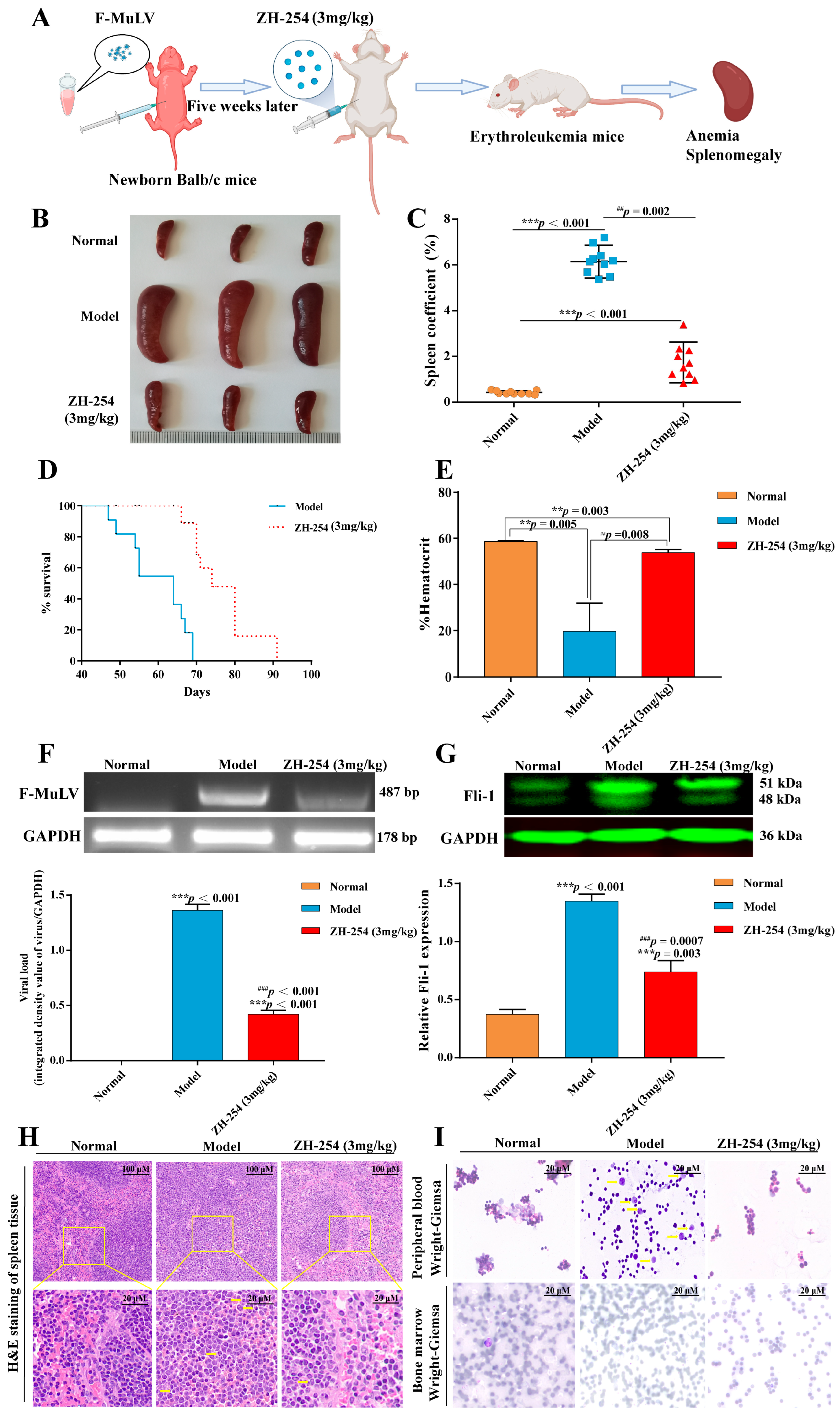
2.2. ZH-254 Could Effectively Prevent the Malignant Development of F-MuLV-Induced Erythroleukemia in Mouse

Around 90 percent of mice successfully exhibited erythroleukemia symptoms after five weeks of continuous injection of the virus suspension, indicating F-MulV-induced erythroleukemia. This was followed by intraperitoneal injection (i.p.) of 3 mg/kg compound ZH-254 seven times every two days (Figure 2A). The results showed that the spleen volume of the model group was significantly (p < 0.001) more prominent than that of the control group and the ZH-254 treatment group (Figure 2B,C). Compared with the model group, the compound treatment could significantly (p < 0.001) prolong the survival time of erythroleukemia mice (Figure 2D). The hematocrit levels of the mice treated with ZH-254 were significantly (p < 0.01) higher than those of the mice in the model group, thus possibly alleviating the anemic state of erythroleukemic mice (Figure 2E).
The spleen viral load (487 bp) measurement showed that the ZH-254 treatment group exhibited weaker bands while the model group possessed strong band intensity. In contrast, no viral RNA was detected in the regular control group, which indicated that the ZH-254 treated mice had a lower (p < 0.01) viral load than that of the model group (Figure 2F). The above results indicated that ZH-254 could effectively prevent the malignant development of F-MuLV-induced erythroleukemia in the mice. Our study found that compared to the normal group of mice, the model group had higher levels of Fli-1 protein in their bone marrow (p < 0.001). The compound-treated group showed that Fli-1 expression was (p < 0.001) activated by F-MuLV. The activation was (p < 0.001) suppressed by ZH-254, which shows that the inhibition of Fli-1 gene expression is an essential molecular mechanism by which the compound prevented F-MuLV-induced erythroleukemia (Figure 2G).
We further analyzed the tissue integrity of the mouse spleen and the degree of tumor cell infiltration, peripheral blood smears, and bone marrow smear to determine the leukemia development. Compared with the normal spleen in the normal group of mice, hematoxylin and eosin (H&E) staining demonstrates that the spleen tissue morphology of the model group of mice was ruptured under low magnification. The red and white bone marrow boundary was not sharp. The whole spleen was infiltrated by tumor cells and showed pathological nuclear division under high magnification; compared with the model group, ZH-254 could reduce the infiltration of leukemia cells, and the tissue structure was more intact (Figure 2H). Giemsa’s staining observed a significant increase in the number of nucleated cells in the peripheral blood field of the model group of erythroleukemic mice; the number of nucleated cells in the visual field was significantly increased (6–8 cells/HP in leukemic mice, 4–5 cells/HP in ZH-254 mice, and 2–3 cells/HP in normal mice). There were more atypical leukemia cells with large cell sizes, varying nuclear sizes, irregular shapes, and easy-to-see mitotic phase. Giemsa’s staining showed noticeable bone marrow cell proliferation in the model group, with granulocytes and erythrocytes multiplying (Table 2). A higher proportion of mature granulocytes were seen post-compound ZH-254 treatment (Figure 2I). An intervention provides evidence that compound ZH-254 inhibits leukemia cell growth and promotes differentiation and maturation of immature erythrocytes, thus alleviating leukemia symptoms and extending survival.

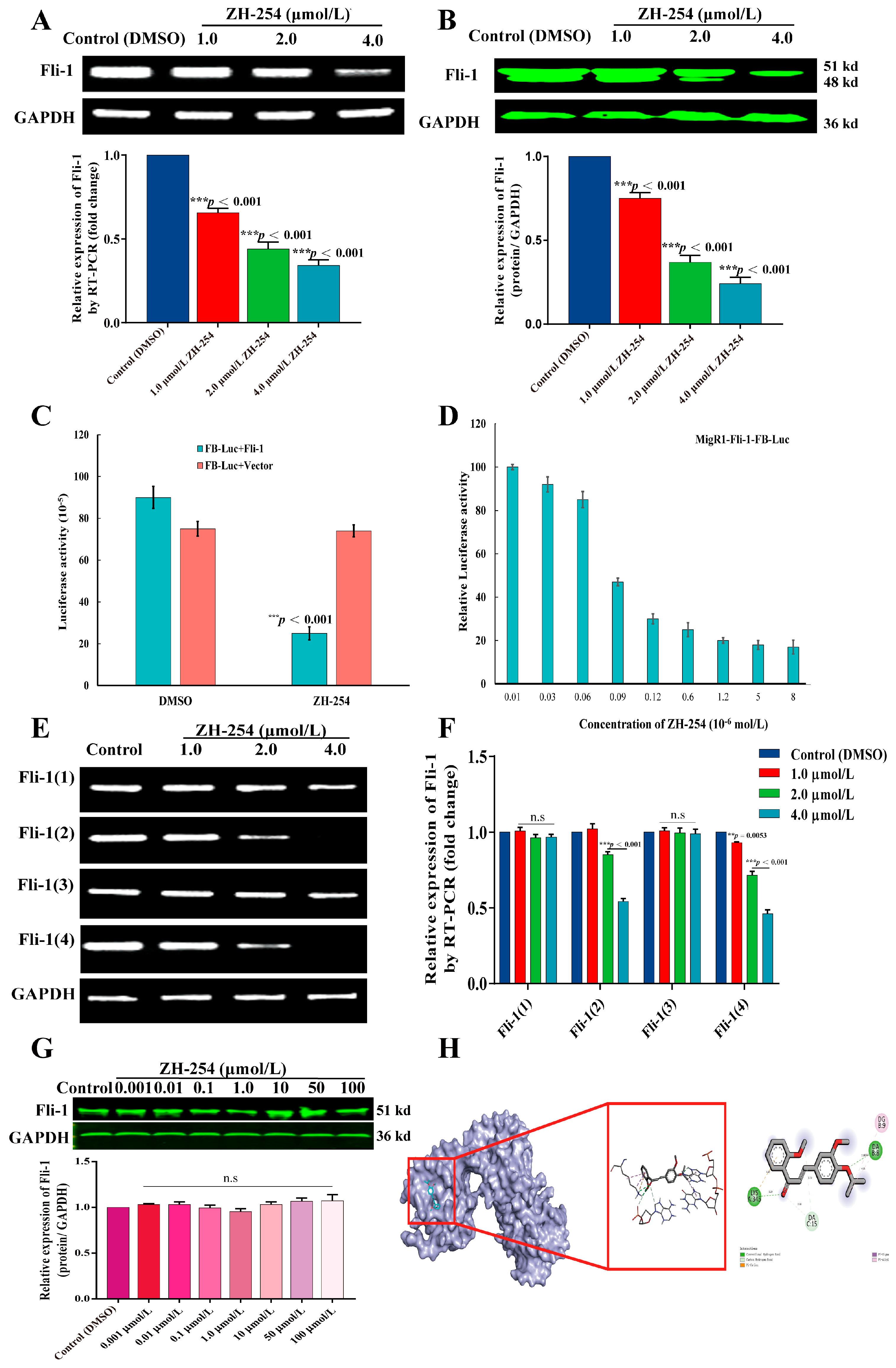
2.3. ZH-254 Downregulated Fli-1 Expression in HEL Cells by Inhibiting Promoter Activity

The occurrence and development of F-MulV-induced erythroleukemia involve the activation of Fli-1 gene expression [16]. Therefore, Fli-1 expression is considered a reference for treating erythroleukemia. The results show that dose-dependent inhibition of Fli-1 expression in HEL cells was found with ZH-254 at both gene and protein levels (Figure 3A,B). A luciferin-based expression assay was previously established to screen Fli-1 targeting regulators from FB-Luc readout reporter plasmids. A minimal promoter is inserted upstream of two consensus Fli-1 binding sites on FB-Luc, allowing it to function as a reporter plasmid. ZH-254 reduced the luciferase activity of the Fli-1 reporter plasmid (FB-Luc) (approximately three-fold times) post-cotransfection with the MigR1-Fli-1 expression vector in HEK-293T cells.
In the presence of Fli-1, ZH-254 can significantly inhibit the expression of Fli-1 promoter-dependent luciferase (Figure 3C). When MigR1-Fli-1 and FB-Luc were co-transfected into HEK293T cells, ZH-254 was concentration-dependent on the inhibition of luciferase activity of the CMV promoter-driven Fli-1 reporter plasmid (Figure 3D). The above results indicated that ZH-254 could downregulate Fli-1 expression in HEL cells by inhibiting promoter activity. In addition, we found four reported subtypes of Fli-1, which were retrieved from NCBI. Therefore, we investigated the regulatory effect of ZH-254 on the expression of the four Fli-1 isoforms. The results showed that ZH-254 significantly inhibited Fli-1 (2) and Fli-1 (4) subtypes in a dose-dependent manner, but Fli-1 (1) and Fli-1 (3) did not exhibit any significant inhibition (Figure 3E,F).
The binding ability of the compound to the Fli-1 protein was further investigated by a cellular thermal shift assay (CETSA) (Figure 3G) and molecular docking experiments (Figure 3H). The results showed that Fli-1 protein expression did not increase significantly even if the concentration was increased, which indicated that ZH-254 did not exhibit the maximum affinity for the Fli-1 protein in living cells; this also confirmed that the Fli-1 protein is not the direct target of compound binding. The binding of ZH-254 and 5JVT strand A showed higher free energy with a delta G = −4.1 kJ/mol using molecular docking experiments, which suggested that the compound binds very weakly to the Fli-1 protein, meaning that the two molecules were unlikely to form a complex. In summary, ZH-254 is a Fli-1 inhibitor that inhibits the Fli-1 promoter but does not bind to the Fli-1 targeting protein. Therefore, the targeted inhibition of the promoter activity of Fli-1 may be the fundamental molecular mechanism for the compound to inhibit the cell growth of erythroleukemia.

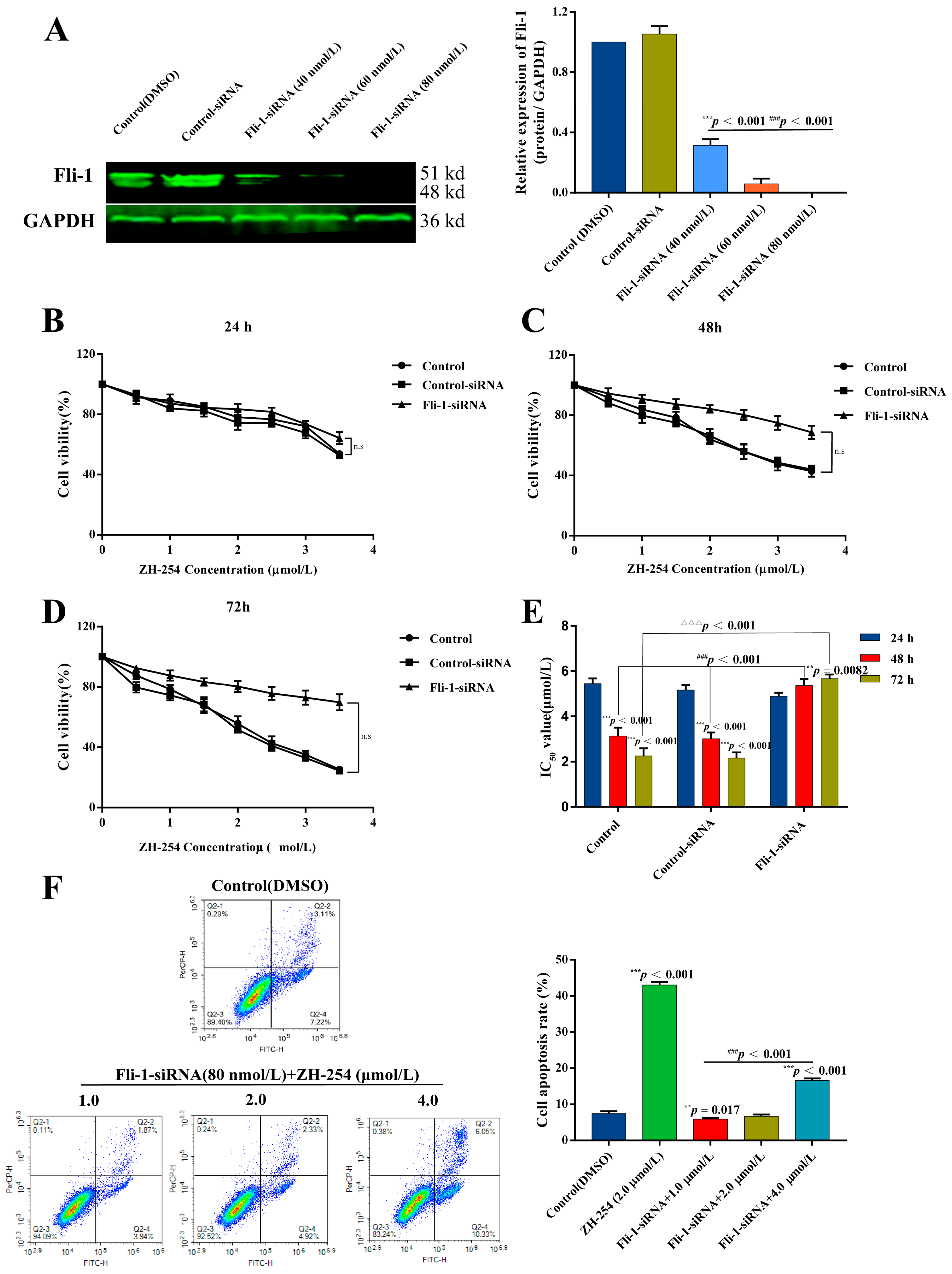
2.4. Validation of ZH-254 Targeting Fli-1 against HEL Cells

The sensitivity of HEL cells with weak expression of Fli-1 induced by siRNA to ZH-254 treatment was further examined to confirm whether Fli-1 is a critical gene in the apoptosis of erythroleukemia cells induced by the compound (Figure 4). The results demonstrated that Fli-1 protein expression gradually decreased with an increase in Fli-1-siRNA silencer concentration, the expression level of Fli-1 was the lowest at the concentration of Fli-1-siRNA of 80 nmol/L, which was determined as the optimal concentration for silencing (Figure 4A). Compared with the control and control siRNA groups at 24 (Figure 4B), 48 (Figure 4C), and 72 h (Figure 4D), the ZH-254 group showed a significantly reduced rate of inducing apoptosis in Fli-1 knockdown cells at 48 (p < 0.05) and 72 h (p < 0.001). It was also evident from the results that the cell proliferation inhibition rate was similar between the control and control siRNA groups, and the auxiliary reagents of siRNA insertion into cellular target genes had a more negligible impact on the apoptosis rate (Figure 4E). The induced apoptosis of ZH-254 was examined in Fli-1 knockdown cells using flow cytometry (Figure 4F). Despite the ability of ZH-254 to induce apoptosis in knockdown HEL cells, the apoptosis rate was significantly (p < 0.001) lower than that of normal cells treated with the compound at the same concentration. The above results have confirmed that Fli-1 is a crucial gene for ZH-254 growth inhibition and -induced apoptosis of HEL cells.

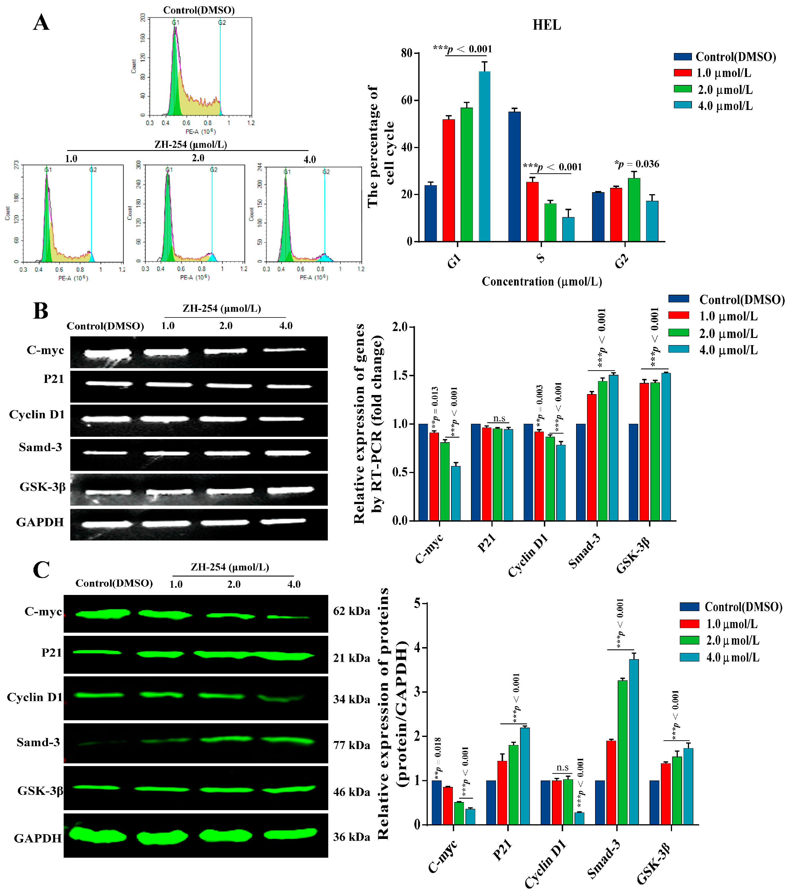
2.5. Effects of ZH-254 on the HEL Cell Cycle and Cycle-Related Genes

A flow cytometric analysis revealed that ZH-254 affected the HEL cell cycle (Figure 5). Compared with the control (DMSO), there was a concentration-dependent increase in the proportion of G1 cells, a gradual decrease in the proportion of cells in the S phase, but an irregular change in the proportion of cells in the G2 phase (Figure 5A). Therefore, ZH-254 also inhibits erythropoiesis by inhibiting the cell cycle in the G1 phase. To understand the genes involved in the process causing an increase in the proportion of cells in the G1 phase by ZH-254, we examined the expressions of the G1 phase of cell-cycle-related genes, including CDK4, C-myc, p21, Cyclin D1, Smad-3, and GSK-3β [17,18] at the mRNA (Figure 5B) and protein (Figure 5C) levels. The results showed that ZH-254 could inhibit the expression of CDK4, C-myc, and Cyclin D1 in HEL cells at the mRNA and protein levels, but this inhibition was more prominent at the protein level. In contrast, ZH-254 promoted the relative expression of Smad-3 and GSK-3 at the mRNA and protein levels. However, the expression of p21 was not changed by ZH-254 at the mRNA level, whereas p21 protein expression was promoted.

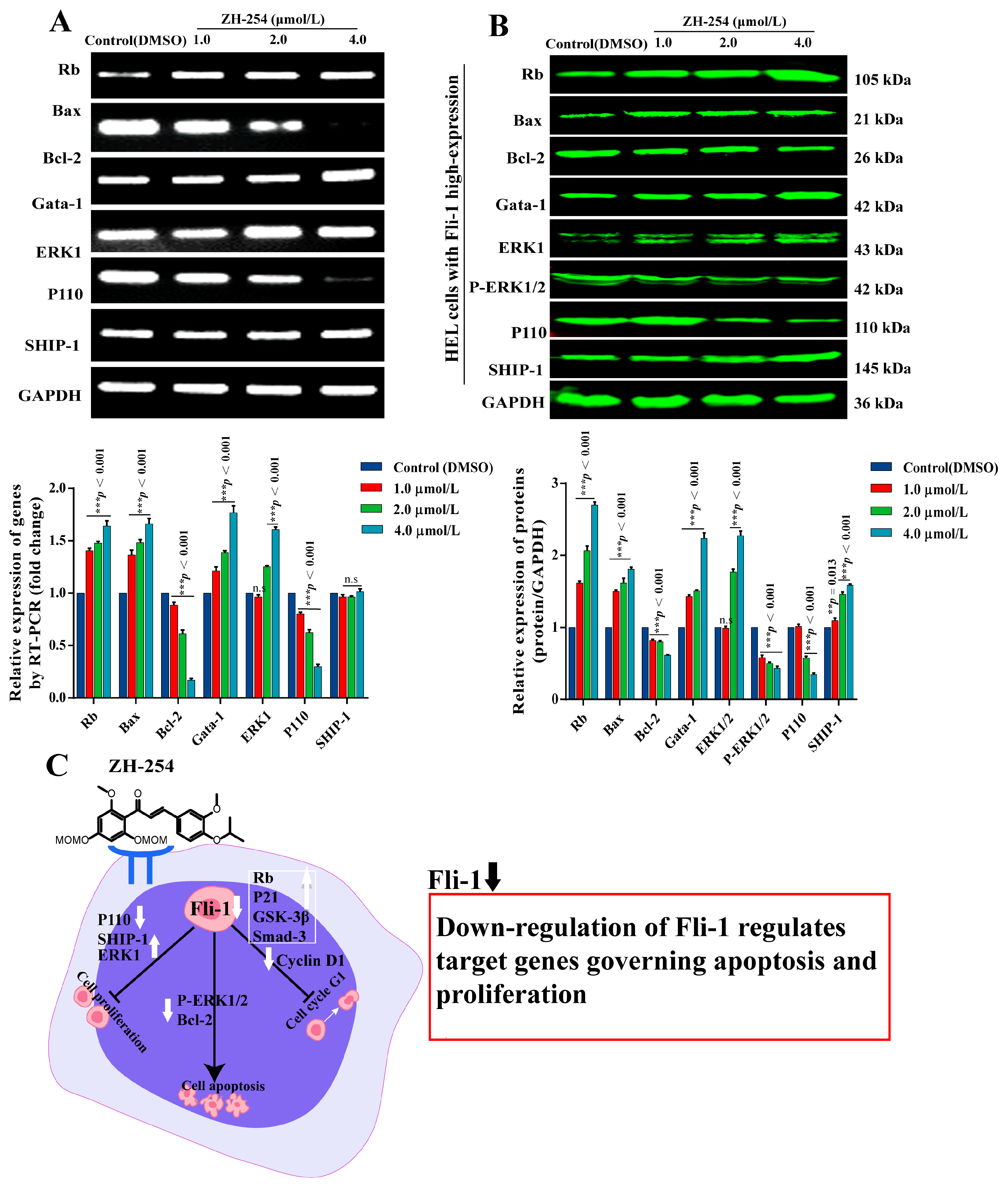
2.6. Regulation of Fli-1 Target Gene Expression in Erythroleukemia Cells by ZH-254

To explore the potential molecular mechanism of ZH-254 inhibition of Fli-1 expression against erythroleukemia cells, we evaluated the expression of the Fli-1 target gene (Figure 6). The results of mRNA-level detection (Figure 6A) showed that the expression of Fli-1 target genes Bcl-2 and P110 gradually decreased with the increase in compound concentration (1, 2, and 4 μmol/L) when compared with the control (DMSO). The expression of target genes Rb, GATA-1, and ERK1 increased gradually with the compound concentration, but the compound did not significantly regulate SHIP-1 expression. At the protein level, Bcl-2 and p110 were downregulated by ZH-254 in a dose-dependent manner, but Rb, GATA-1, ERK1, and SHIP-1 displayed an increasing trend compared with the control (Figure 6B). Moreover, the phosphorylated ERK1 (P-ERK1/2) decreased with increasing ZH-254 concentrations. Thus, the anti-proliferation activity of ZH-254 is associated with regulating Fli-1 target genes and apoptosis, including p110, P-ERK1/2, MDM2, Bcl-2, SHIP-1, and Gata-1, and the phosphorylation level of ERK1 (Figure 6C).

3. Discussion

In this study, we identified a newly synthesized chalcone that showed the potent inhibition of the proliferation of the HEL cells in vitro. The properties of chalcone were validated by apoptosis and blocking the cell cycle; an animal model experiment indicated that the compound prevented the development of F-MuLV-induced erythroleukemia in mice. Our results have confirmed that ZH-254 downregulated Fli-1 expression in HEL cells by inhibiting promoter activity. ZH-254 regulates the expression of Fli-1 target genes including Rb, Bcl-2, Bax, ERK1/2, Gata-1, P110, SHIP-1, and the extracellular regulation of protein kinase 1 phosphorylation; the compound can also regulate the expression of G1-phase-related proteins including CDK4, C-myc, Cyclin D1, Smad-3, GSK-3β, and p21 in the cell cycle pathway. Thus, ZH-254 prevents the malignant development of erythroleukemia by causing Fli-1 expression inactivation through targeted inhibition of its promoter activity, further regulating Fli-1 target genes and G1-phase-related genes.
Fli-1 gene is an E26 family transformation-specific (ETS) transcription factor discovered in recent years, which is a DNA-binding protein involved in regulating various physiological processes such as cell proliferation, differentiation, and apoptosis [19,20]. Further studies have confirmed that aberrant Fli-1 expression is observed in the growth of multiple hematologic malignancies and solid cancers, including leukemia, melanoma, breast cancer, small cell lung cancer, Alzheimer’s disease, neuroblastoma; and rhabdomyosarcoma [7,21,22,23]. Fli-1 is a key regulator in blood cell angiogenesis and inhibits tumorigenesis and development; it could be used as a specific target for treating leukemia. As a therapeutic target, Fli-1 can inhibit the proliferation of tumor cells and tumor blood vessel formation, which will become an excellent target for treating tumors.
Our previous studies found that F-MuLV was inserted near the promoter of the Fli-1 gene in mice. The strong exogenous promoter carried by the virus activated the expression of the Fli-1 gene in mouse bone marrow. Fli-1 aberrant regulation is the most critical event in the induction of F-MuLV-induced erythroleukemia. Its sustained high expression causes erythroid cells to proliferate in response to EPO stimulation and inhibits erythroid cell growth differentiation [7,16]. In addition, activation of the Fli-1 gene by proviral insertion was found in myeloid leukemia induced by the Graffi virus, lymphoma induced by the Cas-Br-E virus, and mouse leukemia induced by the 10A1 virus [11], suggesting that activation of the Fli-1 gene is involved in the development of various types of leukemia or lymphoma [24]. Fli-1 was related to mouse erythroleukemia cells’ malignant transformation and proliferation [25]. Studies have found that Fli-1 is highly expressed in HEL, CB3, CB7, TP1, TP3, HB22.2, Jurkat, and other erythroleukemia cell lines and regulates the survival and proliferation of these cells [26,27,28]. The overexpression of Fli-1 in erythroleukemia cells may directly regulate its target genes and form an anti-apoptotic phenotype. Previous studies have shown that Bcl-2 is activated in most F-MuLV-induced erythroleukemia cell lines. The overexpression of Bcl-2 may delay p53 mutations in leukemia cells [29]. It was also shown that Bcl-2 is an in vivo target of Fli-1-transformed adult erythrocytes. Its overexpression contributes to the proliferation of cells [30,31], so compounds can reduce apoptosis by downregulating Bcl-2 expression. Furthermore, Fli-1 promotes the proliferation of erythroid cells via PI3K-mediated phosphorylation of AKT/PKB and downregulation of SHIP-1 expression [28,32].
Another exciting discovery in our previous study was the active chalcone derivative C10. In leukemia, compound C10 inhibits Fli-1 by modulating signaling pathways related to autophagy and apoptosis, inhibiting erythroleukemia cell proliferation [33]. At the same time, C10 inhibits the proliferation and migration of prostate cancer cells by promoting Fli-1 expression and targeting Fli-1 promoter activity [30]. The two articles were studied the same compound, but its expression of Fli-1 showed two opposite results. Although targeting different types of cancer, C10 has shown potent anticancer activity as an inhibitor of Fli-1 in leukemia and as an agonist of Fli-1 in prostate cancer [30]. Based on these findings, we further investigated the molecular mechanism of Fli-1 targeting by this compound in both cancer cells. It was preliminarily confirmed that Fli-1 exists in HEL cells with four distinguished isoforms, while ZH-254 can only inhibit the expression of two isoforms. However, the functions of the four isoforms of Fli-1 and the molecular mechanisms regulating the occurrence and development of different cancers have not been reported. We should carefully elucidate Fli-1′s dual role in cancer biology in this era of precision medicine. We are still exploring the molecular mechanisms of why Fli-1 functions differently in different cancers, and this is an essential topic that our research group is working on and will focus on in the next few years.

4. Materials and Methods

4.1. Chemicals, Reagents, and Instruments

Roswell Park Memorial Institute (RPMI) 1640 was purchased from Gibco (Waltham, MA, USA). MTT was purchased from Solarbio (Beijing, China). Dimethyl sulfoxide (DMSO) was purchased from Huabio Ltd. (Hangzhou, China). Cell apoptosis and cycle kits were purchased from BD Biosciences (San Jose, CA, USA). The Hoechst staining kit, One-Step TUNEL apoptosis kit, the mitochondrial membrane potential assay kit with JC-1, and the BCA protein assay kit were purchased from Beyotime (Shanghai, China). The CO2 cell incubator was purchased from Thermo Fisher Scientific (Waltham, MA, USA). The allegra X-15R high-speed centrifuge was purchased from Beckman (Brea, CA, USA). The microplate reader was purchased from BioTek (Winooski, VE, USA). ACEA NovoCyte flow cytometry analyzer was purchased from BD Biosciences (San Jose, CA, USA). The Leica inverted fluorescence microscope was purchased from Leica (Wetzlar, Germany). The Odyssey Infrared Imaging System was purchased from Gene Company Limited (LI-COR, HK, China). The U: Genius3 was purchased from Syngene (Synoptics Ltd., Cambridge, UK).

4.2. Antibodies

Antibodies against Fli-1 (ab153909, 1:1000), Rb (ab181616, 1:2000), Gata-1 (ab181544, 1:1000), Bcl-2 (ab32124, 1:1000), p110 (ab151549, 1:1000), SHIP-1 (ab45142, 1:1000), phospho-ERK 1/2 (T202 +Y204) (ab223500, 1:1000), and ERK1 (ab32537, 1:1000) were purchased from Abcam (Cambridge, UK). P53 (AF0879, 1:1000), CDK4 (DF6102, 1:500), C-myc (T0052,1:3000), p21 (AF6290,1:1000), Cyclin D1 (AF0931, 1:1000), Smad-3 (AF6362, 1:1000), and GSK-3β(AF5016,1:1000) were purchased from Affinity (Jiangsu, China). GAPDH (#2118s, 1:1000) was purchased from Cell Signaling Technology (Beverly, MA, USA). The anti-rabbit and anti-mouse IgG (H + L) [Dylight (TM) 800 4×PEG Conjugate] secondary antibodies used in this study were purchased from CST and the dilution ratio was 1: 30,000.

4.3. Cell Culture and Animal Feeding

Various leukemia cell lines, including human erythroleukemia HEL, promyelocytic leukemia HL-60, T-lymphoma Jurkat, and myeloid leukemia K562 cell lines were purchased from ATCC. The cells were preserved in the Key Laboratory of Chemistry for Natural Products of Guizhou Province and the Chinese Academic of Sciences (Guiyang, China). The cells were cultured in RPMI-1640 medium with 10% FBS at 37 °C with a 5% CO2 incubator.
Balb/c mice (20 total; half males and females, seven weeks old) were purchased from HFK Bio-Technology, Co., LTD. Beijing, China [Certificate # SCXK (jing) 2019-0008]. The animals were supplemented with pure feed and distilled water while maintaining a temperature between 20–25 °C, a 55–65% humidity, and an automatic light/darkness cycle of 12 h in an SPF-grade laboratory. The institute’s ethical committee approved all of the experimental operations.

4.4. Compound Treatment

The selected compound was a chalcone derivative named 3,2′-dimethoxy-4-isopropoxy-4′,6′-bis(methoxymethoxy)-chalcone (ZH-254), which is a yellow powder with a purity of 93%. A stock solution of 2 × 10⁴ µmol/L was prepared by dissolving ZH-254 in DMSO.

4.5. Cell Growth Assay

4.5.1. Cell Proliferation Assay

Leukemia cells (HEL, HL-60, Jurkat, and K562) were plated in a 96-well plate in sextuplicate with 5 × 10³ cells/well, and various compound concentrations were added and incubated for 24, 48, and 72 h. Briefly, the morphological changes in the cells after compound treatment were observed using an inverted fluorescence microscope CCD camera (Nikon, Japan). Add 20 µL of MTT solution to each well and incubate at 37 °C for 4 h. After discarding the old medium, add 150 µL DMSO to each well, incubate in a shaker at 37 °C for 15 min to dissolve the Formazan crystals and detect the absorbance of each well at 490 nm.

4.5.2. Cell Apoptosis Assay

The HEL cells were exposed to different doses of ZH-254 (0, 1, 2, and 4 μmol/L) for 48 h. The cells were washed with PBS and mixed with 500 μL of 1× binding buffer to form cell suspensions. Staining was performed with 5 μL Annexin V-FITC and 5 μL PI. The apoptosis rate of each concentration group was quantified by flow cytometry. Staining was performed with 5 μL Annexin V-FITC and 5 μL PI for light avoidance. Finally, the apoptosis rate of each concentration group was assessed by flow cytometry.

4.5.3. Cell Cycle Assay

The procedure for inoculation and treatment of cells is described in Section 4.5.2. The cells were fixed in 70% pre-cooled ethanol at 4 °C for more than 12 h. The effect of different concentrations of ZH-254 on the cell cycle was quantified by flow cytometry.

4.5.4. Hoechst 33258 Staining

Since chromatin condensation and fragmentation are signs of apoptosis [33], we performed this experiment to determine the extent of apoptosis. Briefly, 1 × 10⁶ cells of the HEL cell line were incubated for 24 h in ZH-254 (0, 1, 2, and 4 µmol/L). Then, the cells were collected by centrifugation and washed twice by adding PBS. After being fixed with 4% paraformaldehyde (Beyotime, Shanghai, China) for 30 min, the cells were dyed with Hoechst 33258 (1 mg/mL) at room temperature and protected from light. The nuclear morphology of the HEL cells was observed by fluorescence microscopy.

4.5.5. TUNEL Staining Assay

HEL cells were collected, fixed continuously with 4% PFA for 30 min, and permeated at room temperature with Triton X-100 (Beyotime Biotech) for 5 min. Then, the One-Step TUNEL apoptosis kit (Beyotime, Shanghai, China) was used to prepare the TUNEL test solution. The cells were added to 50 µL of TUNEL detection solution and incubated in the dark at 37 °C for 60 min. Observation of apoptosis under a fluorescent microscope was completed following the protocol [33].

4.5.6. Mitochondrial Membrane Potential (MMP) Assay

The HEL cells were collected, 500 μL of JC-1 staining working solution was added and then incubated again for 20 min in a 37 °C cell incubator, washed twice with 1 × JC-1 staining buffer, and the cells were resuspended. The changes in the mitochondrial membrane potential (MMP) of the cells were detected according to the instructions. The appearance of red fluorescence indicates normal MMP and normal cell status. The presence of green fluorescence indicates a decrease in MMP and that the cells may be in the early stages of apoptosis [34].

4.6. Luciferase Reporter Assay

The effect of ZH-254 on the activity of the Fli-1 promoter was measured according to the following procedure. In brief, the Fli-1 sequence is inserted into the MigR1 luciferase reporter vector. Using Lipofectamine 2000 (Life Technology, Beijing, China), FB-Luc (1.25 µg) and MigR1 (1.25 µg) or MigR1-Fli-1 (1.25 µg), expression vectors were co-transfected into HEK293T cells and incubated for 24 h. After adding ZH-254 for 12 h treatment, the activity of cell luciferase was detected [35].

4.7. ZH-254 In Vivo Activity Assay

4.7.1. In Vivo Assay

An F-MuLV-induced erythroleukemia mouse model was used to determine the activity of ZH-254 in vivo [6]. Newborn Balb/c mice within 2 days of birth were intraperitoneally injected with Friend murine leukemia virus fluid (3000 focus-forming units) at 100 µL/mouse. After 5 weeks of infection, the 3 mg/kg compound ZH-254 was intraperitoneally injected seven times every two days, and the mice were monitored for disease signs. The model and the control groups were continuously injected with normal saline according to the compound injection method. Mice showing signs of terminal leukemia were sacrificed under deep anesthesia, their spleens were removed and weighed, hematocrit was measured, and survival was recorded. The spleen viral load was measured using TRIZOL reagent for RNA extraction from mouse spleen tissue [36].

4.7.2. Hematoxylin and Eosin (H&E) Staining

Fresh tissues were fixed in 4% paraformaldehyde, embedded in paraffin, and stained with H&E using the protocol [37].

4.7.3. Wright–Giemsa Staining

According to the manufacturer’s agreement, the peripheral blood or bone marrow of erythroleukemia mice was coated on a glass sheet and morphologically evaluated with Wright–Giemsa staining solution (Leagene, Beijing, China). The morphology of the cells was examined under a light microscope.

4.8. Validation Assay

4.8.1. Reverse Transcription-Polymerase Chain Reaction (RT-PCR)

A total RNA extraction kit (Thermo Fisher Scientific, Waltham, MA, USA) was used to isolate total RNA in erythroleukemia mouse spleen tissues and cells following the manufacturer’s protocol. After quantification, total RNA (2 µg per sample) was reversely transcribed into cDNA using the HiFiScript cDNA synthesis kit (CW Bio, Beijing, China). Amplification of the target genes was achieved by RT-PCR using cDNA as template and the obtained PCR product was electrophoresed in agarose gel 1% agarose gel at 50 v for 30 min to determine the expression level of the target gene. The gels were assayed by U: Genius3 and the relative expressions were normalized to the housekeeping gene GAPDH using Image J software. The primer sequences are listed in Table 3.

4.8.2. Western Blotting Assay

Western blot was conducted as described [33]. Briefly, the cells were cultured in dishes and treated with various concentrations of ZH-254 for 24 h. The cells were collected and extracted using RIPA reagent 1% PMSF protease inhibitor. The BCA protein assay kit was performed to examine the protein concentration of each sample. Protein (50 μg/per line) was separated by 10% SDS-PAGE and transferred to a 0.45 μm polyvinylidene fluoride (PVDF) membranes. The PVDF membranes were blocked in 5% BSA incubated for 1 h at room temperature and then incubated at 4 °C with anti-Fli-1, anti-Rb, anti-Gata-1, anti-Bcl-2, anti-p110, anti-SHIP-1, anti-phospho-ERK 1/2, anti-ERK1, anti-P53, anti-CDK4, anti-C-myc, anti-p21, anti-Smad-3, and GSK-3β; the anti-GAPDH primary antibody was incubated overnight (loading control). Goat anti-rabbit and anti-mouse Ig G H + L secondary antibodies were used to incubate with PVDF membranes in the dark for 2 h. An Odyssey Infrared Imaging System was used to visualize the blots and the relative expression of the protein was calculated using Image J (V1.8.0.112, NIH, Bethesda, MD USA).

4.8.3. Molecular Docking

To gain an in-depth understanding of the possible binding mode of the compound, ZH-254 and Fli-1 were verified by molecular docking and visual analysis was carried out. ChemBioDraw14 was used to draw the 2D structure of ZH-254 and it was saved in SDF. Format: ChemBio3DUltra software was used to convert the 2D structure of ZH-254 to a 3D structure and it was saved as mol.2 Format. The Fli-1 (PDB entry 5JVT) X-ray structures were downloaded from the Protein Data Bank (PDB, http://www.rcsb.org/, accessed on 18 March 2022). This format was imported into PyMOL software to remove water molecules and small molecule ligands. AutoDockTools software was used to add hydrogen ions and determine the location of the active pocket of the target protein. AutoDockVina software is used for the molecular docking of small molecule ligands and key proteins. Only postures with low energy conformations and good hydrogen bond geometries were considered. Figure 3H was drawn using PyMOL.

4.8.4. Cellular Thermal Shift Assay (CETSA)

The results of the binding of compounds in molecular docking to the target protein were verified using CETSA. The cells were treated with an increased amount of ZH-254 and 0.1% DMSO. After, they underwent incubation for 1 h. After incubation, the cells were washed three times with PBS and heated at 52 °C for 3 min and allowed to cool at room temperature. The cells were treated in a water bath of 37 °C and liquid nitrogen for three cycles [38]. The cells were centrifuged at 12,000 rpm at 4 °C for 5 min to collect the cell precipitation, RIPA containing 1% PMSF was added, and the cells were lysed on ice for 30 min. The supernatant was collected by centrifugation again for Western blotting to block the protein expression level of Fli-1 on the mitochondrial surface. The subsequent steps were performed as described in Section 4.8.2.

4.8.5. Gene Silencing Assay

Small interfering RNAs targeting Fli-1 (Fli-1) and the corresponding non-specific control (NC) siRNAs were obtained from Guangzhou RiboBio Co., Ltd. (Guangzhou, China). The cells were inoculated into 6-well plates at a density of 2.0 × 10⁵ cells/mL and cultured in a 37 °C incubator to reach approximately 60% fusion. Diluted siRNA and Reagent (v4) were mixed and introduced into the cells according to the manufacturer’s instructions. The cells were then cultured in an antibiotic-free medium for 6 h. The transfection complexes were removed, added to a fresh medium, and they continued to be cultured for 24 h. The cells were then further treated with compounds.

4.9. Statistical Analysis

GraphPad Prism 7.0 software (San Diego, CA, USA) was employed to perform the statistical analysis. The differences between the two groups were analyzed using a paired t-test, while the difference among the multiple groups was determined based on a one-way analysis of variance. Mice survival rates were calculated and plotted according to the non-parametric Kaplan–Meier analysis. The presented results are representative of three independent experiments. * p < 0.05 was used as a cut-off to consider the statistical significance.

5. Conclusions

We have synthesized and discovered a new active chalcone derivative, ZH-254, which can prevent the malignant development of erythroleukemia by causing Fli-1 expression inactivation through targeted inhibition of its promoter activity, further regulating Fli-1 target genes and G1-phase-related genes.

6. Patents

The manuscript contains a patent arising from patent application NO. CN202210196035.1.

Author Contributions

Conceptualization, H.L., B.X. and Z.Y.; methodology, M.M. and Y.M.; software, J.Y. and X.W.; validation, B.X., Z.Y. and H.L.; formal analysis, M.M. and M.A.; investigation, B.X.; resources, H.L.; data curation, M.M. and Y.M.; writing—original draft preparation, Y.M., M.M. and H.L.; writing—review and editing, H.L., M.M. and M.A.; visualization, X.W.; supervision, H.L. and Z.Y.; project administration, H.L.; funding acquisition, H.L. All authors have read and agreed to the published version of the manuscript.

Funding

This work was supported by the Science and Technology Planning Project of Guizhou Province (No. QKHZC [2020]4Y161), the Science and Technology Department of Guizhou Province (No. QKHPTRC [2016]5678), and Guizhou province “Hundred” level innovative talent training project (No. QKHPTRC-GCC [2022]034-1).

Data Availability Statement

Not applicable.

Acknowledgments

Figure 2a was created with BioRender.com (https://biorender.com/, accessed on 8 May 2022).

Conflicts of Interest

The authors declare no conflict of interest.

Animal Care

The animal care procedures were carried out following the guidelines of the institution, the animal care committee, and Guizhou Medical University.

References

  1. Fagnan, A.; Bagger, F.O.; Piqué-Borràs, M.R.; Ignacimouttou, C.; Caulier, A.; Lopez, C.K.; Robert, E.; Uzan, B.; Gelsi-Boyer, V.; Aid, Z.; et al. Human erythroleukemia genetics and transcriptomes identify master transcription factors as functional disease drivers. Blood 2020, 136, 698–714. [Google Scholar] [CrossRef] [PubMed]
  2. Siegel, R.L.; Miller, K.D.; Fuchs, H.E.; Jemal, A. Cancer statistics, 2022. CA Cancer J. Clin. 2022, 72, 7–33. [Google Scholar] [CrossRef] [PubMed]
  3. Qiu, H.; Cao, S.; Xu, R. Cancer incidence, mortality, and burden in China: A time-trend analysis and comparison with the United States and United Kingdom based on the global epidemiological data released in 2020. Cancer Commun. 2021, 41, 1037–1048. [Google Scholar] [CrossRef] [PubMed]
  4. Andreotti, G.; Koutros, S.; Hofmann, J.N.; Sandler, D.P.; Lubin, J.H.; Lynch, C.F.; Lerro, C.C.; De Roos, A.J.; Parks, C.G.; Alavanja, M.C.; et al. Glyphosate Use and Cancer Incidence in the Agricultural Health Study. J. Natl. Cancer Inst. 2018, 110, 509–516. [Google Scholar] [CrossRef] [PubMed] [Green Version]
  5. Moon, J.; Yoo, H. Residential radon exposure and leukemia: A meta-analysis and dose-response meta-analyses for ecological, case-control, and cohort studies. Environ. Res. 2021, 202, 111714. [Google Scholar] [CrossRef]
  6. Ko, P.S.; Liu, Y.C.; Yeh, C.M.; Gau, J.P.; Yu, Y.B.; Hsiao, L.T.; Tzeng, C.H.; Chen, P.M.; Chiou, T.J.; Liu, C.J.; et al. The uniqueness of morphological features of pure erythroid leukemia in myeloid neoplasm with erythroid predominance: A reassessment using criteria revised in the 2016 World Health Organization classification. PLoS ONE 2017, 12, e0172029. [Google Scholar] [CrossRef]
  7. Ben-David, Y.; Giddens, E.B.; Bernstein, A. Identification and mapping of a common proviral integration site Fli-1 in erythroleukemia cells induced by Friend murine leukemia virus. Proc. Natl. Acad. Sci. USA 1990, 87, 1332–1336. [Google Scholar] [CrossRef] [Green Version]
  8. Lakhanpal, G.K.; Vecchiarelli-Federico, L.M.; Li, Y.J.; Cui, J.W.; Bailey, M.L.; Spaner, D.E.; Dumont, D.J.; Barber, D.L.; Ben-David, Y. The inositol phosphatase SHIP-1 is negatively regulated by Fli-1 and its loss accelerates leukemogenesis. Blood 2010, 116, 428–436. [Google Scholar] [CrossRef] [Green Version]
  9. Charfi, C.; Voisin, V.; Levros, L.C., Jr.; Edouard, E.; Rassart, E. Gene profiling of Graffi murine leukemia virus-induced lymphoid leukemias: Identification of leukemia markers and Fmn2 as a potential oncogene. Blood 2011, 117, 1899–1910. [Google Scholar] [CrossRef] [Green Version]
  10. Bergeron, D.; Poliquin, L.; Kozak, C.A.; Rassart, E. Identification of a common viral integration region in Cas-Br-E murine leukemia virus-induced non-T-, non-B-cell lymphomas. J. Virol. 1991, 65, 7–15. [Google Scholar] [CrossRef]
  11. Rodenburg, M.; Fischer, M.; Engelmann, A.; Harbers, S.O.; Ziegler, M.; Lohler, J.; Stocking, C. Importance of receptor usage, Fli1 activation, and mouse strain for the stem cell specificity of 10A1 murine leukemia virus leukemogenicity. J. Virol. 2007, 81, 732–742. [Google Scholar] [CrossRef] [PubMed] [Green Version]
  12. Bailly, R.A.; Bosselut, R.; Zucman, J.; Cormier, F.; Delattre, O.; Roussel, M.; Thomas, G.; Ghysdael, J. DNA-binding and transcriptional activation properties of the EWS-FLI-1 fusion protein resulting from the t(11;22) translocation in Ewing sarcoma. Mol. Cell Biol. 1994, 14, 3230–3241. [Google Scholar] [CrossRef] [PubMed]
  13. Zhuang, C.; Zhang, W.; Sheng, C.; Zhang, W.; Xing, C.; Miao, Z. Chalcone: A Privileged Structure in Medicinal Chemistry. Chem. Rev. 2017, 117, 7762–7810. [Google Scholar] [CrossRef] [PubMed]
  14. Thapa, P.; Upadhyay, S.P.; Suo, W.Z.; Singh, V.; Gurung, P.; Lee, E.S.; Sharma, R.; Sharma, M. Chalcone and its analogs: Therapeutic and diagnostic applications in Alzheimer’s disease. Bioorg. Chem. 2021, 108, 104681. [Google Scholar] [CrossRef]
  15. Wen, Z.; Zhang, Y.; Wang, X.; Zeng, X.; Hu, Z.; Liu, Y.; Xie, Y.; Liang, G.; Zhu, J.; Luo, H.; et al. Novel 3′,5′-diprenylated chalcones inhibited the proliferation of cancer cells in vitro by inducing cell apoptosis and arresting cell cycle phase. Eur. J. Med. Chem. 2017, 133, 227–239. [Google Scholar] [CrossRef]
  16. Cui, J.W.; Vecchiarelli-Federico, L.M.; Li, Y.J.; Wang, G.J.; Ben-David, Y. Continuous Fli-1 expression plays an essential role in the proliferation and survival of F-MuLV-induced erythroleukemia and human erythroleukemia. Leukemia 2009, 23, 1311–1319. [Google Scholar] [CrossRef] [Green Version]
  17. Liang, Y.; Qiu, X.; Xu, R.Z.; Zhao, X.Y. Berbamine inhibits proliferation and induces apoptosis of KU812 cells by increasing Smad3 activity. J. Zhejiang Univ. Sci. B 2011, 12, 568–574. [Google Scholar] [CrossRef] [Green Version]
  18. Chen, M.J.; Cheng, A.C.; Lee, M.F.; Hsu, Y.C. Simvastatin induces G1 arrest by up-regulating GSK3beta and down-regulating CDK4/cyclin D1 and CDK2/cyclin E1 in human primary colorectal cancer cells. J. Cell Physiol. 2018, 233, 4618–4625. [Google Scholar] [CrossRef]
  19. Seth, A.; Watson, D.K. ETS transcription factors and their emerging roles in human cancer. Eur. J. Cancer 2005, 41, 2462–2478. [Google Scholar] [CrossRef]
  20. Mhawech-Fauceglia, P.; Herrmann, F.R.; Bshara, W.; Odunsi, K.; Terracciano, L.; Sauter, G.; Cheney, R.T.; Groth, J.; Penetrante, R.; Mhawech-Fauceglia, P. Friend leukaemia integration-1 expression in malignant and benign tumours: A multiple tumour tissue microarray analysis using polyclonal antibody. J. Clin. Pathol. 2007, 60, 694–700. [Google Scholar] [CrossRef]
  21. Yan, X.; Yu, Y.; Li, L.; Chen, N.; Song, W.; He, H.; Dong, J.; Liu, X.; Cui, J. Friend leukemia virus integration 1 is a predictor of poor prognosis of breast cancer and promotes metastasis and cancer stem cell properties of breast cancer cells. Cancer Med. 2018, 7, 3548–3560. [Google Scholar] [CrossRef] [PubMed]
  22. Huang, L.; Zhai, Y.; La, J.; Lui, J.W.; Moore, S.P.G.; Little, E.C.; Xiao, S.; Haresi, A.J.; Brem, C.; Bhawan, J.; et al. Targeting Pan-ETS Factors Inhibits Melanoma Progression. Cancer Res. 2021, 81, 2071–2085. [Google Scholar] [CrossRef] [PubMed]
  23. Li, P.; Wu, Y.; Hamlett, E.D.; Goodwin, A.J.; Halushka, P.V.; Carroll, S.L.; Liu, M.; Fan, H. Suppression of Fli-1 protects against pericyte loss and cognitive deficits in Alzheimer’s disease. Mol. Ther. J. Am. Soc. Gene Ther. 2022, 30, 1451–1464. [Google Scholar] [CrossRef] [PubMed]
  24. Sarrazin, S.; Starck, J.; Gonnet, C.; Doubeikovski, A.; Melet, F.; Morle, F. Negative and translation termination-dependent positive control of FLI-1 protein synthesis by conserved overlapping 5′ upstream open reading frames in Fli-1 mRNA. Mol. Cell Biol. 2000, 20, 2959–2969. [Google Scholar] [CrossRef] [Green Version]
  25. Ben-David, Y.; Gajendran, B.; Sample, K.M.; Zacksenhaus, E. Current insights into the role of Fli-1 in hematopoiesis and malignant transformation. Cell. Mol. Life Sci. CMLS 2022, 79, 163. [Google Scholar] [CrossRef]
  26. Liu, T.; Xia, L.; Yao, Y.; Yan, C.; Fan, Y.; Gajendran, B.; Yang, J.; Li, Y.J.; Chen, J.; Filmus, J.; et al. Identification of diterpenoid compounds that interfere with Fli-1 DNA binding to suppress leukemogenesis. Cell Death Dis. 2019, 10, 117. [Google Scholar] [CrossRef] [Green Version]
  27. Wang, N.; Fan, Y.; Yuan, C.M.; Song, J.; Yao, Y.; Liu, W.; Gajendran, B.; Zacksenhaus, E.; Li, Y.; Liu, J.; et al. Selective ERK1/2 agonists isolated from Melia azedarach with potent anti-leukemic activity. BMC Cancer 2019, 19, 764. [Google Scholar] [CrossRef] [PubMed] [Green Version]
  28. Li, L.; Yu, J.; Cheng, S.; Peng, Z.; Luo, H. Transcription factor Fli-1 as a new target for antitumor drug development. Int. J. Biol. Macromol. 2022, 209, 1155–1168. [Google Scholar] [CrossRef]
  29. Howard, J.C.; Li, Q.; Chu, W.; Zochodne, B.; Kapoor, M.; Ung, Y.; Rosen, K.; Ben-David, Y. Bcl-2 expression in F-MuLV-induced erythroleukemias: A role for the anti-apoptotic action of Bcl-2 during tumor progression. Oncogene 2001, 20, 2291–2300. [Google Scholar] [CrossRef]
  30. Ma, Y.; Xu, B.; Yu, J.; Huang, L.; Zeng, X.; Shen, X.; Ren, C.; Ben-David, Y.; Luo, H. Fli-1 Activation through Targeted Promoter Activity Regulation Using a Novel 3′, 5′-diprenylated Chalcone Inhibits Growth and Metastasis of Prostate Cancer Cells. Int. J. Mol. Sci. 2020, 21, 2216. [Google Scholar] [CrossRef]
  31. Lesault, I.; Quang, C.T.; Frampton, J.; Ghysdael, J. Direct regulation of BCL-2 by FLI-1 is involved in the survival of FLI-1-transformed erythroblasts. EMBO J. 2002, 21, 694–703. [Google Scholar] [CrossRef] [Green Version]
  32. Song, J.; Yuan, C.; Yang, J.; Liu, T.; Yao, Y.; Xiao, X.; Gajendran, B.; Xu, D.; Li, Y.J.; Wang, C.; et al. Novel flavagline-like compounds with potent Fli-1 inhibitory activity suppress diverse types of leukemia. FEBS J. 2018, 285, 4631–4645. [Google Scholar] [CrossRef] [PubMed] [Green Version]
  33. Zhang, Y.Q.; Wen, Z.H.; Wan, K.; Yuan, D.; Zeng, X.; Liang, G.; Zhu, J.; Xu, B.; Luo, H. A novel synthesized 3′, 5′-diprenylated chalcone mediates the proliferation of human leukemia cells by regulating apoptosis and autophagy pathways. Biomed. Pharmacother. 2018, 106, 794–804. [Google Scholar] [CrossRef] [PubMed]
  34. Cui, W.; Wu, X.; Shi, Y.; Guo, W.; Luo, J.; Liu, H.; Zheng, L.; Du, Y.; Wang, P.; Wang, Q.; et al. 20-HETE synthesis inhibition attenuates traumatic brain injury-induced mitochondrial dysfunction and neuronal apoptosis via the SIRT1/PGC-1α pathway: A translational study. Cell Prolif. 2021, 54, e12964. [Google Scholar] [CrossRef] [PubMed]
  35. Tang, X.; Liang, Y.; Sun, G.; He, Q.; Qu, H.; Gao, P. UBQLN4 is activated by C/EBPbeta and exerts oncogenic effects on colorectal cancer via the Wnt/beta-catenin signaling pathway. Cell Death Discov. 2021, 7, 398. [Google Scholar] [CrossRef]
  36. Dignam, J.D.; Lebovitz, R.M.; Roeder, R.G. Accurate transcription initiation by RNA polymerase II in a soluble extract from isolated mammalian nuclei. Nucleic Acids Res. 1983, 11, 1475–1489. [Google Scholar] [CrossRef] [Green Version]
  37. Restall, B.S.; Haven, N.J.M.; Kedarisetti, P.; Martell, M.T.; Cikaluk, B.D.; Silverman, S.; Peiris, L.; Deschenes, J.; Zemp, R.J. Virtual hematoxylin and eosin histopathology using simultaneous photoacoustic remote sensing and scattering microscopy. Opt. Express 2021, 29, 13864–13875. [Google Scholar] [CrossRef]
  38. Dziekan, J.M.; Yu, H.; Chen, D.; Dai, L.; Wirjanata, G.; Larsson, A.; Prabhu, N.; Sobota, R.M.; Bozdech, Z.; Nordlund, P. Identifying purine nucleoside phosphorylase as the target of quinine using cellular thermal shift assay. Sci. Transl. Med. 2019, 11, eaau3174. [Google Scholar] [CrossRef]
Figure 1. ZH-254 induced inhibition of HEL cell proliferation and apoptosis. (A) Chemical structure of ZH-254. (B) Cell viability curves of HEL cell growth after ZH-254 treatment by MTT assay. (C) The apoptosis-inducing effect of different concentrations of ZH-254 on normal HEL cells was detected by flow cytometry. (D) Comparison of the effects of ZH-254 on the early and late apoptosis rates of HEL cells; (E) Microscopic observation of the cell state of HEL cells after treatment with ZH-254 for 48 h; ZH-254 induced apoptosis as visualized by Hoechst 33258 (Beyotime, Shanghai, China); ZH-254 induced apoptosis as visualized by TUNEL (Beyotime, Shanghai, China); effect of ZH-254 on the membrane potential of human erythroleukemia HEL cells. The above images are all magnified at 200×. The data are presented as mean ± SEM. ** p < 0.01, *** p < 0.001.
Catalysts 13 00084 g001
Figure 2. Compound ZH-254 inhibits leukemogenesis in F-MuLV-induced erythroleukemia. (A) A schematic diagram of the modeling process of erythroleukemia mice. (B) Illustration of splenomegaly and statistical chart of splenic coefficients (C). (D) Kaplan–Meier survival curves for each experimental group (E) Statistics of the hematocrit rate of these leukemic mice. (F) Effect of ZH-254 on splenic viral load. (G) Fli-1 protein in the mice bone marrow. (H) H&E staining of spleen tissue of erythroleukemia mice. (I) Peripheral blood Giemsa stain; bone marrow Giemsa stain. Compared with the control group, ** p < 0.01 (n = 10) and *** p < 0.001 (n = 10) are compared to the control group; ## p < 0.01 (n = 10), ### p < 0.001 (n = 10) compared with the ZH-254 group.
Catalysts 13 00084 g002
Figure 3. ZH-254 inhibits Fli-1 expression in HEL cells. (A,B) Expression and statistical results of Fli-1 at the mRNA and protein levels in HEL cells. (C) Import MigR1-Fli-1 or MigR1-Vector into HEK293T cells with or without ZH-254. After 12 h, the luciferase activity was detected by the dual-luciferase report detection system. (D) When MigR1-Fli-1 is co-transfected with FB-Luc, the concentration of ZH-254 affects the luciferase activity of the Fli-1 reporting plasmid driven. (E) Effect of ZH-254 on Fli-1 isoforms in HEL cells and statistical analysis of the mRNA expressions of Fli-1 isoforms. (F) The histograms show relative Fli-1 expression in HEL cells, calculated using Image J. Use GAPDH as a housekeeper gene. (G) CETSA to detect the binding of ZH-254 to the Fli-1 protein. (H) Three-dimensional binding mode of ZH-254 into Fli-1; two-dimensional binding mode of the co-crystallized ZH-254 shows different interactions with amino acids in Fli-1. ** p < 0.01, *** p < 0.001.
Catalysts 13 00084 g003
Figure 4. Validation of ZH-254 targeting Fli-1 against HEL cells. (A) Western blotting was performed to detect Fli-siRNA to silence the expression of Fli-1 in HEL cells. Statistical analysis of the gray value of the Fli-1 protein band using the Image J software (V1.8.0.112, NIH, Bethesda, MD USA). *** p < 0.001 compared with the control (DMSO) group, ### p < 0.001 compared with the control siRNA group. GAPDH is used as a reference for Fli-1. (BD) Proliferation inhibition curves of HEL cells treated with ZH-254 for 24 h, 48 h and 72 h. (E) Half-inhibitory concentration (IC50 value) of HEL cells treated with ZH-254 was statistically calculated at various periods. ** p < 0.01, *** p < 0.001 compared with the control and control siRNA group at 24 h, ### p < 0.001 compared with the control and control siRNA at 48 h, ∆∆∆ p < 0.001 compared with the control and control siRNA at 72 h. (F) Flow cytometry was performed to detect apoptosis in ZH-254-treated HEL cells. *** p < 0.001 compared with the control siRNA group, ### p < 0.01 compared with ZH-254.
Catalysts 13 00084 g004
Figure 5. Effect of ZH-254 on HEL cell cycle, which is involved in expressing G1-cycle-related proteins. (A) ZH-254 on HEL cell cycle by flow cytometry. Statistical analysis of the proportion of cells in different cell cycle phases in different periods. (B,C) Regulation of ZH-254 on G1-phase-related gene and protein expression was assessed by RT-PCR and Western blotting. Quantitative analysis with Image J software (V1.8.0.112, NIH, Bethesda, MD USA). * p < 0.05, *** p < 0.001.
Catalysts 13 00084 g005
Figure 6. Regulation of Fli-1 target gene expression in HEL cells by ZH-254. The regulation of Fli-1 target genes by ZH-254 was verified by RT-PCR (A) and Western blotting (B). Image J (V1.8.0.112, NIH, Bethesda, MD USA) was used to quantify these effects. (C) A proposed signaling pathway suggests that ZH-254 causes apoptosis inhibits proliferation, and arrests cell cycle progression in HEL cells. *** p < 0.001is the threshold value used to consider the statistical significance.
Catalysts 13 00084 g006
Table 1. The effect of ZH-254 on the growth of leukemic cells.
Cell Lines
HELK562JurkatHL-60
Inhibition rate (%)77.05 ± 0.2834.57 ± 3.6160.37 ± 0.3153.29 ± 0.41
The inhibition rate =1 − (OD processing/OD control) × 100%.
Table 2. The proportion of bone marrow granulocyte of F-MuLV-induced erythroleukemia mice (%) (X ± S, n = 6).
The Proportion of Marrow Granulocyte (%)Normal (n = 6)Model (n = 6)ZH-254 (3 mg/kg) (n = 6)
mature granulocyte23.83 ± 1.4754.00 ± 2.45 **66.17 ± 5.78 **,##
Promyelocyte15.31 ± 4.4933.17 ± 2.32 **20.18 ± 2.71 *,##
Myelocyte and metamyelocyte8.20 ± 1.6016.83 ± 3.97 *13.17 ± 2.48 *,#
* p < 0.05, ** p < 0.01, compared with the control group; # p < 0.05, ## p < 0.01, compared with the ZH-254 group. Error bars show means ± SD for ten mice per experimental group.
Table 3. The sequences of the primers used in this study.
GenesPrimerPrimer Sequence (5′→3′)
Fli-1(total)ForwardCAGCCCCACAAGATCAACCC
ReverseCACCGGAGACTCCCTGGAT
Fli-1(1)ForwardATGGACGGGACTATTAAGGA
ReverseTCATGTCGGCCTTGGGGAGAT
Fli-1(2)ForwardACTGCCTCGGGGAGTCCTGAC
ReverseCTTGACGTTGACCCTCACTG
Fli-1(3)ForwardGTGGACTGCAGCGTTAGCAA
ReverseGGCCATTCTTCTCGTCCATA
Fli-1(4)ForwardCAATCCTCACGATTGAGTGTC
ReverseGGACTTTTGTTGAGGCCAGA
CDK4ForwardATGGCTACCTCTCGATATGAGC
ReverseCATTGGGGACTCTCACACTCT
C-mycForwardTCCTGGCAAAAGGTCAGAGT
ReverseGTTGTGTGTTCGCCTCTTGA
P21ForwardGCAGACCAGCATGACAGATTT
ReverseGGATTAGGGCTTCCTCTTGGA
Cyclin D1ForwardGCTGCGAAGTGGAAACCATC
ReverseCCTCCTTCTGCACACATTTGAA
Smad-3ForwardTGGACGCAGGTTCTCCAAAC
ReverseCCGGCTCGCAGTAGGTAAC
GSK-3βForwardATGGCAGCAAGGTAACCACAG
ReverseTCTCGGTTCTTAAATCGCTTGTC
RbForwardCTCTCGTCAGGCTTGAGTTTG
ReverseGACATCTCATCTAGGTCAACTGC
Bcl-2ForwardGGTGGGGTCATGTGTGTGG
ReverseCGGTTCAGGTACTCAGTCATCC
Gata-1ForwardCTGTCCCCAATAGTGCTTATGG
ReverseGAATAGGCTGCTGAATTGAGGG
ERKForwardTACACCAACCTCTCGTACATCG
ReverseCATGTCTGAAGCGCAGTAAGATT
P110ForwardTATTTGGACTTTGCGACAAGACT
ReverseTCGAACGTACGGTCTGGATAG
SHIP-1ForwardGCGTGCTGTATCGGAATTGC
ReverseTGGTGAAGAACCTCATGGAGAC
Fr.MuLV envForward
Reverse
TGGAATCCTGTGGCATCCATGAAC
TAAAACGCAGCTCAGTAACAGTCCG
GAPDHForwardGGAGCGAGATCCCTCCAAAAT
ReverseGGCTGTTGTCATACTTCTCATGG
Disclaimer/Publisher’s Note: The statements, opinions and data contained in all publications are solely those of the individual author(s) and contributor(s) and not of MDPI and/or the editor(s). MDPI and/or the editor(s) disclaim responsibility for any injury to people or property resulting from any ideas, methods, instructions or products referred to in the content.

