Article

Sepiolite-Supported WS2 Nanosheets for Synergistically Promoting Photocatalytic Rhodamine B Degradation

1
Key Laboratory of Special Functional Materials for Ecological Environment and Information, Ministry of Education, Hebei University of Technology, Tianjin 300130, China
2
Institute of Power Source and Ecomaterials Science, Hebei University of Technology, Tianjin 300130, China
3
Department of Chemical & Biological Engineering, University of British Columbia, 2360 East Mall, Vancouver, BC V6T 1Z3, Canada
*
Authors to whom correspondence should be addressed.
Catalysts 2022, 12(11), 1400; https://doi.org/10.3390/catal12111400
Submission received: 28 September 2022 / Revised: 23 October 2022 / Accepted: 3 November 2022 / Published: 9 November 2022

Abstract

:
Pristine tungsten disulfide (WS2) nanosheets are extremely prone to agglomeration, leading to blocked active sites and the decrease of catalytic activity. In this work, highly dispersed WS2 nanosheets were fabricated via a one-step in situ solvothermal method, using sepiolite nanofibers as a functional carrier. The ammonium tetrathiotungstate was adopted as W and S precursors, and N,N-dimethylformamide could provide a neutral reaction environment. The electron microscope analysis revealed that the WS2 nanosheets were stacked compactly in the shape of irregular plates, while they were uniformly grown on the surface of sepiolite nanofibers. Meanwhile, the BET measurement confirmed that the as-prepared composite has a larger specific surface area and is more mesoporous than the pure WS2. Due to the improved dispersion of WS2 and the synergistic effect between WS2 and the mesoporous sepiolite mineral which significantly facilitated the mass transport, the WS2/sepiolite composite exhibited ca. 2.6 times the photocatalytic efficiency of the pure WS2 for rhodamine B degradation. This work provides a potential method for low-cost batch preparation of high-quality 2D materials via assembling on natural materials.

1. Introduction

Since the last century, the environmental pollution caused by excessive discharge of organic pollutants in wastewater has led to the inadequacy of freshwater resources and threatened human health [1,2]. To solve these issues, photocatalysis could be regarded as a green and economical method. In fact, the rapid recombination rate of photogenerated electron-hole pairs and low solar light energy utilization efficiency significantly reduced the photocatalytic efficiency [3]. Compared with the prior photocatalysts containing ZnO and CdS, transition metal dichalcogenides (TMDCs) have received substantial interest due to their sandwich-like layered structure, strong photocatalytic activities, and favorable optical and electronic properties. WS2 as a typical TMDC has been studied deeply due to its suitable bandgap, excellent stability, and nontoxic features [4,5]. However, pristine WS2 has high specific surface energy and poor dispersion, and the spontaneous agglomeration of WS2 with high specific surface energy always leads to a decrease in exposed active sites, which causes an unsatisfactory photocatalytic degradation effect. Thus, there has been little study on photocatalytic degradation of organic wastewater by pure WS2, which has seriously hindered its practical application.
Fortunately, the development of supported WS2 photocatalysts has become a feasible and promising approach. The active components dispersed on the surface of the support could increase the specific surface area and improve the catalytic efficiency of the active components per unit mass. A series of materials were combined with pure WS2 to improve the photocatalytic performance. In previous reports, many carriers have been adopted to support WS2, such as graphene [6], reduced graphene oxide [7], TiO2 [8], CdS [9], ZnO [10], Bi2MoO6 [11], etc. However, the high fabrication cost and complex process made these carriers unsuitable for broad applications. Natural minerals have the advantages of low cost, environmental friendliness, special morphology, and unique properties, which make them a very promising candidate for carriers. To date, different mineral-based composites with highly dispersed active compounds have been successfully synthesized.
Mineral materials are widely available and have been reported as catalyst carriers. Sepiolite (Sep) is a kind of natural magnesium silicate clay mineral, which is abundant in nature. Ascribed to the 1D fibrous structure, it has a strong adsorption capacity and high specific surface area [12,13,14,15]. As a carrier, sepiolite can improve the dispersion of materials. In addition, the interface effect between sepiolite and the loaded material can also enhance the photocatalytic performance. Sep has attracted much attention in the field of adsorbent and functional carrier for wastewater treatment [16,17,18,19].
In our previous studies, we have successfully synthesized ultrathin MoS2 nanosheets with the assistance of sepiolite and tourmaline [20,21]. Herein, a novel WS2/Sep composite with few layered WS2 nanosheets was fabricated via an environmentally friendly and simple solvothermal route. The ultrathin WS2 nanosheets that uniformly grow on the surface of the Sep led to the exposure of more surface-active sites and the solution to agglomeration. Furthermore, the WS2/Sep composite exhibited much higher photocatalytic efficiency toward rhodamine B (RhB) degradation than the pristine WS2, and the synergistic effect between WS2 and Sep was also noted. This work offers a new insight for low-cost preparation of dispersed 2D material using natural minerals.

2. Results and Discussion

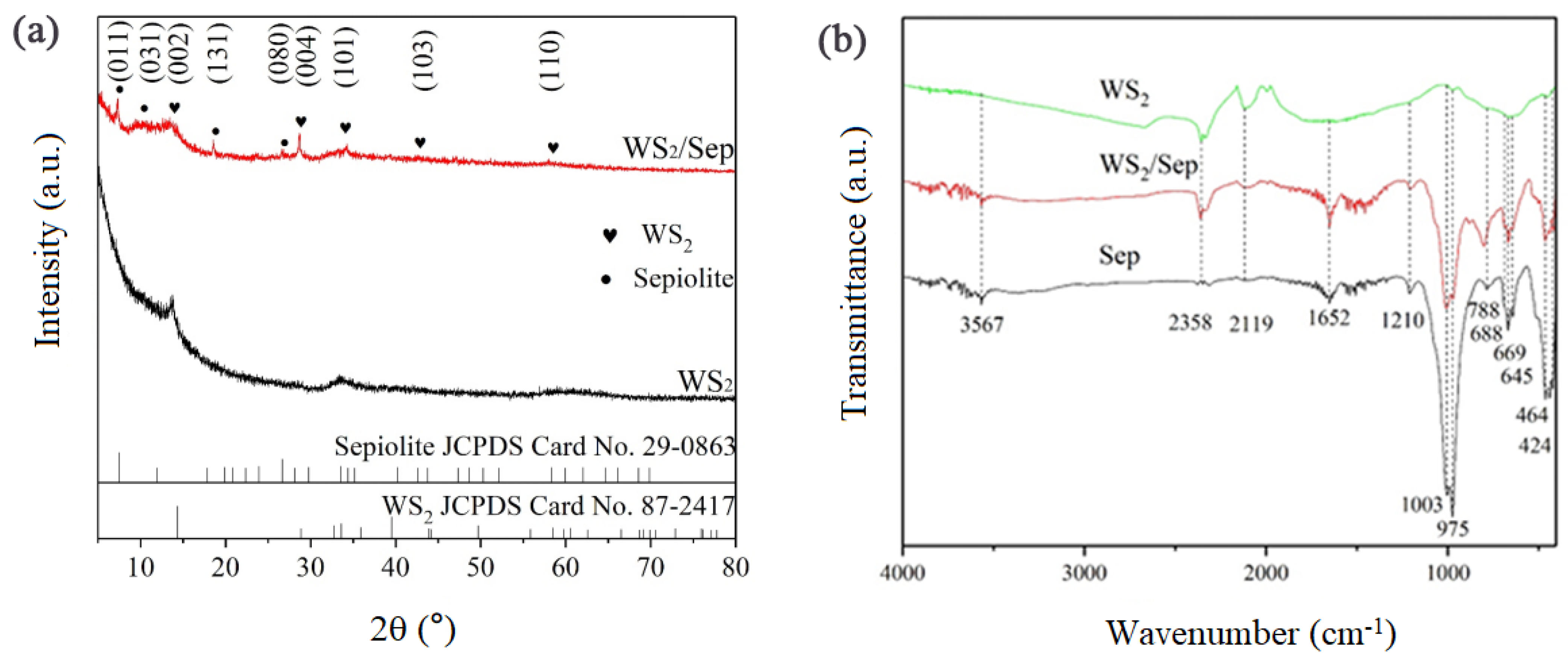
2.1. Crystal Phase and Groups Analysis

XRD patterns were recorded to examine the phase of the prepared pure WS2 and WS2/Sep composite (Figure 1a). All of the diffraction peaks of both the pure WS2 nanosheets and WS2/Sep composite matched well with the 2H hexagonal phase (JCPDS Card No.87-2417) [22]. The XRD patterns showed that the main peaks of WS2 of the WS2/Sep were located at 2θ = 13.775°, 28.6989°, 34.248°, and 47.105°, corresponding to the (002), (004), (101), and (103) facets of WS2, and the diffraction peaks at 7.423°, 11.982°, 20.785°, and 26.667° corresponded well to the (011), (031), (131), and (080) facets of the Sep, respectively. The d-spacing value of the composite corresponding to the peak at 13.775° was calculated to be 6.4 Å, which was close to that of the pure WS2. The few broad bread-like peaks could be attributed to the partial crystallization of the WS2 nanosheets. After the solvothermal treatment, WS2 was evenly dispersed on the support, and the diffraction peaks corresponding to WS2 were sharp and symmetric, further confirming the great crystallization of composite. Furthermore, the FT-IR was used to identify the functional groups of the WS2/Sep sample (Figure 1b). The broad absorption peaks at around 3567 cm−1 and 1652 cm−1 were ascribed to the stretching vibration of the hydroxyl group of crystal water. The peaks at 1003, 975, 424, and 788 cm−1 were ascribed to the stretching of the Si-O band in the Si-O-Si groups of sepiolite. The peaks at 669, 688, and 645 cm−1 corresponded to the bending vibration of Mg3OH. The peaks at 464 were attributed to Si-O-Al (octahedral) and Si-O-Si [23,24,25]. The two intensive peaks at around 2358 and 2119 cm−1 originated from the characteristic peaks of hexagonal WS2. The results of XRD and FT-IR revealed the WS2/Sep maintained the original crystal structure and surface functional groups of WS2 and sepiolite.

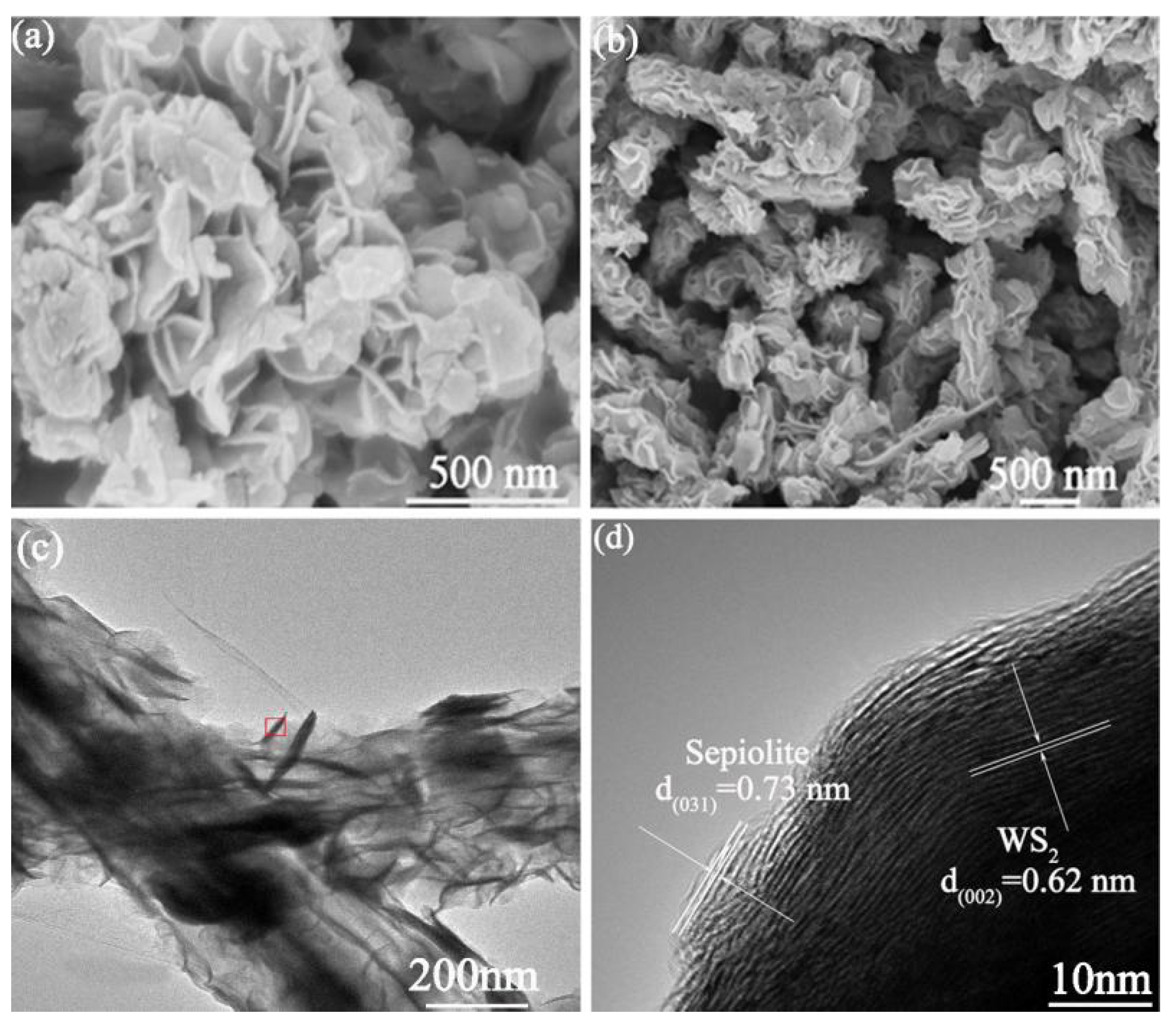
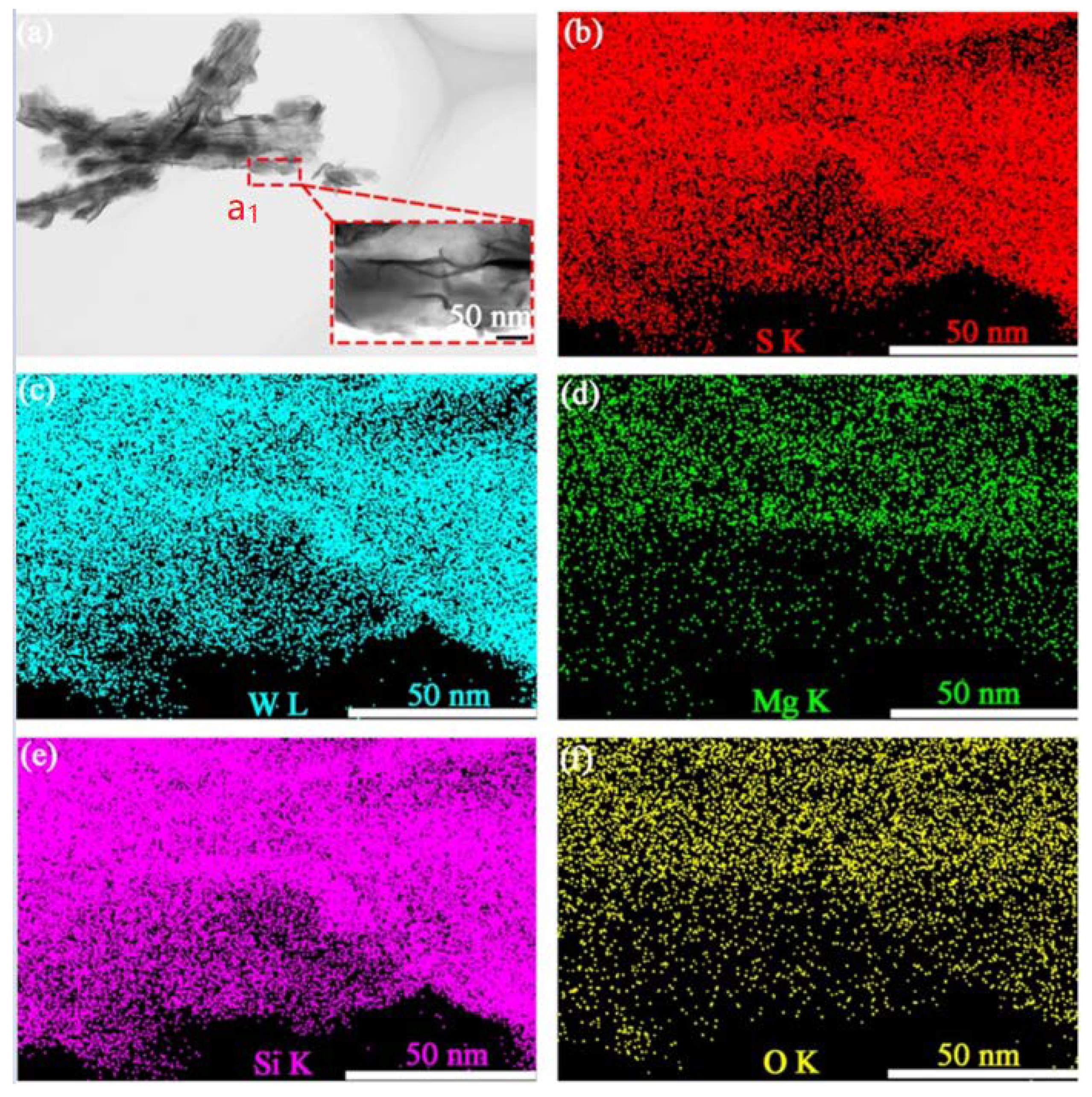
2.2. Morphology and EDS Analysis

SEM, TEM, and HRTEM were used to observe the microstructure of the pure WS2 and WS2/Sep (Figure 2). The pure WS2 with the shape of irregular plates consisted of WS2 nanosheets stacked compactly (Figure 2a). In Figure 2b, one can observe the WS2 nanosheets were uniformly anchored on the surface of Sep nanofibers, and a bark-like structure was formed intimately; the WS2 nanosheets in the WS2/Sep composite owned better dispersion. Compared with the pure WS2 nanosheets, the mineral material (i.e., Sep) as a support of the composite could significantly reduce the agglomeration of the WS2 nanosheets. Thus, more surface active sites of WS2 could be exposed outside. The above results provided direct evidence that the WS2 phase had been uniformly loaded on the Sep nanofibers.
TEM and HRTEM observations were adopted to deeply investigate the microstructure of the WS2/Sep composite (Figure 2c,d), which further illustrated that the WS2 phase had been loaded successfully on the surface of the Sep nanofibers. Moreover, the HRTEM images (Figure 2d) indicated a lattice space distance of 0.62 nm, which corresponded to the (002) facet of 2H-WS2 [26,27]. Lattice spacing consistent with the d-spacing of Sep (031) was also detected. In addition, the elemental mapping images (Figure 3) also confirmed that WS2 nanosheets were successfully assembled on the surface of Sep mineral.

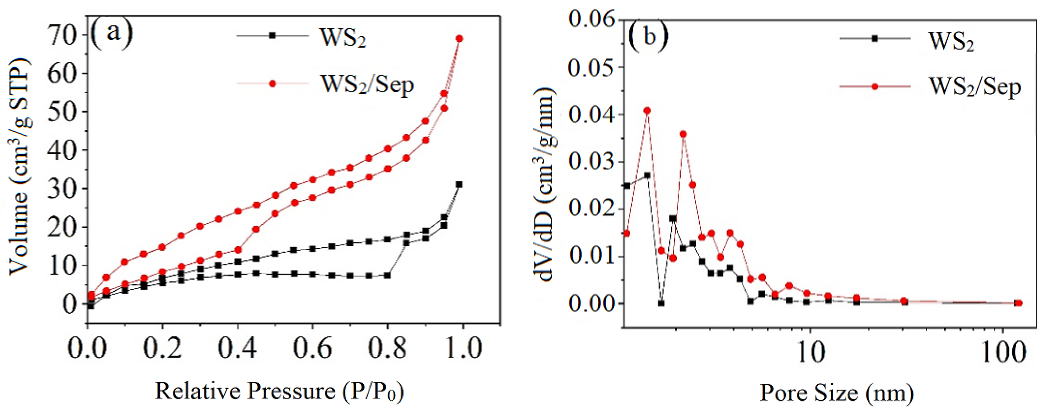
2.3. Nitrogen Adsorption-Desorption Analysis

Nitrogen adsorption-desorption isotherms and pore size distribution curves were adopted to further compare the surface area and pore structure between the WS2/Sep composite and pure WS2 (Figure 4). Both the samples presented Langmuir type IV isotherms with H3 hysteresis type loops, suggesting the existence of slit-like mesoporous structures because of the stacking of sheets. This was consistent with the electron microscopy images. The WS2/Sep composite possessed a specific surface area of 45.9 m2/g, which was much larger than that of the pure WS2 (25.3 m2/g). Meanwhile, the pore volume of the WS2/Sep (0.107 cm3/g) was also much larger than that of the WS2 (0.048 cm3/g). Consequently, the composite could expose more surface active sites and increase the number of mesopores, which tended toward improving catalytic activity [28].

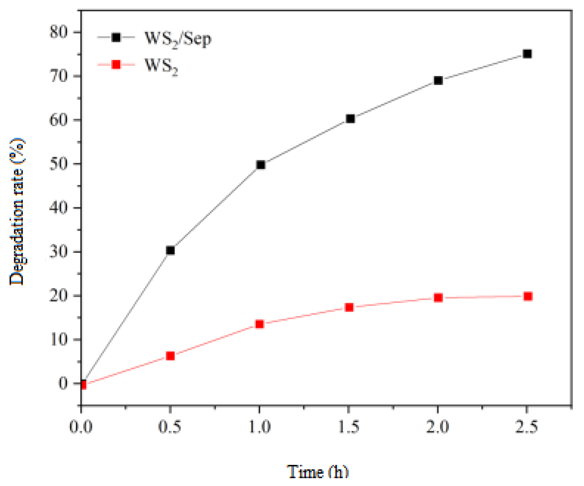
2.4. Photocatalytic Performance of RhB Degradation

Comparative experiments were performed to evaluate the performance of the WS2/Sep nanocomposite for RhB degradation, which is shown in Figure 5. It can be clearly seen that the WS2/Sep composite exhibited much higher photocatalytic efficiency than the pure WS2. After 150 min catalysis, the RhB degradation rate for the pure WS2 only reached ~18%, while it achieved ca. 76% for the WS2/Sep nanocomposites. The 4.2-fold improved activity could be mainly attributed to the better dispersion of the WS2 nanosheets [20]. In addition, the excellent synergies between the WS2 nanosheets and Sep nanofibers could also accelerate the photocatalysis, which will be further discussed in the following section.

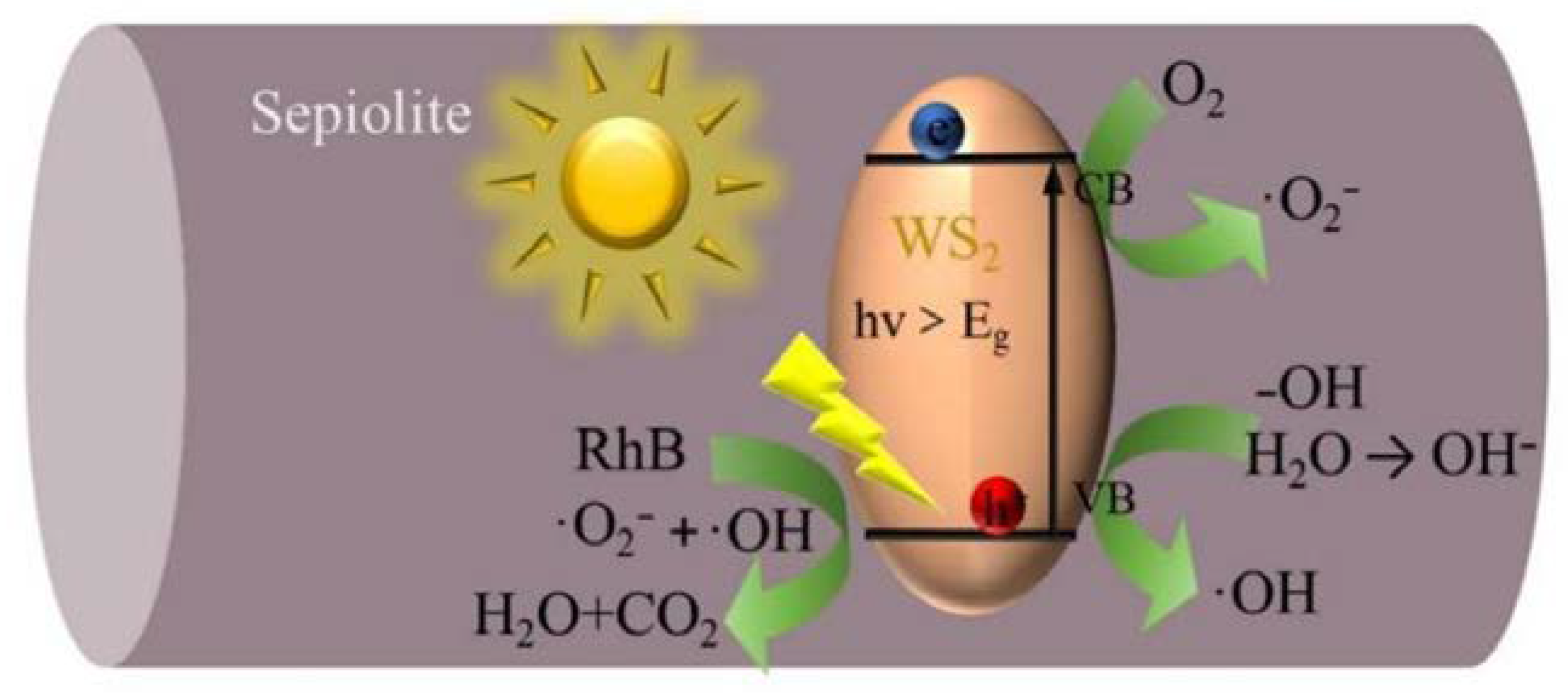
2.5. The Possible RhB Degradation Mechanism for the WS2/Sep Composite

Figure 6 shows the possible process of the photocatalytic RhB degradation in which the synergic effect between the Sep and the grown WS2 in the WS2/sepiolite nanocomposite is illustrated. Under irradiation, the holes (h+) and electrons (e) were produced. Then, the dissolved O2 would react with e and OH− would react with h+ to generate superoxide anion radical (·O2) and hydroxyl radical (·OH), respectively [29,30]. Finally, the RhB molecules adsorbed on the surface-active sites of the WS2 were degraded to CO2 and H2O [31,32]. During this process, the excellent adsorption of Sep was conductive to transferring the dissolved O2 and RhB molecules to the active sites of the WS2, and the abundant hydroxyl (–OH) groups on the Sep surface also benefited the production of ·OH [33,34,35]. Thus, in addition to the improved dispersion of WS2, the synergic effects provided by the Sep during photocatalysis also enabled the WS2/Sep composite to exhibit much higher photocatalytic activity than the pure WS2 [36]. Furthermore, the presence of mesopores favored multilight scattering, resulting in enhanced harvesting of the exciting light, and accordingly, improved photocatalytic activity [37,38]. These mesopores also facilitated fast mass transport and thus enhanced the performance [39,40].

3. Experiment

3.1. Chemicals and Reagents

The Sep was provided by LB Nanomaterials Technology Co., Ltd. (Henan, China), and the chemical analysis of the Sep was determined as SiO2 of 53.56 wt%, MgO of 36.79 wt%, CaO of 5.53 wt%, Fe2O3 of 1.17 wt%, and other impurities of 2.95 wt%. The ammonium tetrathiotungstate (H8N2S4W) and dimethylformamide (DMF) were supplied by Sigma-Aldrich Co., Ltd (Shanghai, China). The (RhB) was purchased from Kewei Chemical Group Co., Ltd. (Tianjin, China). All chemicals used in the experiments were of analytical grade. These materials were used without further purification. Deionized (DI) water was used in all experiments.

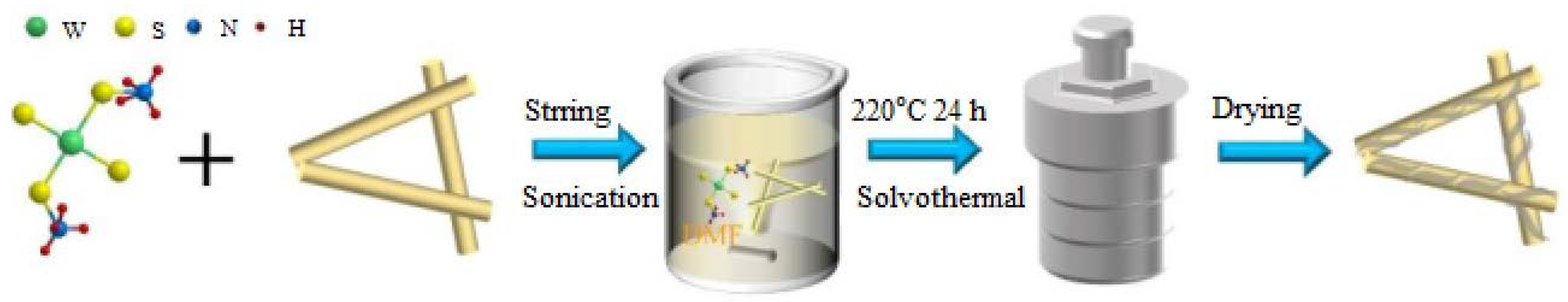
3.2. Synthesis of WS2/Sep Nanocomposite

The WS2/Sep nanocomposite was fabricated by a solvothermal method (Figure 7). A total of 30 mg of H8N2S4W was dissolved in 15 mL of N,N-dimethylformamide (DMF) and stirred for 0.5 h. Then, 10 mg of Sep powder was added into the solution and stirred continuously for 0.5 h. Next, the above mixture was sonicated for 10 min. Later, the above suspension was transferred into a 25 mL Teflon-sealed autoclave and heated to 220 °C for 24 h. After cooling down to room temperature, the final product was obtained by filtration, washed several times with DI water, and dried in a vacuum oven at 80 °C for 12 h. The preparation process of the pure WS2 was similar to that of the WS2/Sep nanocomposite but without the addition of Sep.

3.3. Physicochemical Characterizations of the Synthesized WS2/Sep Composite

X-ray powder diffraction (XRD) analysis was performed with the working conditions of Cu Kα radiation (λ = 1.54 Å), 40 mA, and 40 kV on a Smart Lab 9 KW X-ray diffractometer from Rigaku (Tokyo, Japan). Fourier-transform infrared spectroscopy (FTIR) spectra of the samples were recorded in a transmission mode from 400 to 4000 cm−1 on a Tensor II Fourier transform infrared spectrometer manufactured by Bruker (Saarbrucken, Germany). The morphologies of the as-synthesized samples were observed by using a S-4800 scanning electron microscope working at 5 kV, from JEOL (Tokyo, Japan). Transmission electron microscope (TEM) images, high-resolution TEM (HRTEM) images, and energy-dispersive X-ray spectroscopy (EDS) were carried out on a JEM 2100F transmission electron microscope, from JEOL (Tokyo, Japan), with an accelerating voltage of 200 kV. The N2 adsorption-desorption tests were conducted on Autosorb-iQ2, from Quantachrome (Boynton Beach, FL, USA). All samples were outgassed in nitrogen flow at 200 °C for 4 h before measurements. The specific surface area (SSA) was evaluated using the Brunauer-Emmett-Teller (BET) method, and the total pore volume was calculated at the relative pressure of approximately 0.99. The pore size distribution was computed using the Barrett-Joyner-Halenda (BJH) method.

3.4. Photocatalytic Performance Tests

Photocatalytic activity of the as-prepared samples was assessed by the reduction of RhB in aqueous solutions, using a 500 W Xe lamp equipped with a cut-off filter (λ > 420 nm). In a typical evaluation procedure, 20 mg of sample was added into 100 mL of RhB solution with a concentration of 20 mg/L at room temperature [41]. The suspension solution stirred vigorously in the dark for 0.5 h to reach the equilibrium of adsorption/desorption before visible light irradiation. At an interval of 0.5 h, 5.6 mL of the suspension was collected and centrifuged for absorbance analysis. The concentration of dye solution was analyzed by recording the UV–vis spectra at wavelength of 553 nm using a Shimadzu UV-1800 spectrophotometer from Shimadzu (Shimane, Japan).

4. Conclusions

In summary, a novel WS2/Sep composite as a photocatalyst was successfully prepared via a facile solvothermal method. The physicochemical characterization results confirmed that the bark-like WS2 nanosheets uniformly grew on the Sep nanofibers, which led to a larger surface area and more exposed active sites. In a typical photocatalytic application, the as-synthesized WS2/Sep exhibited considerably improved photocatalytic performance for RhB degradation over the pure WS2, which could be mainly attributed to the better dispersion of WS2 nanosheets and the synergistic effect between the WS2 nanosheets and the Sep nanofibers’ support during the photocatalysis. This work is believed to provide new ideas for the low-cost batch preparation of high-quality two-dimensional materials based on natural minerals. However, further investigation of the as-developed composite photocatalyst with respect to reusability and the versatility for the degradation of other organic pollutants are still needed to see if there are any limitations to its practical applications.

Author Contributions

Conceptualization, F.W. and J.B.; methodology, F.W. and J.B.; software, J.B. and K.C.; validation, F.W. and J.B.; formal analysis, F.W. and J.B.; investigation, J.B. and K.C.; resources, F.W.; data curation, J.B. and F.W.; writing—original draft preparation, J.B.; K.C.; X.X., B.F.; and F.W.; writing—review and editing, F.W. and B.F.; visualization, F.W. and B.F.; supervision, F.W.; project administration, F.W.; funding acquisition, F.W. All authors have read and agreed to the published version of the manuscript.

Funding

This work was financially supported by the National Key R&D Program of China (No. 2021YFC1910605), the National Natural Science Foundation of China (No. 51874115), the Introduced Overseas Scholars Program of Hebei province, China (No. C201808), and the Excellent Young Scientist Foundation of Hebei province, China (No. E2018202241).

Data Availability Statement

Data will be available upon request from the corresponding authors.

Acknowledgments

Authors acknowledge the financial support by the National Key R&D Program of China (No. 2021YFC1910605), the National Natural Science Foundation of China (No. 51874115), the Introduced Overseas Scholars Program of Hebei province, China (No. C201808), and the Excellent Young Scientist Foundation of Hebei province, China (No. E2018202241).

Conflicts of Interest

The authors declare no conflict of interest.

References

  1. Yahya, N.; Aziz, F.; Jamaludin, N.A.; Mutalib, M.A.; Ismail, A.F.; Salleh, W.N.W.; Jaafar, J.; Yusof, N.; Ludin, N.A. A review of integrated photocatalyst adsorbents for wastewater treatment. J. Environ. Chem. Eng. 2018, 6, 7411–7425. [Google Scholar] [CrossRef]
  2. Yaqoob, A.A.; Parveen, T.; Umar, K.; Mohamad Ibrahim, M.N. Role of nanomaterials in the treatment of wastewater: A review. Water 2020, 12, 495. [Google Scholar] [CrossRef] [Green Version]
  3. Liao, G.; Li, C.; Li, X.; Fang, B. Emerging polymeric carbon nitride Z-scheme systems for photocatalysis. Cell Rep. Phys. Sci. 2021, 2, 100355. [Google Scholar] [CrossRef]
  4. Cong, C.; Shang, J.; Wang, Y.; Yu, T. Optical properties of 2D semiconductor WS2. Adv. Opt. Mater. 2018, 6, 1700767. [Google Scholar] [CrossRef]
  5. Manzeli, S.; Ovchinnikov, D.; Pasquier, D.; Yazyev, O.V.; Kis, A. 2D transition metal dichalcogenides. Nat. Rev. Mater. 2017, 2, 17033. [Google Scholar] [CrossRef]
  6. Yuan, L.; Chung, T.F.; Kuc, A.; Wan, Y.; Xu, Y.; Chen, Y.P.; Heine, T.; Huang, L. Photocarrier generation from interlayer charge-transfer transitions in WS2-graphene heterostructures. Sci. Adv. 2018, 4, e1700324. [Google Scholar] [CrossRef] [Green Version]
  7. Wang, X.; Gu, D.; Li, X.; Lin, S.; Zhao, S.; Rumyantseva, M.; Gaskov, A. Reduced graphene oxide hybridized with WS2 nanoflakes based heterojunctions for selective ammonia sensors at room temperature. Sens. Actuators B Chem. 2019, 282, 290–299. [Google Scholar] [CrossRef]
  8. Lu, Z.; Cao, Z.; Hu, E.; Hu, K.; Hu, X. Preparation and tribological properties of WS2 and WS2/TiO2 nanoparticles. Tribol. Int. 2019, 130, 308–316. [Google Scholar] [CrossRef]
  9. Su, L.; Luo, L.; Song, H.; Wu, Z.; Tu, W.; Wang, Z.J.; Ye, J. Hemispherical shell-thin lamellar WS2 porous structures composited with CdS photocatalysts for enhanced H2 evolution. Chem. Eng. J. 2020, 388, 124346. [Google Scholar] [CrossRef]
  10. Pataniya, P.M.; Late, D.; Sumesh, C.K. Photosensitive WS2/ZnO nano-heterostructure-based electrocatalysts for hydrogen evolution reaction. ACS Appl. Energy Mater. 2021, 4, 755–762. [Google Scholar] [CrossRef]
  11. Gao, J.; Liu, C.; Wang, F.; Jia, L.; Duan, K.; Liu, T. Facile synthesis of heterostructured WS2/Bi2MoO6 as high-performance visible-light-driven photocatalysts. Nanoscale Res. Lett. 2017, 12, 377. [Google Scholar] [CrossRef] [PubMed] [Green Version]
  12. Chen, Q.; Zhu, R.; Liu, S.; Wu, D.; Fu, H.; Zhu, J.; He, H. Self-templating synthesis of silicon nanorods from natural sepiolite for high-performance lithium-ion battery anodes. J. Mater. Chem. A 2018, 6, 6356–6362. [Google Scholar] [CrossRef]
  13. Li, Z.; Gómez-Avilés, A.; Sellaoui, L.; Bedia, J.; Bonilla-Petriciolet, A.; Belver, C. Adsorption of ibuprofen on organo-sepiolite and on zeolite/sepiolite heterostructure: Synthesis, characterization and statistical physics modeling. Chem. Eng. J. 2019, 371, 868–875. [Google Scholar] [CrossRef]
  14. Santos, S.C.R.; Boaventura, R.A.R. Adsorption of cationic and anionic azo dyes on sepiolite clay: Equilibrium and kinetic studies in batch mode. J. Environ. Chem. Eng. 2016, 4, 1473–1483. [Google Scholar] [CrossRef]
  15. Zhang, J.; Yan, Z.; Ouyang, J.; Yang, H.; Chen, D. Highly dispersed sepiolite-based organic modified nanofibers for enhanced adsorption of Congo red. Appl. Clay Sci. 2018, 157, 76–85. [Google Scholar] [CrossRef]
  16. Ma, Y.; Zhang, G. Sepiolite nanofiber-supported platinum nanoparticle catalysts toward the catalytic oxidation of formaldehyde at ambient temperature: Efficient and stable performance and mechanism. Chem. Eng. J. 2016, 288, 70–78. [Google Scholar] [CrossRef]
  17. Selvitepe, N.; Balbay, A.; Saka, C. Optimisation of sepiolite clay with phosphoric acid treatment as support material for CoB catalyst and application to produce hydrogen from the NaBH4 hydrolysis. Int. J. Hydrogen Energy 2019, 44, 16387–16399. [Google Scholar] [CrossRef]
  18. Xu, X.; Chen, W.; Zong, S.; Ren, X.; Liu, D. Atrazine degradation using Fe3O4-sepiolite catalyzed persulfate: Reactivity, mechanism and stability. J. Hazard. Mater. 2019, 377, 62–69. [Google Scholar] [CrossRef]
  19. Zhou, F.; Yan, C.; Sun, Q.; Komarneni, S. TiO2/Sepiolite nanocomposites doped with rare earth ions: Preparation, characterization and visible light photocatalytic activity. Micropor. Mesopor. Mater. 2019, 274, 25–32. [Google Scholar] [CrossRef]
  20. Wang, F.; Hao, M.; Liu, W.; Yan, P.; Fang, B.; Li, S.; Liang, J.; Zhu, M.; Cui, L. Low-cost fabrication of highly dispersed atomically-thin MoS2 nanosheets with abundant active Mo-terminated edges. Nano Mater. Sci. 2021, 3, 205–212. [Google Scholar] [CrossRef]
  21. Hao, M.; Li, H.; Cui, L.; Liu, W.; Fang, B.; Liang, J.; Xie, X.; Wang, D.; Wang, F. Higher photocatalytic removal of organic pollutants using pangolin-like composites made of 3–4 atomic layers of MoS2 nanosheets deposited on tourmaline. Environ. Chem. Lett. 2021, 19, 3573–3582. [Google Scholar] [CrossRef]
  22. Sun, Y.; Wu, J.; Zhang, L. Fabrication of Ag-WS2 composites with preferentially oriented WS2 and its anisotropic tribology behavior. Mater. Lett. 2020, 260, 126975. [Google Scholar] [CrossRef]
  23. Arabkhani, P.; Javadian, H.; Asfaram, A.; Sadeghfar, F.; Sadegh, F. Synthesis of magnetic tungsten disulfide/carbon nanotubes nanocomposite (WS2/Fe3O4/CNTs-NC) for highly efficient ultrasound-assisted rapid removal of amaranth and brilliant blue FCF hazardous dyes. J. Hazard. Mater. 2021, 420, 126644. [Google Scholar] [CrossRef] [PubMed]
  24. Uğurlu, M.; Karaoğlu, M.H. TiO2 supported on sepiolite: Preparation, structural and thermal characterization and catalytic behaviour in photocatalytic treatment of phenol and lignin from olive mill wastewater. Chem. Eng. J. 2011, 166, 859–867. [Google Scholar] [CrossRef]
  25. Zhang, Y.; Wang, D.; Zhang, G. Photocatalytic degradation of organic contaminants by TiO2/sepiolite composites prepared at low temperature. Chem. Eng. J. 2011, 173, 1–10. [Google Scholar] [CrossRef]
  26. Anto Jeffery, A.; Nethravathi, C.; Rajamathi, M. Two-dimensional nanosheets and layered hybrids of MoS2 and WS2 through exfoliation of ammoniated MS2 (M = Mo, W). J. Phys. Chem. C 2014, 118, 1386–1396. [Google Scholar] [CrossRef]
  27. Yang, J.; Voiry, D.; Ahn, S.J.; Kang, D.; Kim, A.Y.; Chhowalla, M.; Shin, H.S. Two-dimensional hybrid nanosheets of tungsten disulfide and reduced graphene oxide as catalysts for enhanced hydrogen evolution. Angew. Chem. Int. Ed. 2013, 52, 13751–13754. [Google Scholar] [CrossRef]
  28. Liu, C.; Chai, B.; Wang, C.; Yan, J.; Ren, Z. Solvothermal fabrication of MoS2 anchored on ZnIn2S4 microspheres with boosted photocatalytic hydrogen evolution activity. Int. J. Hydrogen Energy 2018, 43, 6977–6986. [Google Scholar] [CrossRef]
  29. Wu, Y.; Liu, Z.; Li, Y.; Chen, J.; Zhu, X.; Na, P. Construction of 2D-2D TiO2 nanosheet/layered WS2 heterojunctions with enhanced visible-light-responsive photocatalytic activity. Chin. J. Catal. 2019, 40, 60–69. [Google Scholar] [CrossRef]
  30. Feng, Z.; Zeng, L.; Chen, Y.; Ma, Y.; Zhao, C.; Jin, R.; Lu, Y.; Wu, Y.; He, Y. In situ preparation of Z-scheme MoO3/g-C3N4 composite with high performance in photocatalytic CO2 reduction and RhB degradation. J. Mater. Res. 2017, 32, 3660–3668. [Google Scholar] [CrossRef]
  31. Baral, A.; Das, D.; Minakshi, M.; Ghosh, M.; Padhi, D. Probing environmental remediation of RhB organic dye using α-MnO2 under visible-light irradiation: Structural, photocatalytic and mineralization studies. ChemistrySelect 2016, 1, 4277–4285. [Google Scholar] [CrossRef]
  32. Zhang, Q.; Tai, M.; Zhou, Y.; Zhou, Y.; Wei, Y.; Tan, C.; Wu, Z.; Li, J.; Lin, H. Enhanced Photocatalytic Property of γ-CsPbI3 Perovskite Nanocrystals with WS2. ACS Sustain. Chem. Eng. 2019, 8, 1219–1229. [Google Scholar] [CrossRef]
  33. Elangovan, E.; Sivakumar, T.; Brindha, A.; Thamaraiselvi, K.; Sakthivel, K.; Kathiravan, K.; Aishwarya, S. Visible Active N-Doped TiO2/WS2 Heterojunction Nano Rods: Synthesis, Characterization and Photocatalytic Activity. J. Nanosci. Nanotechnol. 2019, 19, 4429–4437. [Google Scholar] [CrossRef] [PubMed]
  34. Fu, S.; Yuan, W.; Liu, X.; Yan, Y.; Liu, H.; Li, L.; Zhao, F.; Zhou, J. A novel 0D/2D WS2/BiOBr heterostructure with rich oxygen vacancies for enhanced broad-spectrum photocatalytic performance. J. Colloid. Interface Sci. 2020, 569, 150–163. [Google Scholar] [CrossRef]
  35. Xiang, Q.; Cheng, F.; Lang, D. Hierarchical Layered WS2/Graphene-Modified CdS Nanorods for Efficient Photocatalytic Hydrogen Evolution. ChemSusChem 2016, 9, 996–1002. [Google Scholar] [CrossRef] [PubMed]
  36. Liu, X.; Xing, Z.; Zhang, Y.; Li, Z.; Wu, X.; Tan, S.; Yu, X.; Zhu, Q.; Zhou, W. Fabrication of 3D flower-like black N-TiO2-x@MoS2 for unprecedented-high visible-light-driven photocatalytic performance. Appl. Catal. B Environ. 2017, 201, 119–127. [Google Scholar] [CrossRef]
  37. Liu, Y.; Shen, S.; Li, Z.; Ma, D.; Xu, G.; Fang, B. Mesoporous g-C3N4 nanosheets with improved photocatalytic performance for hydrogen evolution. Mater. Charact. 2021, 174, 111031. [Google Scholar] [CrossRef]
  38. Liu, Y.; Xu, G.; Ma, D.; Li, Z.; Yan, Z.; Xu, A.; Zhong, W.; Fang, B. Synergistic effects of g-C3N4 three-dimensional inverse opals and Ag modification on high-efficiency photocatalytic H2 evolution. J. Clean. Prod. 2021, 328, 129745. [Google Scholar] [CrossRef]
  39. Fang, B.; Kim, J.; Kim, M.; Yu, J. Hierarchical nanostructured carbons with meso-macroporosity: Design, characterization and applications. Acc. Chem. Res. 2013, 46, 1397–1406. [Google Scholar] [CrossRef] [PubMed]
  40. Fang, B.; Daniel, L.; Bonakdarpour, A.; Govindarajan, R.; Sharman, J.; Wilkinson, D. Dense Pt nanowire electrocatalysts for improved fuel cell performance using a graphitic carbon nitride-decorated hierarchical nanocarbon support. Small 2021, 17, 2102288. [Google Scholar] [CrossRef]
  41. Ji, H.; Fei, T.; Zhang, L.; Yan, J.; Fan, Y.; Huang, J.; Song, Y.; Man, Y.; Tang, H.; Xu, H.; et al. Synergistic effects of MoO2 nanosheets and graphene-like C3N4 for highly improved visible light photocatalytic activities. Appl. Surf. Sci. 2018, 457, 1142–1150. [Google Scholar] [CrossRef]
Figure 1. (a) XRD patterns of the pure WS2 and WS2/Sep nanocomposites obtained via solvothermal at 220 °C (b) and FT-IR spectra of the pure WS2, sepiolite, and WS2/Sep nanocomposites.
Catalysts 12 01400 g001
Figure 2. Morphology and structure of the pure WS2 and WS2/sepiolite nanocomposite. (a,b) SEM images of the pure WS2 and WS2/sepiolite nanocomposite obtained via solvothermal; (c,d) TEM and HRTEM images of WS2/Sep.
Catalysts 12 01400 g002
Figure 3. EDS mapping analysis of element distribution (bf) in marked area (red rectangle in (a)) for the WS2/Sep nanocomposite: S (b), W (c), Mg (d), Si (e), and O (f).
Catalysts 12 01400 g003
Figure 4. Nitrogen adsorption-desorption isotherms (a) and the corresponding pore size distribution curves (b) of WS2 and WS2/sepiolite composite.
Catalysts 12 01400 g004
Figure 5. Photocatalytic activity of the WS2/Sep composite and pure WS2.
Catalysts 12 01400 g005
Figure 6. Possible RhB degradation mechanism of the WS2/Sep composite.
Catalysts 12 01400 g006
Figure 7. Schematic illustration for the synthesis process of WS2/Sep composite.
Catalysts 12 01400 g007
Publisher’s Note: MDPI stays neutral with regard to jurisdictional claims in published maps and institutional affiliations.

