Article

A Colorimetric CO2 Hydration Assay for Facile, Accurate, and Precise Determination of Carbonic Anhydrase Activity

1
Division of Applied Life Science and ABC-RLRC, Gyeongsang National University, Jinju 52828, Korea
2
Division of Life Science, Research Institute of Life Science and ABC-RLRC, Gyeongsang National University, Jinju 52828, Korea
*
Author to whom correspondence should be addressed.
Catalysts 2022, 12(11), 1391; https://doi.org/10.3390/catal12111391
Submission received: 27 September 2022 / Revised: 27 October 2022 / Accepted: 7 November 2022 / Published: 8 November 2022
(This article belongs to the Section Biocatalysis)

Abstract

:
A simple, reliable, and universal method is demanded for routine determination of carbonic anhydrase (CA) activity, overcoming the limitations of previous assays that are inaccurate, complicated, expensive, or limited to a specific enzyme family. The most widely used Wilbur–Anderson assay was modified to improve the speed, accuracy, and precision by employing a temperature controllable UV/Vis spectrophotometer and the pH indicator phenol red. The experimental setting, measurement, and data analysis were facile and straightforward. The assay was validated using a commercially available bovine CA, showing that the obtained activity was directly proportional to the amount of enzyme. The measured activity (2540 WAU mg−1) agreed well with the previously reported data. The comparison results with esterase assay showed that the CO2 hydration assay should not be substituted by the esterase assay in the measurement of CA activity. The simple and reliable colorimetric method can be widely adopted for the routine determination of CO2 hydration activity, substituting for the traditional Wilbur–Anderson assay.

1. Introduction

Carbonic anhydrase (CA, EC 4.2.1.1) is an enzyme that rapidly catalyzes the reversible hydration of carbon dioxide (CO2) [1]. CAs are classified into eight families (α, β, γ, δ, ζ, η, θ, and ι) that have convergently evolved [2]. CA is a metabolically and physiologically important enzyme involved in photosynthesis, respiration, CO2 transportation, pH regulation, and biomineralization. Recently, CA enzymes have been studied and engineered to develop biocatalysts for CO2 capture and utilization [3]. In addition, because of its physiological importance in pathogenic bacteria and cancer cells, CA has been the target for developing antibiotics and anticancer drugs [4,5]. With the increasing attention on this enzyme, facile and reliable methods are highly demanded for a routine determination of CA activity.
Currently, the most widely used assays for CA activity rely on two different reactions: ester hydrolysis and CO2 hydration, which are both catalyzed at the same active site of CA [6]. The esterase assay is a colorimetric method that generally employs 4-nitrophenyl acetate (p-NPA) as the substrate. The esterase assay is simple and reliable; however, it is primarily limited to the activity measurement of α-CAs because only the α-CAs clearly exhibit esterase activity, although there are a few reports of p-NPA-hydrolyzing CAs from η-, θ-, and ι-families [7,8,9,10].
Two methods are widely used for the measurement of CO2 hydration activity. One is the stopped-flow spectroscopic assay. This method is unique in that it enables the determination of steady-state kinetic parameters such as Kcat and KM by allowing precise and accurate measurement of initial rates of CO2 hydration at different substrate concentrations [11]. However, a stopped-flow spectrophotometer is uncommon and highly expensive. Moreover, the preparation for the experiment is not quite simple, and the data analysis is relatively complicated. The other method is the electrometric method known as the Wilbur–Anderson (WA) assay using a pH meter [12]. This method does not require any special equipment, and the preparation for the experiment is simple, making it the most widely used assay to date for routine determination of CA activity. However, this method has a severe drawback: it is difficult to reliably measure the enzyme activity due to (i) the slow response of pH electrode [13,14], (ii) the relatively large reaction volume (5–10 mL) required to accommodate a common pH electrode [15,16], which prevents the reaction solution from fast homogenous mixing, and (iii) the adsorption of proteins and chemicals on the electrode membrane, which may interfere with the correct measurement.
In this paper, we introduce a modified WA assay that allows facile, accurate, and precise measurement of CO2 hydration activity at a relatively low cost. This method has already been successfully used in previous studies [14,17,18,19], but the detailed experimental settings and protocols for the measurement and the data analysis as well as the validation of the assay have not been published elsewhere.

2. Results and Discussion

2.1. General Description of the Assay

During the CO2 hydration reaction, protons are released, which causes a decrease in the pH of the solution:
CO2(aq) + H2O → HCO3 + H+
The pH change is directly measured using a pH meter in the WA method [12]. In contrast, the pH change is indirectly monitored with a spectrophotometer as the color change of a pH indicator in our approach. Tris (pKa = 8.1) with a pH of 8.3 at 25 °C is used as the reaction buffer, and phenol red (pKa = 7.9) is used as the pH indicator because its pKa value is close to that of Tris, allowing accurate reflection of the pH change by the color change. The total reaction volume is approximately 1 mL, which is much smaller than that used in the previous WA method. This novel colorimetric method is based on the technique developed by Datta and Shepard II [13]. In the previous method, CO2 gas was directly injected into the cuvette inside the spectrophotometer, which required a complicated experimental setting. Instead, our approach adopts a more straightforward preparation, utilizing the separately prepared CO2-saturated water as the substrate, the same as the WA assay. We present the detailed experimental and analytical procedures below.

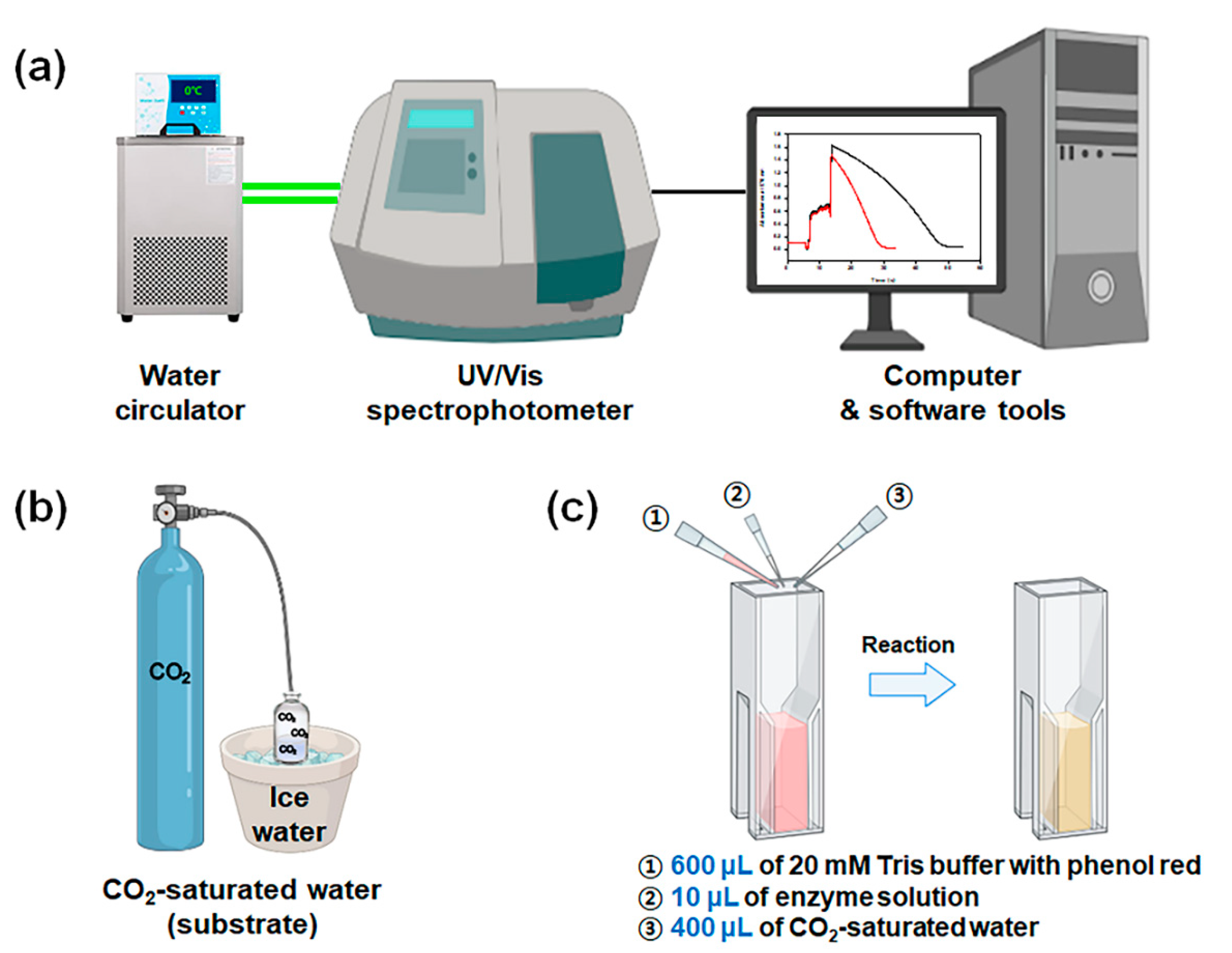
2.2. Experimental Settings and Spectrophotometric Monitoring of the Reaction

2.2.1. Equipment Setting

The spectrophotometer was connected to a computer for data communication. The computer was equipped with a software tool for real-time measurement and recording (Figure 1a). In the software tool, kinetics mode was selected for time-course measurement of absorbance change, and the data acquisition interval was set to 0.1 s. The wavelength was set to 570 nm, which is the absorption maximum of phenol red. Deionized water was used for the baseline correction at 570 nm. The temperature of the cuvette holder was set to 0 °C using the water circulator.

2.2.2. Preparation of Materials

Disposable cuvettes were prepared according to the expected number of measurements. The optical sides of the cuvettes were treated with an anti-fog agent prior to the measurement to prevent the formation of fog outside the cuvette due to the temperature difference between the cuvette holder and the environment. Both the reaction buffer (20 mM Tris, 100 μM phenol red, pH 8.3 adjusted using H2SO4) and enzyme solution were placed on ice water and equilibrated at 0 °C. To prepare CO2-saturated water, a glass serum bottle containing approximately 50 mL of deionized water was placed on ice water, and then pure CO2 gas was bubbled into the bottle at a flow rate of approximately 2 L min−1 (Figure 1b). This CO2 bubbling was started at least 30 min before the first measurement and was continued until all the sample measurements were completed.

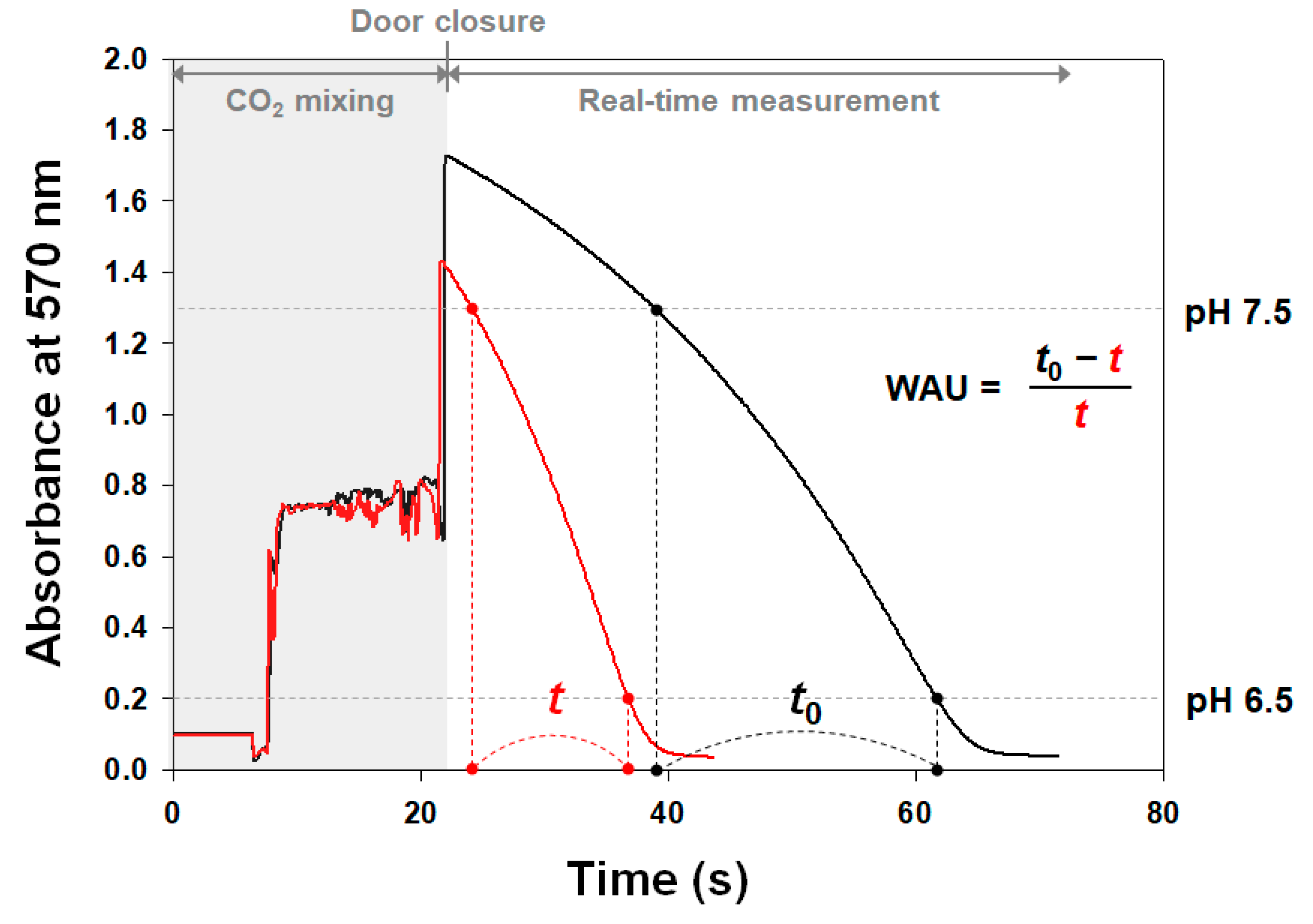
2.2.3. Initiation and Measurement of the Reaction

First, an empty cuvette was placed in the cell holder, and then both 600 μL of reaction buffer and 10 μL of enzyme solution were sequentially added to the cuvette. Second, the data acquisition was started on the computer software, and then 400 μL of CO2-saturated water was added to the cuvette and thoroughly mixed by repetitive pipetting to initiate the reaction (Figure 1c). Then, the spectrophotometer door was immediately closed, and the reaction was monitored at 570 nm in real time. When the absorbance decreased to a minimum value, the measurement was stopped, and the data was saved for further analysis. For the measurement of the noncatalyzed reaction (blank), the same volume of the corresponding buffer was added instead of the enzyme solution.

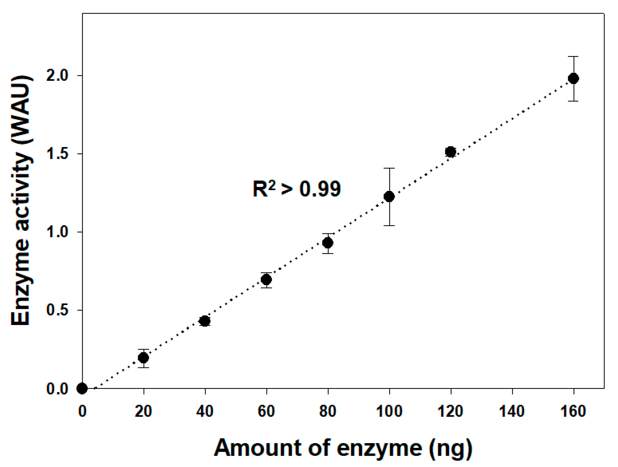
2.3. Data Analysis and Determination of Enzyme Unit

The Wilbur–Anderson unit (WAU) was calculated from the obtained data. First, the amount of time taken for the pH to drop from 7.5 to 6.5 was acquired for the activity calculation (Figure 2). As the pH change was indirectly measured via the absorbance change, the absorbance values corresponding to pH 7.5 and pH 6.5 were required for the calculation. These values were predetermined by measuring the absorbance of a mixture of 600 μL of reaction buffer and 400 μL of deionized water with the pH adjusted to 7.5 or 6.5 using HCl. In our experimental setting, the absorbance values corresponding to pH 7.5 and pH 6.5 were 1.3 and 0.2, respectively. The obtained times for the enzyme sample and blank were designated t and t0, respectively. In our experiments, the t0 value was determined to be 23.80 ± 0.53 s when a 20 mM sodium phosphate buffer (pH 7.5) was used as the blank buffer. Next, the WAU was calculated as follows:
WAU   = t 0 t t
Depending on the total reaction volume, a different WAU value can be obtained even though the same amount of enzyme is used. This problem has not been addressed well in previous studies and can lead to a wrong conclusion in a comparative study on CA activity performed under a different experimental setting [14]. For example, suppose that the same amount of enzyme is added to a 5 mL reaction solution (3 mL of Tris buffer/2 mL of CO2-saturated water) instead of a 1 mL reaction solution. In that case, it will show only one-fifth of the activity obtained from the 1 mL reaction solution because the concentration of enzyme will be diluted by one-fifth. Thus, if necessary, the WAU value obtained by our method can be further corrected for comparison with previously published data.

2.4. Validation of the Assay

Commercially available bovine CA (bCA) was used for the validation of our method. The bCA powder was weighed and dissolved in 20 mM sodium phosphate buffer (pH 7.5) to prepare bCA solution with 1 mg mL−1 concentration. By dilution, enzyme solutions were prepared with various concentrations from 2 μg mL−1 to 16 μg mL−1, and their activities for CO2 hydration were measured. The obtained WAU values were directly proportional to the amount of enzyme with the coefficient of determination (R2) above 0.99, demonstrating the validity of the method (Figure 3). When the activity was higher than 2 WAU, the absorbance data without missing points from 1.3 (corresponding to pH 7.5) could not be reliably obtained due to the fast reaction, suggesting that enzyme samples with high activity need to be appropriately diluted before the measurement. The activity of bCA was determined to be 12,700 WAU mg−1 based on the 1 mL reaction volume, and this could be translated to 2540 WAU mg−1 based on a 5 mL reaction volume. This value coincides nicely with the data (≥2000 WAU mg−1) provided by the company and the previously published data (2600–3300 WAU mg−1), both obtained by using the electrometric method in a 5 mL reaction volume [15]. The limit of detection (LOD) and the limit of quantitation (LOQ) were determined to be 15.0 ng and 45.4 ng, respectively, using the linear regression method [20].

2.5. Advantages and Limitations of the Assay

The limitations of the WA assay that arise by using a pH electrode can be completely resolved by our method. In contrast to a slow response of pH electrode to pH change, the absorbance change on the spectrophotometer can be immediately recorded and visualized. In the traditional WA assay, stirring may be required to increase the mixing speed. However, stirring can decrease the accuracy of measurement by accelerating the mass transfer at the air–liquid interface (CO2(aq) → CO2(g)), and thus reducing the substrate concentration. In contrast, fast homogenous mixing is possible by simple repetitive pipetting by virtue of the small reaction volume in our assay. These advantages lead to the reliable measurement of CA activity with the improved accuracy and precision.
Our method has more advantages over the original WA method as follows: (i) Fast and simple measurement: a single measurement maximally takes only 2 min. When using the original method, the reaction vial and pH electrode should be thoroughly washed with distilled water after each measurement. These steps are time-consuming and laborious. (ii) Small quantity of enzyme: due to the small reaction volume, only a small amount of enzyme sample is required for a single measurement. (iii) Easy monitoring: the absorbance change by the reaction can be visualized in real time, which can be easily monitored and compared with previous data. This greatly accelerates our decision on whether the sample should be re-measured due to an unexpected problem.
In addition to free CA enzymes, the activity of immobilized enzymes can be also measured using our method. However, the particle size of immobilized enzymes should be small enough to be suspended in the reaction solution without settlement until the reaction is finished. For example, bacterial cells expressing CA [14] or silica particles with immobilized CA [18] can be used in our method. If the activity of the enzyme-immobilized particles is too low, then a higher number of particles should be added to the reaction solution. In this case, a high turbidity caused by the particles can severely interfere with the accurate measurement of absorbance change at 570 nm. Therefore, activity measurement of large-sized particles or particles with low activity should be done by using the traditional WA method with proper stirring.
Conventionally, the WA assay has been conducted at the low temperature condition, because under higher temperature conditions, the enzymatic reaction is too fast to be reliably measured. Practically, the measurements have been conducted on ice water because the temperature can be held constant easily by using ice; at any other temperature, temperature controller is definitely required to keep the temperature constant. In our method, the temperature controller is employed, so in principle, the reaction temperature is not restricted to 0 °C. It can be possible to adopt a higher temperature condition; however, in this case, the experimental settings might need to be re-optimized.

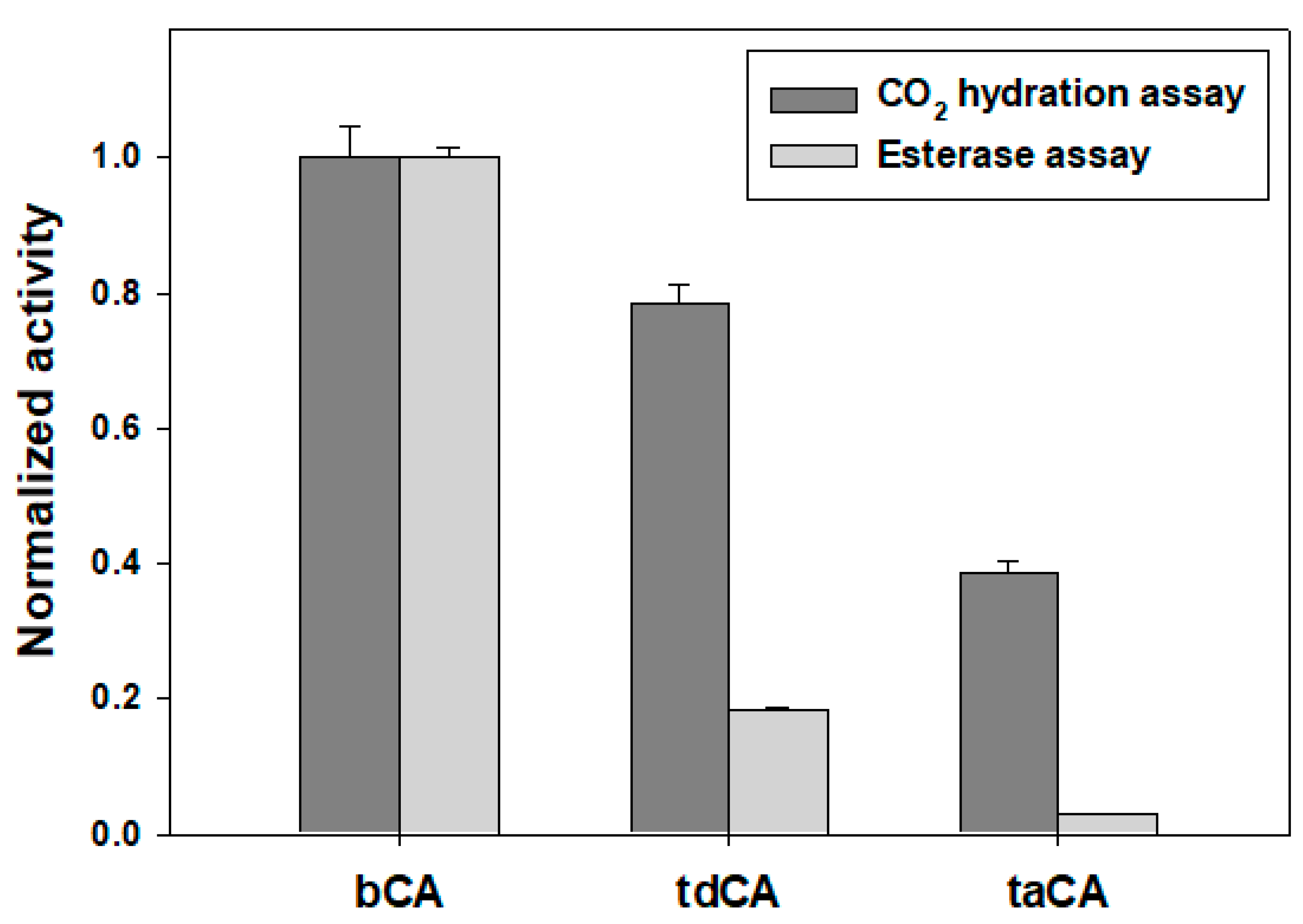
2.6. Comparison with the Esterase Assay

Among the eight distinct families, the α-family CAs are the most studied, and many recent studies have been conducted using α-CAs as the model or the target CAs. As α-CAs can catalyze ester hydrolysis in addition to the CO2 hydration, the facile esterase assay has been routinely utilized to assess and compare the activities of α-CAs. In this study, we compared the activities of several α-CAs including bCA, Thermosulfurimonas dismutans CA (tdCA), and Thermovibrio ammonificans CA (taCA) by using both the CO2 hydration assay and the esterase assay. The measured activity of ester hydrolysis was not commensurate with that of CO2 hydration (Figure 4). For example, the activity of bCA was 2.6-fold higher than that of taCA when assessed by the CO2 hydration assay; however, bCA showed 35.4-fold higher activity than taCA when the esterase assay was used (Figure 4). Although the discrepancy between the CO2 hydration activity and the esterase activity of α-CA has been shown in previous studies [17,21,22,23,24], this limitation is not well-known and may lead to serious misinterpretation of enzyme activity. To conclude, the CO2 hydration assay should not be substituted by the esterase assay.

3. Experimental

3.1. Materials for CO2 Hydration Assay

A UV/Vis spectrophotometer (UV-1800; Shimadzu, Kyoto, Japan) with temperature controllable cuvette holder was used for the assay. A circulating cooling bath (Jeio Tech, Daejeon, Korea) filled with an anti-freezing solution was attached to the spectrophotometer. The spectrophotometer was connected to a desktop computer with the software UVProbe 2.60 (Shimadzu). Disposable acrylic semi-micro cuvettes (Sarstedt, Nümbrecht, Germany) with a path length of 10 mm were used. Phenol red, sulfuric acid (H2SO4), and hydrochloric acid (HCl) were purchased from Sigma-Aldrich (St. Louis, MO, USA). Sodium hydroxide (NaOH) was purchased from Daejung Chemicals (Siheung, Korea). Tris was purchased from Duchefa Biochemie (Haarlem, Netherlands). A glass serum bottle (125 mL) was purchased from Sigma-Aldrich. Gas bubbling was conducted using compressed CO2 in a gas cylinder with a purity of 99.5% (Daepoong, Jinju, Korea). A bovine CA (bCA, Sigma-Aldrich, Cat. #C3934) was used for the assay validation. An anti-fog detergent was purchased from Sonax (Neuburg an der Donau, Germany).

3.2. Preparation of Recombinant CAs for Comparison of Assays

The tdCA and taCA were produced by recombinant Escherichia coli BL21 (DE3) strains and purified by His6-tag affinity chromatography as previously described [25]. The enzymes, including bCA, were dialyzed against 20 mM sodium phosphate buffer supplemented with 300 mM NaCl (pH 7.5). Molar concentration of enzyme was determined by using the absorbance of denatured enzyme solution in 6 M guanidine hydrochloride/20 mM sodium phosphate (pH 7.5) and the molar extinction coefficient at 280 nm calculated by ProtParam (http://web.expasy.org/protparam/, (accessed on 25 September 2022); 50,420 M−1 cm−1 for bCA, 35,535 M−1 cm−1 for tdCA, and 38,515 M−1 cm−1 for taCA).

3.3. Esterase Assay

Esterase activity was measured by the colorimetric method as previously described [17]. The reaction buffer (20 mM sodium phosphate supplemented with 300 mM NaCl, pH 7.5), the enzyme solution (10 μM), and the substrate solution (30 mM p-NPA in acetonitrile, Sigma-Aldrich) were sequentially mixed with a volume ratio of 8:1:1 in the disposable cuvette. The absorbance change at 405 nm by p-NPA hydrolysis was monitored at 25 °C inside the spectrophotometer. The enzyme-catalyzed rate of p-NPA hydrolysis was determined by subtracting the uncatalyzed rate of p-NPA hydrolysis, which was obtained by adding the corresponding blank buffer instead of the enzyme solution, from the measured rate.

4. Conclusions

The novel colorimetric method provides a fast and simple means for the determination of CO2 hydration activity of CA in a more reliable manner than the existing methods using a pH electrode. In addition to the improved speed, accuracy, and precision, our method requires only a small quantity of enzyme and allows easy monitoring of the reaction. However, caution is needed when immobilized enzymes are used for the measurement. This assay can substitute the traditional WA assay and can be widely adopted for the routine measurement of CO2 hydration activity.

Author Contributions

B.H.J. contributed to the conception and design of the study. Material preparation, data collection and analysis were performed by J.H.K. and B.H.J. The first draft of the manuscript was written by J.H.K. and the manuscript was revised by B.H.J. All authors have read and agreed to the published version of the manuscript.

Funding

This work was supported by the National Research Foundation grants (NRF-2021R1F1A1057310 and NRF-2021R1A5A8029490) funded by the Ministry of Science and ICT, Korea.

Data Availability Statement

All data generated during the current study are available from the corresponding author on request.

Conflicts of Interest

The authors declare no competing interest.

References

  1. Tripp, B.C.; Smith, K.; Ferry, J.G. Carbonic anhydrase: New insights for an ancient enzyme. J. Biol. Chem. 2001, 276, 48615–48618. [Google Scholar] [CrossRef] [Green Version]
  2. Hirakawa, Y.; Senda, M.; Fukuda, K.; Yu, H.Y.; Ishida, M.; Taira, M.; Kinbara, K.; Senda, T. Characterization of a novel type of carbonic anhydrase that acts without metal cofactors. BMC Biol. 2021, 19, 105. [Google Scholar] [CrossRef]
  3. Talekar, S.; Jo, B.H.; Dordick, J.S.; Kim, J. Carbonic anhydrase for CO2 capture, conversion and utilization. Curr. Opin. Biotechnol. 2022, 74, 230–240. [Google Scholar] [CrossRef] [PubMed]
  4. Supuran, C.T. Inhibition of bacterial carbonic anhydrases and zinc proteases: From orphan targets to innovative new antibiotic drugs. Curr. Med. Chem. 2012, 19, 831–844. [Google Scholar] [CrossRef]
  5. Thiry, A.; Supuran, C.T.; Masereel, B.; Dogne, J.M. Recent developments of carbonic anhydrase inhibitors as potential anticancer drugs. J. Med. Chem. 2008, 51, 3051–3056. [Google Scholar] [CrossRef] [PubMed]
  6. Angeli, A.; Carta, F.; Supuran, C.T. Carbonic anhydrases: Versatile and useful biocatalysts in chemistry and biochemistry. Catalysts 2020, 10, 1008. [Google Scholar] [CrossRef]
  7. Innocenti, A.; Supuran, C.T. Paraoxon, 4-nitrophenyl phosphate and acetate are substrates of α- but not of β-, γ- and ζ-carbonic anhydrases. Bioorg. Med. Chem. Lett. 2010, 20, 6208–6212. [Google Scholar] [CrossRef] [PubMed]
  8. Giovannuzzi, S.; De Luca, V.; Nocentini, A.; Capasso, C.; Supuran, C.T. Coumarins inhibit η-class carbonic anhydrase from Plasmodium falciparum. J. Enzyme Inhib. Med. Chem. 2022, 37, 680–685. [Google Scholar] [CrossRef]
  9. Kikutani, S.; Nakajima, K.; Nagasato, C.; Tsuji, Y.; Miyatake, A.; Matsuda, Y. Thylakoid luminal θ-carbonic anhydrase critical for growth and photosynthesis in the marine diatom Phaeodactylum tricornutum. Proc. Natl. Acad. Sci. USA 2016, 113, 9828–9833. [Google Scholar] [CrossRef] [PubMed] [Green Version]
  10. Jensen, E.L.; Clement, R.; Kosta, A.; Maberly, S.C.; Gontero, B. A new widespread subclass of carbonic anhydrase in marine phytoplankton. ISME J. 2019, 13, 2094–2106. [Google Scholar] [CrossRef]
  11. Khalifah, R.G. The carbon dioxide hydration activity of carbonic anhydrase: I. Stop-flow kinetic studies on the native human isoenzymes b and c. J. Biol. Chem. 1971, 246, 2561–2573. [Google Scholar] [CrossRef]
  12. Wilbur, K.M.; Anderson, N.G. Electrometric and colorimetric determination of carbonic anhydrase. J. Biol. Chem. 1948, 176, 147–154. [Google Scholar] [CrossRef]
  13. Datta, P.K.; Shepard II, T.H. Carbonic anhydrase: A spectrophotometric assay. Arch. Biochem. Biophys. 1959, 79, 136–145. [Google Scholar] [CrossRef]
  14. Jo, B.H.; Moon, H.J.; Cha, H.J. Engineering the genetic components of a whole-cell catalyst for improved enzymatic CO2 capture and utilization. Biotechnol. Bioeng. 2020, 117, 39–48. [Google Scholar] [CrossRef]
  15. Jo, B.H.; Kim, I.G.; Seo, J.H.; Kang, D.G.; Cha, H.J. Engineered Escherichia coli with periplasmic carbonic anhydrase as a biocatalyst for CO2 sequestration. Appl. Environ. Microbiol. 2013, 79, 6697–6705. [Google Scholar] [CrossRef] [PubMed] [Green Version]
  16. Li, C.X.; Jiang, X.C.; Qiu, Y.J.; Xu, J.H. Identifcation of a new thermostable and alkali-tolerant α-carbonic anhydrase from Lactobacillus delbrueckii as a biocatalyst for CO2 biomineralization. Bioresourc. Bioproc. 2015, 2, 44. [Google Scholar] [CrossRef] [Green Version]
  17. Jo, B.H.; Im, S.K.; Cha, H.J. Halotolerant carbonic anhydrase with unusual N-terminal extension from marine Hydrogenovibrio marinus as novel biocatalyst for carbon sequestration under high-salt environments. J. CO2 Util. 2018, 26, 415–424. [Google Scholar] [CrossRef]
  18. Kim, S.; Joo, K.I.; Jo, B.H.; Cha, H.J. Stability-controllable self-immobilization of carbonic anhydrase fused with a silica-binding tag onto diatom biosilica for enzymatic CO2 capture and utilization. ACS Appl. Mater. Interfaces 2020, 12, 27055–27063. [Google Scholar] [CrossRef] [PubMed]
  19. Wi, S.; Hwang, I.S.; Jo, B.H. Engineering a plant viral coat protein for in vitro hybrid self-assembly of CO2-capturing catalytic nanofilaments. Biomacromolecules 2020, 21, 3847–3856. [Google Scholar] [CrossRef] [PubMed]
  20. Shrivastava, A.; Gupta, V. Methods for the determination of limit of detection and limit of quantitation of the analytical methods. Chron. Young Sci. 2011, 2, 21–25. [Google Scholar] [CrossRef]
  21. Jo, B.H.; Seo, J.H.; Yang, Y.J.; Baek, K.; Choi, Y.S.; Pack, S.P.; Oh, S.H.; Cha, H.J. Bioinspired silica nanocomposite with autoencapsulated carbonic anhydrase as a robust biocatalyst for CO2 sequestration. ACS Catal. 2014, 4, 4332–4340. [Google Scholar] [CrossRef]
  22. Jo, B.H.; Park, T.Y.; Park, H.J.; Yeon, Y.J.; Yoo, Y.J.; Cha, H.J. Engineering de novo disulfide bond in bacterial α-type carbonic anhydrase for thermostable carbon sequestration. Sci. Rep. 2016, 6, 29322. [Google Scholar] [CrossRef] [PubMed] [Green Version]
  23. Warden, A.C.; Williams, M.; Peat, T.S.; Seabrook, S.A.; Newman, J.; Dojchinov, G.; Haritos, V.S. Rational engineering of a mesohalophilic carbonic anhydrase to an extreme halotolerant biocatalyst. Nat. Commun. 2015, 6, 10278. [Google Scholar] [CrossRef] [PubMed] [Green Version]
  24. Zhang, S.; Zhang, Z.; Lu, Y.; Rostam-Abadi, M.; Jones, A. Activity and stability of immobilized carbonic anhydrase for promoting CO2 absorption into a carbonate solution for post-combustion CO2 capture. Bioresour. Technol. 2011, 102, 10194–10201. [Google Scholar] [CrossRef]
  25. Jo, B.H.; Hwang, I.S. Characterization and high-level periplasmic expression of thermostable α-carbonic anhydrase from Thermosulfurimonas dismutans in Escherichia coli for CO2 capture and utilization. Int. J. Mol. Sci. 2020, 21, 103. [Google Scholar] [CrossRef]
Figure 1. The facile, precise, and accurate assay for the colorimetric determination of CO2 hydration activity. (a) Instrumental setup for spectrophotometer equipped with temperature-controllable cell holder. (b) Preparation of CO2-saturated water as the enzyme substrate. (c) Components of the CO2 hydration reaction. Figures were created with BioRender.com.
Catalysts 12 01391 g001
Figure 2. Data analysis for the calculation of enzyme activity in Wilbur–Anderson unit (WAU). The graph shows (black line) the noncatalyzed reaction and (red line) the enzyme-catalyzed reaction that were separately measured at 570 nm in a time-dependent manner.
Catalysts 12 01391 g002
Figure 3. Experimental validation of the assay. The direct proportionality between the amount of enzyme and the measured activity is shown. Error bars represent standard deviations from two independent experiments.
Catalysts 12 01391 g003
Figure 4. Activities of α-CAs measured by two different assays. The measured activities were normalized to the activity of bCA in each assay. Error bars represent standard deviations from two independent experiments.
Catalysts 12 01391 g004
Publisher’s Note: MDPI stays neutral with regard to jurisdictional claims in published maps and institutional affiliations.

