Article

Thermal Stability and Utilization of 1D-Nanostructured Co3O4 Rods Derived by Simple Solvothermal Processing

1
Department of Inorganic Chemical Technology and Nonmetals, Faculty of Chemical Engineering and Technology, University of Zagreb, Marulićev trg 20, 10000 Zagreb, Croatia
2
Department of Chemistry and Applied Bioscience, ScopeM, Eidgenössische Technische Hochschule Zürich, Auguste-Piccard-Hof 1, 8093 Zürich, Switzerland
*
Author to whom correspondence should be addressed.
Catalysts 2022, 12(10), 1162; https://doi.org/10.3390/catal12101162
Submission received: 9 September 2022 / Revised: 29 September 2022 / Accepted: 30 September 2022 / Published: 2 October 2022
(This article belongs to the Special Issue Nanomaterials for Photocatalysis)

Abstract

:
For p-type semiconductor nanoparticles, such as the cobalt oxide spinel, enhancing the nanoparticle geometry can expose more of the surface and bring up the sensitivity and applicability, pointing to even more advantageous behaviour in comparison to n-type semiconductors which are known for a somewhat faster reactivity. Here, we present a strategy that relies on fostering a simple synthetic route that can deliver reasonably or comparably performing p-type-semiconducting partially 1D-Co3O4 material prepared under less technically and economically demanding conditions. Structurally monophasic Co3O4 nanoparticles with a spinel structure were indicated by powder X-ray diffraction, while the presence of traces of organic-phase residuals in otherwise chemically homogeneous material was observed by Fourier-transform infrared spectroscopy. Scanning electron microscopy further showed that the observed fine nanoparticle matter formed agglomerates with the possible presence of rod-like formations. Interestingly, using transmission electron microscopy, it was possible to reveal that the agglomerates of the fine nanoparticulated material were actually nanostructured, i.e., the presence of 1D-shaped Co3O4 rods embedded in fine nanoparticulated matrix was confirmed. In conjunction with the N2 adsorption–desorption isotherms, discussion about the orientation, exposure of nanostructured rod domains, and derivative geometry parameters was possible. The nanostructured Co3O4 material was shown to be stable up to 800 °C whereat the decomposition to CoO takes place. The specific surface area of the nanostructured sample was raised. For the purpose of testing the photoactivity of the prepared samples, simple sorption/photodegradation tests using methylene blue as the model pollutant were performed. The degradation performance of the prepared nanostructured Co3O4 was better described by a pseudo-second-order fit, suggesting that the prepared material is worth further development toward improved and stable immobilized photocatalysts.

1. Introduction

Semiconducting materials based on mixed metal oxides are convenient for application as gas sensors, photocatalysts, or photovoltaics, due to some of their unique properties but also some quite general advantages. Namely, gas sensors require materials with a vapour-sensitive surface, photocatalytic systems require materials with an oxygen-sensitive surface, and photovoltaic devices require materials with a photon-sensitive surface. The unique properties are specific electronic structure properties yielding specific bandgaps and specific surface reactivity conditions, while general advantages are the ability to be derived over a low-price synthesis, into a robust material, whose thermodynamics and reaction mechanisms are well known and are widely applicable.
Among various metal-oxide-based materials for interaction with harmful or toxic gases, various n-type semiconductor materials (SnO2, ZnO, TiO2, WO3, In2O3, and Fe2O3) [1,2,3] and p-type semiconductor materials (NiO, CuO, Mn3O4, and Co3O4) [4,5,6,7] have emerged. Having the equivalent morphologies, n-type semiconductors are known for a somewhat faster reactivity (response), leading to a considerably lower amount of p-type semiconductor-related reports [7]. Among those, a cobalt oxide (Co3O4) spinel is found. It is a mixed-valence spinel of CoO and Co2O3 with a high-oxygen-content, p-type semiconductor with an indirect band gap in the range between 1.6 and 2.2 eV. Its p-type conductivity originates from oxygen over-stoichiometry caused by metal vacancies as well as excess interstitial oxygen. As previously suggested, Co3O4 is studied for application in energy storage [8], catalysis [9], sensors [10], electrochemistry [11], magnetism [12], and gas sensing. For example, catalytic and sensing fields of application for Co3O4 heavily rely on surface performance which, in turn, heavily relates to morphologic properties and thermal stability properties. Specifically, for photocatalytic performance, operation occurs under ambient conditions; for sensing performance, the operation is usually at elevated temperatures above 200 °C; for catalytic performance, operations at even higher temperatures are required [8,9,10]. We bring about the necessity to enhance the investigation of morphology development and thermal stability in order to show the importance of Co3O4 p-type material for the abovementioned application. Namely, when electrons become trapped, charge carrier pairs are created. Under ambient conditions, oxygen species readily adsorb at the Co3O4 surface, causing the creation of additional charge carriers at the surface. Oxidizing gases other than the atmospheric oxygen, O2, for example, ozone or nitrous oxide, generally tend to enhance this effect, while reducing gases tend to decrease this effect, both of which affect surface conductivity, i.e., yield a qualitative and quantitative electric signal [7,13]. Finally, the materials can be utilised in constituents of the photovoltaic devices.
There are a lot of routes reported on how to prepare Co3O4 nanoparticles [14]. In general, the majority of them rely on complex, toxic, or expensive precursors, time-demanding or high-temperature processing, etc. [14]. Therefore, the solvothermal route has been recognised and is a significant milestone for simple, affordable, and convenient preparation of Co3O4 nanoparticles with high purity and homogeneity [14]. To reach the area of advanced materials, conventional Co3O4 nanoparticles have to be nanostructured to increase the material’s specific surface area and pore volume [15,16]. In the following sections, we discuss Co3O4 nanostructures according to their performance for superstructured morphologies from zero-dimensional (0D) nanoparticles to three-dimensional (3D) networks, but with a focus on 1D nanoformations. Among nanostructured Co3O4 materials, the architectures of 1D Co3O4 nanorods, hollow nanotubes, porous nanowires, and nanofibers were evidenced as promising for advanced materials application. Of all the three mentioned areas where the Co3O4 nanostructures can be utilised, the material performance greatly depends on the material’s morphology and assembly. Thus, reports on the morphological control, including crystal size, external shape, surface structure, crystal orientation, controllable pore, stacking manners, aspect ratios, and even crystalline densities, are highly interesting [17,18].
Porous structures were found to be beneficial for gas sensing on behalf of increasing the surface reactive sites and facilitating the diffusion of target gases. One-dimensional nanofibers containing widely necked particles were more advantageous for achieving a high gas-sensing response with the less-elongated nanofibers showing more narrow inter-particle contacts [19]; in addition, hydrothermally derived mesoporous, macroporous, and 1D Co3O4 materials can have several times more effective gas-sensing than their isotropic 0D nanoparticle counterparts [20]. Choi et al. controlled the solvothermal reaction to yield cobalt-containing precursors in the form of nanorods, nanosheets, and nanocubes and converted all of them into nanostructured Co3O4 without morphological variation; they observed better gas-sensing responses, which was attributed to the less agglomerated nanostructures [21].
Co3O4 has been considered as a convenient candidate for the photodegradation of organic dyes in wastewater treatment due to its thermodynamic stability and other favourable properties such as low cost, easy preparation, and environmental friendliness [22]. As conventional wastewater treatment cannot completely degrade the pollutants down to non-hazardous substances, advanced oxidation processes (AOPs) were introduced as convenient tools for eliminating various organic pollutants from water under UV–VIS irradiation in the presence of photocatalysts [23]. In contrast to the widely used TiO2-based materials, Co3O4 has a narrower band gap (in the VIS part of the solar spectra, around 2 eV), better surface availability, and beneficial physical and chemical properties. The water contaminated with organic dyes discharged by various industries such as paint, textiles, paper, and plastics poses a great threat to the environment [24]. Co3O4 nanoparticles exhibit enhanced catalytic activity compared to bulk Co3O4, because of their large surface-to-volume ratio, size- and shape-dependent properties, and high concentration of under-coordinated active surface sites [22,25]. In comparison to other metal oxides, the performance of Co3O4 nanoparticles has been shown as advantageous many times [26,27,28]. Therefore, it is of great importance to control the preparation of nanosized Co3O4. Nanostructured Co3O4 has even been used in all-oxide heterojunction photovoltaics [29,30].
All the above results suggest that porous 1D nanostructured Co3O4 can easily serve as a highly performing material because of its high specific surface area. To prepare specifically 1D Co3O4 nanoparticles, one can use the template approach, which is, again, complex or slow, whereas direct chemical deposition, electrospinning, hydrothermal, and solvothermal synthesis are more favourable. Nanostructured Co3O4 can be prepared via intermediate cobalt carbonate, cobalt hydroxide, and cobalt carbonate-hydroxide, where, during their thermal annealing, the morphology becomes preserved [31,32,33]. It was shown that on behalf of co-precipitation followed by calcination, small nanorods 6–8 nm in diameter and 20–30 nm in length can be prepared [34]. It was found that the temperature of the solvothermal processing was the most important parameter for achieving the rhombic shape of the nanostructured 1D particles [35].
Here, we report on the simple course of solvothermal synthesis of Co3O4 that allowed the preparation of nanostructured formations in the matrix. The characterisation methods focused on describing compositional and structural aspects of the derived material. The foremost important was the description of the morphologic parameters with respect to thermal stability, both of which are fundamental for photocatalytic or photovoltaic or sensing applications. Photocatalytic efficiency was confirmed under some restrictions, but more importantly, high sorption efficiency was confirmed, which can be further optimized.

2. Results and Discussion

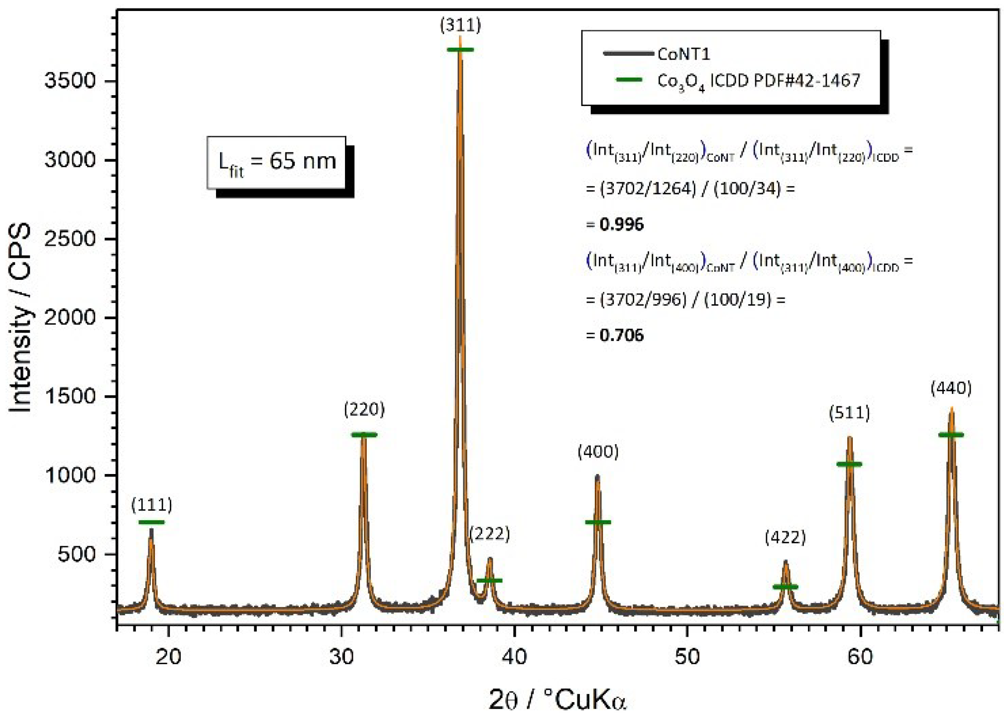
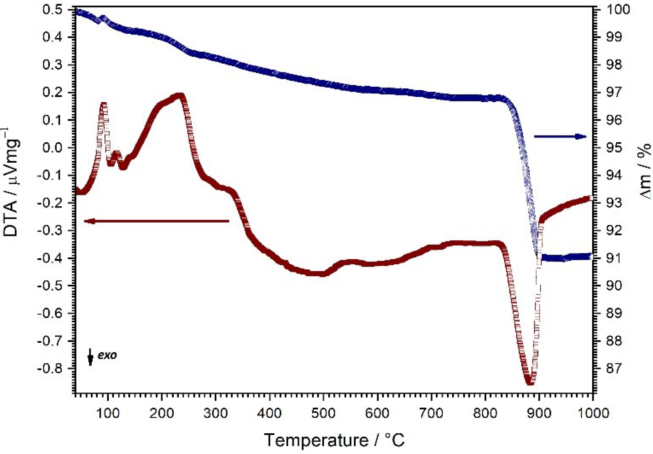
2.1. Phase Development and Thermal Evolution

Figure 1 presents the PXRD pattern of the sample synthesized by the solvothermal method. Observed diffraction peaks at 19.00, 31.27, 36.85, 38.54, 44.81, 55.66, 59.36, 65.24, and 68.63°2θ correspond to (111), (220), (311), (222), (400), (422), (511), (440), and (531) planes of cubic Co3O4 (ICDD PDF#42-1467). There are no residual peaks corresponding to other cobalt oxide phases or unassigned peaks. As can be seen, the diffraction peaks are broad due to the nanosized crystallites. The average spinel crystallite size was calculated by the Scherrer method and yielded 65 nm. The diffractogram was fitted and, using ICDD data, the aspect ratio of the peaks was compared. The comparison coefficient of 0.996 for the (220) plane suggests complete concordance with the isotropic particle configuration of the ICDD reference. However, the comparison coefficient of 0.706 for the (400) plane suggests the opposite. It seems that the particles in the samples are statistically not isotropic. At this point, the attempts to describe the exact aspect ratio to 1D or 2D configuration would be too speculative.
Figure 2 shows the FTIR spectra of the synthesized nanoparticles in the wavenumber range of 700–500 cm−1. Two strong bands at 660 and 560 cm−1 were observed. Bands at 660 and 560 cm−1 are due ν(Co–O) modes and are typical for crystalline Co3O4 [36]. Namely, Co3O4 is a mixed-valence compound having a spinel structure with oxygen atoms arranged in a cubic close-packed structure where Co2+ occupies tetragonal while Co3+ takes the octahedral interstices [17]. Therefore, the peak at 660 cm−1 is attributed to the tetrahedrally coordinated Co2+, while the peak at 560 cm−1 can be assigned to octahedrally coordinated Co3+ [37].
Thermal evolution and thermal behaviour of the derived nanoparticles were investigated by simultaneous thermal analysis that inherently provides us with more relevant information. Namely, the whole course of the sample evolution was monitored rather than properties at a given discrete state as with other methods, which makes the pinpointing of applicability ranges much more possible in order for adjustments in future experiments. Figure 3 shows TG and DTA curves in the temperature range between 25 °C and 1000 °C, recorded with a constant heating rate of 10 °C min1. The removal of the persisting solvents and adsorbed water is responsible for the first two endothermic peaks on the DTA curve, which are accompanied with minor mass loss observable on the TG curve. Following this, a somewhat stronger endotherm and mass loss are observed and ascribed to the decomposition of the remaining impurities related with precursors residuals. With the further increase in the heating, in the temperature range between 820 °C and 910 °C, one can observe a strong exotherm followed by considerable mass loss. These features are a consequence of the cobalt oxide spinel decomposition to cobalt (II) oxide [38]. Octahedral Co3+ cations undergo reduction due to the surface oxygen release at elevated temperatures in lean oxygen environments [39]. The reaction entropy is dominated by the oxygen gas release and the Gibbs energy of reaction products is decreased [40]. The sample mass loss in this temperature range is 6%, which indeed roughly fits the oxygen loss in the spinel decomposition process following the equation:
Co3O4 → 3CoO + ½O2

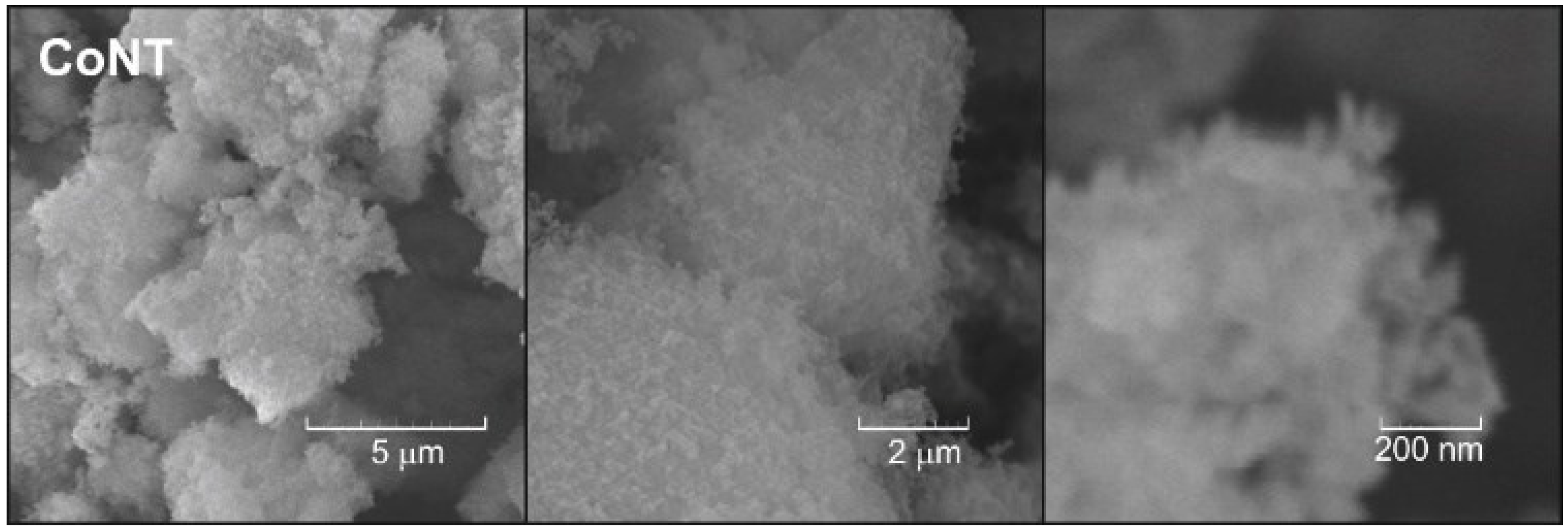
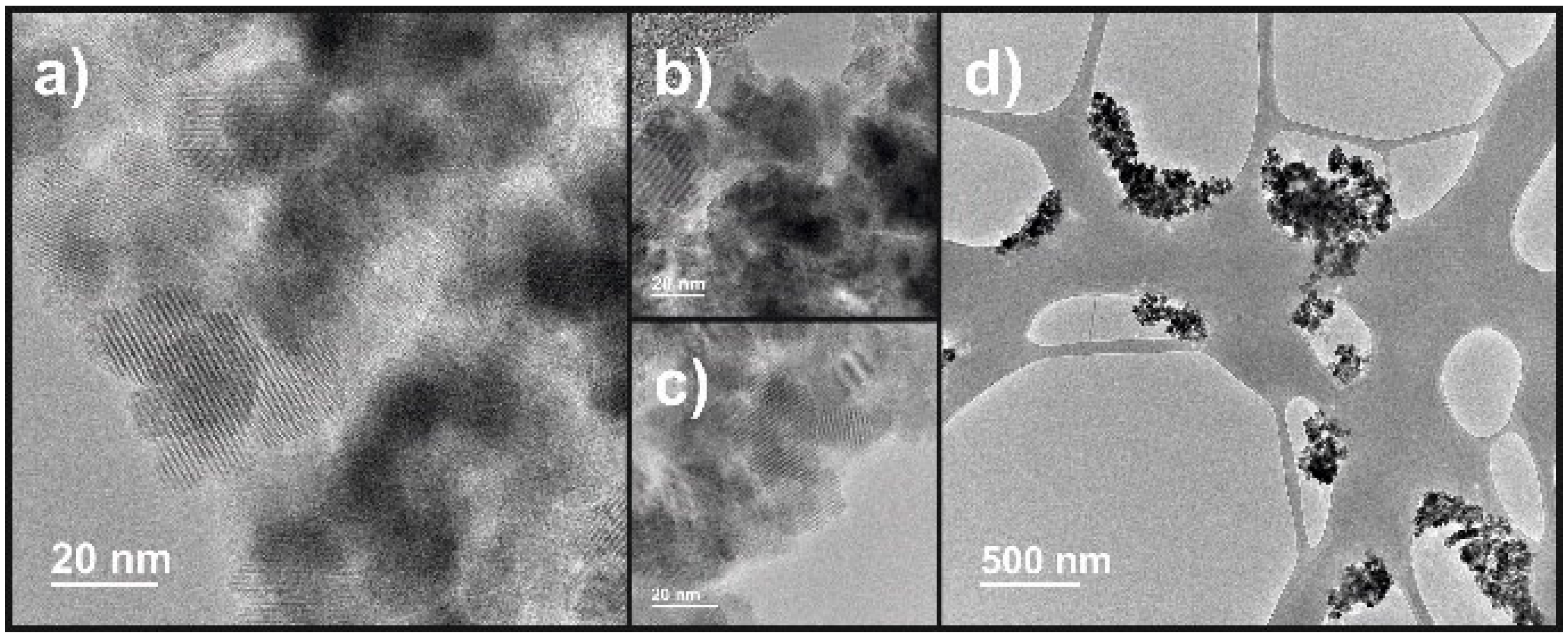
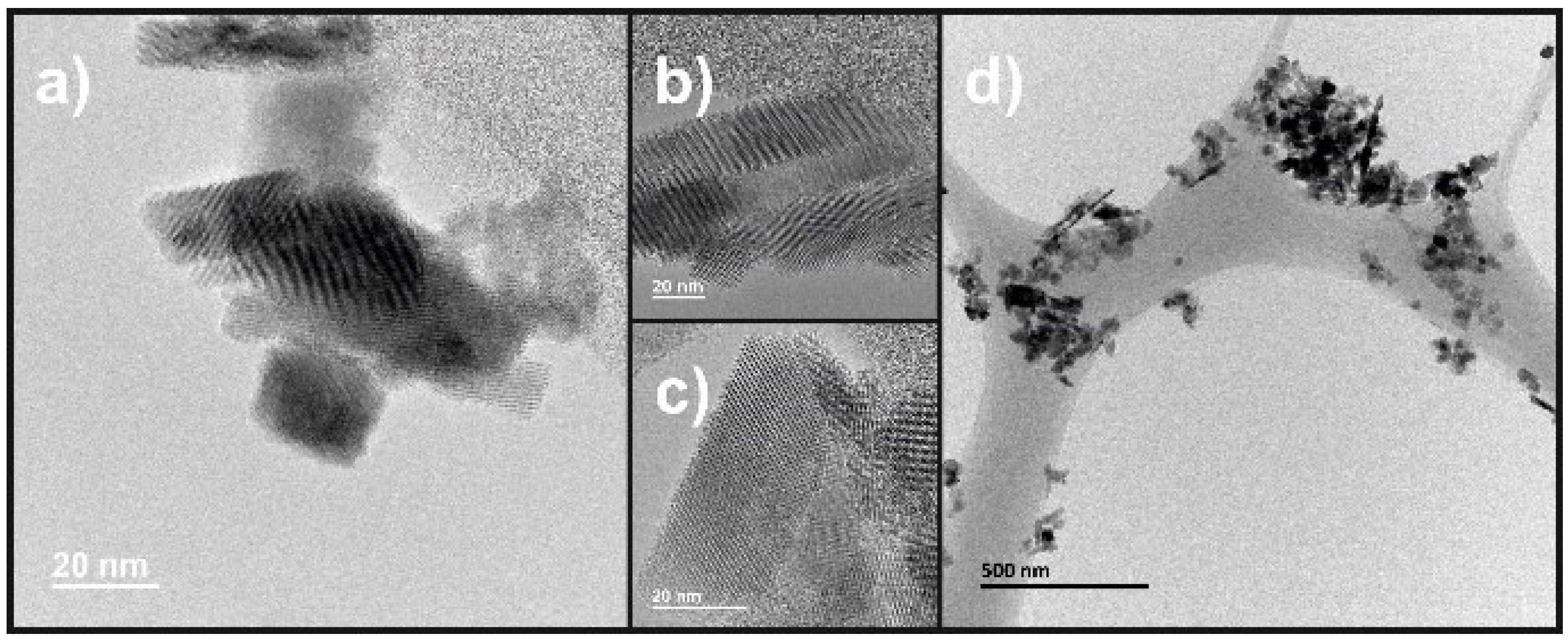
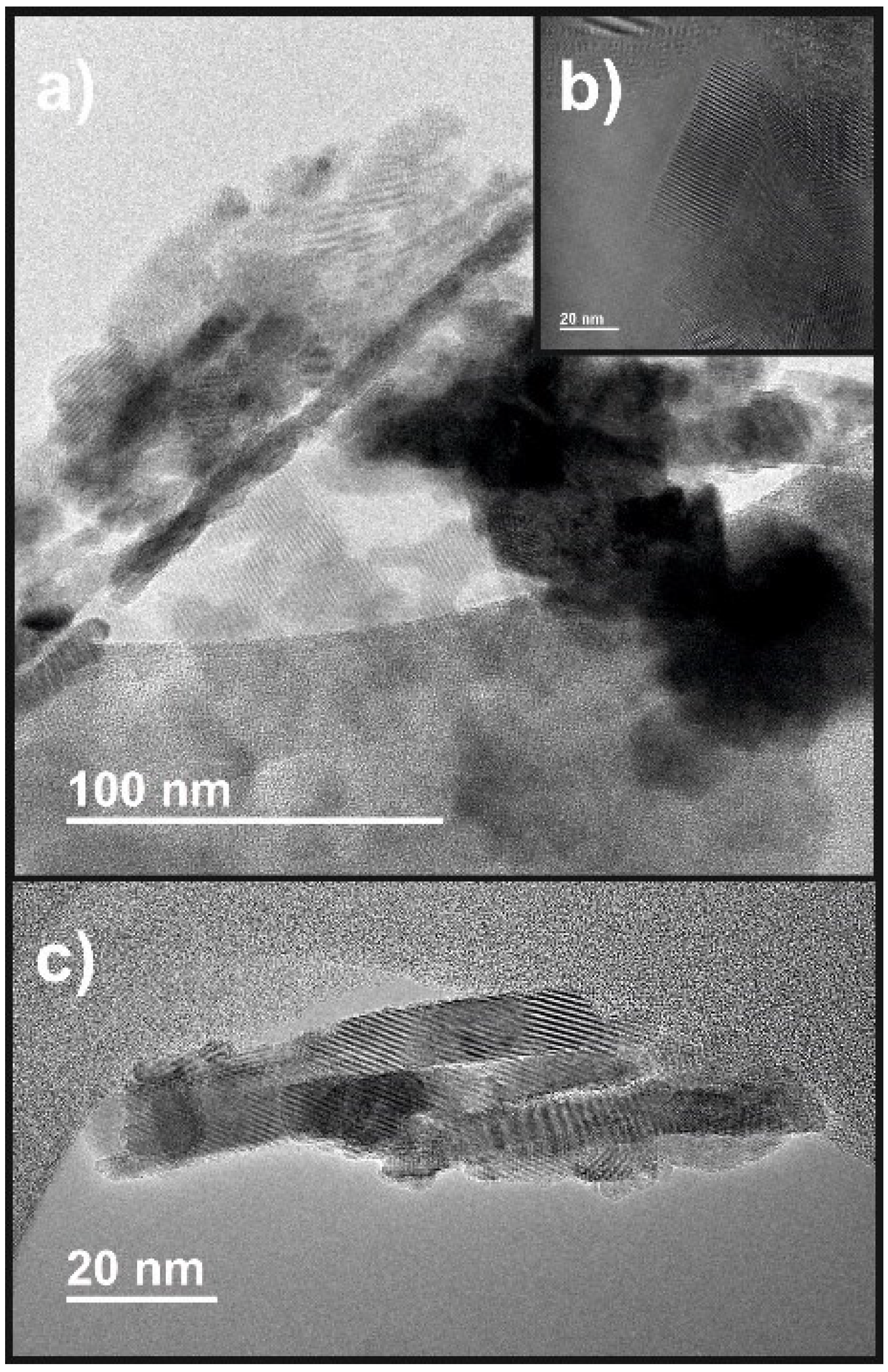
2.2. Morphological Characteristics

Figure 4 shows representative SEM micrographs taken at different magnifications. As can be seen, the sample consists of agglomerated particles. Higher magnifications enable closer inspection that reveals that the agglomerates actually consist of much finer particles with nonuniform nonisotropic particle shape. The agglomerates are similar in size; the magnification of the SEM micrographs is not sufficient to discuss the particle size distribution of the domains within the agglomerates beyond any doubt. However, it may be said that the particles forming agglomerates seem to have a narrow particle size distribution in the submicron range. Interestingly, one can observe that the mentioned domains may be elongated in shape. For more detail, TEM is necessary.
The transmission electron microscopy is much more revealing towards shape and size distribution of the particles that form the agglomerates. Only for the purpose of comparison of nanostructured cobalt oxide with conventional cobalt oxide nanoparticles in TEM, we prepared conventional cobalt oxide nanoparticles at a much lower temperature than of the solvothermal reaction. Figure 5a–d show those nanoparticles; the agglomerates are dispersed relatively successfully, so the domain particles are predominately isotropic with a quite narrow particle size distribution with size values centred at 25 nm. Nanoformations with specific aspect ratios (1D, 2D) are not evidenced.
Figure 6a–d show TEM micrographs for the nanostructured cobalt oxide spinel. In this case, the micrographs show a completely different appearance of the particles, solvothermally derived at the temperature of 235 °C. The average particle size distributions seem to be somewhat greater than in the case of conventional nanoparticles. In this case, one may say that some matrix is retained with more or less the same nanoparticle shape and size distribution; however, a plethora of nanoformations with a specific aspect ratio, i.e., nanorods, that are embedded in the matrix are observed. The nanorods actually range from elongated particles, nanoplates, or nanorods about 30 nm in diameter and 2–3 times the length, to nanowires about 10 nm in diameter and up to 10 times the length.
This nanostructured 1D morphology is particularly obvious from the micrographs in Figure 7a–c. Microscopy results point to lower sizes of the particles than the PXRD for the crystallites. Considering microscopy does not give average nor statistical values and considering that Scherrer is not sensitive to the crystallite aspect ratio, we think that the discrepancy between PXRD crystallite and TEM particle size determination does not exist. In addition, TEM diffraction was omitted as PXRD did not even suggest the presence of phases other than the cobalt oxide spinel. Overall, from the micrographs of the cobalt oxide spinel, one must expect considerable specific surface and thereof surface reactivity.
The nitrogen adsorption–desorption isotherm of the prepared nanostructured powder is shown in Figure 8. The isotherms follow type V adsorption–desorption, which complies with the mesoporous microstructure. Adsorption in mesoporous materials normally occurs in multilayers and is followed by capillary condensation that takes place in the mesopores. Such a condensation process is accompanied with characteristic hysteresis of the isotherms. In this particular case, the appearance of the hysteresis loop resembles the H1 type. Agglomerates or spherical particles arranged in a uniform way often make relatively connected pores with cylindrical geometry with relatively high pore size and high pore uniformity, all of which is typical for the H1 hysteresis loop [41]. Using the BET method, the specific surface area of 44.7 m2 g−1 was calculated. Using the BJH method from the desorption branch of nitrogen isotherms, the pore size distribution of the nanofeatured powder was calculated. The material shows a wide bimodal pore size distribution with an average pore diameter of 20 nm, which is consistent with the nanostructured cobalt oxide spinel nanoformations embedded within the isotropic and agglomerated cobalt oxide spinel nanoparticles.

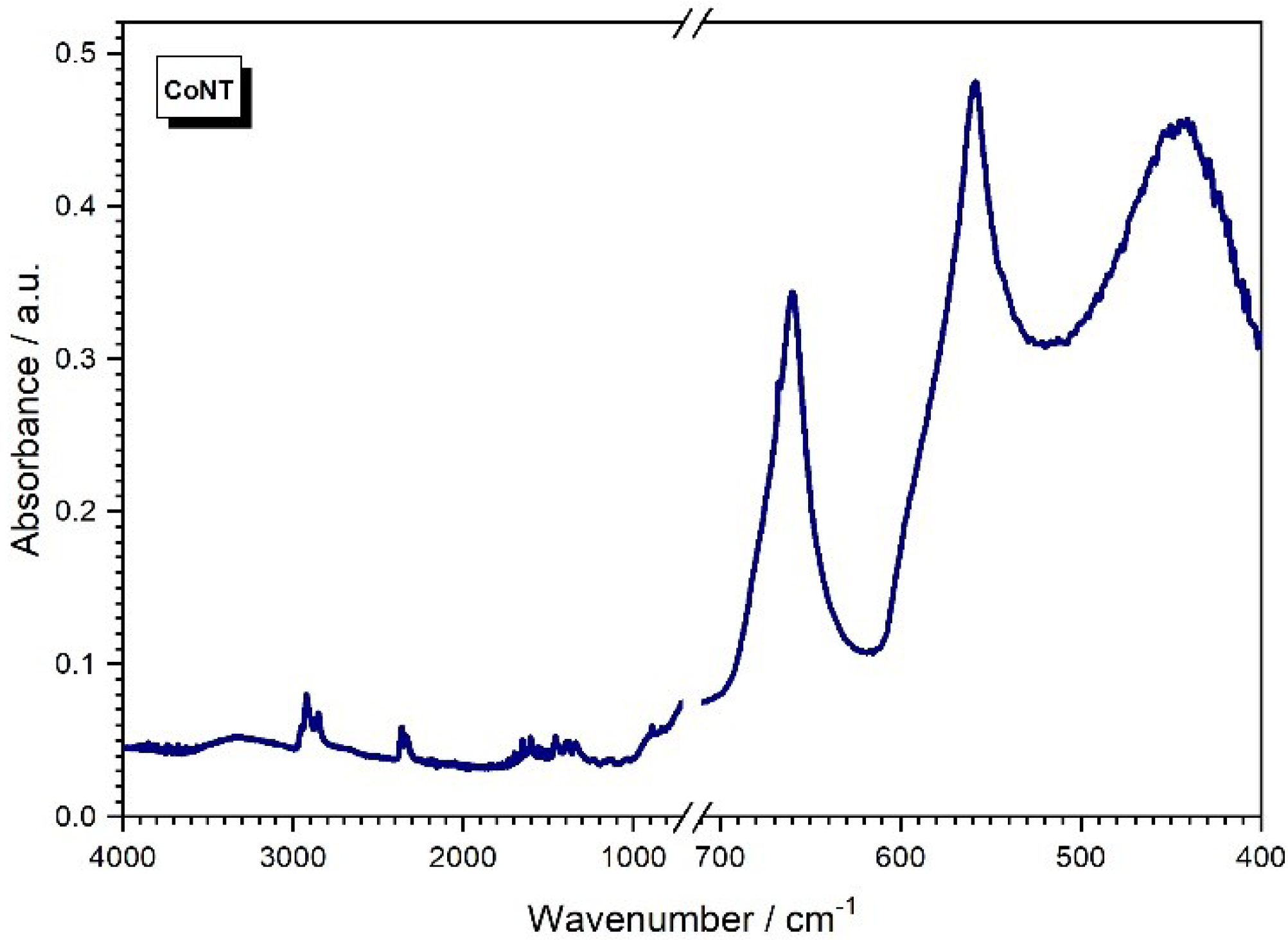
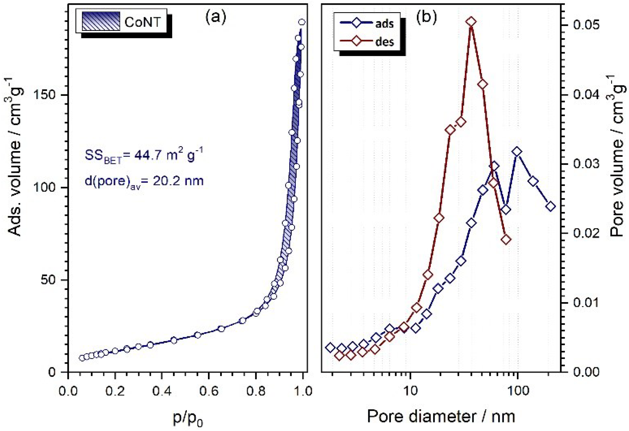
2.3. Intermediate Functional Properties

We performed only a simple study to prove the activity of the surface by testing the photoactivity. For the test, the photodegradation efficiency experiment was performed using the simplest setup and basic dye. The change in the dye concentration vs. reaction time (30 min in dark and 60 min of irradiation) was measured. Figure 9 presents the MB dye degradation ratio. Considerable degradation activity was noted in the first 30 min of the process and was attributed to sorption in the dark. After an additional 60 min of irradiation, the nanostructured cobalt oxide photocatalyst reached almost complete photodegradation. Both the adsorption and the photodegradation part of the experiment were governed by the small amount of the sample available, so the experimental setup with respect to the available reaction chamber was scaled down to the best of our capabilities. Therefore, we were able to collect a relatively small amount of concentration measurement of the dye for the experiment graph (Figure 9). The data were fitted to pseudo-first- and pseudo-second-order kinetic processes. The constant (k) was obtained from the slope of the linear fit −ln(C/Co) vs. reaction time, and for the pseudo-second from the linear fit (1/C − 1/Co) vs. reaction time. The kinetic constant of k = 0.00155 ± 0.00021 L∙mg–1∙min–1 and the R2 = 94.569% were better for the pseudo-second-order, indicating that our photocatalytic process was governed by more factors than just the pollutant concentration, such as light intensity and by-products formation [42]. We consider that it serves as a point for indicating the sorption and photocatalytic activity, but does not allow discussion of the reaction mechanisms. Namely, it is necessary to point out that the experiment surely suffers from some loss (filter-related losses for each measurement of the concentration) of the powdered material, so the results must be taken relatively, i.e., absolute values for adsorption in the dark may be marginally underestimated but the absolute values for subsequent photodegradation are surely underestimated. Hence, the values for sorption activity are probably quite believable but the values for the extent of the photocatalytic activity of the nanostructured cobalt oxide spinel can only serve as an indicator. Because of the abovementioned boundaries, conducting and comparing the sorption/degradation experiment for the isotropic and nanostructured cobalt oxide spinel nanoparticles does not make sense. Despite the goal of this experiment being just to point to the existence of the surface-related photoactivity of the cobalt oxide spinel nanoparticles, the observed sorption activity is likely a far better indicator of the enhanced specific surface properties of the nanostructured cobalt oxide spinel.
In conclusion, previous characterisation results, including sorption/catalytic efficiency, nominate the developed nanostructured cobalt oxide materials for the photocatalytic) application, especially for the degradation of harmful organic dyes. We wanted to test the gas-sensing performance as well but we encountered the boundaries of our system. Basically, the mass yield of our reactor was small, and thus, we were unable to collect sufficient mass to go through an optimisation process (trials and errors to match optimal content of the additives) for the preparation of films (of sufficient lateral size to accommodate heaters for sensing at temperatures normally above 300 °C and electrodes for electric performance testing) by tape casting (inherently a material-inefficient processing for small batches) [43]. Thus, some compromises in the context of the reactors for synthesis and testing are necessary to facilitate the desired scale-up of the material for gas sensing application, which nevertheless, are considered worth further development. It would be great if we could have performed sensing performance tests, but we consider that the demonstrated photocatalytically beneficial performance proved the viability of the prepared material. Reaching out to the synthesis and deposition route of Co3O4 films in a single step would resolve the mentioned limitations and allow more reasonable scale-up efforts.

3. Materials and Methods

3.1. Materials

The modified procedure was used for the synthesis [36]. Briefly, 10 mL of 0.1 M ammonia (NH4OH, p.a. Kemika, Zagreb, Croatia) solution was slowly added to the 10 mL of 0.025 M nitrate solution prepared by dissolving cobalt nitrate hexahydrate (Co(NO3)2 × 6H2O, p.a. Kemika, Zagreb, Croatia) in demineralized water. After stirring for 30 min, the mixture was transferred in a 50 mL centrifuge tube and cobalt hydroxide precipitate was separated by centrifugation at 3500 rpm for 5 min. The precipitate was washed with deionized water and centrifuged. This procedure was repeated 6 times. Then, the precipitate was transferred in a 20 mL Teflon-lined autoclave. An amount of 0.3 g of sodium nitrate (NaNO3, p.a. Kemika, Zagreb, Croatia), 8 mL of methanol (CH3OH, p.a. Kemika, Zagreb, Croatia), and 8 mL of deionized water were then added. An autoclave was filled to 80% of the total volume and sealed, placed in a laboratory furnace, and heated at 235 °C for 36 h. After cooling, the washing procedure was repeated six more times as described previously. The derived materials were then dried at reduced pressure.

3.2. Methods

The powder X-ray diffraction (PXRD) was accomplished using the diffractometer XRD6000 (Shimadzu, Kyoto, Japan) with CuKα radiation at an accelerating voltage of 40 kV and current of 30 mA. Data were collected between 15 and 70° 2θ, in a step scan mode with steps of 0.02° and a counting time of 0.6 s. The average crystallite size was calculated from the broadening of the (311) diffraction peak using the Scherrer equation [44]: d = kλ/(βcosθ), where d is the average crystallite diameter, k is the Scherrer constant (0.94), λ is the X-ray wavelength (0.15418 nm), β is the full-width at half-height of the (311) diffraction peak corrected for instrumental broadening, and θ is the diffraction angle.
IR spectra were acquired using the Fourier Transform Infrared (FTIR) spectrometer Vertex 70 (Bruker, Billerica, MA, USA) in Attenuated Total Reflectance (ATR) mode. The samples were pressed on a diamond prism and the absorbance data were collected between 400 and 4000 cm−1 with a spectral resolution of 1 cm−1 and average of 64 scans.
The morphology was investigated with a Vega EasyProbe3 (Tescan, Brno, Czech Republic) Scanning Electron Microscope (SEM) operating at 30 kV. Samples for SEM characterization were fixed on a sample holder using double-sided carbon conductive tape, and then gold-coated using a SC 7620 sputter coater (Quorum, East Sussex, Laughton, UK).
The morphology was investigated with a JEM-ARM200F (Jeol, Tokyo, Japan) Transmission Electron Microscope (TEM). Powder samples for TEM characterization were ultrasonically dispersed and were placed on a copper mesh sample holder and subjected to analysis.
Simultaneous Thermogravimetric and Differential Thermal Analysis (TG-DTA) was accomplished using a STA 409C (Netzsch, Burlington, MA, USA). For the thermal analysis, ~50 mg of material was placed in Pt crucibles and heated at a rate of 10 °C min−1 to 1300 °C in a synthetic air flow of 30 cm3 min−1, and α-alumina was used as a reference.
The surface area was determined by nitrogen gas adsorption–desorption isotherms obtained at 77 K on ASAP-2000 equipment (Micromeritics Corporation, Norcross, GA, USA) using the Brunauer–Emmet–Teller model (BET). The samples were previously degassed at 200 °C under a dynamic vacuum of 1.3·10−2 Pa. Pore size distributions were calculated from desorption isotherms by the Barrett–Joyner–Halenda (BJH) model.
The photocatalytic activity was assessed using the methylene blue (MB) degradation test. For the photodegradation kinetics investigation, 50 mL of 25 mgL−1 of MB solution was poured in a borosilicate cylindrical glass vessel with a 50 mm diameter and 120 mm height. A quartz glass tube was placed in the vessel. An amount of 50 mg of Co3O4 was added to solution; thus, the photocatalyst concentration was 1 g L−1. A Pen Ray lamp (Analytik Jena GMBH, Upland, CA, USA), with a radiation wavelength of 365 nm and emission intensity of 2 mW cm−2, was placed inside the quartz tube. The solution was first stirred for 30 min to establish adsorption equilibrium. After 30 min, the lamp was switched on and the photodegradation experiment was carried out at 25 °C. In order to determine the methylene blue concentration, aliquots of 4 mL were withdrawn from the suspension at appropriate time intervals using a filter. The concentrations were determined using a UV–VIS spectrophotometer Cary 1E (Varian Inc., Palo Alto, CA, USA). For monitoring the concentration of MB, and hence its degradation, the absorption peak height at a wavelength of 664 nm (where the maximum was) was used. The methylene blue concentration was computed using the previously established calibration curve.

4. Conclusions

A facile and affordable course of synthesis for the preparation of nanostructured cobalt oxide powders was shown in this study.
Diffraction analysis indicated the crystallisation of structurally monophasic Co3O4 nanoparticles with a spinel structure. Infrared spectroscopy showed traces of organic phase in an otherwise chemically homogeneous material.
Scanning electron microscopy showed that the fine nanoparticle matter formed agglomerates. Transmission electron microscopy revealed that the agglomerates of the fine nanoparticulated material actually consisted of 1D-shaped cobalt oxide spinel rods embedded in a fine nanoparticulated matrix. Nitrogen adsorption–desorption isotherms gave a sample-specific surface area of 44.7 m2 g1 and enabled discussion of the orientation of the nanostructured material domains, exposure of nanostructured rods, and derivative geometry parameters. Thermal analysis showed that the nanostructured Co3O4 material was stable up to 800 °C where decomposition to CoO was observed.
On behalf of basic sorption/degradation tests, it was shown that the prepared nanostructured Co3O4 materials had a chemical and (micro)structural background suitable for considering application as photocatalysts for the removal of the wastewater micropollutants, but also for continuation of the development of a single-step preparing route that would particularly favour gas-sensing application.

Author Contributions

Conceptualization, V.M. and S.K.; methodology, V.M., S.K., M.P. and I.P.; software, V.M., M.P. and I.P.; validation, V.M. and I.P.; formal analysis, V.M., M.P. and I.P.; investigation, V.M. and S.K.; resources, V.M. and S.K.; data curation, V.M. and I.P.; writing—original draft preparation, V.M. and S.K.; writing—review and editing, V.M.; visualization, V.M.; supervision, V.M.; project administration, V.M. and S.K.; funding acquisition, V.M. and S.K. All authors have read and agreed to the published version of the manuscript.

Funding

This work has been funded by the projects PZS-2019-02-1555 PV-WALL in the Research Cooperability Program of the Croatian Science Foundation funded by the European Union from the European Social Fund under the Operational Programme Efficient Human Resources 2014-2020 (TEM characterisation), UIP-2019-04-2367 SLIPPERY SLOPE (synthesis of material with reduced dimensionality), IP-2018-01-2963 HOUDINI (solvothermal synthesis), of the Croatian Science Foundation, and project KK.01.2.1.02.0316 by the European Regional Development Fund under the call Increasing the development of new products and services arising from research and development activities—phase II (photocatalytic test).

Data Availability Statement

The data behind this research are available upon reasonable request to corresponding author.

Acknowledgments

The sustenance of the University of Zagreb is gratefully acknowledged.

Conflicts of Interest

The authors declare no conflict of interest.

References

  1. Wagner, T.; Haffer, S.; Weinberger, C.; Klaus, D.; Tiemann, M. Mesoporous materials as gas sensors. Chem. Soc. Rev. 2013, 42, 4036–4053. [Google Scholar] [CrossRef]
  2. Wang, C.X.; Yin, L.W.; Zhang, L.Y.; Xiang, D.; Gao, R. Metal oxide gas sensors: Sensitivity and influencing factors. Sensors 2010, 10, 2088–2106. [Google Scholar] [CrossRef] [Green Version]
  3. Gu, C.D.; Zheng, H.; Wang, X.L.; Tu, J.P. Superior ethanol-sensing behavior based on SnO2 mesocrystals incorporating orthorhombic and tetragonal phases. RSC Adv. 2015, 5, 9143–9153. [Google Scholar] [CrossRef]
  4. San, X.G.; Wang, G.S.; Liang, B.; Ma, J.; Meng, D.; Shen, Y.B. Flower-like NiO hierarchical microspheres self-assembled with nanosheets: Surfactant-free solvothermal synthesis and their gas sensing properties. J. Alloys Compd. 2015, 636, 357–362. [Google Scholar] [CrossRef]
  5. Liu, X.C.; Hu, M.; Wang, Y.F.; Liu, J.F.; Qin, Y.X. High sensitivity NO2 sensor based on CuO/p-porous silicon heterojunction at room temperature. J. Alloys Compd. 2016, 685, 364–369. [Google Scholar] [CrossRef]
  6. Yoon, J.W.; Kim, H.J.; Jeong, H.M.; Lee, J.H. Gas sensing characteristics of p-type Cr2O3 and Co3O4 nanofibers depending on inter-particle connectivity. Sens. Actuator B Chem. 2014, 202, 263–271. [Google Scholar] [CrossRef]
  7. Kim, H.J.; Lee, J.H. Highly sensitive and selective gas sensors using p-type oxide semiconductors: Overview. Sens. Actuator B Chem. 2014, 192, 607–627. [Google Scholar] [CrossRef]
  8. Ge, X.; Gu, C.D.; Wang, X.L.; Tu, J.P. Correlation between microstructure and electrochemical behavior of the mesoporous Co3O4 sheet and its ionothermal synthesized hydrotalcite-like a-Co(OH)2 precursor. J. Phys. Chem. C 2014, 118, 911–923. [Google Scholar] [CrossRef]
  9. Liang, Y.Y.; Li, Y.G.; Wang, H.L.; Zhou, J.G.; Wang, J.; Reigier, T.; Dai, H.J. Co3O4 nanocrystals on graphene as a synergistic catalyst for oxygen reduction reaction. Nat. Mater. 2011, 10, 780–786. [Google Scholar] [CrossRef] [PubMed] [Green Version]
  10. Xu, J.M.; Zhang, J.; Wang, B.B.; Liu, F. Shape-regulated synthesis of cobalt oxide and its gas-sensing property. J. Alloys Compd. 2015, 619, 361–367. [Google Scholar] [CrossRef]
  11. Cheng, J.P.; Chen, X.; Wu, J.S.; Liu, F.; Zhang, X.B.; Dravid, V.P. Porous cobalt oxides with tunable hierarchical morphologies for supercapacitor electrodes. CrystEngComm 2012, 14, 6702–6709. [Google Scholar] [CrossRef]
  12. Qiu, H.J.; Mu, Y.P.; Zhang, H.J.; Wang, Y. Designed synthesis of cobalt-oxidebased nanomaterials for superior electrochemical energy storage devices. Nano Res. 2015, 8, 321–339. [Google Scholar] [CrossRef]
  13. Vetter, S.; Haffer, S.; Wagner, T.; Tiemann, M. Nanostructured Co3O4 as a CO gas sensor: Temperature-dependent behavior. Sens. Actuator B Chem. 2015, 206, 133–138. [Google Scholar] [CrossRef]
  14. Farhadi, S.; Pourzare, K.; Sadeghinejad, S. Simple preparation of ferromagnetic Co3O4 nanoparticles by thermal dissociation of the [CoII(NH3)6](NO3)2 complex at low temperature. J. Nanostruct. Chem. 2013, 3, 16. [Google Scholar] [CrossRef] [Green Version]
  15. Akamatsu, T.; Itoh, T.; Izu, N.; Shin, W. NO and NO2 sensing properties of WO3 and Co3O4 based gas sensor. Sensors 2013, 13, 12467–12481. [Google Scholar] [CrossRef] [Green Version]
  16. Shaalan, M.N.; Rashad, M.; Moharram, A.H.; Abdel-Rahim, M.A. Promising methane gas sensor synthesized by microwave-assisted Co3O4 nanoparticles. Mat. Sci. Semicond. Proc. 2016, 46, 1–5. [Google Scholar] [CrossRef]
  17. Wang, X.; Tian, W.; Zhai, T.Y.; Zhi, C.Y.; Bando, Y.; Golberg, D. Cobalt(II,III) oxide hollow structures fabrication, properties and applications. J. Mater. Chem. 2012, 22, 23310–23326. [Google Scholar] [CrossRef]
  18. Ma, L.Y.; Niu, B.; Xu, H.Y.; Cao, B.Q.; Wang, J.Q. Microwave hydrothermal synthesis of nanoporous cobalt oxides and their gas sensing properties. Mater. Res. Bull. 2011, 46, 1097–1101. [Google Scholar] [CrossRef]
  19. Yoon, J.W.; Choi, J.K.; Lee, J.H. Design of a highly sensitive and selective C2H5OH sensor using p-type Co3O4 nanofibers. Sens. Actuator B Chem. 2012, 161, 570–577. [Google Scholar] [CrossRef]
  20. Nguye, H.; El-Safty, S.A. Meso- and macroporous Co3O4 nanorods for effective VOC gas sensor. J. Phys. Chem. C 2011, 115, 8466–8474. [Google Scholar] [CrossRef]
  21. Choi, K.I.; Kim, H.R.; Kim, K.M.; Liu, D.; Cao, G.; Lee, J.H. C2H5OH sensing characteristics of various Co3O4 nanostructures prepared by solvothermal reaction. Sens. Actuator B Chem. 2010, 146, 183–189. [Google Scholar] [CrossRef]
  22. Zhuo, L.; Ge, J.; Cao, L.; Wang, B. Solvothermal Synthesis of CoO, Co3O4, Ni(OH)2 and Mg(OH)2. Cryst. Growth Des. 2009, 9, 1–6. [Google Scholar] [CrossRef]
  23. Sun, L.; Li, H.; Ren, L.; Hu, C. Synthesis of Co3O4 nanostructures using a solvothermal approach. Solid State Sci. 2009, 11, 108–112. [Google Scholar] [CrossRef]
  24. Lou, X.; Han, J.; Chu, W.; Wang, X.; Cheng, Q. Synthesis and photocatalytic property of Co3O4 nanorods. Mater. Sci. Eng. B 2007, 137, 268–271. [Google Scholar] [CrossRef]
  25. Kohan, M.G.; Solomon, G.; You, S.; Yusupov, K.; Concina, I.; Vomiero, A. Vertically aligned Co3O4 nanorods as platform for inverted all-oxide heterojunctions. Nano Select. 2021, 2, 967–978. [Google Scholar] [CrossRef]
  26. Soni, V.; Xia, C.; Cheng, C.K.; Nguyen, V.H.; Nguyen, D.L.T.; Bajpai, A.; Kim, S.Y.; Van Le, Q.; Khan, A.A.P.; Singh, P.; et al. Advances and recent trends in cobalt-based cocatalysts for solar-to-fuel conversion. Appl. Mater. Today 2021, 24, 101074. [Google Scholar] [CrossRef]
  27. Sonu; Dutta, V.; Sharma, S.; Raizada, P.; Hosseini-Bandegharaei, A.; Gupta, V.K.; Singh, P. Review on augmentation in photocatalytic activity of CoFe2O4 via heterojunction formation for photocatalysis of organic pollutants in water. J. Saudi Chem. Soc. 2019, 23, 1119–1136. [Google Scholar] [CrossRef]
  28. Chandel, N.; Sharma, K.; Sudhaik, A.; Raizada, P.; Hosseini-Bandegharaei, A.; Thakur, V.K.; Singh, P. Magnetically separable ZnO/ZnFe2O4 and ZnO/CoFe2O4 photocatalysts supported onto nitrogen doped graphene for photocatalytic degradation of toxic dyes. Arab. J. Chem. 2020, 2, 4324–4340. [Google Scholar] [CrossRef]
  29. Yan, W.; Xu, Y.; Hao, S.; He, Z.; Wang, L.; Wei, Q.; Xu, Q.; Tang, H. Promoting charge separation in hollow-structured C/MoS2@ZnIn2S4/Co3O4 photocatalysts via double heterojunctions for enhamced photocatalytic hydrogen evolution. Inorg. Chem. 2022, 61, 4275–4734. [Google Scholar] [CrossRef]
  30. Chen, X.; Cheng, J.P.; Shou, Q.L.; Liu, F.; Zhang, X.B. Effect of calcinations temperature on the porous structure of cobalt oxide micro-flowers. Cryst. Eng. Comm. 2012, 14, 1271–1276. [Google Scholar] [CrossRef]
  31. Cheng, J.P.; Liu, L.; Zhang, J.; Liu, F.; Zhang, X.B. Influences of anion exchange and phase transformation on the supercapacitive properties of a-Co(OH)2. J. Electroanal. Chem. 2014, 722, 23–31. [Google Scholar] [CrossRef]
  32. Yuan, Y.F.; Xia, X.H.; Wu, J.B.; Huang, X.H.; Pei, Y.B.; Yang, J.L.; Guo, S.Y. Hierarchically porous Co3O4 film with mesoporous walls prepared via liquid crystalline template for supercapacitor application. Electrochem. Commun. 2011, 13, 1123–1126. [Google Scholar] [CrossRef]
  33. Patil, D.; Patil, P.; Subramanian, V.; Joy, P.A.; Potdar, H.S. Highly sensitive and fast responding CO sensor based on Co3O4 nanorods. Talanta 2010, 81, 37–43. [Google Scholar] [CrossRef] [PubMed]
  34. Wen, Z.; Zhu, L.P.; Mei, W.M.; Hu, L.; Li, Y.G.; Sun, L.W.; Cai, H.; Ye, Z.Z. Rhombus-shaped Co3O4 nanorod arrays for high-performance gas sensor. Sens. Actuator B Chem. 2013, 186, 172–179. [Google Scholar] [CrossRef]
  35. Warang, T.; Patel, N.; Santini, A.; Bazzanella, N.; Kale, A.; Miotello, A. Pulsed laser deposition of Co3O4 nanoparticles assembled coating: Role of substrate temperature to tailor disordered to crystalline phase and related photocatalytic activity in degradation of methylene blue. Appl. Catal. A Gen. 2012, 423, 21–27. [Google Scholar] [CrossRef]
  36. Xia, F.; Ou, E.; Wang, L.; Wang, J. Photocatalytic degradation of dyes over cobalt doped mesoporous SBA-15 under sunlight. Dye. Pigment. 2008, 76, 76–81. [Google Scholar] [CrossRef]
  37. Makhlouf, M.T.; Abu-Zied, B.M.; Mansoure, T.H. Direct Fabrication of Cobalt Oxide Nanoparticles Employing Sucrose as a Combustion Fuel. J. Nanopart. Res. 2013, 2013, 384350. [Google Scholar] [CrossRef] [Green Version]
  38. Kaczmarczyk, J.; Zasada, F.; Janas, J.; Indyka, P.; Piskorz, W.; Kotarba, A.; Sojka, Z. Thermodynamic Stability, Redox Properties, and Reactivity of Mn3O4, Fe3O4, and Co3O4 Model Catalysts for N2O Decomposition: Resolving the Origins of Steady Turnover. ACS Catal. 2016, 6, 1235–1246. [Google Scholar] [CrossRef]
  39. Wang, L.; Maxisch, T.; Ceder, G. A First-Principles Approach to Studying the Thermal Stability of Oxide Cathode Materials. Chem. Mater. 2007, 19, 543–552. [Google Scholar] [CrossRef]
  40. Sing, K.S.W.; Everett, D.H.; Haul, R.A.W.; Moscou., L.; Pierotti, R.A.; Rouquerol, J.; Siemieniewska, T. Reporting Physisorption Data for Gas/Solid Systems with Special Reference to the Determination of Surface Area and Porosity. Pure Appl. Chem. 1985, 57, 603–619. [Google Scholar] [CrossRef]
  41. Kumarage, G.W.C.; Comini, E. Low-Dimensional Nanostructures Based on Cobalt Oxide (Co3O4) in Chemical-Gas Sensing. Chemosensors 2021, 9, 197. [Google Scholar] [CrossRef]
  42. Verma, M.; Mitan, M.; Kim, H.; Vaya, D. Efficient photocatalytic degradation of Malachite green dye using facilely synthesized cobalt oxide nanomaterials using citric acid and oleic acid. J. Phys. Chem. Solids 2021, 155, 110125. [Google Scholar] [CrossRef]
  43. Suwanchawalit, C. High Photocatalytic Performance of Magnetic CoFe2O4-Graphene Nanocomposite for Organic Dye Removal. Aust. J. Basic Appl. Sci. 2015, 9, 159–165. [Google Scholar]
  44. Klug, H.P.; Alexander, L.E. X-Ray Diffraction Procedures, 2nd ed.; John Wiley & Sons Inc.: New York, NY, USA, 1974; pp. 687–703. [Google Scholar]
Figure 1. Powder X-ray diffraction pattern of the prepared Co3O4 spinel.
Catalysts 12 01162 g001
Figure 2. Infrared ATR spectrum of the prepared Co3O4 nanoparticles.
Catalysts 12 01162 g002
Figure 3. TG and DTA curves of the prepared Co3O4 nanoparticles.
Catalysts 12 01162 g003
Figure 4. Scanning electron microscopy micrographs of the prepared Co3O4 nanoparticles.
Catalysts 12 01162 g004
Figure 5. Transmission electron microscopy micrographs of the conventional Co3O4 nanoparticles: (a) Co3O4 nanoparticle matrix; (b) Co3O4 nanoparticle agglomerate; (c) Co3O4 nanoparticles fringes, and (d) Co3O4 nanoparticle agglomerates at lower magnification.
Catalysts 12 01162 g005
Figure 6. Transmission electron microscopy micrographs of the nanostructured Co3O4 formations: (a) 1D Co3O4 nanoformations; (b) ordered 1D Co3O4 nanorods; (c) fringes for 1D Co3O4 nanoformations, and (d) 1D Co3O4 nanoformations agglomerates at lower magnification.
Catalysts 12 01162 g006
Figure 7. Transmission electron microscopy high-magnification micrographs of the prepared 1D nanostructured Co3O4 formations: (a) 1D Co3O4 nanoformations in nanoparticulated matrix; (b) visible fringes for 1D Co3O4 at higher magnification; (c) 1D Co3O4 nanoformations at higher magnification.
Catalysts 12 01162 g007
Figure 8. (a) N2 adsorption–desorption isotherms of Co3O4 nanoformations; (b) pore size distribution calculated from desorption branch.
Catalysts 12 01162 g008
Figure 9. Indicative values for the sorption (−30–0 min, in dark) and photocatalytic degradation (0–60 min, irradiated) of the MB dye by the nanostructured cobalt oxide spinel photocatalyst for the case of: (a) pseudo-first-order fit of the photodegradation; (b) pseudo-second-order fit of the photodegradation. Inset: 500–700 nm segment of MB solution Vis spectra taken at various times.
Catalysts 12 01162 g009
Publisher’s Note: MDPI stays neutral with regard to jurisdictional claims in published maps and institutional affiliations.

