
Recent Progress in WS2-Based Nanomaterials Employed for Photocatalytic Water Treatment


Abstract
:1. Introduction
2. Background
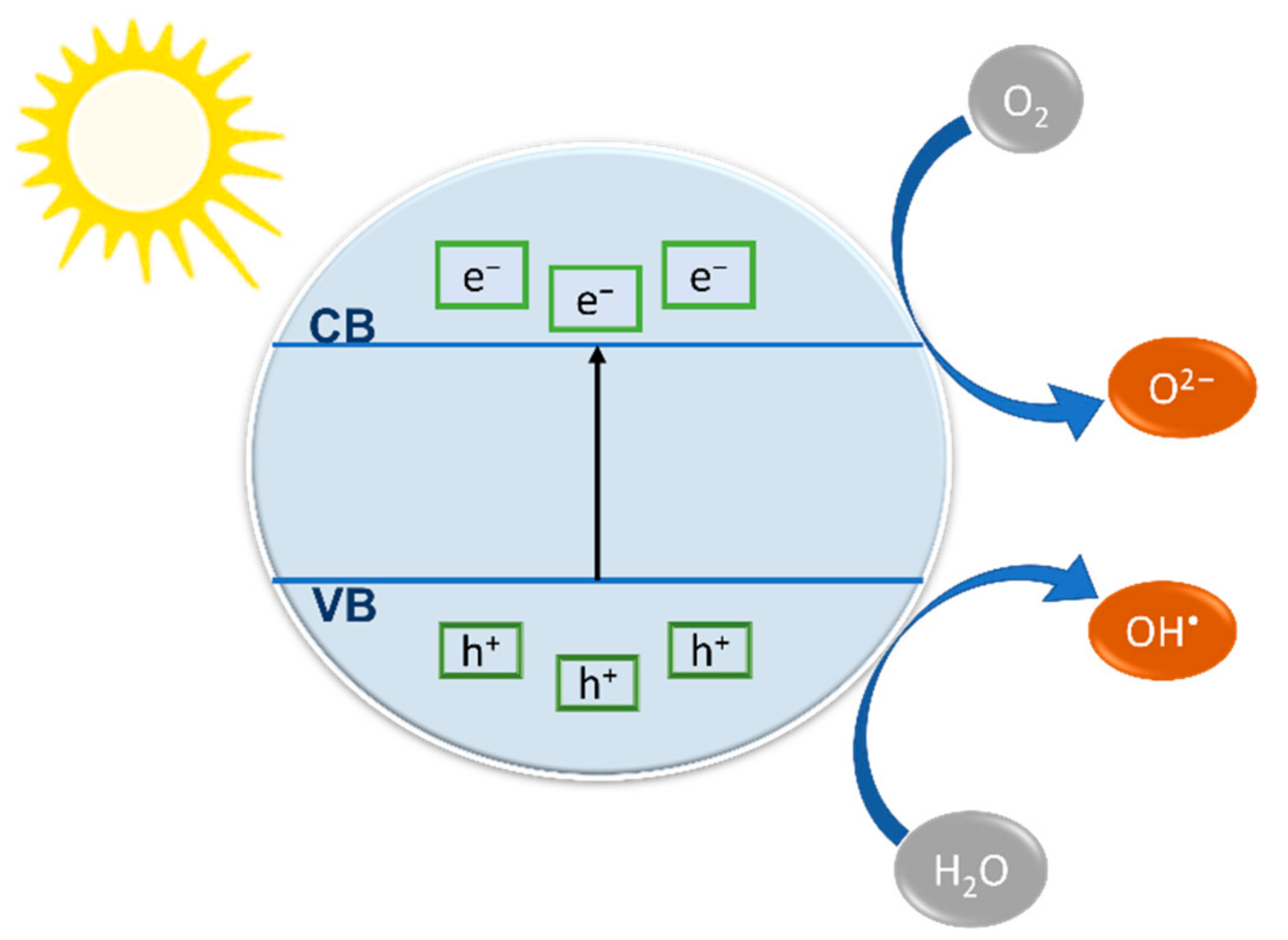
2.1. Fundamentals of Photocatalytic Degradation of Pollutants in Water
2.2. The Most Common Photocatalysts
3. WS2 as a Photocatalyst
3.1. Structure
3.2. Properties
3.3. Synthesis
4. Photocatalytic Water Treatment Using WS2 and Heterostructures
4.1. Photocatalytic Degradation of Organic Substances
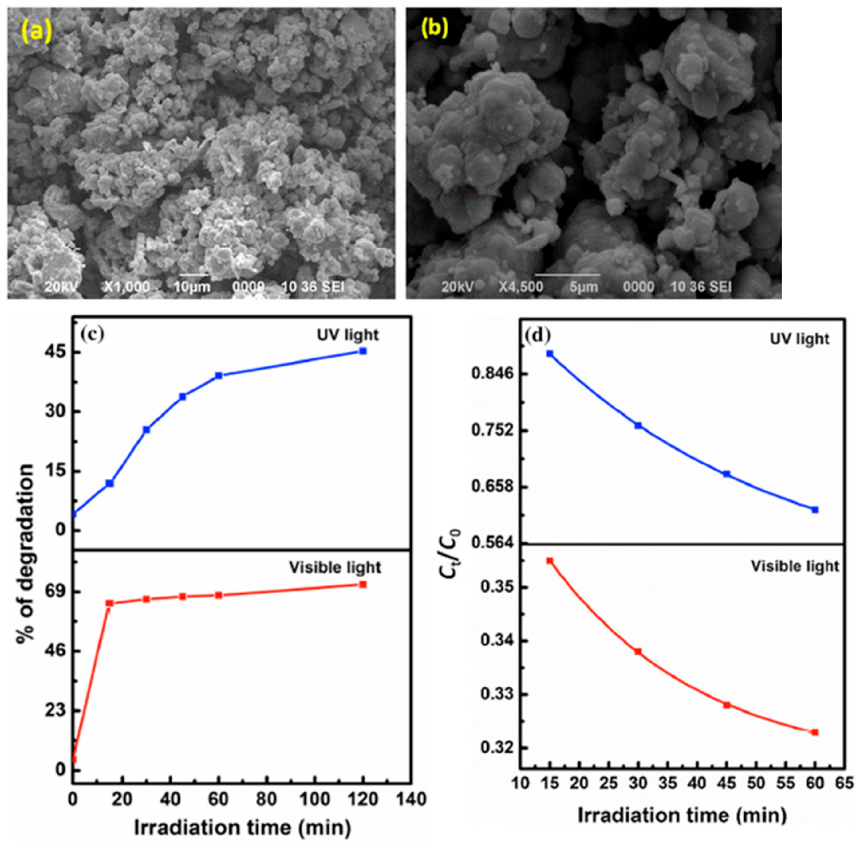
4.1.1. Photocatalytic Degradation of Organic Substance Using WS2 Nanostructures
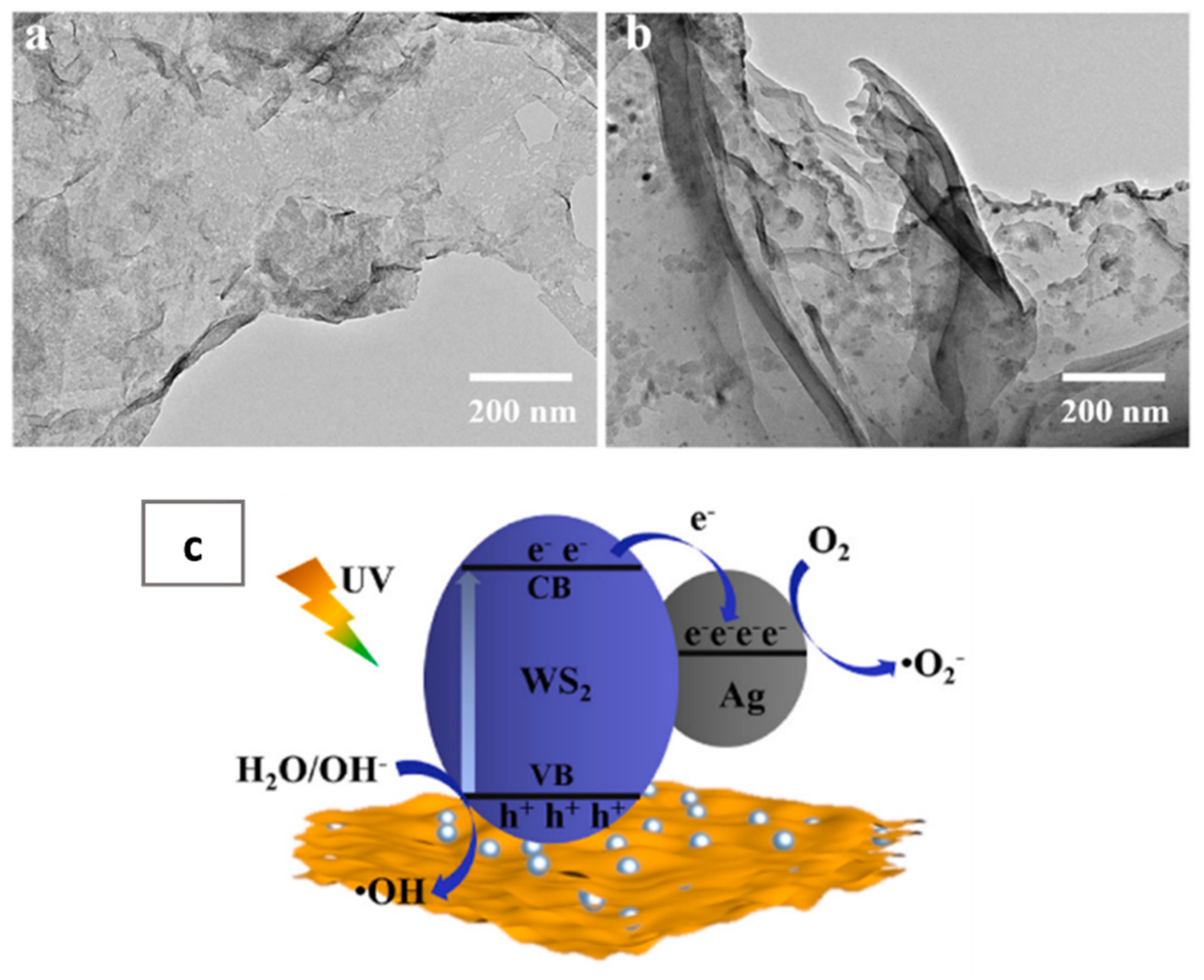
4.1.2. Photocatalytic Degradation of Organic Substance Using WS2 HeterostructureWS2/Ag
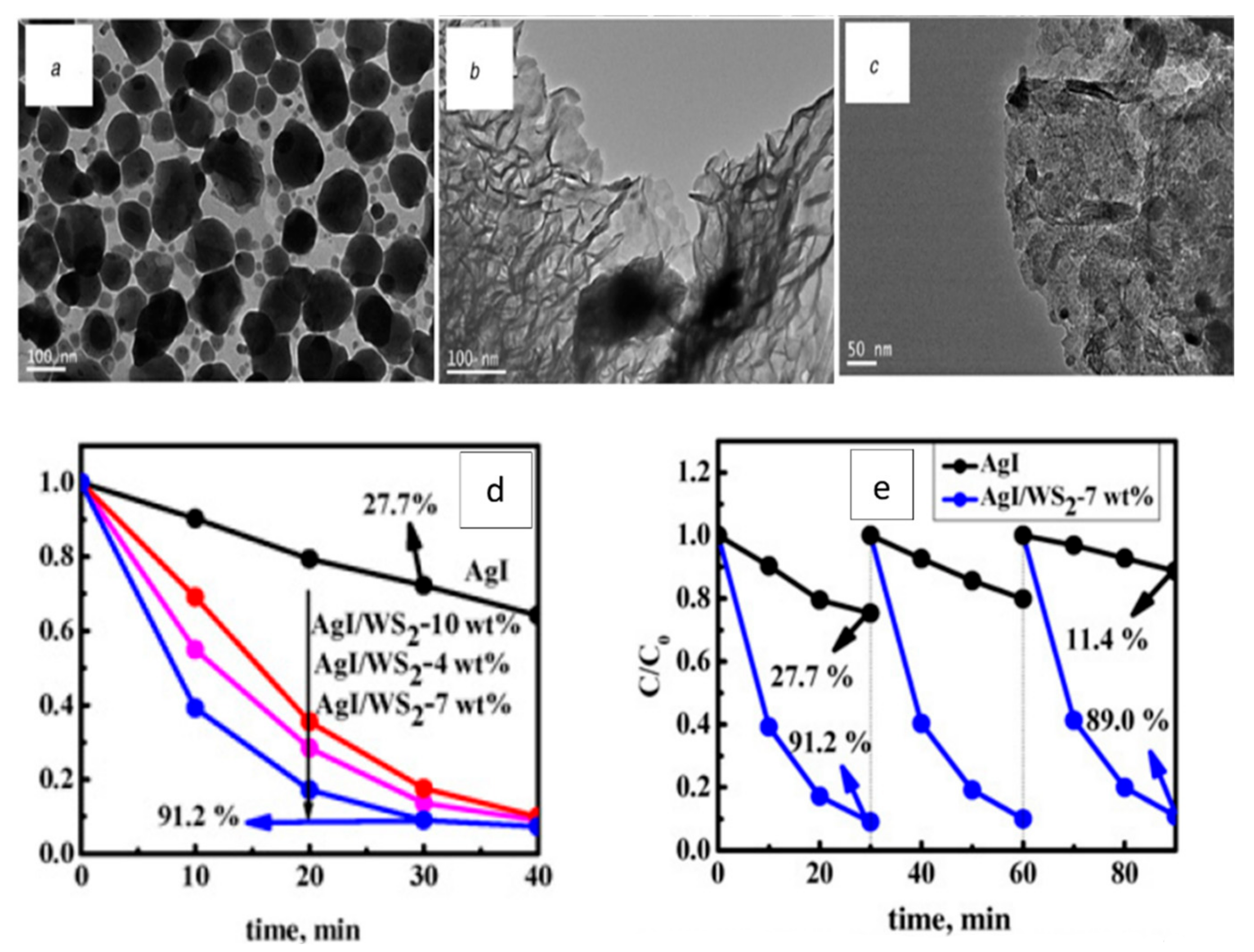
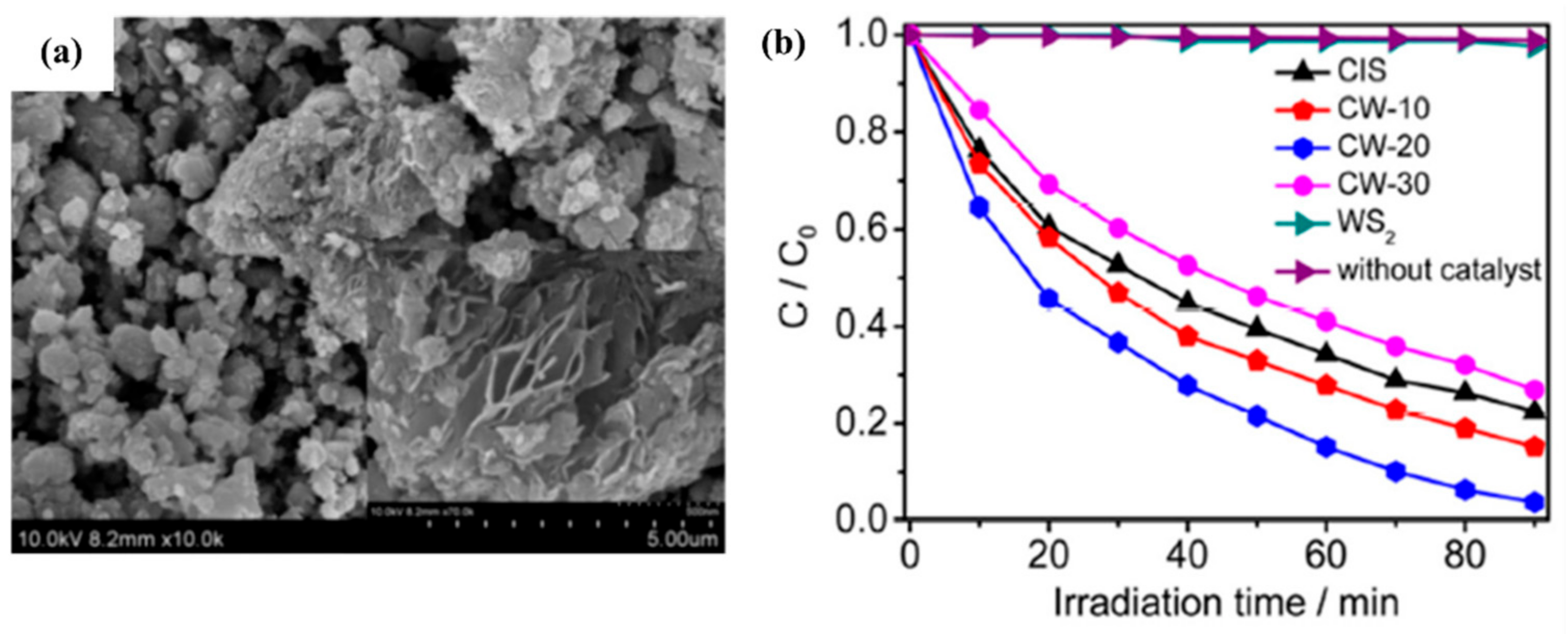
AgI/WS2
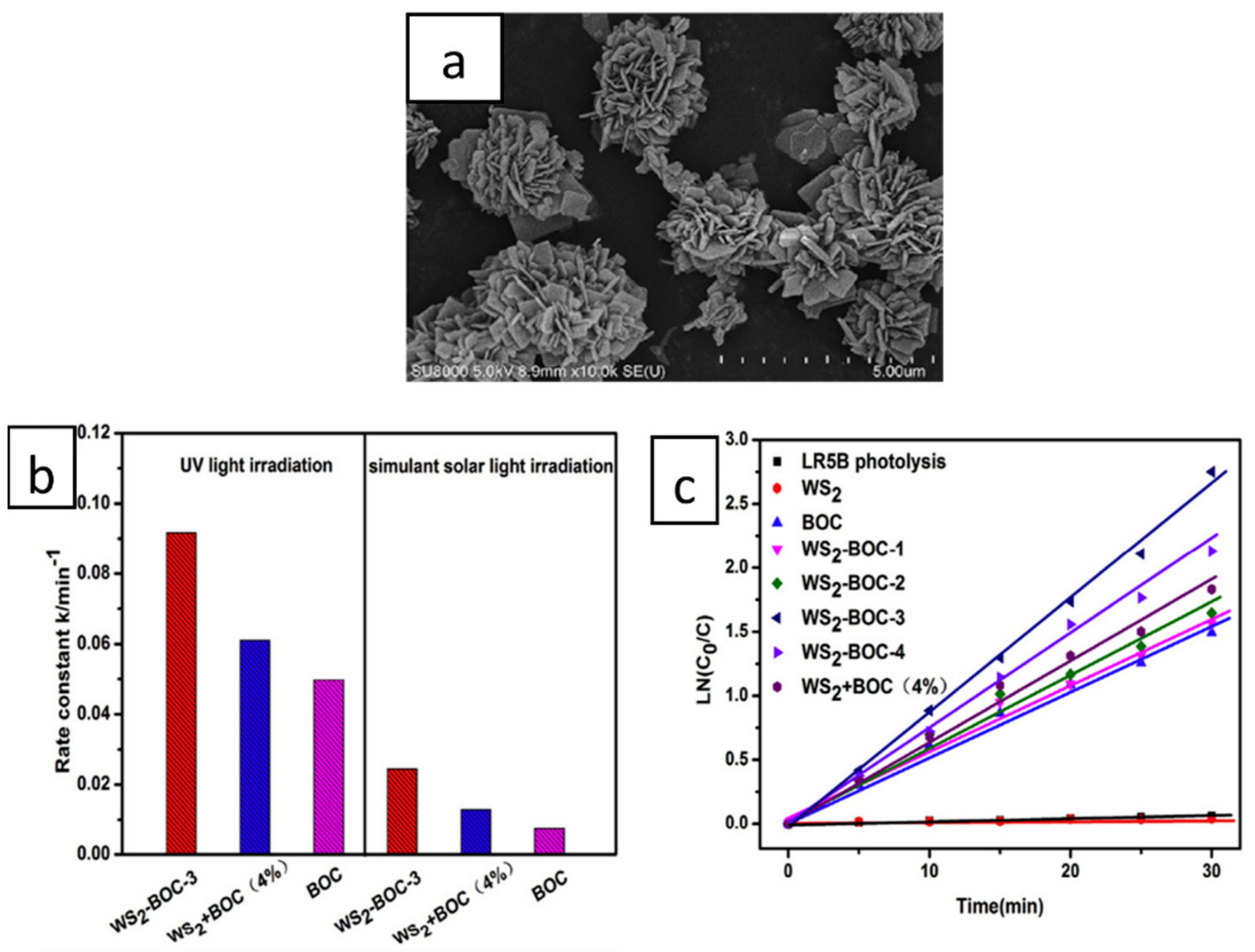
WS2/Bi2O2CO3
WS2/BiOBr
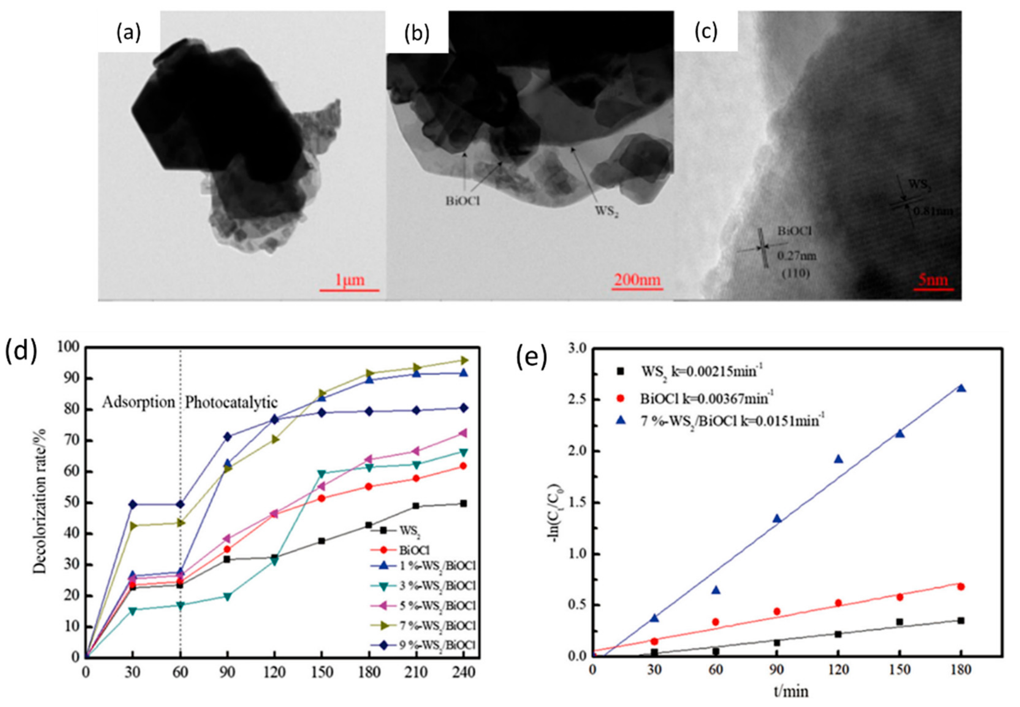
BiOCl/WS2
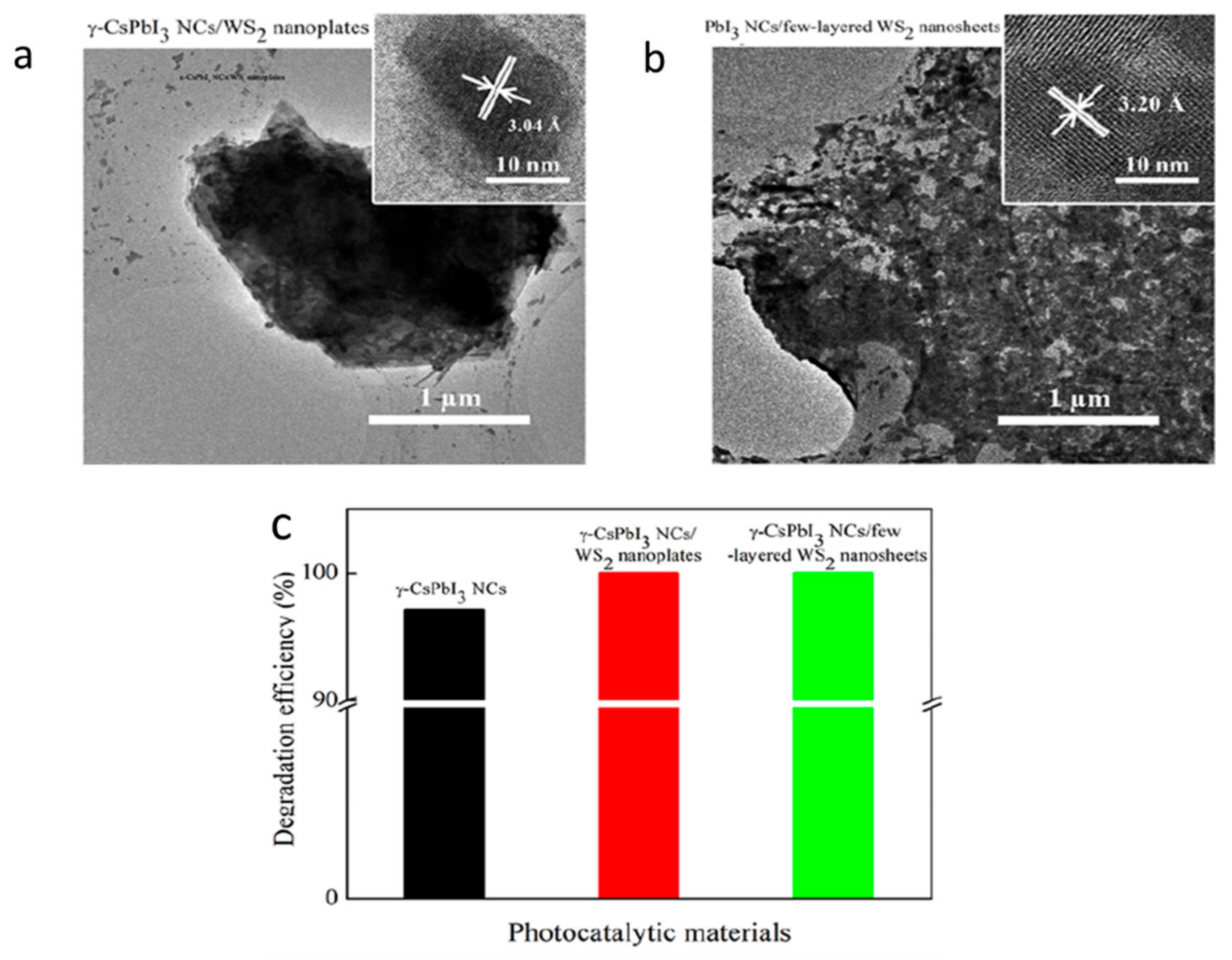
γ-CsPbI3/WS2
WS2/g-C3N4
WS2/Fe3O4
WS2/MoS2
WS2/MoS2/BiOCl
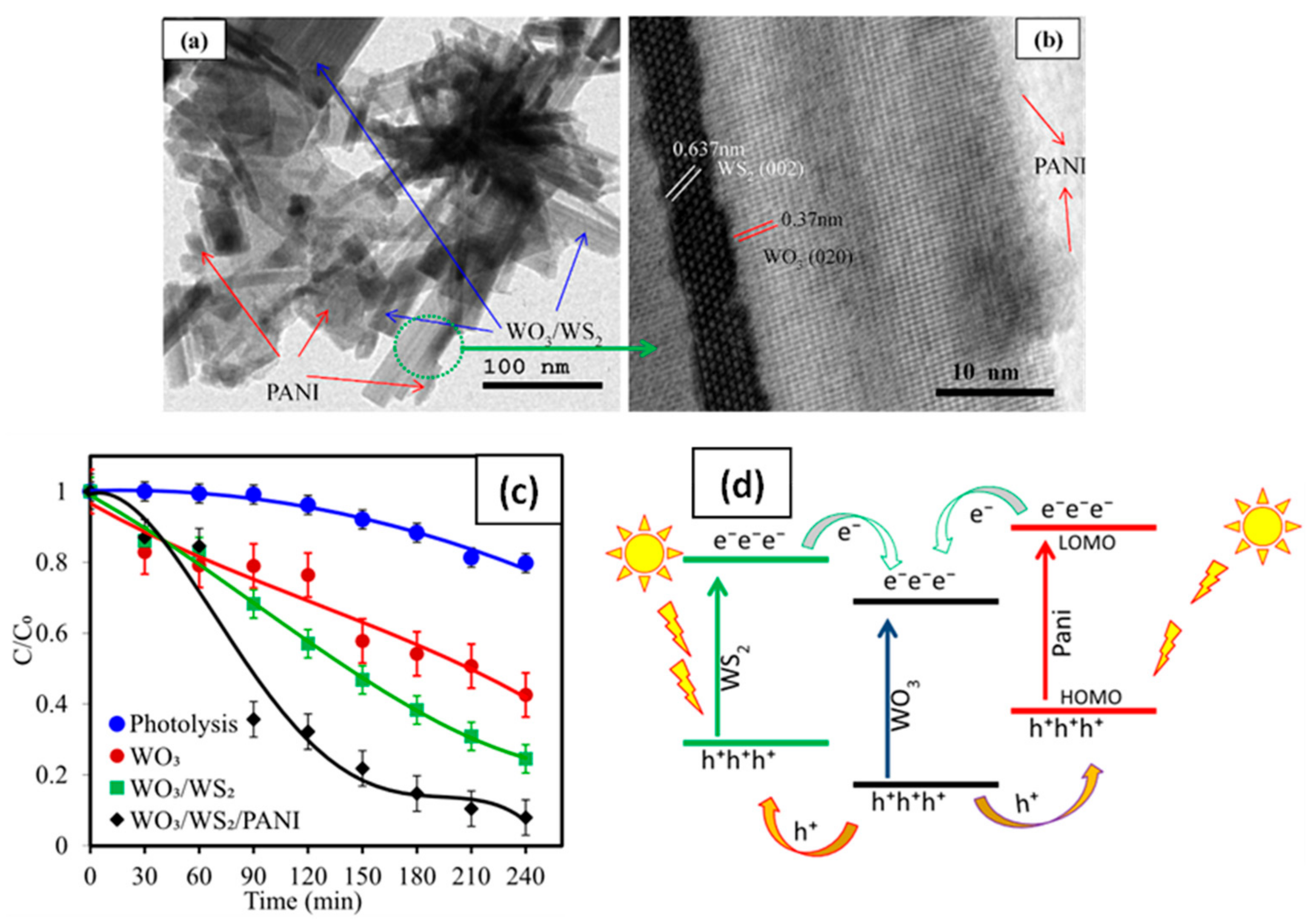
WS2/WO3
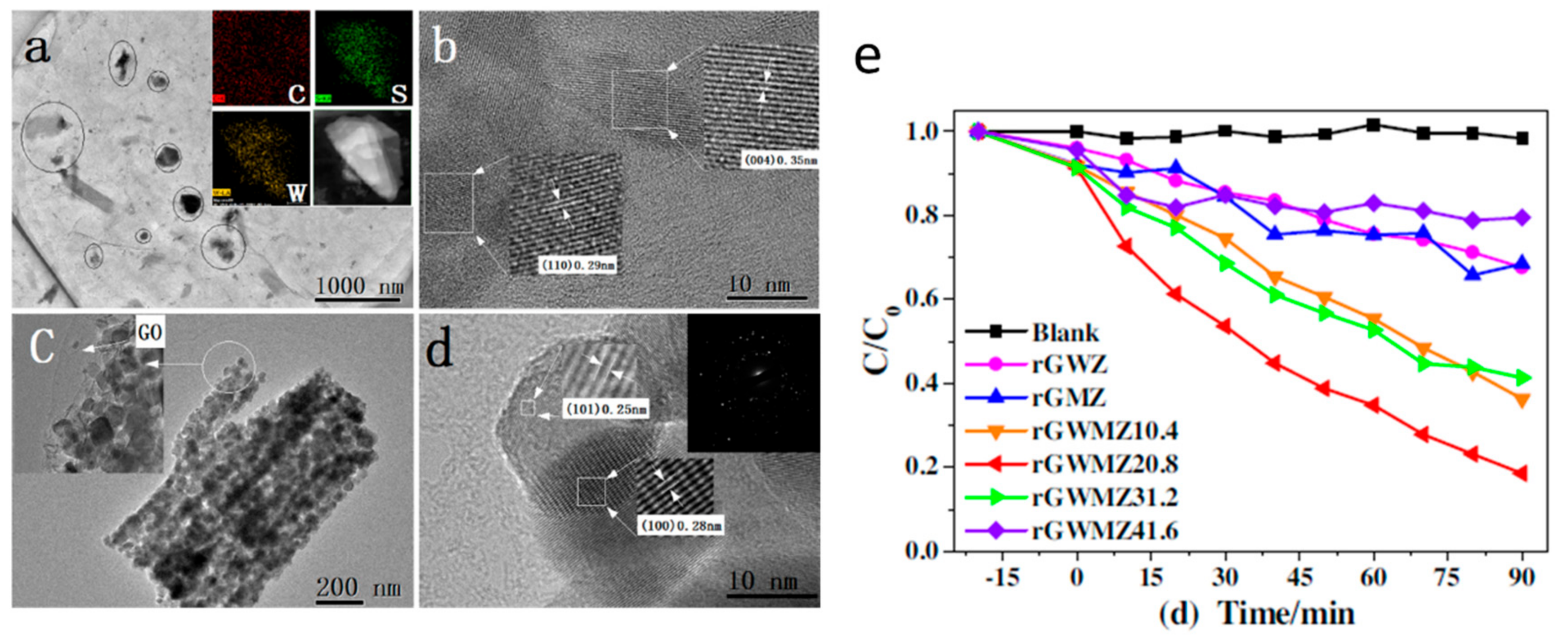
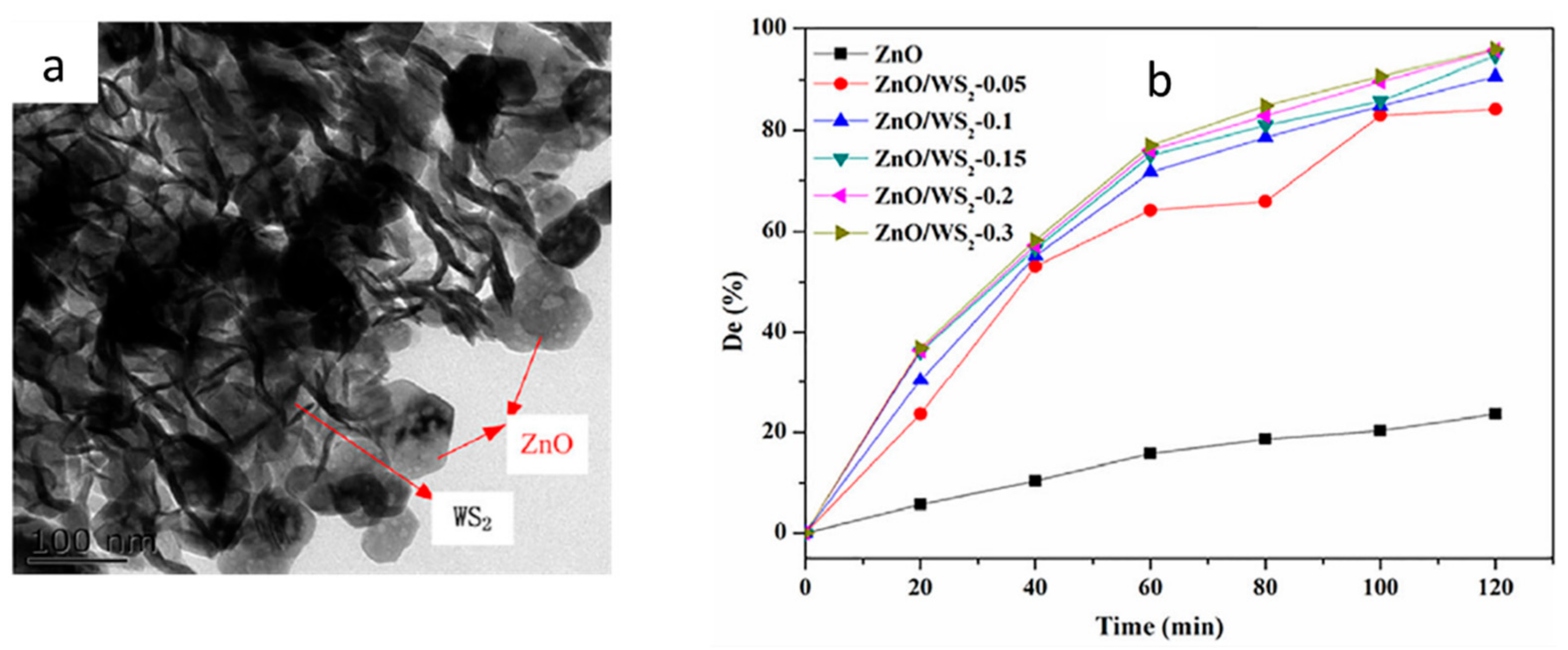
WS2/ZnO
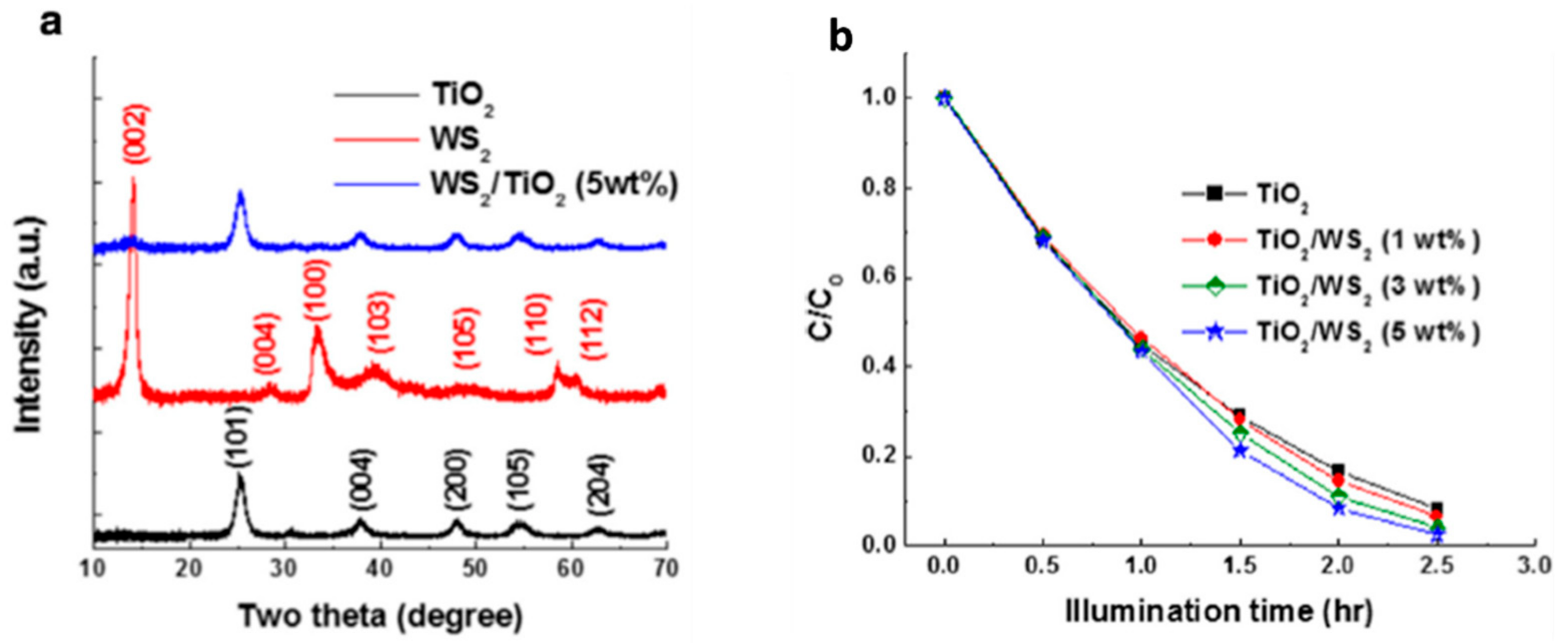
WS2/TiO2
4.2. Microorganisms’ Disinfection
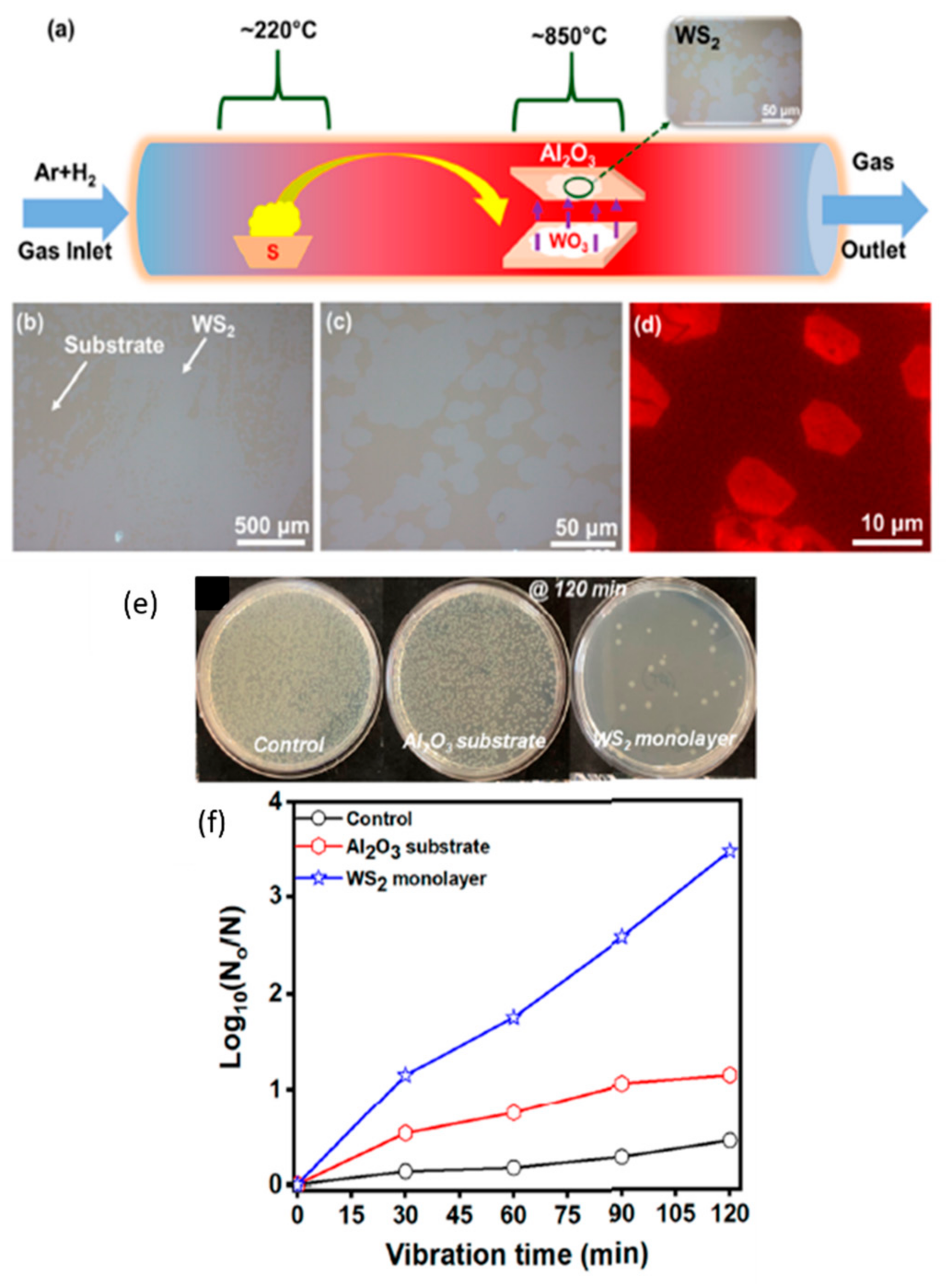
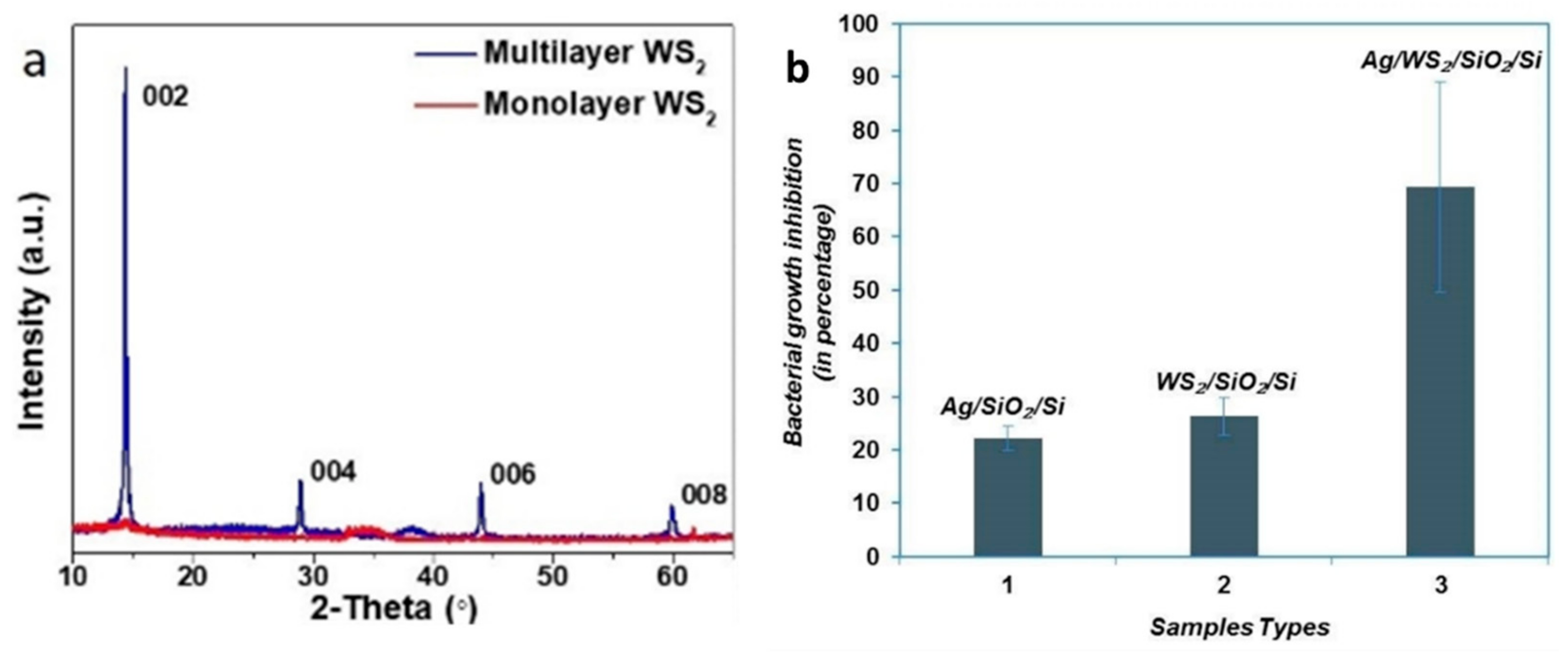
4.2.1. WS2 Monolayer
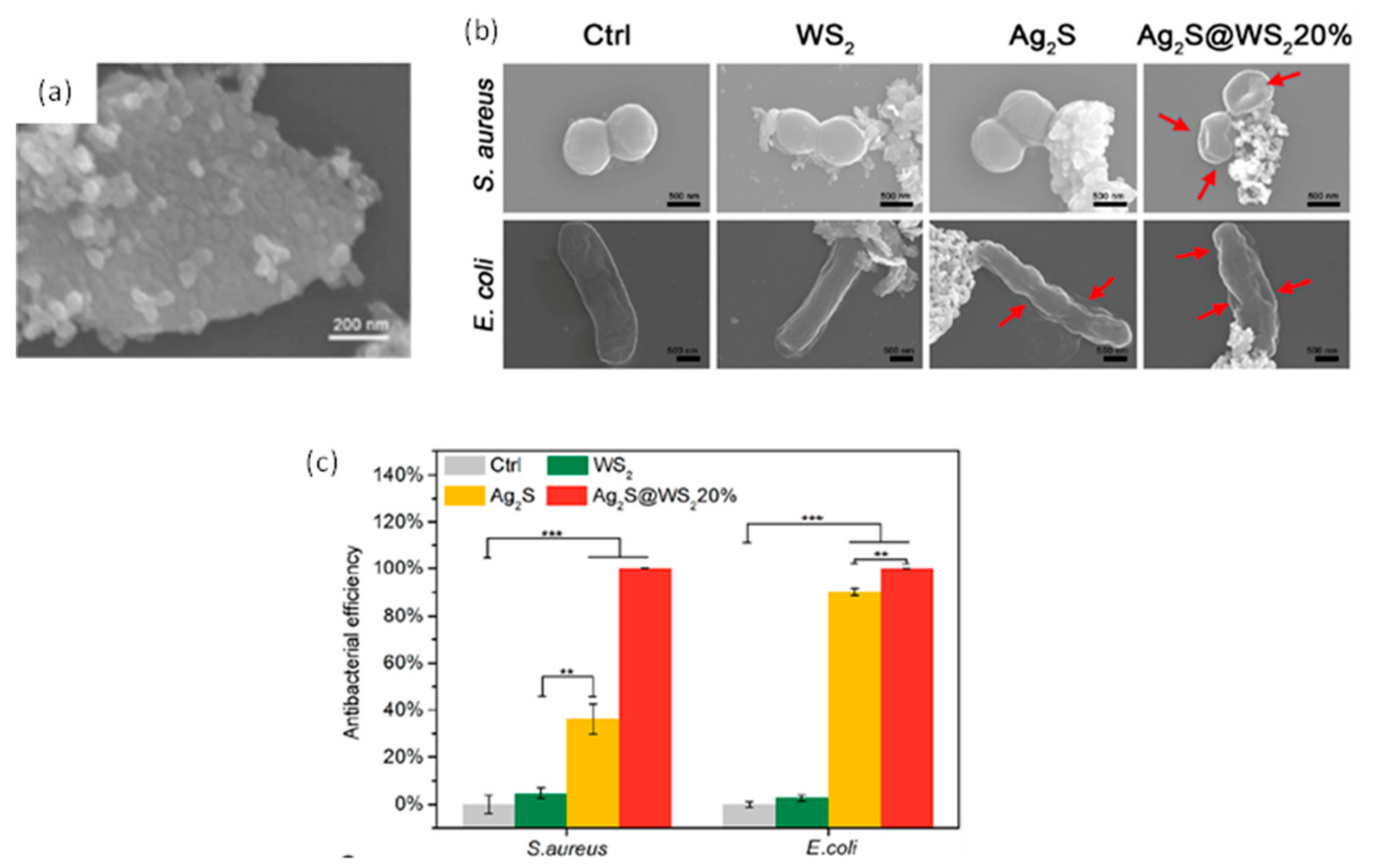
4.2.2. Ag2S@WS2
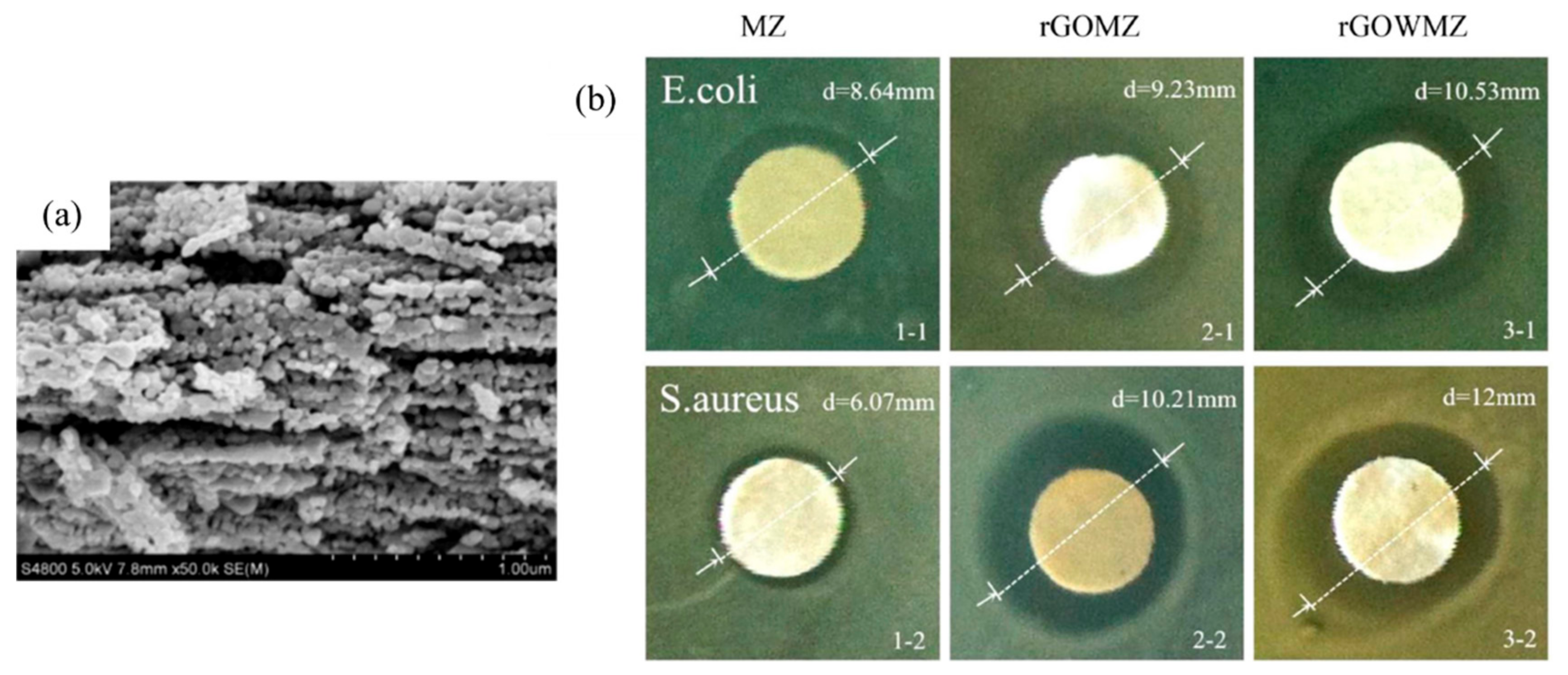
4.2.3. Graphene oxide/WS2/Mg-doped ZnO
4.2.4. WS2/Ag Nanoparticle
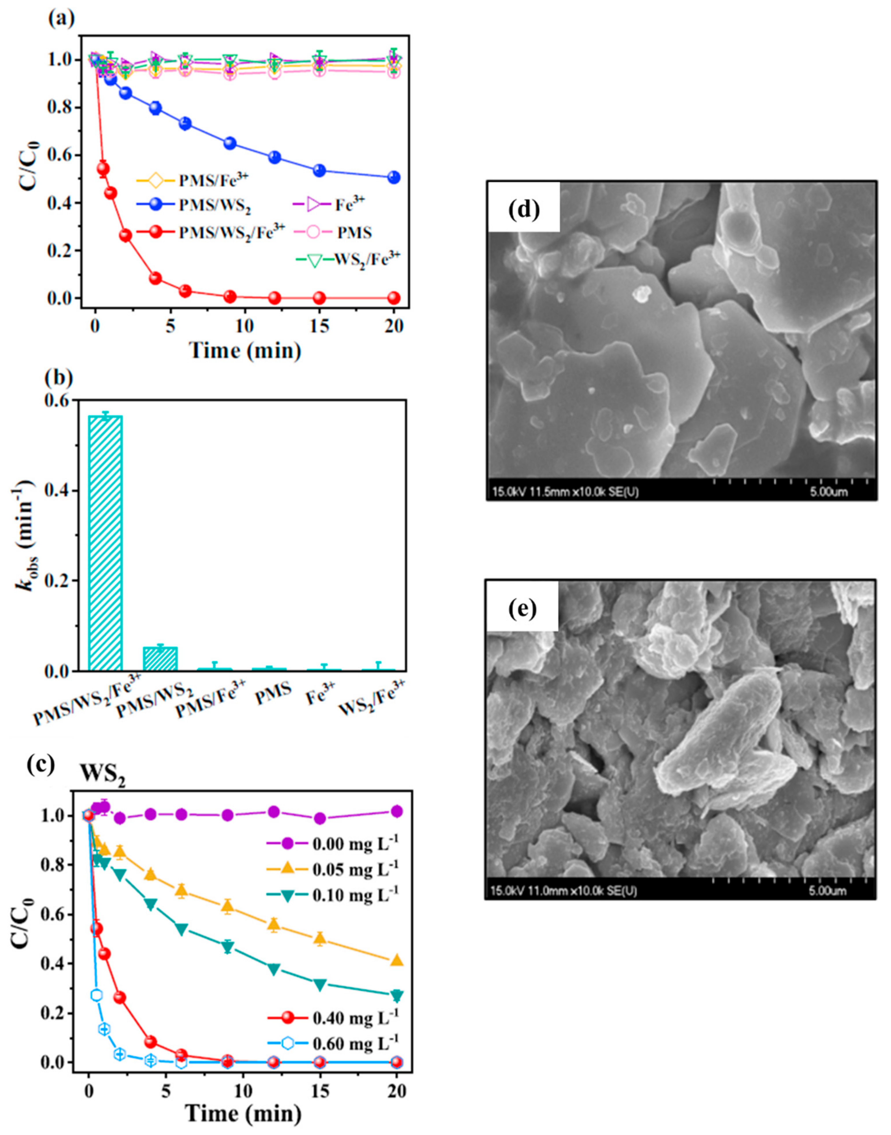
4.2.5. PMS/WS2/Fe3+
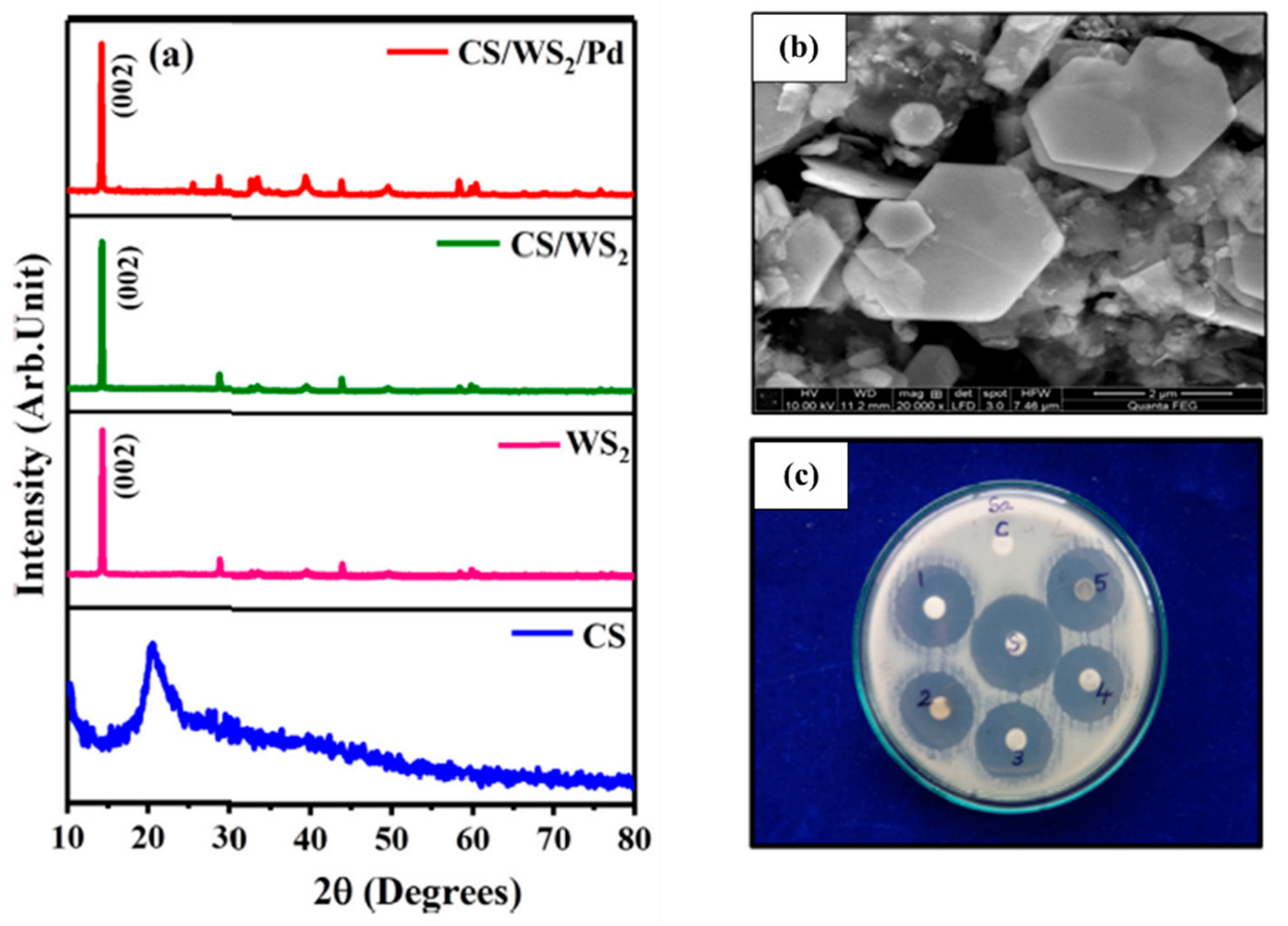
4.2.6. CS/WS2/Pd
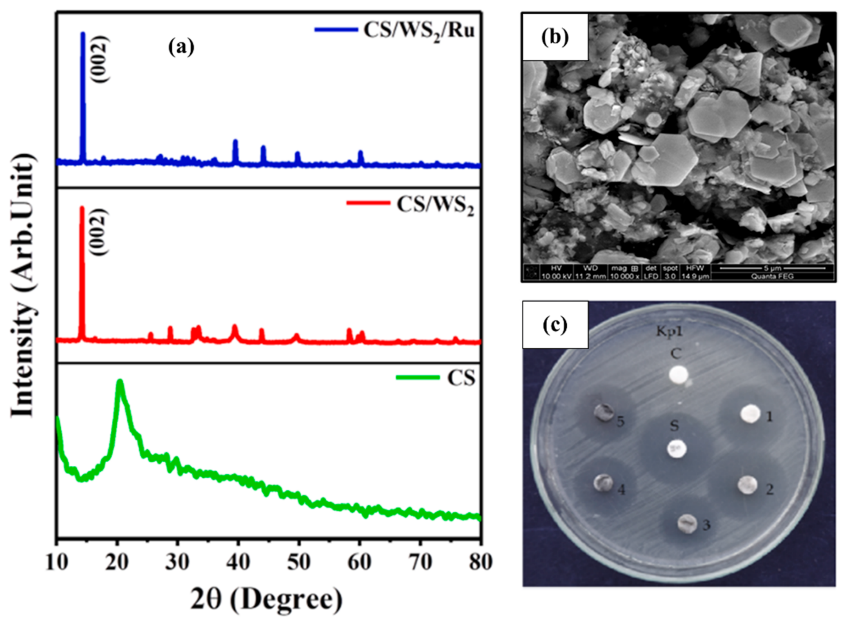
4.2.7. CS/WS2/Ru
4.3. Heavy Metals Reduction
4.3.1. CaIn2S4/WS2
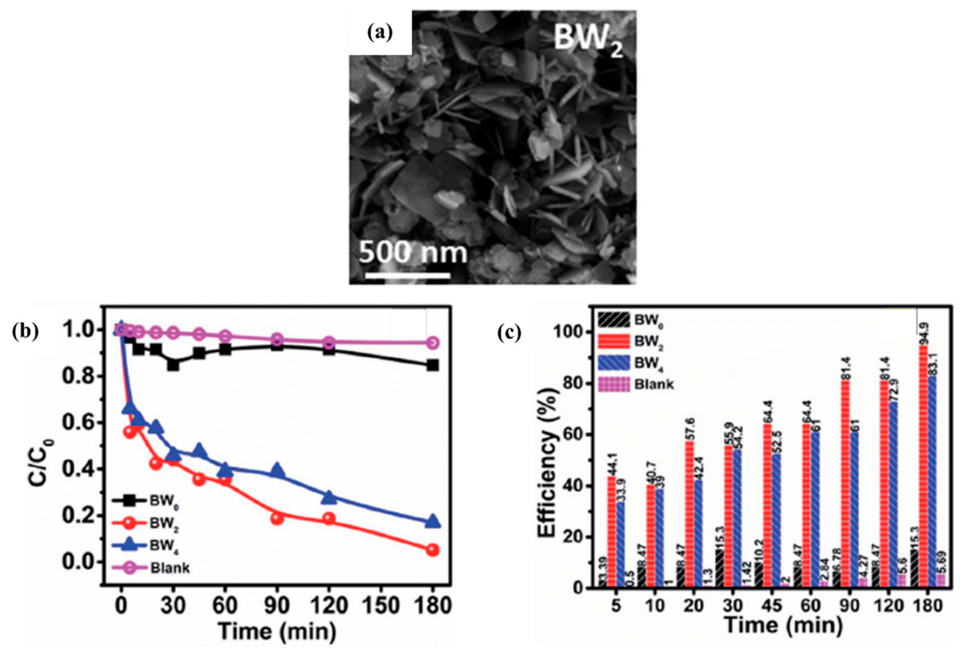
4.3.2. WS2/BiOCl
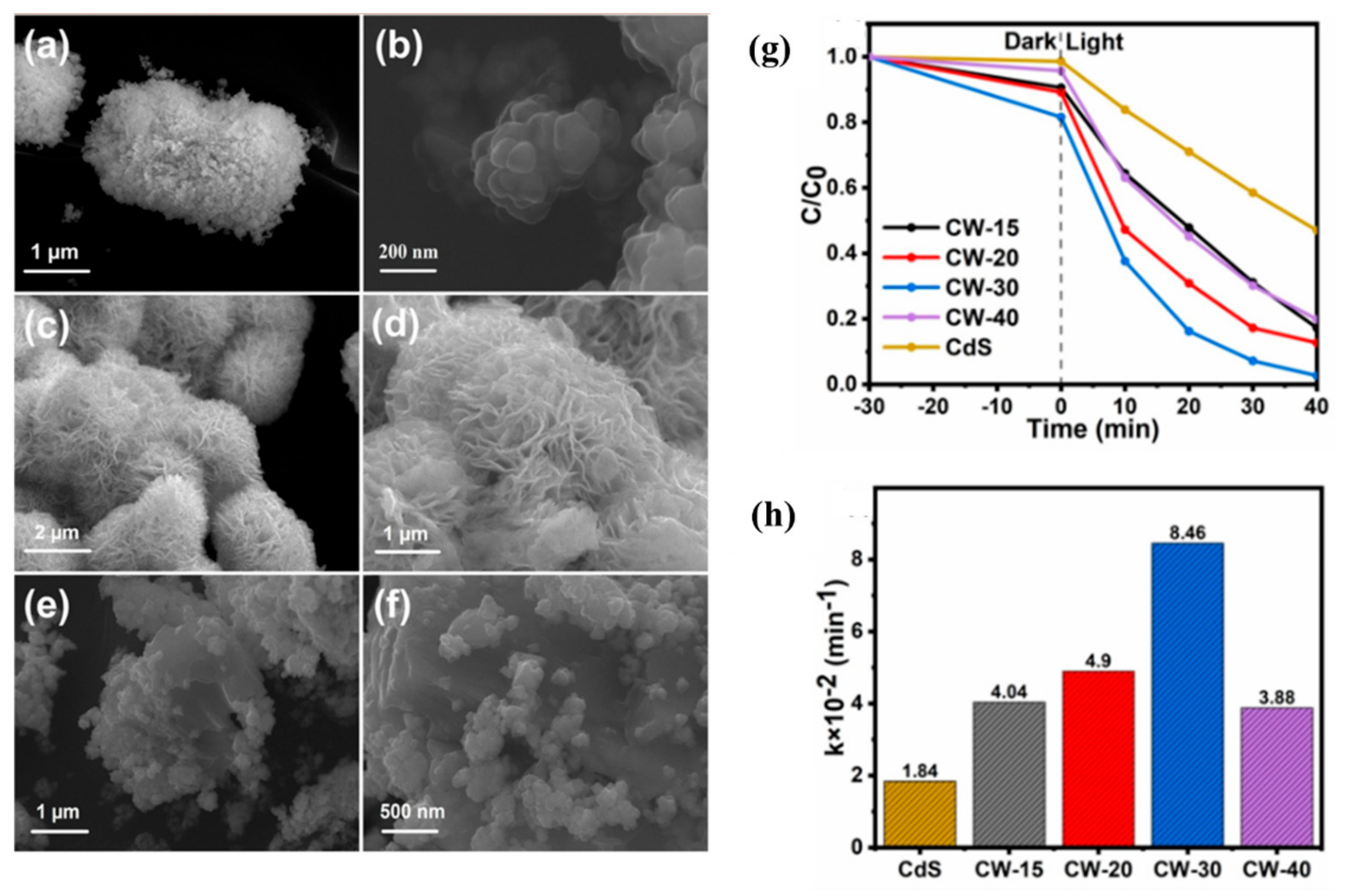
4.3.3. CdS/WS2
4.4. Pharmaceuticals Photodegradation
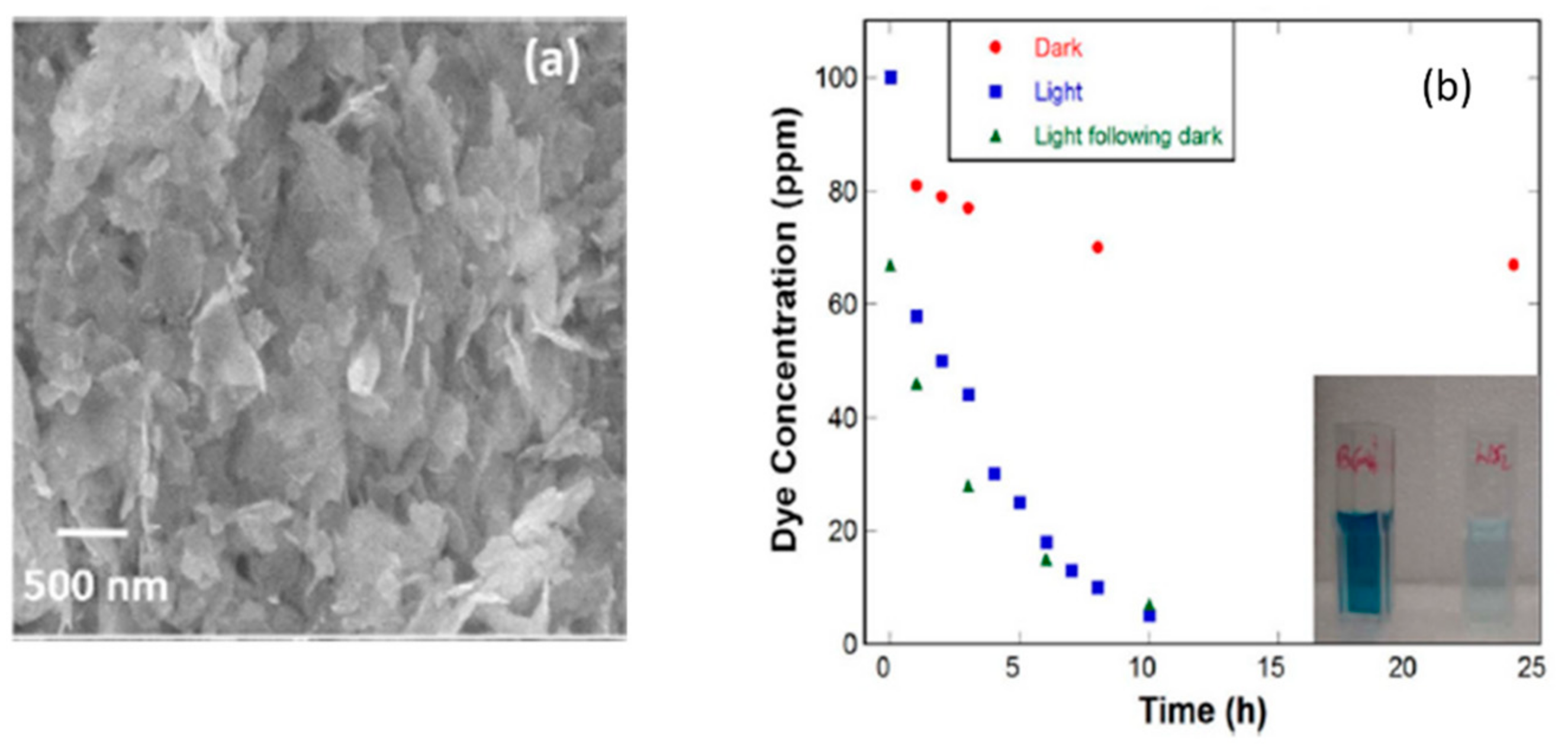
4.4.1. WS2 Nanosheets
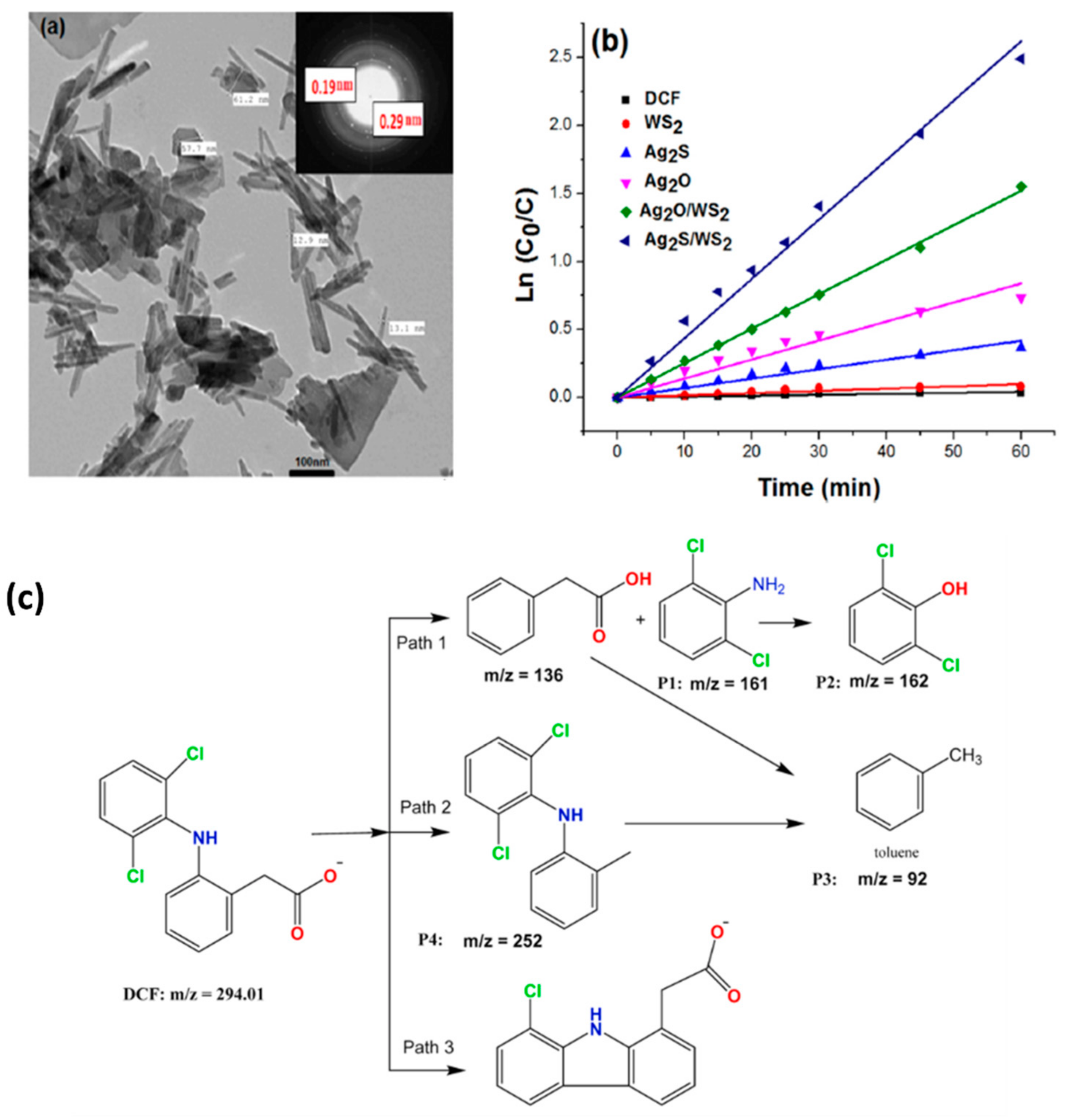
4.4.2. Ag@Ag2S/WS2 and Ag@Ag2O/WS2
4.4.3. PMS/WS2/Fe3+
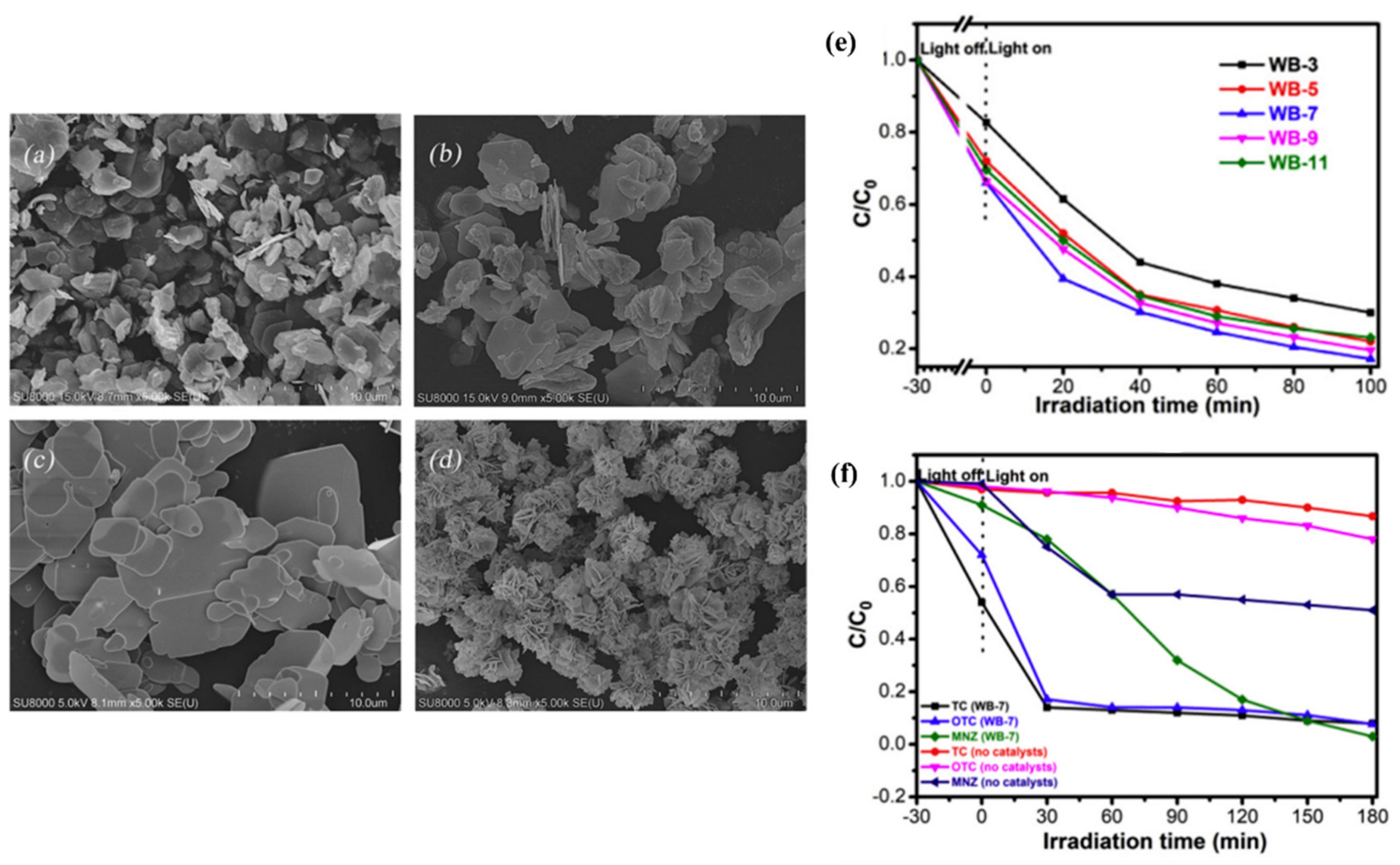
4.4.4. WS2/BiOBr
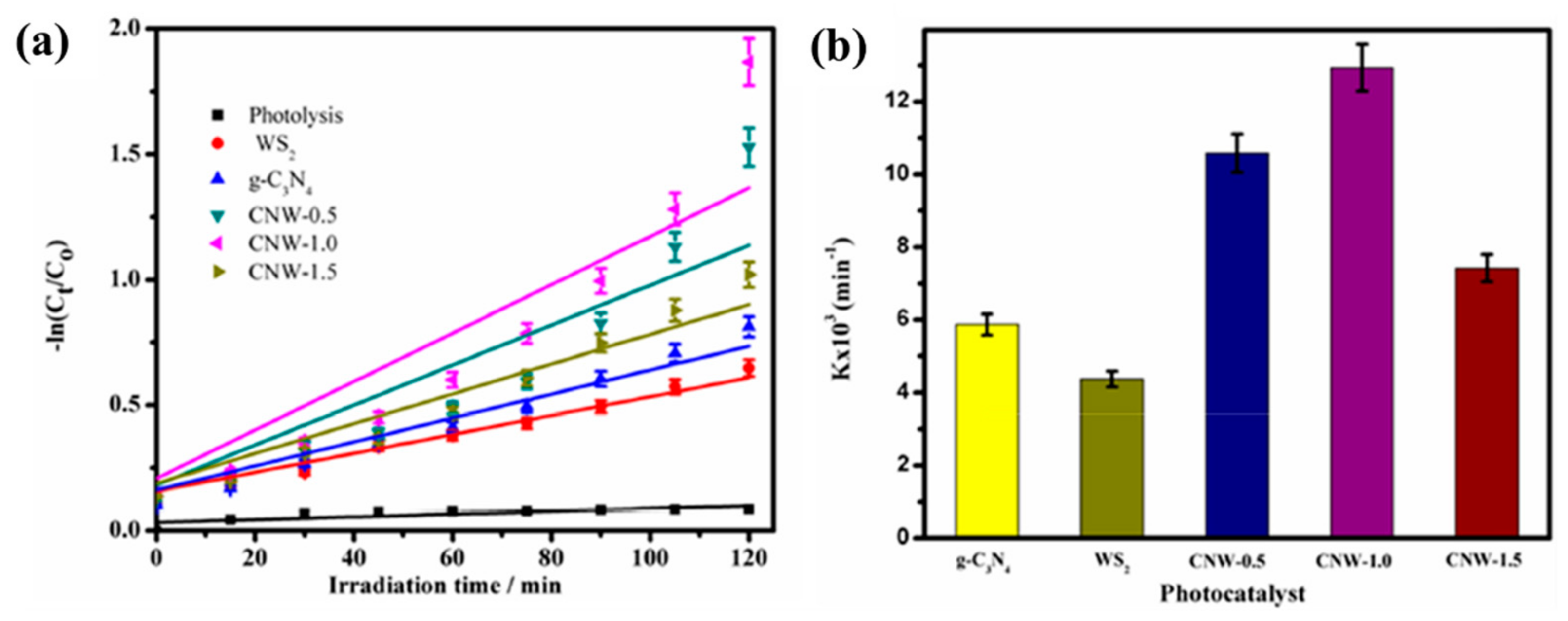
4.4.5. g-C3N4/WS2
5. Conclusions
Author Contributions
Funding
Data Availability Statement
Conflicts of Interest
References
- Jing, L.; Zhou, W.; Tian, G.; Fu, H. Surface tuning for oxide-based nanomaterials as efficient photocatalysts. Chem. Soc. Rev. 2013, 42, 9509–9549. [Google Scholar] [CrossRef] [PubMed]
- Alharbi, N.S.; Hu, B.; Hayat, T.; Rabah, S.O.; Alsaedi, A.; Zhuang, L.; Wang, X. Efficient elimination of environmental pollutants through sorption-reduction and photocatalytic degradation using nanomaterials. Front. Chem. Sci. Eng. 2020, 14, 1124–1135. [Google Scholar] [CrossRef]
- Garg, A.; Singhania, T.; Singh, A.; Sharma, S.; Rani, S.; Neogy, A.; Wang, X. Photocatalytic Degradation of Bisphenol-A using N, Co Codoped TiO2 Catalyst under Solar Light. Sci. Rep. 2019, 9, 765. [Google Scholar] [CrossRef] [PubMed]
- Qutub, N.; Singh, P.; Sabir, S.; Sagadevan, S.; Oh, W.-C. Enhanced photocatalytic degradation of Acid Blue dye using CdS/TiO2 nanocomposite. Sci. Rep. 2022, 12, 5759. [Google Scholar] [CrossRef] [PubMed]
- Zhang, J.; Zhang, L.; Ma, X.; Ji, Z. A study of constructing heterojunction between two-dimensional transition metal sulfides (MoS2 and WS2) and (101), (001) faces of TiO2. Appl. Surf. Sci. 2018, 430, 424–437. [Google Scholar] [CrossRef]
- Ahmad, R.; Ahmad, Z.; Khan, A.U.; Mastoi, N.R.; Aslam, M.; Kim, J. Photocatalytic systems as an advanced environmental remediation: Recent developments, limitations and new avenues for applications. J. Environ. Chem. Eng. 2016, 4, 4143–4164. [Google Scholar] [CrossRef]
- Li, S.; Wang, C.; Cai, M.; Yang, F.; Liu, Y.; Chen, J.; Zhang, P.; Li, X.; Chen, X. Facile fabrication of TaON/Bi2MoO6 core–shell S-scheme heterojunction nanofibers for boosting visible-light catalytic levofloxacin degradation and Cr(VI) reduction. Chem. Eng. J. 2022, 428, 131158. Available online: https://reader.elsevier.com/reader/sd/pii/S138589472102739X?token=D799FE216F506B28FC9A76E8F9EFC7EEA687C2DB4A2C5E4FC1B6619450B2316178BE5B5C01908DFCF9277A19062DAC7F&originRegion=eu-west-1&originCreation=20211216102211 (accessed on 16 December 2021). [CrossRef]
- Wang, W.; Zeng, Z.; Zeng, G.; Zhang, C.; Xiao, R.; Zhou, C.; Xiong, W.; Yang, Y.; Liu, Y.; Huang, D.; et al. Sulfur doped carbon quantum dots loaded hollow tubular g-C3N4 as novel photocatalyst for destruction of Escherichia coli and tetracycline degradation under visible light. Chem. Eng. J. 2019, 378, 122132. Available online: https://reader.elsevier.com/reader/sd/pii/S1385894719315268?token=E75BCF52D7CADB9C79C9153BB600C5717FE868F4BDB3A4177DF72392C836E5B307F44EBC39C8E62F8F3DF39E862C8B55&originRegion=eu-west-1&originCreation=20211216104206 (accessed on 16 December 2021). [CrossRef]
- Sahu, K.; Kuriakose, S.; Singh, J.; Satpati, B.; Mohapatra, S. Facile synthesis of ZnO nanoplates and nanoparticle aggregates for highly efficient photocatalytic degradation of organic dyes. J. Phys. Chem. Solids 2018, 121, 186–195. Available online: https://reader.elsevier.com/reader/sd/pii/S0022369717324393?token=5DC0699F806F4278530EE3223AEF547BF5347A5CAF0686D2B3FE4D7A973AA8408AC6906B379906548B22375646CF2A55&originRegion=eu-west-1&originCreation=20211216105136 (accessed on 16 December 2021). [CrossRef]
- George, R.; Bahadur, N.; Singh, N.; Singh, R.; Verma, A.; Shukla, A.K. Environmentally Benign TiO2 Nanomaterials for Removal of Heavy Metal Ions with Interfering Ions Present in Tap Water. Mater. Today Proc. 2016, 3, 162–166. [Google Scholar] [CrossRef]
- Pelaez, M.; Nolan, N.T.; Pillai, S.C.; Seery, M.K.; Falaras, P.; Kontos, A.G.; Dunlop, P.S.M.; Hamilton, J.W.J.; Byrne, J.A.; O’Shea, K.; et al. A review on the visible light active titanium dioxide photocatalysts for environmental applications. Appl. Catal. B Environ. 2012, 125, 331–349. [Google Scholar] [CrossRef]
- Garg, A.; Basu, S.; Shetti, N.P.; Reddy, K.R. 2D materials and its heterostructured photocatalysts: Synthesis, properties, functionalization and applications in environmental remediation. J. Environ. Chem. Eng. 2021, 9, 106408. [Google Scholar] [CrossRef]
- Wang, Y.; Xiao, X.; Lu, M.; Xiao, Y. 3D network-like rGO-MoSe2 modified g-C3N4 nanosheets with Z-scheme heterojunction: Morphology control, heterojunction construct, and boosted photocatalytic performances. J. Alloys Compd. 2021, 893, 163197. [Google Scholar] [CrossRef]
- Wang, Y.; Lei, S.; Zhang, X.; Zhou, S. First-principles study of nitrogen defect g-C3N4/WS2 heterojunction on photocatalytic activity. Curr. Appl. Phys. 2022, 39, 70–76. [Google Scholar] [CrossRef]
- Choi, W.; Choudhary, N.; Han, G.H.; Park, J.; Akinwande, D.; Lee, Y.H. Recent development of two-dimensional transition metal dichalcogenides and their applications. Mater. Today 2017, 20, 116–130. [Google Scholar] [CrossRef]
- Zhang, D.; Liu, T.; Cheng, J.; Liang, S.; Chai, J.; Yang, X.; Wang, H.; Zheng, G.; Cao, M. Controllable synthesis and characterization of tungsten disulfide nanosheets as promising nanomaterials for electronic devices. Ceram. Int. 2019, 45, 12443–12448. [Google Scholar] [CrossRef]
- Shalaby, M.S.; Sołowski, G.; Abbas, W. Recent Aspects in Membrane Separation for Oil/Water Emulsion. Adv. Mater. Interfaces 2021, 8, 2100448. [Google Scholar] [CrossRef]
- Zhang, N.; Yang, X.; Wang, Y.; Qi, Y.; Zhang, Y.; Luo, J.; Cui, P.; Jiang, W. A review on oil/water emulsion separation membrane material. J. Environ. Chem. Eng. 2022, 10, 107257. [Google Scholar] [CrossRef]
- Mishra, A.K.; Lakshmi, K.V.; Huang, L. Eco-friendly synthesis of metal dichalcogenides nanosheets and their environmental remediation potential driven by visible light. Sci. Rep. 2015, 5, 15718. [Google Scholar] [CrossRef]
- Nawaz, A.; Goudarzi, S.; Saravanan, P.; Zarrin, H. Z-scheme induced g-C3N4/WS2 heterojunction photocatalyst with improved electron mobility for enhanced solar photocatalysis. Sol. Energy. 2021, 228, 53–67. [Google Scholar] [CrossRef]
- Ma, S.; Zeng, L.; Tao, L.; Tang, C.Y.; Yuan, H.; Long, H.; Cheng, P.K.; Chai, Y.; Chen, C.; Fung, K.H.; et al. Enhanced Photocatalytic Activity of WS2 Film by Laser Drilling to Produce Porous WS2/WO3 Heterostructure Methods and mechanisms for improvement of photocatalytic activity, are important and popular research topics for renewable energy production and waste water treatment OPEN. Sci. Rep. 2017, 7, 3125. Available online: https://nature.com/scientificreports/ (accessed on 1 June 2022). [PubMed]
- Hazarika, S.J.; Mohanta, D. Inorganic fullerene-type WS2 nanoparticles: Processing, characterization and its photocatalytic performance on malachite green. Appl. Phys. A 2017, 123, 381. [Google Scholar] [CrossRef]
- Hamd, W.; Daher, E.A.; Tofa, T.S.; Dutta, J. Recent Advances in Photocatalytic Removal of Microplastics: Mechanisms, Kinetic Degradation, and Reactor Design. Front. Mar. Sci. 2022, 9, 885614. [Google Scholar] [CrossRef]
- Kozlova, E.A.; Parmon, V.N. Heterogeneous semiconductor photocatalysts for hydrogen production from aqueous solutions of electron donors. Russ. Chem. Rev. 2017, 86, 870–906. Available online: https://iopscience.iop.org/article/10.1070/RCR4739 (accessed on 20 August 2022). [CrossRef]
- Ohtani, B. Revisiting the fundamental physical chemistry in heterogeneous photocatalysis: Its thermodynamics and kinetics Physical Chemistry Chemical Physics. R. Soc. Chem. 2014, 16, 1788–1897. Available online: https://pubs.rsc.org/en/content/articlehtml/2014/cp/c3cp53653j (accessed on 20 August 2022).
- Subhiksha, V.; Kokilavani, S.; Sudheer Khan, S. Recent advances in degradation of organic pollutant in aqueous solutions using bismuth based photocatalysts: A review. Chemosphere 2022, 290, 133228. [Google Scholar] [CrossRef] [PubMed]
- Adhikari, S.; Kim, D.H. Synthesis of Bi2S3/Bi2WO6 hierarchical microstructures for enhanced visible light driven photocatalytic degradation and photoelectrochemical sensing of ofloxacin. Chem. Eng. J. 2018, 354, 692–705. [Google Scholar] [CrossRef]
- Guo, Q.; Zhou, C.; Ma, Z.; Yang, X. Fundamentals of TiO2 Photocatalysis: Concepts, Mechanisms, and Challenges Advanced Materials. Adv. Mater. 2019, 31, 1901997. Available online: https://onlinelibrary.wiley.com/doi/full/10.1002/adma.201901997 (accessed on 20 August 2022). [CrossRef]
- Li, K.; An, X.; Park, K.H.; Khraisheh, M.; Tang, J. A critical review of CO2 photoconversion: Catalysts and reactors. Catal. Today 2014, 224, 3–12. [Google Scholar] [CrossRef]
- Al Jitan, S.; Palmisano, G.; Garlisi, C. Synthesis and surface modification of TiO2-based photocatalysts for the conversion of CO2 Catalysts. Multidiscip. Digit. Publ. Inst. 2020, 10, 227. Available online: https://www.mdpi.com/2073-4344/10/2/227/htm (accessed on 20 August 2022).
- Shtyka, O.; Ciesielski, R.; Kedziora, A.; Maniukiewicz, W.; Dubkov, S.; Gromov, D.; Maniecki, T. Photocatalytic Reduction of CO2 Over Me (Pt, Pd, Ni, Cu)/TiO2 Catalysts. Top. Catal. 2020, 63, 113–120. Available online: https://link.springer.com/article/10.1007/s11244-020-01241-y (accessed on 21 August 2022). [CrossRef]
- Zou, H.; Yan, X.; Ren, J.; Wu, X.; Dai, Y.; Sha, D.; Pan, J.; Liu, J. Photocatalytic activity enhancement of modified g-C3N4 by ionothermal copolymerization. J. Mater. 2015, 1, 340–347. [Google Scholar] [CrossRef] [Green Version]
- Ismael, M. A review on graphitic carbon nitride (g-C3N4) based nanocomposites: Synthesis, categories, and their application in photocatalysis. J. Alloys Compd. 2020, 846, 156446. [Google Scholar] [CrossRef]
- Chen, S.; Pan, Y.; Wang, D.; Deng, H. Structural Stability and Electronic and Optical Properties of Bulk WS2 from First-Principles Investigations. J. Electron. Mater. 2020, 49, 7363–7369. [Google Scholar] [CrossRef]
- Lv, R.; Robinson, J.A.; Schaak, R.E.; Sun, D.; Sun, Y.; Mallouk, T.E.; Terrones, M. Transition metal dichalcogenides and beyond: Synthesis, properties, and applications of single- and few-layer nanosheets. Acc. Chem. Res. 2015, 48, 56–64. Available online: https://pubmed.ncbi.nlm.nih.gov/25490673/ (accessed on 9 May 2022). [CrossRef] [PubMed]
- Zeng, Z.; Yin, Z.; Huang, X.; Li, H.; He, Q.; Lu, G.; Boey, H.; Zhang, H. Single-layer semiconducting nanosheets: High-yield preparation and device fabrication. Angew. Chem. 2011, 50, 11093–11097. Available online: https://pubmed.ncbi.nlm.nih.gov/22021163/ (accessed on 9 May 2022). [CrossRef] [PubMed]
- Gutiérrez, H.R.; Perea-López, N.; Elías, A.L.; Berkdemir, A.; Wang, B.; Lv, R.; López-Urías, R.; Crespi, V.H.; Terrones, H.; Terrones, S. Extraordinary room-temperature photoluminescence in triangular WS2 monolayers. Nano. Lett. 2013, 13, 3447–3454. Available online: https://eres.qnl.qa/login?url=https://search.ebscohost.com/login.aspx?direct=true&db=cmedm&AN=23194096&site=ehost-live (accessed on 9 May 2022). [CrossRef] [PubMed]
- Yun, W.S.; Han, S.W.; Hong, S.C.; Kim, I.G.; Lee, J.D. Thickness and strain effects on electronic structures of transition metal dichalcogenides: 2H-MX2 semiconductors (M = Mo, W; X = S, Se, Te). Phys. Rev. B 2012, 85, 33305. [Google Scholar] [CrossRef]
- Liu, L.; Kumar, S.B.; Ouyang, Y.; Guo, J. Performance Limits of Monolayer Transition Metal Dichalcogenide Transistors. IEEE Trans. Electron. Devices 2011, 58, 3042–3047. [Google Scholar] [CrossRef]
- Al-Hilli, A.A.; Evans, B.L. The preparation and properties of transition metal dichalcogenide single crystals. J. Cryst. Growth 1972, 15, 93–101. [Google Scholar] [CrossRef]
- Jin, Q.; Dai, X.; Song, J.; Pu, K.; Wu, X.; An, J.; Zhao, T. High photocatalytic performance of g-C3N4/WS2 heterojunction from first principles. Chem. Phys. 2021, 545, 111141. [Google Scholar] [CrossRef]
- Bin Rafiq, M.K.S.; Amin, N.; Alharbi, H.F.; Luqman, M.; Ayob, A.; Alharthi, Y.S.; Alharthi, N.H.; Bais, B.; Akhtaruzzaman, M. WS2: A New Window Layer Material for Solar Cell Application. Sci. Rep. 2020, 10, 771. [Google Scholar] [CrossRef] [PubMed] [Green Version]
- Green, M.A. Thin-film solar cells: Review of materials, technologies and commercial status. J. Mater. Sci. Mater. Electron. 2007, 18, 15–19. [Google Scholar] [CrossRef]
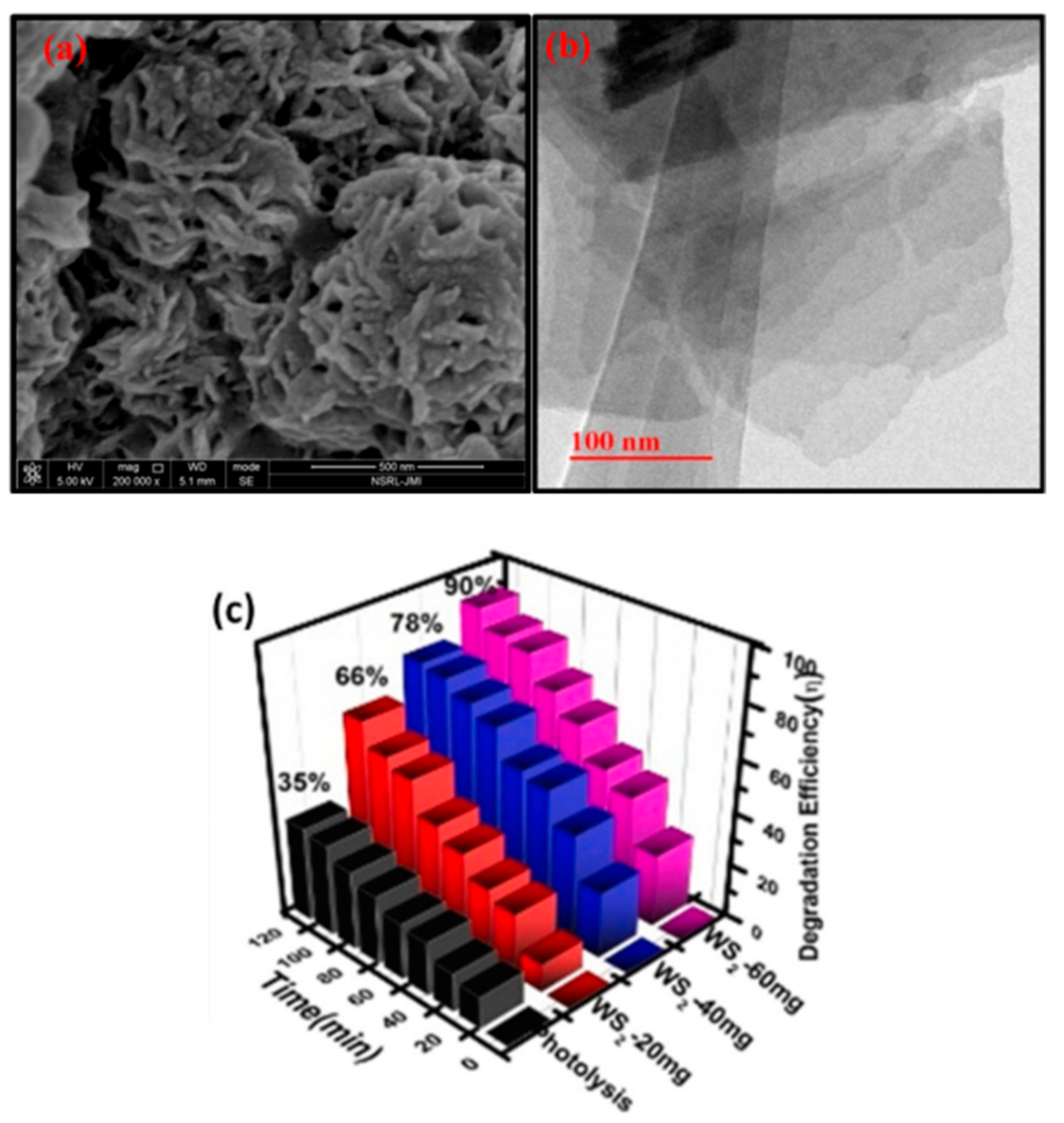
- Fatima, T.; Husain, S.; Narang, J.; Khanuja, M.; Shetti, N.P.; Reddy, K.R. Novel tungsten disulfide (WS2) nanosheets for photocatalytic degradation and electrochemical detection of pharmaceutical pollutants. J. Water Process. Eng. 2022, 47, 102717. [Google Scholar] [CrossRef]
- Voiry, D.; Yamaguchi, H.; Li, J.; Silva, R.; Alves, D.C.B.; Fujita, T.; Chen, M.; Asefa, T.; Shenoy, V.B.; Eda, G.; et al. Enhanced catalytic activity in strained chemically exfoliated WS2 nanosheets for hydrogen evolution. Nat. Mater. 2013, 12, 850–855. [Google Scholar] [CrossRef]
- Cao, S.; Liu, T.; Hussain, S.; Zeng, W.; Peng, X.; Pan, F. Hydrothermal synthesis of variety low dimensional WS2 nanostructures. Mater. Lett. 2014, 129, 205–208. [Google Scholar] [CrossRef]
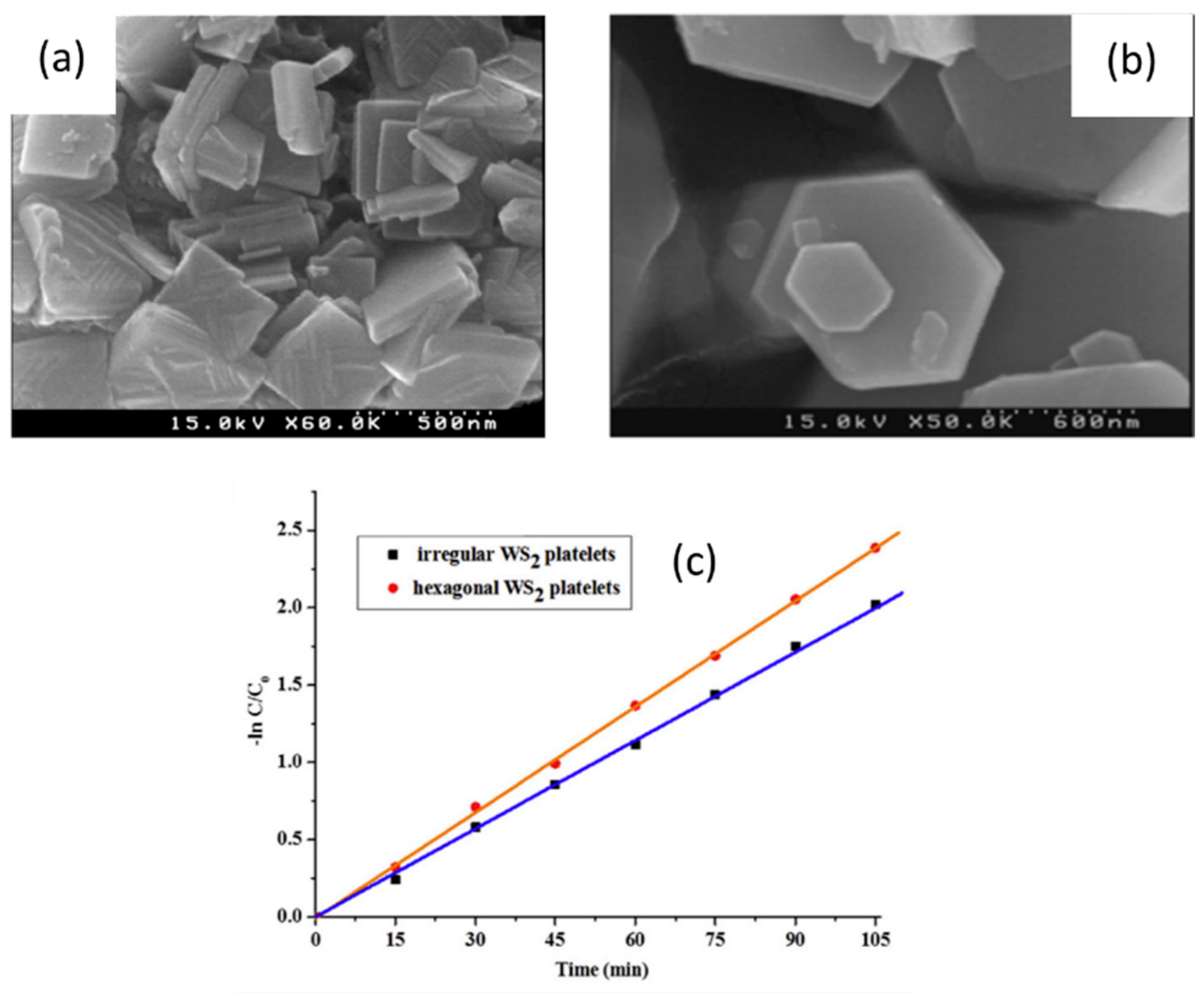
- Vattikuti, S.V.P.; Byon, C.; Chitturi, V. Selective hydrothermally synthesis of hexagonal WS2 platelets and their photocatalytic performance under visible light irradiation. Superlattices Microstruct. 2016, 94, 39–50. [Google Scholar] [CrossRef]
- Joseph, A.; Aneesh, P.M. Efficient degradation of methylene blue: A comparative study using hydrothermally synthesised SnS2, WS2 and VS2 nanostructures. Mater. Res. Bull. 2022, 146, 111623. [Google Scholar] [CrossRef]
- Liu, X.; Chen, X.; Wang, S.; Yan, L.; Yan, J.; Guo, H.; Yang, F.; Lin, J. Promoting the photocatalytic H2 evolution activity of CdLa2S4 nanocrystalline using few-layered WS2 nanosheet as a co-catalyst. Int. J. Hydrogen Energy 2022, 47, 2327–2337. [Google Scholar] [CrossRef]
- Kasinathan, K.; Marimuthu, K.; Murugesan, B.; Sathaiah, M.; Subramanian, P.; Sivakumar, P.; Swaminathan, U.; Subbiah, R. Fabrication of eco-friendly chitosan functionalized few-layered WS2 nanocomposite implanted with ruthenium nanoparticles for in vitro antibacterial and anticancer activity: Synthesis, characterization, and pharmaceutical applications. Int. J. Biol. Macromol. 2021, 190, 520–532. [Google Scholar] [CrossRef]
- Ling, X.; Lee, Y.H.; Lin, Y.; Fang, W.; Yu, L.; Dresselhaus, M.S.; Kong, J. Role of the seeding promoter in MoS2 growth by chemical vapor deposition. Nano. Lett. 2014, 14, 464–472. [Google Scholar] [CrossRef] [PubMed]
- Mzerd, A.; Sayah, D.; Tedenac, J.C.; Boyer, A. Optimal crystal growth conditions of thin films of Bi2Te3 semiconductors. J. Cryst. Growth 1994, 140, 365–369. [Google Scholar] [CrossRef]
- Thakur, D.; Sharma, M.; Vaish, R.; Balakrishnan, V. WS2Monolayer for Piezo-Phototronic Dye Degradation and Bacterial Disinfection. ACS Appl. Nano Mater. 2021, 4, 7879–7887. [Google Scholar] [CrossRef]
- Chen, F.; Xia, Y.; Lv, Q.; Mao, S.; Li, Y. Growth and Optoelectronic Properties of Large-Scale Bilayer WS2Ribbons with Unusual Shapes via Chemical Vapor Deposition. J. Phys. Chem. C 2022, 126, 1099–1106. [Google Scholar] [CrossRef]
- Thakur, D.; Kumar, P.; Sabarigresan, M.; Ramadurai, R.; Balakrishnan, V. Layer number dependent optical and electrical properties of CVD grown two-dimensional anisotropic WS2. Surf. Interfaces 2021, 26, 101308. [Google Scholar] [CrossRef]
- Meng, L.; Yu, Y.; Yan, W.; Li, H.; Zhao, Q.; Yan, X. Growth mechanism of two-dimensional WS2 film under the modulation of liquid metal. Phys. E Low-Dimens. Syst. Nanostruct. 2021, 134, 114885. [Google Scholar] [CrossRef]
- Coleman, J.N.; Lotya, M.; O’Neill, A.; Bergin, S.D.; King, P.J.; Khan, U.; Young, K.; Gaucher, A.; De, S.; Smith, R.J.; et al. Two-dimensional nanosheets produced by liquid exfoliation of layered materials. Science 2011, 331, 568–571. [Google Scholar] [CrossRef]
- Smith, R.J.; King, P.J.; Lotya, M.; Wirtz, C.; Khan, U.; De, S.; O’Neill, A.; Duesberg, G.S.; Grunlan, J.C.; Moriarty, G.; et al. Large-scale exfoliation of inorganic layered compounds in aqueous surfactant solutions. Adv. Mater. 2011, 23, 3944–3948. Available online: https://onlinelibrary.wiley.com/doi/full/10.1002/adma.201102584 (accessed on 1 June 2022). [CrossRef]
- Mohan, V.B.; Lau K tak Hui, D.; Bhattacharyya, D. Graphene-based materials and their composites: A review on production, applications and product limitations. Compos. Part B Eng. 2018, 142, 200–220. [Google Scholar] [CrossRef]
- Bonaccorso, F.; Bartolotta, A.; Coleman, J.N.; Backes, C. 2D-Crystal-Based Functional Inks. Adv. Mater. 2016, 28, 6136–6166. Available online: https://onlinelibrary.wiley.com/doi/full/10.1002/adma.201506410 (accessed on 1 June 2022). [CrossRef]
- Bonaccorso, F.; Colombo, L.; Yu, G.; Stoller, M.; Tozzini, V.; Ferrari, A.C.; Ruoff, R.S.; Pellegrini, V. Graphene, related two-dimensional crystals, and hybrid systems for energy conversion and storage. Science 2015, 347, 6217. [Google Scholar] [CrossRef] [PubMed]
- Hu, G.; Kang, J.; Ng, L.W.T.; Zhu, X.; Howe, R.C.T.; Jones, C.G.; Hersam, M.C.; Hasan, T. Functional inks and printing of two-dimensional materials. Chem. Soc. Rev. 2018, 47, 3265–3300. Available online: https://pubs.rsc.org/en/content/articlehtml/2018/cs/c8cs00084k (accessed on 1 June 2022). [CrossRef] [PubMed] [Green Version]
- Ashfaq, M.; Talreja, N.; Chauhan, D.; Viswanathan, M.R. Synthesis of Cu-doped 2D-WS2 nanosheet-based nano-antibiotic materials for inhibiting E. Coli and S. aureus bacterial strains. New J. Chem. 2022, 46, 5581–5587. [Google Scholar] [CrossRef]
- Esfandiari, M.; Kamaei, S.; Rajabali, M.; Mohajerzadeh, S. High-performance large-area WS2-based transistors by a novel tin-oxide assisted liquid-phase exfoliation: Doping adjustment by plasma treatment. 2D Mater. 2021, 8, 025013. [Google Scholar] [CrossRef]
- Tayebi, M.; Masoumi, Z.; Lee, B.K. Ultrasonically prepared photocatalyst of W/WO3 nanoplates with WS2 nanosheets as 2D material for improving photoelectrochemical water splitting. Ultrason. Sonochem. 2021, 70, 105339. [Google Scholar] [CrossRef]
- Ren, X.; Wang, B.; Huang, Z.; Qiao, H.; Duan, C.; Zhou, Y.; Zhong, J.; Wang, Z.; Qi, X. Flexible self-powered photoelectrochemical-type photodetector based on 2D WS2-graphene heterojunction. FlatChem 2021, 25, 100215. [Google Scholar] [CrossRef]
- Zhu, D.; Zhou, Q. Action and mechanism of semiconductor photocatalysis on degradation of organic pollutants in water treatment: A review. Environ. Nanotechnol. Monit. Manag. 2019, 12, 100255. [Google Scholar] [CrossRef]
- Wang, H.; Zhang, L.; Chen, Z.; Hu, J.; Li, S.; Wang, Z.; Liu, J.; Wang, X. Semiconductor heterojunction photocatalysts: Design, construction, and photocatalytic performances. Chem. Soc. Rev. 2014, 43, 5234–5244. [Google Scholar] [CrossRef]
- Fan, Y.; Chen, G.; Li, D.; Li, F.; Luo, Y.; Meng, Q. Enhancement of photocatalytic H2 evolution on hexagonal CdS by a simple calcination method under visible light irradiation. Mater. Res. Bull. 2011, 46, 2338–2341. [Google Scholar] [CrossRef]
- Zong, X.; Wu, G.; Yan, H.; Ma, G.; Shi, J.; Wen, F.; Wang, L.; Li, C. Photocatalytic H2 evolution on MoS2/CdS catalysts under visible light irradiation. J. Phys. Chem. C 2010, 114, 1963–1968. [Google Scholar] [CrossRef]
- Es’haghzade, Z.; Pajootan, E.; Bahrami, H.; Arami, M. Facile synthesis of Fe3O4 nanoparticles via aqueous based electro chemical route for heterogeneous electro-Fenton removal of azo dyes. J. Taiwan Inst. Chem. Eng. 2017, 71, 91–105. [Google Scholar] [CrossRef]
- Yao, L.; Zhang, L.; Wang, R.; Chou, S.; Dong, Z.L. A new integrated approach for dye removal from wastewater by polyoxometalates functionalized membranes. J. Hazard. Mater. 2016, 301, 462–470. [Google Scholar] [CrossRef] [PubMed]
- Tekin, G.; Ersöz, G.; Atalay, S. Degradation of benzoic acid by advanced oxidation processes in the presence of Fe or Fe-TiO2 loaded activated carbon derived from walnut shells: A comparative study. J. Environ. Chem. Eng. 2018, 6, 1745–1759. [Google Scholar] [CrossRef]
- Akerdi, A.G.; Es’Haghzade, Z.; Bahrami, S.H.; Arami, M. Comparative study of GO and reduced GO coated graphite electrodes for decolorization of acidic and basic dyes from aqueous solutions through heterogeneous electro-Fenton process. J. Environ. Chem. Eng. 2017, 5, 2313–2324. [Google Scholar] [CrossRef]
- Byrne, C.; Subramanian, G.; Pillai, S.C. Recent advances in photocatalysis for environmental applications. J. Environ. Chem. Eng. 2018, 6, 3531–3555. [Google Scholar] [CrossRef]
- Akerdi, A.G.; Bahrami, S.H. Application of heterogeneous nano-semiconductors for photocatalytic advanced oxidation of organic compounds: A review. J. Environ. Chem. Eng. 2019, 7, 103283. [Google Scholar] [CrossRef]
- Ashraf, W.; Fatima, T.; Srivastava, K.; Khanuja, M. Superior photocatalytic activity of tungsten disulfide nanostructures: Role of morphology and defects. Appl. Nanosci. 2019, 9, 1515–1529. [Google Scholar] [CrossRef]
- Koyyada, G.; Prabhakar Vattikuti, S.V.; Shome, S.; Shim, J.; Chitturi, V.; Jung, J.H. Enhanced solar light-driven photocatalytic degradation of pollutants and hydrogen evolution over exfoliated hexagonal WS2 platelets. Mater. Res. Bull. 2019, 109, 246–254. [Google Scholar] [CrossRef]
- Wu, S.C.; Cheng, P.; Han, J.J.; Chen, Y.; Yan, X.; Guo, X.J.; Lang, W.Z. Construction of two-dimensional Ag/WS2 hybrid membranes with self-cleaning ability by photocatalysis for efficient water filtration. J. Membr. Sci. 2022, 641, 119865. [Google Scholar] [CrossRef]
- Wu, X.F.; Li, H.; Zhang, Y.; Zhang, J.R.; Su, J.Z.; Feng, Y.M.; Zhang, W.G.; Sun, L.S.; Sun, X.G. Synthesis of AgI/WS2 hybrids as a novel photocatalyst with efficient degradation of rhodamine B. Micro Nano Lett. 2019, 14, 173–177. [Google Scholar] [CrossRef]
- Li, L.; Cai, J.; Yan, Y.; Zhao, F.; Zhou, J. Flower-like direct Z-scheme WS2/Bi2O2CO3 photocatalyst with enhanced photocatalytic activity. J. Alloys Compd. 2019, 810, 151872. [Google Scholar] [CrossRef]
- Fu, S.; Yuan, W.; Liu, X.; Yan, Y.; Liu, H.; Li, L.; Zhao, F.; Zhou, J. A novel 0D/2D WS2/BiOBr heterostructure with rich oxygen vacancies for enhanced broad-spectrum photocatalytic performance. J. Colloid Interface Sci. 2020, 569, 150–163. [Google Scholar] [CrossRef] [PubMed]
- Ashraf, W.; Bansal, S.; Singh, V.; Barman, S.; Khanuja, M. BiOCl/WS2hybrid nanosheet (2D/2D) heterojunctions for visible-light-driven photocatalytic degradation of organic/inorganic water pollutants. RSC Adv. 2020, 10, 25073–25088. [Google Scholar] [CrossRef] [PubMed]
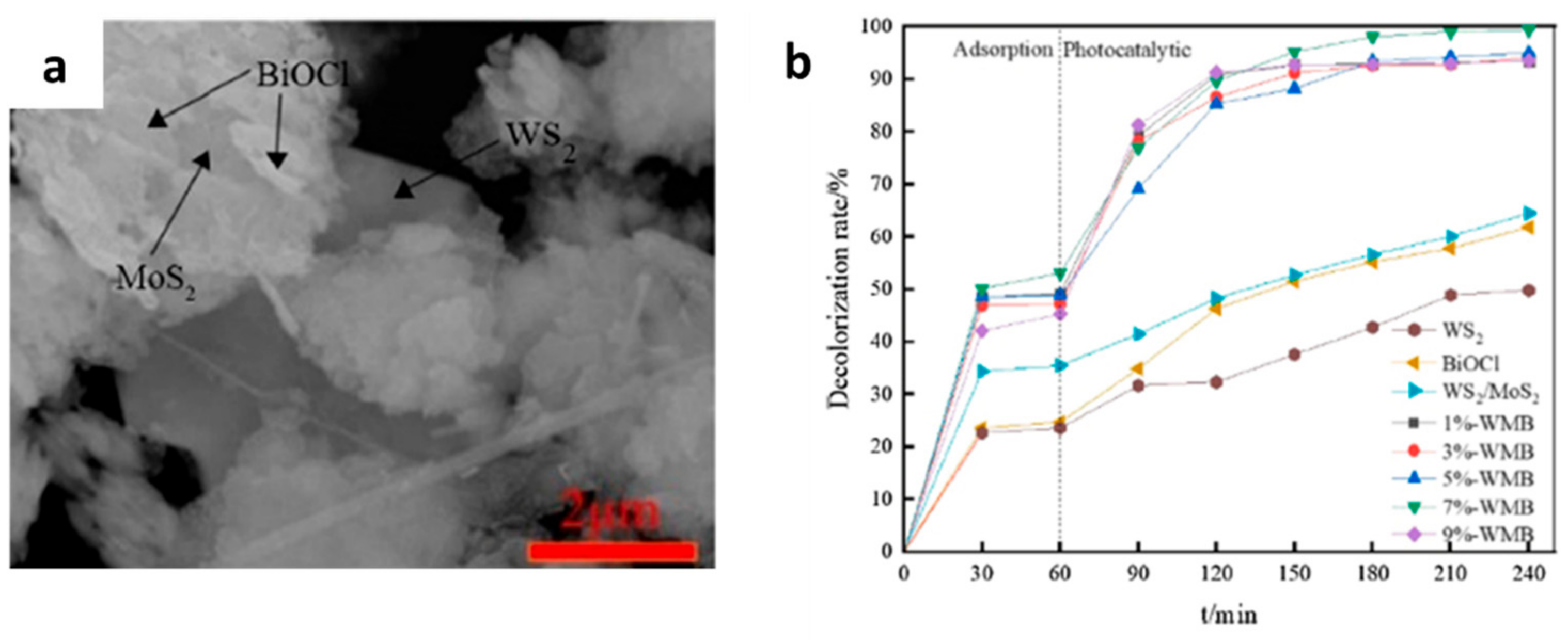
- Qi, S.; Liu, X.; Ma, N.; Xu, H. Construction and photocatalytic properties of WS2/BiOCl heterojunction. J. Nanoparticle Res. 2020, 22, 357. [Google Scholar] [CrossRef]
- Zhang, Q.; Tai, M.; Zhou, Y.; Zhou, Y.; Wei, Y.; Tan, C.; Wu, Z.; Li, J.; Lin, H. Enhanced Photocatalytic Property of γ-CsPbI3 Perovskite Nanocrystals with WS2. ACS Sustain. Chem. Eng. 2020, 8, 1219–1229. [Google Scholar] [CrossRef]
- Tran, H.H.; Truong, D.H.; Truong, T.T.; Xuan Dieu Nguyen, T.; Jin, Y.S.; Kim, S.J.; Vo, V. A Facile Synthesis of WS2/g-C3N4 Composites with Improved Photocatalytic Activity. Bull. Korean Chem. Soc. 2018, 39, 965–971. [Google Scholar] [CrossRef]
- Zeng, P.; Ji, X.; Su, Z.; Zhang, S. WS2/g-C3N4 composite as an efficient heterojunction photocatalyst for biocatalyzed artificial photosynthesis. RSC Adv. 2018, 8, 20557–20567. [Google Scholar] [CrossRef]
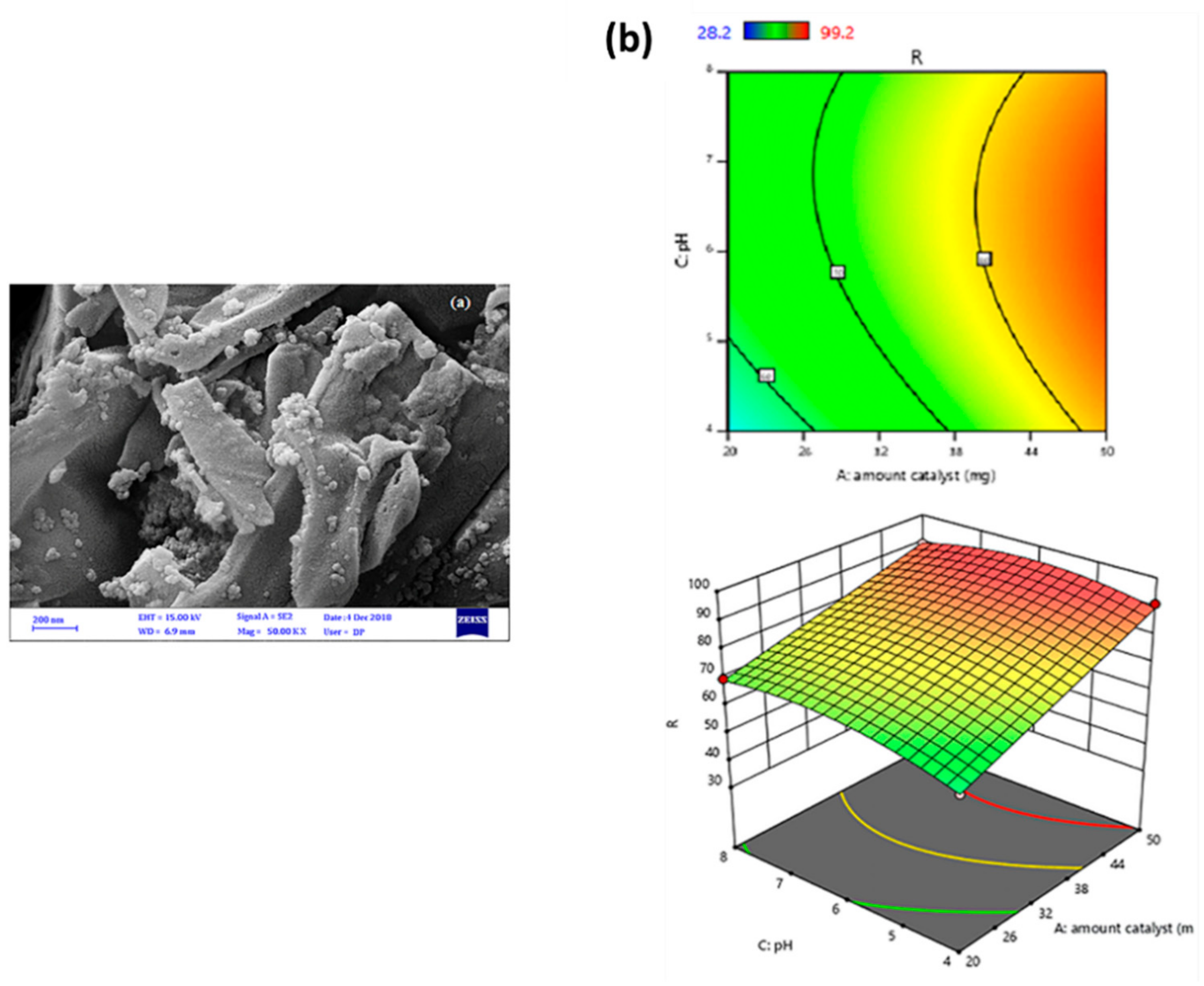
- Merci, S.; Saljooqi, A.; Shamspur, T.; Mostafavi, A. Investigation of photocatalytic chlorpyrifos degradation by a new silica mesoporous material immobilized by WS2 and Fe3O4 nanoparticles: Application of response surface methodology. Appl. Organomet. Chem. 2020, 34, e5343. [Google Scholar] [CrossRef]
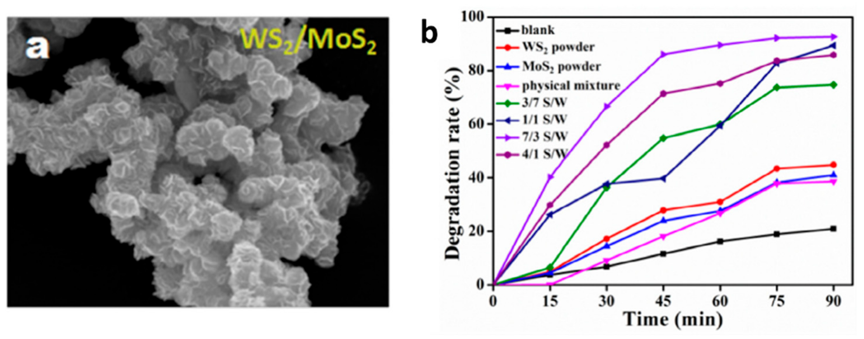
- Zhao, Y.; Liu, J.; Zhang, X.; Wang, C.; Zhao, X.; Li, J.; Jin, H. Convenient Synthesis of WS2-MoS2 Heterostructures with Enhanced Photocatalytic Performance. J. Phys. Chem. C 2019, 123, 27363–27368. [Google Scholar] [CrossRef]
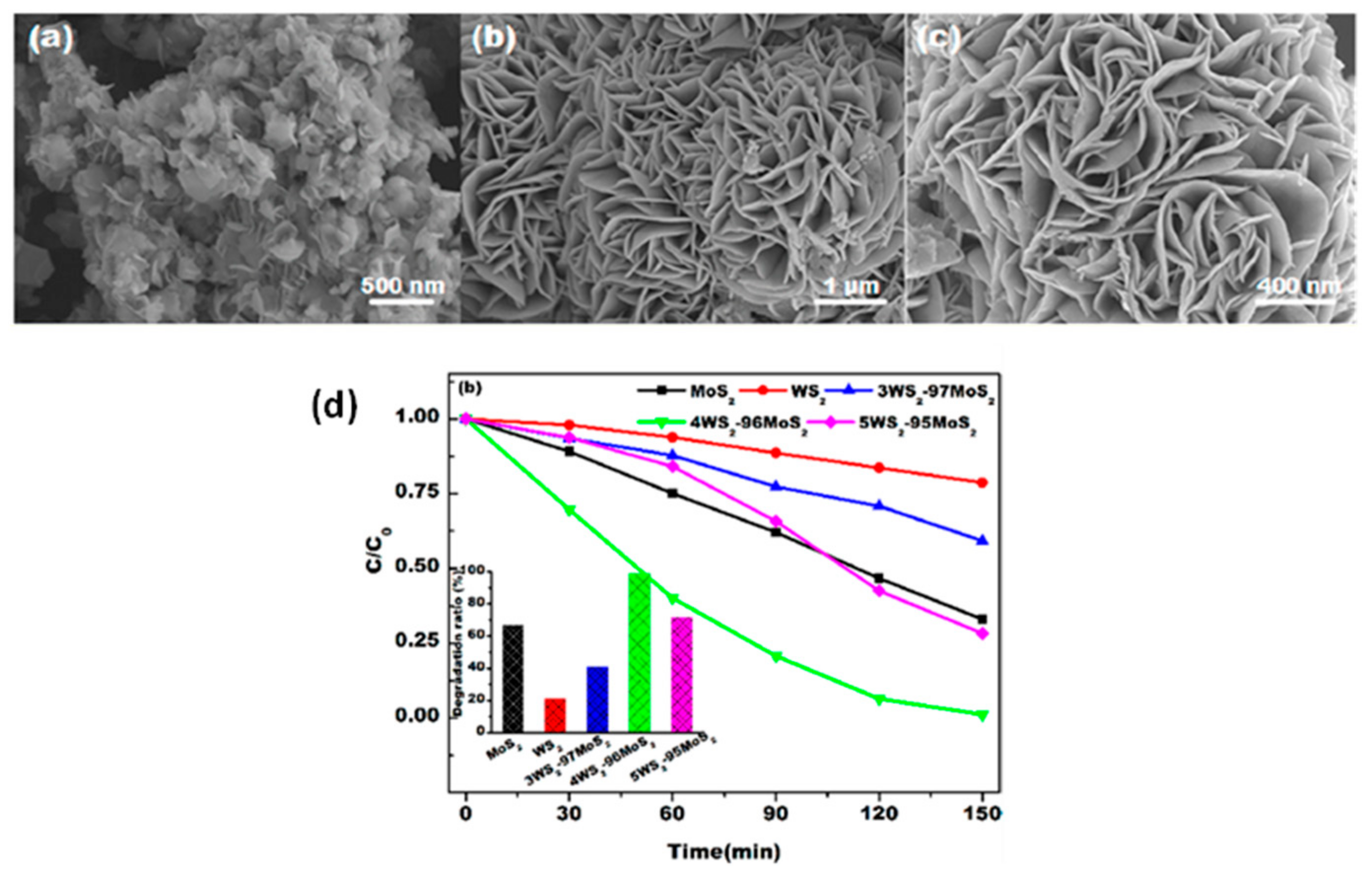
- Luo, S.; Dong, S.; Lu, C.; Yu, C.; Ou, Y.; Luo, L.; Sun, J.; Sun, J. Rational and green synthesis of novel two-dimensional WS2/MoS2 heterojunction via direct exfoliation in ethanol-water targeting advanced visible-light-responsive photocatalytic performance. J. Colloid Interface Sci. 2018, 513, 389–399. [Google Scholar] [CrossRef]
- Qi, S.; Liu, X.; Ma, N.; Xu, H. Construction and photocatalytic properties of WS2/MoS2/BiOCl heterojunction. Chem. Phys. Lett. 2021, 763, 138203. [Google Scholar] [CrossRef]
- Barakat, M.A.; Kumar, R.; Almeelbi, T.; Al-Mur, B.A.; Eniola, J.O. Sustainable visible light photocatalytic scavenging of the noxious organic pollutant using recyclable and reusable polyaniline coupled WO3/WS2 nanohybrid. J. Clean. Prod. 2022, 330, 129942. [Google Scholar] [CrossRef]
- Yu, W.; Chen, X.; Mei, W.; Chen, C.; Tsang, Y. Photocatalytic and electrochemical performance of three-Dimensional reduced graphene Oxide/WS2/Mg-doped ZnO composites. Appl. Surf. Sci. 2017, 400, 129–138. [Google Scholar] [CrossRef]
- Zhang, X.; Qiu, F.; Rong, X.; Xu, J.; Rong, J.; Zhang, T. Zinc oxide/graphene-like tungsten disulphide nanosheet photocatalysts: Synthesis and enhanced photocatalytic activity under visible-light irradiation. Can. J. Chem. Eng. 2018, 96, 1053–1061. [Google Scholar] [CrossRef]
- Cho, E.C.; Chang-Jian, C.W.; Zheng, J.H.; Huang, J.H.; Lee, K.C.; Ho, B.C.; Hsiao, Y.S. Microwave-assisted synthesis of TiO2/WS2 heterojunctions with enhanced photocatalytic activity. J. Taiwan Inst. Chem. Eng. 2018, 91, 489–498. [Google Scholar] [CrossRef]
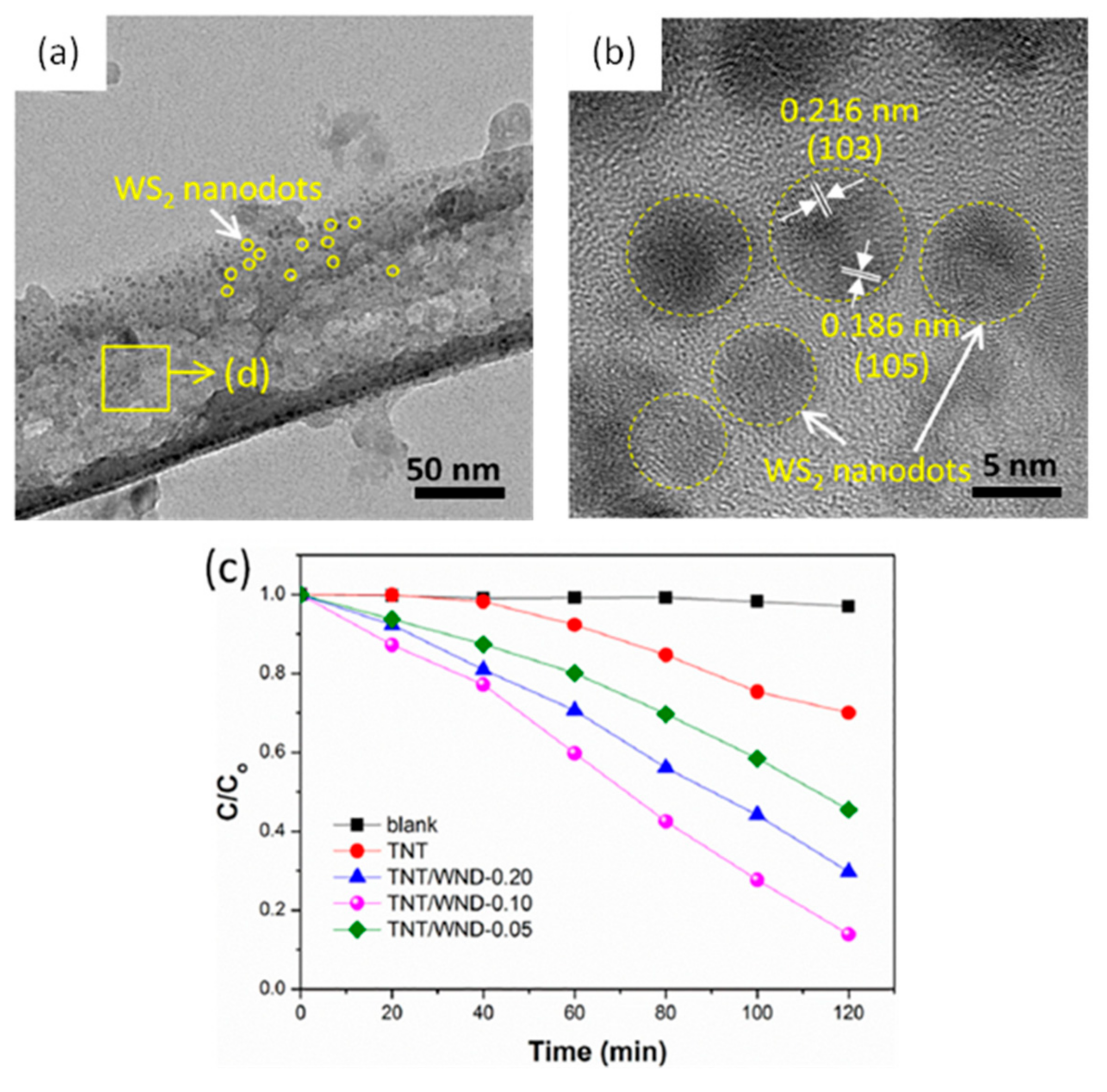
- Wu, Y.; Liu, Z.; Li, Y.; Chen, J.; Zhu, X.; Na, P. WS2 nanodots-modified TiO2 nanotubes to enhance visible-light photocatalytic activity. Mater. Lett. 2019, 240, 47–50. [Google Scholar] [CrossRef]
- Liao, Y.; Yao, Y.; Yu, Y.; Zeng, Y. Enhanced Antibacterial Activity of Curcumin by Combination With Metal Ions. Colloids Interface Sci. Commun. 2018, 25, 1–6. [Google Scholar] [CrossRef]
- Schwarzenbach, R.P.; Egli, T.; Hofstetter, T.B.; Von Gunten, U.; Wehrli, B. Global water pollution and human health. Annu. Rev. Environ. Resour. 2010, 35, 109–136. [Google Scholar] [CrossRef]
- Santosham, M.; Chandran, A.; Fitzwater, S.; Fischer-Walker, C.; Baqui, A.H.; Black, R. Progress and barriers for the control of diarrhoeal disease. Lancet 2010, 376, 63–67. [Google Scholar] [CrossRef]
- Staedel, C.; Darfeuille, F. MicroRNAs and bacterial infection. Cell. Microbiol. 2013, 15, 1496–1507. [Google Scholar] [CrossRef]
- WHO. Guidelines for Drinking-Water Quality; World Health Organization: Geneva, Switzerland, 2011; Volume 216, pp. 303–304. [Google Scholar]
- Rengifo-Herrera, J.A.; Mielczarski, E.; Mielczarski, J.; Castillo, N.C.; Kiwi, J.; Pulgarin, C. Escherichia coli inactivation by N, S co-doped commercial TiO2 powders under UV and visible light. Appl. Catal. B Environ. 2008, 84, 448–456. [Google Scholar] [CrossRef]
- Rengifo-Herrera, J.A.; Pierzchała, K.; Sienkiewicz, A.; Forró, L.; Kiwi, J.; Pulgarin, C. Abatement of organics and Escherichia coli by N, S co-doped TiO2 under UV and visible light. Implications of the formation of singlet oxygen (1O2) under visible light. Appl. Catal. B Environ. 2009, 88, 398–406. [Google Scholar] [CrossRef]
- Lin, Y.; Han, D.; Li, Y.; Tan, L.; Liu, X.; Cui, Z.; Yang, X.; Li, Z.; Liang, Y.; Zhu, S.; et al. Ag2S@WS2 Heterostructure for Rapid Bacteria-Killing Using Near-Infrared Light. ACS Sustain. Chem. Eng. 2019, 7, 14982–14990. [Google Scholar] [CrossRef]
- Chen, C.; Yu, W.; Liu, T.; Cao, S.; Tsang, Y. Graphene oxide/WS2/Mg-doped ZnO nanocomposites for solar-light catalytic and anti-bacterial applications. Sol. Energy Mater. Sol. Cells 2017, 160, 43–53. [Google Scholar] [CrossRef]
- Kumar, P.; Kataria, S.; Roy, S.; Jaiswal, A.; Balakrishnan, V. Photocatalytic Water Disinfection of CVD Grown WS2 Monolayer Decorated with Ag Nanoparticles. ChemistrySelect 2018, 3, 7648–7655. [Google Scholar] [CrossRef]
- Luo, H.; Liu, C.; Cheng, Y.; Zeng, Y.; He, D.; Pan, X. Fe(III) greatly promotes peroxymonosulfate activation by WS2 for efficient carbamazepine degradation and Escherichia coli disinfection. Sci. Total Environ. 2021, 787, 147724. [Google Scholar] [CrossRef]
- Kasinathan, K.; Marimuthu, K.; Murugesan, B.; Samayanan, S.; Cai, Y.; Rathinam, C. Facile synthesis of highly biologically active chitosan functionalized 2D WS2 nanocomposite anchored with palladium nanoparticles for antibacterial and anticancer activity: In-vitro biomedical evaluation. J. Mol. Liq. 2021, 335, 116582. [Google Scholar] [CrossRef]
- Fu, F.; Wang, Q. Removal of heavy metal ions from wastewaters: A review. J. Environ. Manag. 2011, 92, 407–418. [Google Scholar] [CrossRef]
- Litter, M.I. Mechanisms of removal of heavy metals and arsenic from water by TiO2-heterogeneous photocatalysis. Pure Appl. Chem. 2015, 87, 557–567. [Google Scholar] [CrossRef]
- Engates, K.E.; Shipley, H.J. Adsorption of Pb, Cd, Cu, Zn, and Ni to titanium dioxide nanoparticles: Effect of particle size, solid concentration, and exhaustion. Environ. Sci. Pollut. Res. 2011, 18, 386–395. [Google Scholar] [CrossRef]
- Skubal, L.R.; Meshkov, N.K.; Rajh, T.; Thurnauer, M. Cadmium removal from water using thiolactic acid-modified titanium dioxide nanoparticles. J. Photochem. Photobiol. A Chem. 2002, 148, 393–397. [Google Scholar] [CrossRef]
- Regmi, P.; Garcia Moscoso, J.L.; Kumar, S.; Cao, X.; Mao, J.; Schafran, G. Removal of copper and cadmium from aqueous solution using switchgrass biochar produced via hydrothermal carbonization process. J. Environ. Manag. 2012, 109, 61–69. [Google Scholar] [CrossRef] [PubMed]
- Gao, X.; Meng, X. Photocatalysis for heavy metal treatment: A review. Processes 2021, 9, 1729. [Google Scholar] [CrossRef]
- Meng, X.; Zhang, Z.; Li, X. Synergetic photoelectrocatalytic reactors for environmental remediation: A review. J. Photochem. Photobiol. C Photochem. Rev. 2015, 24, 83–101. [Google Scholar] [CrossRef]
- Kubota, Y.; Watanabe, K.; Tsuda, O.; Taniguchi, T. Deep ultraviolet light-emitting hexagonal boron nitride synthesized at atmospheric pressure. Science 2007, 317, 932–934. [Google Scholar] [CrossRef]
- Xie, W.; Zhang, M.; Liu, D.; Lei, W.; Sun, L.; Wang, X. Photocatalytic TiO2/porous BNNSs composites for simultaneous LR2B and Cr (VI) removal in wool dyeing bath. J. Photochem. Photobiol. A Chem. 2017, 333, 165–173. [Google Scholar] [CrossRef]
- Liu, B.; Liu, X.; Li, L.; Zhuge, Z.; Li, Y.; Li, C.; Gong, Y.; Niu, L.; Xu, S.; Sun, C.Q. CaIn2S4 decorated WS2 hybrid for efficient Cr(VI) reduction. Appl. Surf. Sci. 2019, 484, 300–306. [Google Scholar] [CrossRef]
- Wang, G.; Dou, K.; Cao, H.; Du, R.; Liu, J.; Tsidaeva, N.; Wang, W. Designing Z-scheme CdS/WS2 heterojunctions with enhanced photocatalytic degradation of organic dyes and photoreduction of Cr (VI): Experiments, DFT calculations and mechanism. Sep. Purif. Technol. 2022, 291, 120976. [Google Scholar] [CrossRef]
- Kumar, E.; Holt, W. V Impacts of Endocrine Disrupting Chemicals on Reproduction in Wildlife. In Advances in Experimental Medicine and Biology; Springer: New York, NY, USA, 2014; Volume 753, pp. 55–70. [Google Scholar]
- Cesaro, A.; Belgiorno, V. Removal of Endocrine Disruptors from Urban Wastewater by Advanced Oxidation Processes (AOPs): A Review. Open Biotechnol. J. 2016, 10, 151–172. [Google Scholar] [CrossRef]
- Petrie, B.; Barden, R.; Kasprzyk-Hordern, B. A review on emerging contaminants in wastewaters and the environment: Current knowledge, understudied areas and recommendations for future monitoring. Water Res. 2015, 72, 3–27. [Google Scholar] [CrossRef]
- Torad, E.; Mohamed, M.M.; Khalil, M.M.H.; Ismail, E.H. Optimal design of silver@silver sulfide-modified WS2 and its application in photocatalytic diclofenac degradation and H2 generation. J. Environ. Chem. Eng. 2021, 9, 106446. [Google Scholar] [CrossRef]
- Fu, S.; Liu, X.; Yan, Y.; Li, L.; Liu, H.; Zhao, F.; Zhou, J. Few-layer WS2 modified BiOBr nanosheets with enhanced broad-spectrum photocatalytic activity towards various pollutants removal. Sci. Total Environ. 2019, 694, 133756. [Google Scholar] [CrossRef] [PubMed]


| Precursors | Conditions | Morphology | Application | Ref. |
|---|---|---|---|---|
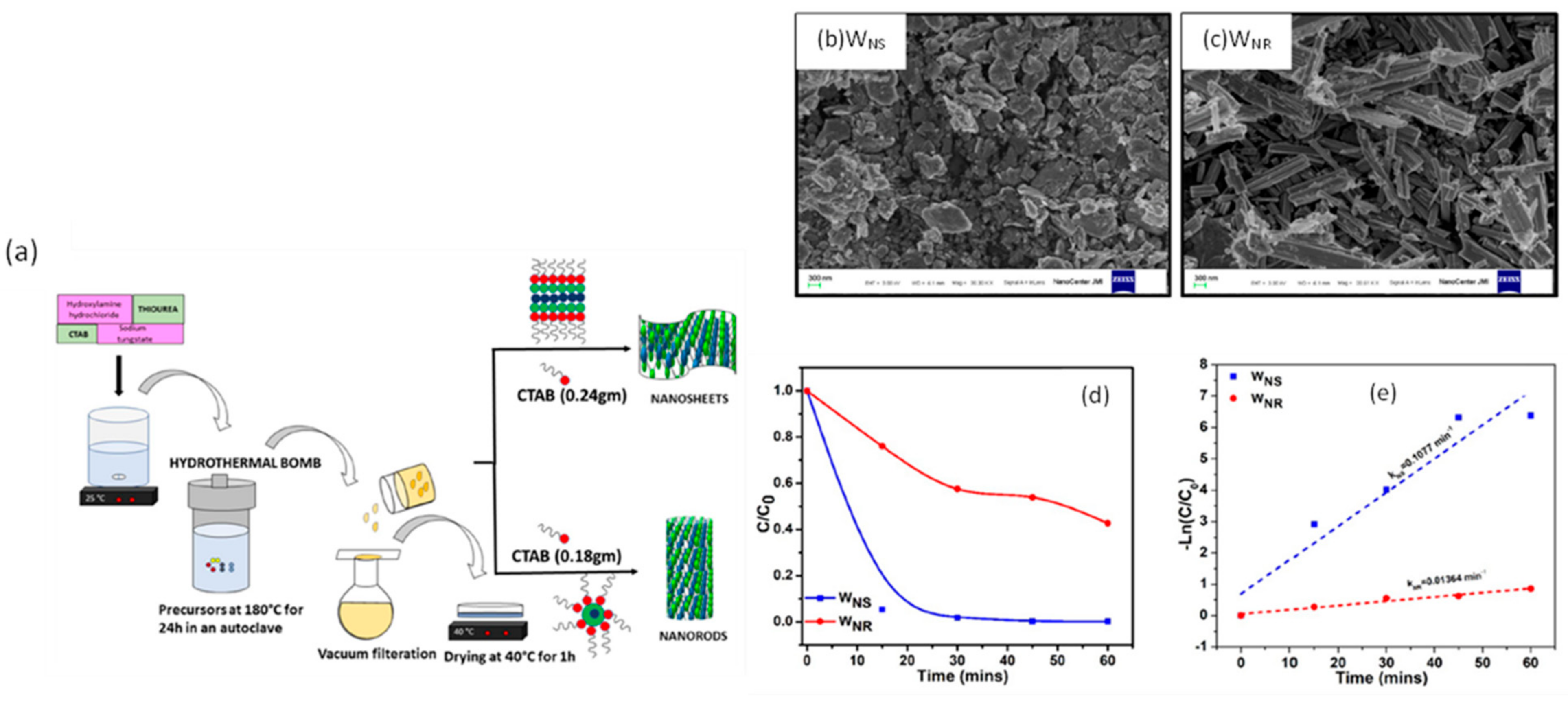
| Sodium tungstate, thiourea, hydroxylamine hydrochloride | Autoclave, 60 °C for 8 h | Nanosheets | Photocatalytic degradation of antibiotic nitrofurantoin (NFT) | [44] |
| Sodium tungstate, thioacetamide, oxalic acid. | Autoclave, 200 °C for 24 h. | Nanoflakes | Photodegradation of methylene blue | [48] |
| Sodium tungstate, thiourea, hydroxylamine hydrochloride, CTAB, hydrochloric acid. | Autoclave, 180 °C for 24 h. | Nanosheets | Hydrogen evolution | [49] |
| Sodium tungstate, L-cysteine. | Autoclave, 180 °C for 24 h. | Nanosheets | Biomedical evaluation | [50] |
| Precursors | Conditions | Substrate | Morphology | Ref. |
|---|---|---|---|---|
| WO3 and sulfur | Ar + H2 (95% + 5%) environment at 100 SCCM, 850 °C for WO3 and 220 °C for sulfur 10 min | c-plane sapphire | Monolayer thin film | [53] |
| WO3 and sulfur | Ar 75 SCCM, 250 °C for S, and 920 °C for 15 min | Si/SiO | Bilayer ribbons | [54] |
| WO3 and sulfur | Ar + H2 (95% + 5%) 50 SCCM, 850oC for 10 min | SiO2/Si | Nanorods | [55] |
| WO3 and sulfur | Ar 150 SCCM, 880 °C for 15 min | SiO2/Si with Au foil on the center | 2D film | [56] |
| Processing | Solvent | Ref. |
|---|---|---|
| Sonication for 3 h. Centrifugation at 3000 rpm for 20 min. | N-Methyl-2-pyrrolidone (NMP) | [63] |
| Oxygen plasma treatment followed by Sonication at 400 W. | Dimethyl sulfoxide (DMSO) and water | [64] |
| Sonication for 5 days. Centrifugation in 3500 rpm for 60 min. | 35 vol% ethanol @ water | [65] |
| Ultrasonication for 6 h in a water bath. | N-Methyl-2-pyrrolidone (NMP) | [66] |
| WS2-Based Photocatalyst | Organic Pollutants | Removal Efficiency | Ref. |
|---|---|---|---|
| Few-layer WS2 nanosheets | BG | 95%, 10 h | [19] |
| Inorganic fullerene (IF)-type WS2 nanoparticles | MG | 712% | [22] |
| Hexagonal-shaped WS2 platelets | RhB | 98%, 105 min | [47] |
| WS2 nanosheets, WS2 nanorods | MB 4-CP | WS2(NS): ~100%, 60 min WS2(NR): ~50%, 60 min WS2(NS): ~100%, 120 min WS2(NR): ~69%, 120 min | [77] |
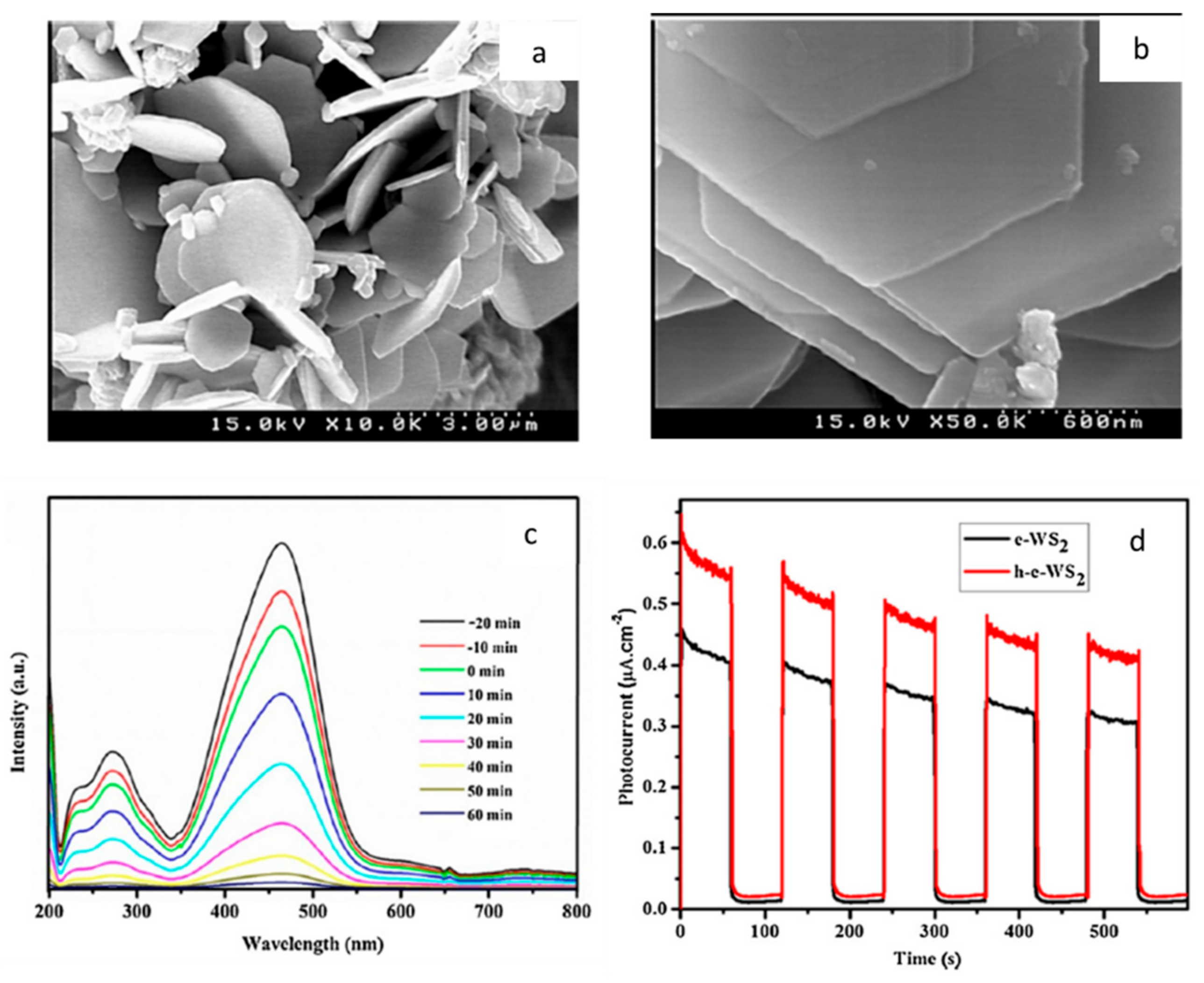
| WS2 (e-h-WS2) | MO CR RhB PR | 99%, 60 min 99%, 110 min 99.9%, 100 min 99%, 110 min | [78] |
| WS2/Ag | Evans blue | Dye rejection rate: 90.2% | [79] |
| AgI/WS2 | RhB | 91.2%, 30 min | [80] |
| WS2/Bi2O2CO3 | LR5B | 91%, 90 min | [81] |
| WS2/BiOBr | RhB | 95% | [82] |
| BiOCl/WS2 | MG | 99%, 60 min | [83] |
| BiOCl/WS2 | MB | 95.83%, 240 min | [84] |
| γ-CsPbI3/WS2 | MB | 100%, 30 min | [85] |
| WS2/g-C3N4 | MB | 85.3%, 6 h | [86] |
| WS2/g-C3N4 | MB | 95.5%, 2 h | [20] |
| WS2/g-C3N4 | RhB | 100%, 1 h | [87] |
| WS2/Fe3O4 | CP | 91% | [88] |
| WS2/MoS2 | MB | 100%, 150 min | [89] |
| WS2/MoS2 | RhB MB | 93%, 90 min 85%, 90 min | [90] |
| WS2/MoS2/BiOCl | MB | 99.36%, 240 min | [91] |
| WS2/WO3 | 2-CP | 100% | [92] |
| WS2/ZnO (rGWMZ) | RhB | 100%, 5 min | [93] |
| ZnO/WS2 | RhB | 95.71%, 120 min | [94] |
| WS2/TiO2 | MB | 100%, 480 min | [95] |
| WS2 nanodots-modified TiO2 nanotubes | RhB | 86.1%, 120 min | [96] |
| WS2-Based Photocatalyst | Microorganism | Inactivation Performance | Ref. |
|---|---|---|---|
| WS2 monolayer | E. coli bacteria | ~99%, 120 min | [53] |
| Ag2S@WS2 | S. aureus E. coli | 99.93%, 20 min 99.84%, 20 min | [104] |
| Graphene oxide/WS2/Mg-doped ZnO | S. aureus, E. coli | Inhibition rings size: 6.07 and 8.64 mm, respectively | [105] |
| WS2/Ag nanoparticles | E. coli | 96%, 3 h | [106] |
| PMS/WS2/Fe3+ | E. coli | 97.3%, 1 min | [107] |
| CS/WS2/Pd | E. coli S. aureus B. subtilis K. pneumoniae | Zone of inhibition: 38 mm 32 mm 28 mm 27 mm | [108] |
| CS/WS2/Ru | E. coli S. aureus B. subtilis K. pneumoniae | Zone of inhibition: 37 mm 31 mm 29 mm 27 mm | [50] |
| WS2-Based Photocatalyst | Metal Ion | Removal Efficiency | Ref. |
|---|---|---|---|
| CaIn2S4/WS2 | Cr (VI) | 98% | [118] |
| WS2/BiOCl | Cr (VI) | 94.9% | [83] |
| CdS/WS2 | Cr (VI) | Reduction rate of 0.0846/min | [119] |
| WS2-Based Photocatalyst | Pharmaceutical | Removal Efficiency | Ref. |
|---|---|---|---|
| WS2 nanosheets | Nitrofurantoin (NFT) | 90%, 120 min | [44] |
| Ag@Ag2S/WS2, Ag@Ag2O/WS2 | Diclofenac (DCF) | 92%, 80%, respectively | [123] |
| PMS/WS2/Fe3+ | Carbamazepine (CBZ) | ~100%, 10 min | [107] |
| WS2/BiOBr | Ciprofloxacin (CIP) Tetracycline (TC) Metronidazole (MNZ) Oxytetracycline (OTC) | 83% 92% 97% 92% | [124] |
| g-C3N4/WS2 | Tetracycline (TC) | 84.5%, 2 h | [20] |
Publisher’s Note: MDPI stays neutral with regard to jurisdictional claims in published maps and institutional affiliations. |
© 2022 by the authors. Licensee MDPI, Basel, Switzerland. This article is an open access article distributed under the terms and conditions of the Creative Commons Attribution (CC BY) license (https://creativecommons.org/licenses/by/4.0/).
Share and Cite
Yousef, A.; Thiehmed, Z.; Abdul Shakoor, R.; Altahtamouni, T. Recent Progress in WS2-Based Nanomaterials Employed for Photocatalytic Water Treatment. Catalysts 2022, 12, 1138. https://doi.org/10.3390/catal12101138
Yousef A, Thiehmed Z, Abdul Shakoor R, Altahtamouni T. Recent Progress in WS2-Based Nanomaterials Employed for Photocatalytic Water Treatment. Catalysts. 2022; 12(10):1138. https://doi.org/10.3390/catal12101138
Chicago/Turabian StyleYousef, Aseel, Zeineb Thiehmed, Rana Abdul Shakoor, and Talal Altahtamouni. 2022. "Recent Progress in WS2-Based Nanomaterials Employed for Photocatalytic Water Treatment" Catalysts 12, no. 10: 1138. https://doi.org/10.3390/catal12101138