Article

Novel Transaminase and Laccase from Streptomyces spp. Using Combined Identification Approaches

by
Erica E. Ferrandi
1,
Jelena Spasic
2,
Lidija Djokic
2,
Yevhen Vainshtein
3,
Ramsankar Senthamaraikannan
4,
Sandra Vojnovic
2,
Christian Grumaz
3,
Daniela Monti
1,* and
Jasmina Nikodinovic-Runic
2,*
1
Istituto di Scienze e Tecnologie Chimiche (SCITEC), Consiglio Nazionale delle Ricerche, via Mario Bianco 9, 20131 Milan, Italy
2
Institute of Molecular Genetics and Genetic Engineering, University of Belgrade, Vojvode Stepe 444a, 11221 Belgrade, Serbia
3
Fraunhofer Institute for Interfacial Engineering (IGB), In-Vitro Diagnostics, Nobelstrasse 12, 70569 Stuttgart, Germany
4
AMBER Centre, Trinity College Dublin, College Green, Dublin 2, Ireland
*
Authors to whom correspondence should be addressed.
Catalysts 2021, 11(8), 919; https://doi.org/10.3390/catal11080919
Submission received: 29 June 2021 / Revised: 24 July 2021 / Accepted: 26 July 2021 / Published: 29 July 2021

Abstract

:
Three Streptomyces sp. strains with a multitude of target enzymatic activities confirmed by functional screening, namely BV129, BV286 and BV333, were subjected to genome sequencing aiming at the annotation of genes of interest, in-depth bioinformatics characterization and functional expression of the biocatalysts. A whole-genome shotgun sequencing followed by de novo genome assembly and annotation was performed revealing genomes of 6.4, 9.4 and 7.3 Mbp, respectively. Functional annotation of the proteins of interest resulted in between 2047 and 2763 putative targets. Among the various enzymatic activities that the three Streptomyces strains demonstrated to produce by functional screening, we focused our attention on transaminases (TAs) and laccases due to their high biocatalytic potential. Bioinformatics search allowed the identification of a putative TA from Streptomyces sp. BV333 as a potentially novel broad substrate scope TA and a putative laccase from Streptomyces sp. BV286 as potentially novel blue multicopper oxidase. The two sequences were cloned and overexpressed in Escherichia coli and the two novel enzymes, transaminase Sbv333-TA and laccase Sbv286-LAC, were characterized. Interestingly, both enzymes resulted to be exceptionally thermostable, Sbv333-TA showing a melting temperature (TM = 85 °C) only slightly lower compared to the TM of the most thermostable transaminases described to date (87–88 °C) and Sbv286-LAC being even thermoactivated at temperature >60 °C. Moreover, Sbv333-TA showed a broad substrate scope and remarkably demonstrated to be active in the transamination of β-ketoesters, which are rarely accepted by currently known TAs. On the other hand, Sbv286-LAC showed an improved activity in the presence of the cosolvent acetonitrile. Overall, it was shown that a combination of approaches from standard microbiological and biochemical screens to genome sequencing and analysis is required to afford novel and functional biocatalysts.

1. Introduction

Actinomycetes, especially Streptomycetes, have been in the focus of systems biology and genome metabolic modelling approaches to further intensify their explorations in terms of antibiotic discovery. However, their particular development and specificity of the niches they occupy led our research towards exploring them as a potential source of novel biocatalysts [1,2].
The enzyme market is fast-growing and was valued at USD 7082 million in 2017, and is projected to reach USD 10,519 million in 2024, while the cost of enzymes for biofuel applications alone should total USD 1.0 billion in 2020 [3,4]. More stringent environmental norms coupled with health and environmental awareness are contributing towards intensified research and development efforts in widening the diversity of enzyme applications beyond the laboratory scale. Therefore, carbohydrate hydrolases and proteases are expected to dominate the enzyme market due to their applications in the production of biofuels and detergents, however, other enzyme classes, including laccases and transaminases, are gaining importance due to their increasing usage in food and beverage, textile, pharmaceutical/cosmetic and recycling industries [5].
Streptomyces spp. are usually saprophytic soil-dwelling aerobic filamentous bacteria playing a crucial role in soil ecology by effectively hydrolyzing a wide range of polysaccharides (cellulose, chitin, xylan, and agar) and other natural macromolecules [6]. This biodegradation efficiency is based on the secretion of potent hydrolases (enzymes classified as EC 3 in the EC number classification) such as cellulases, lipases, proteases, xylanases, and cutinases, while other enzymes of environmental and industrial importance have also been identified from this genus [7,8,9]. Of special interest are laccases and aminotransferases.
Aminotransferases (ATAs) or transaminases (TAs) are pyridoxal-5’-phosphate (PLP)-dependent enzymes capable of performing the transfer of an amino group between an amino donor and a prochiral ketone generating optically pure chiral amines. The numerous examples of ATA applications in the synthesis of chiral amines in the pharmaceutical and fine chemical industries demonstrate the high biocatalytic potential of these enzymes, the most remarkable example being the industrial application of ATAs in the production of the anti-hyperglycaemic drug sitagliptin [10,11]. To date, despite ATAs being quite ubiquitous in nature, ATAs exploited for biocatalytic application are mainly from bacteria and fungi. Curiously, to the best of our knowledge only a few ATAs have been identified and characterized in Streptomycetes. For example, two ATAs from S. griseus were reported as capable to catalyse the transamination of ω-aminoacids [12,13], while an ATA from S. venezuelae was shown to be involved in the biosynthesis of the rare sugar desosamine [14].
Laccases are blue multicopper oxidoreductases able to catalyze the oxidation of a wide range of substrates [15]. These enzymes find a broad range of applications in the paper and pulp, textile, petrochemical and pharmaceutical industries [16]. Moreover, they can be used for the treatment of industrial wastes by performing detoxifications and bioremediation processes or surface modifications of plastic materials [17]. Bacterial laccases, showing a thermophilic character and working in a broad pH range, result to be particularly attractive for industrial applications [17]. Streptomyces laccases are among the best-characterized bacterial laccases and have been identified for example in S. cyaneus [18], S. coelicolor [19], and S. sviceus [20,21].
We previously conducted an extensive activity screen of our in-house library of Streptomyces spp. rhizosphere isolates, and showed their biocatalytic potential [2]. The goal of this study was to identify and evaluate potentially novel biocatalysts from selected Streptomyces spp. Thus, three of the strains with target activities confirmed by functional screening, namely Streptomyces sp. BV129, BV286, and BV333 were subjected to genome sequencing aiming at the annotation of genes of interest, and in-depth bioinformatic characterization that led to cloning and heterologous expression in E. coli of one aminotransferase (Sbv333-TA) and one laccase (Sbv286-LAC) sequence.

2. Results

Three Actinobacteria strains, namely Streptomyces sp. BV129, Streptomyces sp. BV286 and Streptomyces sp. BV333, were selected in this study to identify and evaluate potentially novel biocatalysts. These strains were selected due to the fact that they showed excellent activities during the phenotypic screen [2]. BV129, BV286 and BV333 strains grew well on the mannitol–soy flower medium and sporulated well after 7 days at 30 °C (Figure 1a). BV286 extruded deep brown pigment on this solid medium even after 24 h growth. Further morphological differences were revealed by SEM (Figure 1b) which revealed chains of smooth and oval spores for BV129 and BV333, while BV286 only sporadically sporulated within this time frame.

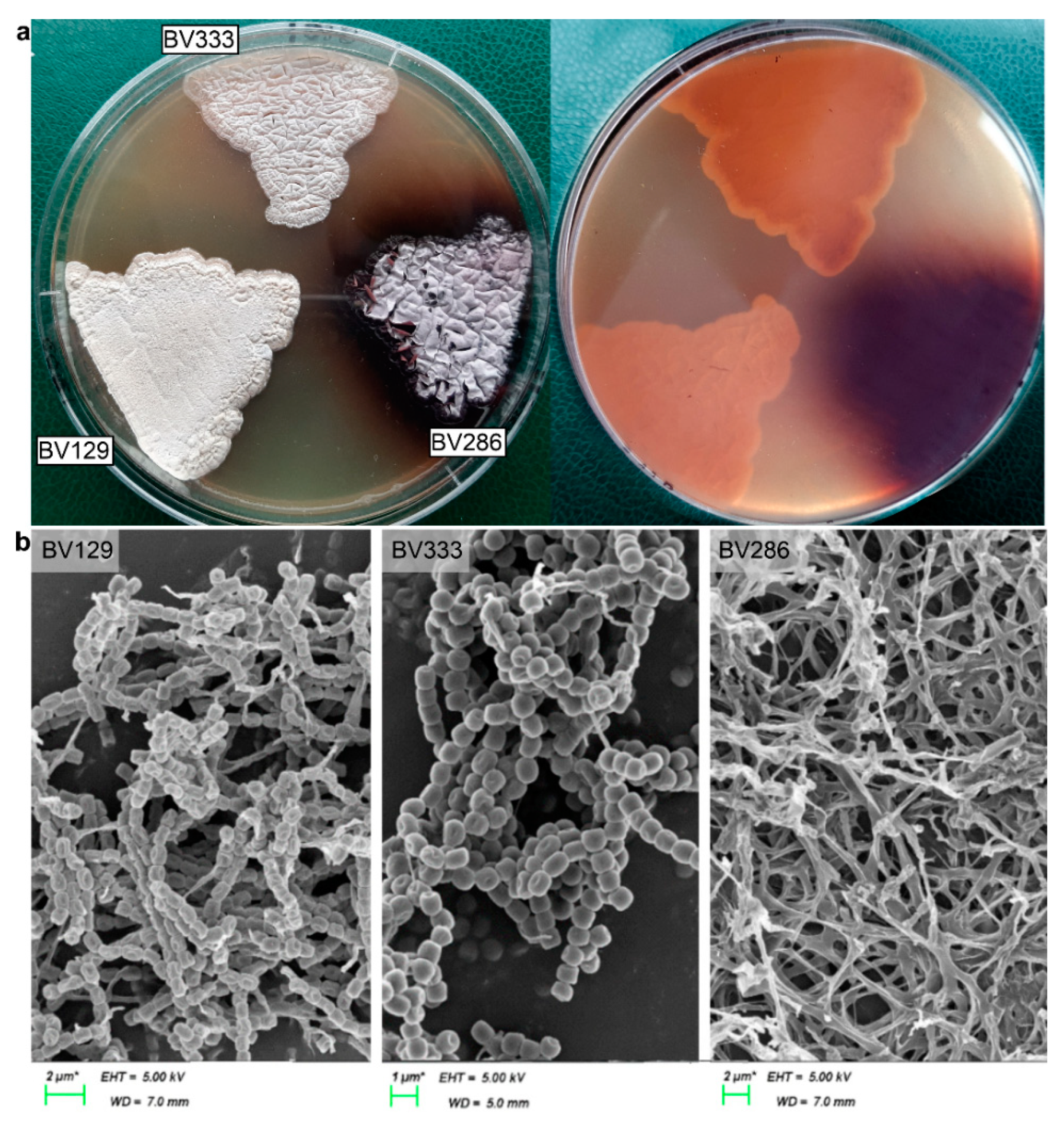
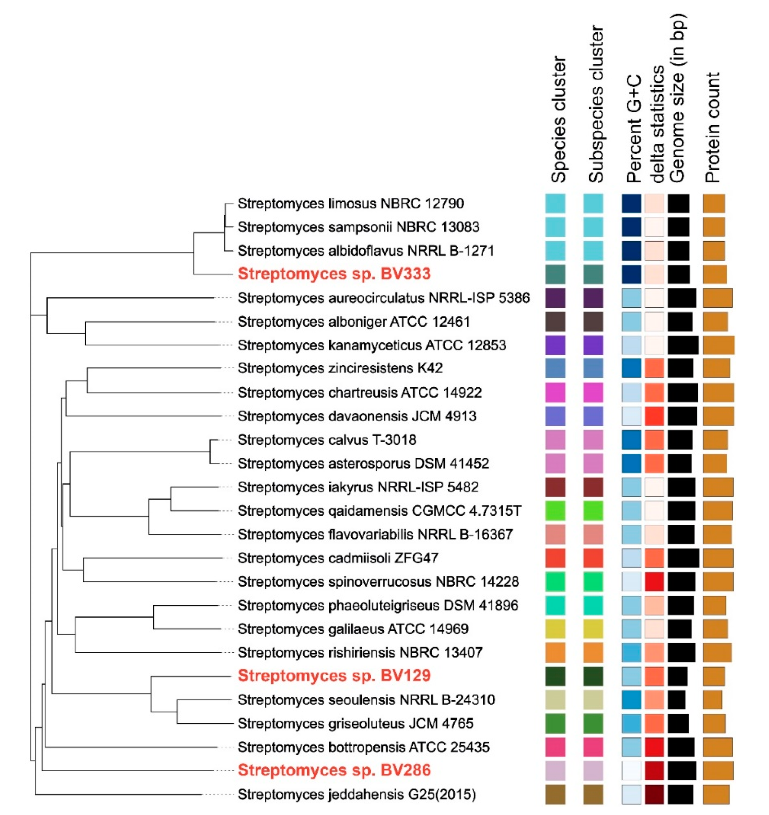
2.1. Genome Sequencing and Analysis

A whole-genome shotgun sequencing (WGS) followed by de novo genome assembly and annotation was performed on these three strains. Genomes were assembled using ABySS v.1.5.2 and annotated with Prokka 1.12 and analyzed by a variety of bioinformatics tools (Table S1). Corresponding assembly statistics are represented in Table 1. The resulting whole-genome shotgun project, including assembly and raw sequence reads, was deposited at NCBI (PRJNA739376; https://www.ncbi.nlm.nih.gov/bioproject/739376).
To reliably classify the Streptomyces strains, we performed a phylogenetic analysis based on whole genome-based sequence comparison using a TYGS (The Type (Strain) Genome Server) [22]. TYGS is a web-based bioinformatics pipeline utilizing well-established tools to perform sequence-based taxonomical classification (the full list of tools used for the analysis with corresponding references is given in Table S1). The phylogenetic tree inferred with the FastME 2.1.6.1 [23] from the type-based species clustering from the whole-genome comparison is depicted in Figure 2. Based on the phylogenetic analysis, all three strains belong to different species clusters and do not share common characteristics, such as GC content or the number of predicted genes. Nevertheless, the phylogenetic analysis based on de novo assembled sequences reveals a higher similarity between BV129 and BV286 strain (Figure 2).
A gene-annotation-based search was carried out by using Prokka [24] to find putative gene coding for biotechnologically important enzymatic activities (Table 2). In particular, the search was focused on (i) hydrolases, e.g., lipases, cutinases, cellulases, gelatinase, PHA-depolymerase, and proteases, (ii) oxidoreductases, e.g., lignin peroxidase, laccases and tyrosinases, and (iii) aminotransferases. According to gene annotation, the most abundant enzymes among the selected ones are hydrolases belonging to a rather broad EC category “proteases” (Table 2), followed by “lipases” and “cellulases”, while other hydrolases are much less abundant or not present. One or two laccase-coding genes are predicted in all strains, while aminotransferases are largely represented with >30 annotated genes in each strain, this fact being not surprising due to their functional role in the cell metabolism. Nevertheless, many enzymes which are not predicted by the algorithm underlying Prokka were previously identified by alternative methods [2]. Indeed, activities such as gelatinase, PHA-depolymerase and lignin peroxidase were confirmed in the functional screen but revealed non-present by sequence analysis. One of the possible explanations is the way Prokka processes the data. It is a computational pipeline, utilizing different tools for every step of its analysis. For example, for the genes/CDS prediction, Prokka uses the Prodigal [25]. At the later stages of the Prokka pipeline, the prediction results first are refined with the BLAST and then made compatible with an NCBI genome submission policy using the tbl2asn tool. Such conversion sometimes removes additional information from the protein annotation as not compliant with the submission standards.

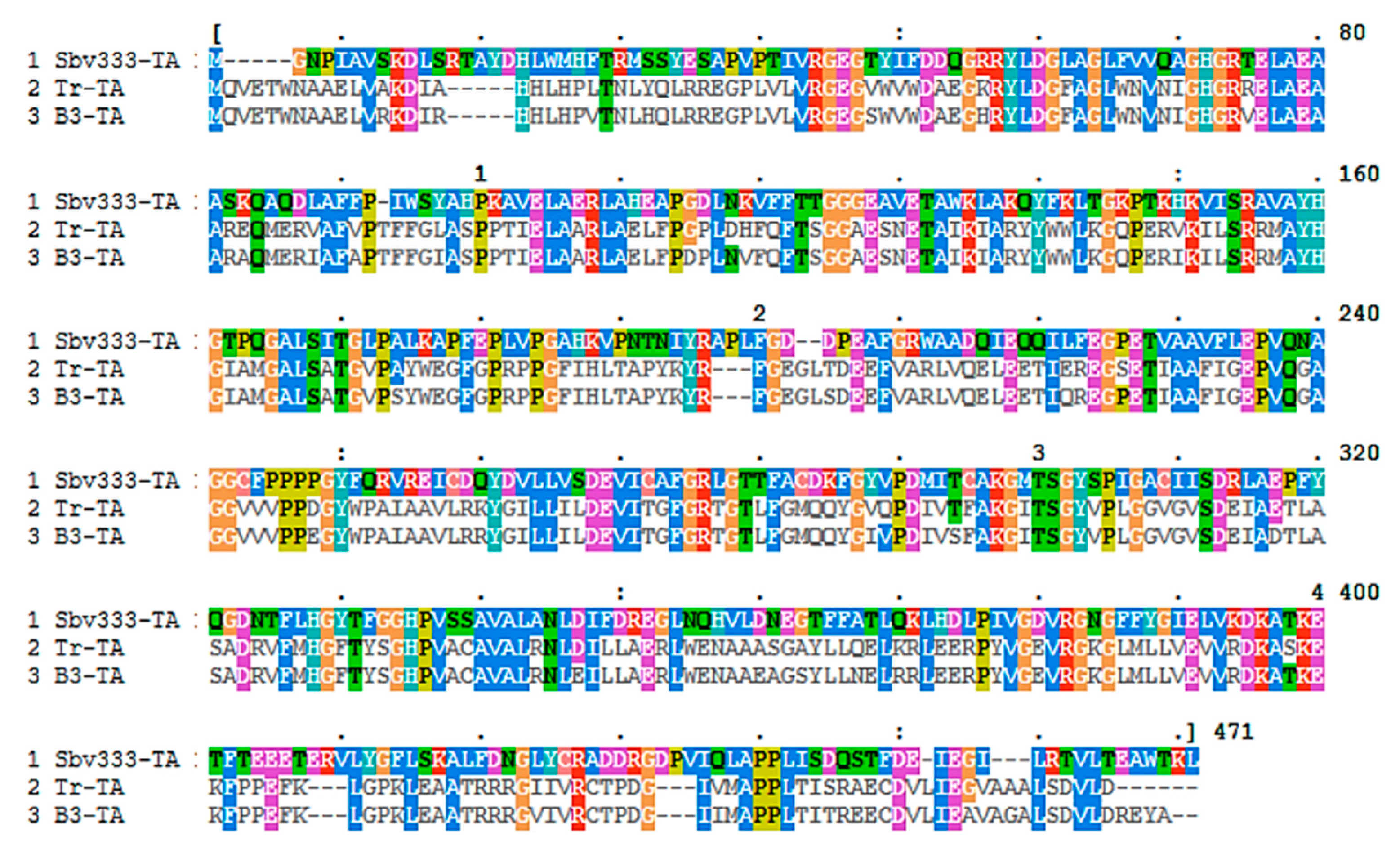
2.2. Bioinformatics Analysis of Transaminases and Laccases

A deeper bioinformatic analysis of the three genome sequences of Streptomyces sp. BV129, BV286 and BV333 was carried out in the search of genes coding for transaminases (ATAs) and laccases. Known ATA sequences, either (S)- or (R)-selective (Table S2), or laccase sequences (Table S3) were used to perform a multiple sequence alignment with the predicted ORFs of the above mentioned Streptomyces genomes by using the LAST program (http://last.cbrc.jp/).
Concerning the search for (S)-selective ATAs, 11 potential (S)-selective transaminases were identified in the genome of Streptomyces strain BV129, 6 sequences in the genome of strain BV286 and 10 sequences in the genome of strain BV333, respectively. Based on the LAST alignment score, 8 of these candidates (BV129_00199, BV129_05035, BV286_01057, BV286_02269, BV286_05820, BV333_00884, BV333_03408, BV333_03485) were selected for further bioinformatics analysis. Five of the eight selected sequences were discarded since they lacked the high-(S)-selective-ATAs conserved residues suggested by Steffen-Munsberg et al. [26]. The three remaining sequences shared high sequence identity (88% similarity of BV129_05035 sequence and 91% BV286_05820 sequence compared to BV333_03408, respectively). Therefore, only one of these was selected for further characterization, i.e., the BV333_03408 sequence, encoding for a protein of 459 aa (named Sbv333-TA). Remarkably, BLAST analysis revealed that Sbv333-TA is identical (100% query cover) to an uncharacterized protein annotated as an aspartate aminotransferase from Streptomyces sp. M10 (WP_047470642.1). It is also noteworthy that the alignment of Sbv333-TA with available ATA genes from other sources revealed a high sequence similarity with ATA sequences from extremophiles. In particular, Sbv333-TA showed high sequence similarity with the TA from Thermomicrobium roseii (Tr-TA, WP_015922033) (43% identity, 89% query cover) [27] and a TA from hot spring metagenomes (B3-TA, KX505389) (41% identity, 94% query cover) [28] (Figure 3). Concerning the search for (R)-selective transaminases, only two sequences showing similarity with known (R)-selective ATAs were found, but the alignment scores were low and they lacked the important conserved residues suggested by Hohne et al. [29].
Concerning the search for laccase homologs, five sequences (BV129_02175, BV129_04916, BV286_03089, BV333_01214 and BV333_01561) showing similarity with known laccases were found in the genomes of the selected Streptomyces isolates and in particular three of them showed high LAST alignment score (BV129_04916, BV286_03089, and BV333_01561). These three sequences were quite similar to each other, sharing around 72–74% identity, therefore, among them, the 975 bp-long BV286_03089 sequence, corresponding to a protein of 325 amino acids, was selected for cloning ad expression in E. coli. By BLAST analysis, it was shown that BV286_03089 has similarities with a putative copper oxidase from Streptomyces umbrinus (Genbank GHH35467.1) (92% identity) while a search in the PDB database revealed that the closest homologue to this sequence is the laccase Ssl1 from Streptomyces sviceus (82% identity) (pdb 6YZY). In addition, a Clustal Omega alignment was carried out to compare BV286_03089 with other already characterized laccase sequences (Table S3). This analysis confirmed the high similarity of BV286_03089 with different Streptomyces laccases (79–82% identity) and low identity with other bacterial laccases (18%–25% identity).

2.3. Cloning, Expression and Functional Analysis of Novel Transaminase and Laccase

The two selected sequences, BV333_03408 and BV286_03089, potentially coding for an (S)-ATA and a laccase, respectively, were cloned into the pETite vector in frame with a C-term His-tag sequence and the resulting plasmids (pETite-Sbv333-TA and pETite-Sbv286-LAC; Table S4) were subsequently transformed and overexpressed in E. coli Rosetta cells.
As shown by SDS-PAGE analysis, Sbv333-TA was successfully produced by this host, but mostly accumulated in the cells as inclusion bodies (Figure S1), thus leading to poor recovery yields (about 4 mg L−1 after Ni-NTA purification). To overcome these solubility issues, plasmid pETite-Sbv333-TA was expressed in E. coli BL21(DE3) containing the plasmid pGro7 (Takara Bio Inc.; Table S4) which allows the co-expression of the target protein with the chaperon proteins GroES and GroEL. This expression system significantly improved the solubility of Sbv333-TA yielding 70 mg L−1 of pure enzyme. On the contrary, the laccase from Streptomyces sp. BV286 (Sbv286-LAC) was obtained in soluble form in E. coli Rosetta cells. Expression conditions were further optimized, and the expression levels were higher when cells were cultivated in terrific broth (TB) media and at 17 °C for 72 h. After successful expression in E. coli Rosetta, Sbv286-LAC was purified using QIAGEN Ni-NTA column and 12 mg of pure protein were obtained from 1 L culture (Figure S2). Cell lysate from cells maintained at 17 °C for 72 h after induction contained the highest concentration of Sbv286-LAC and a lesser concentration of other proteins in comparison to lysates obtained from other induction conditions.

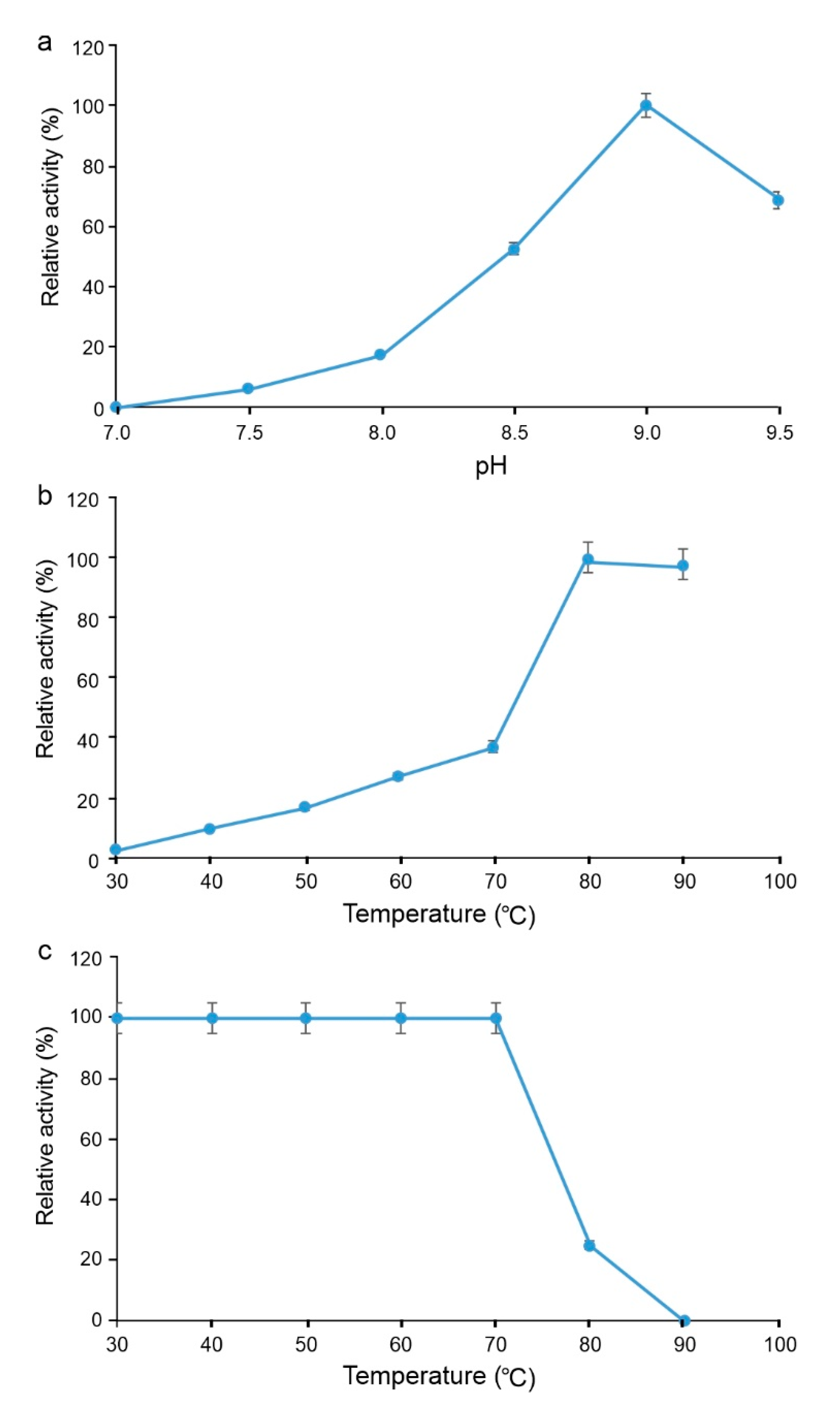
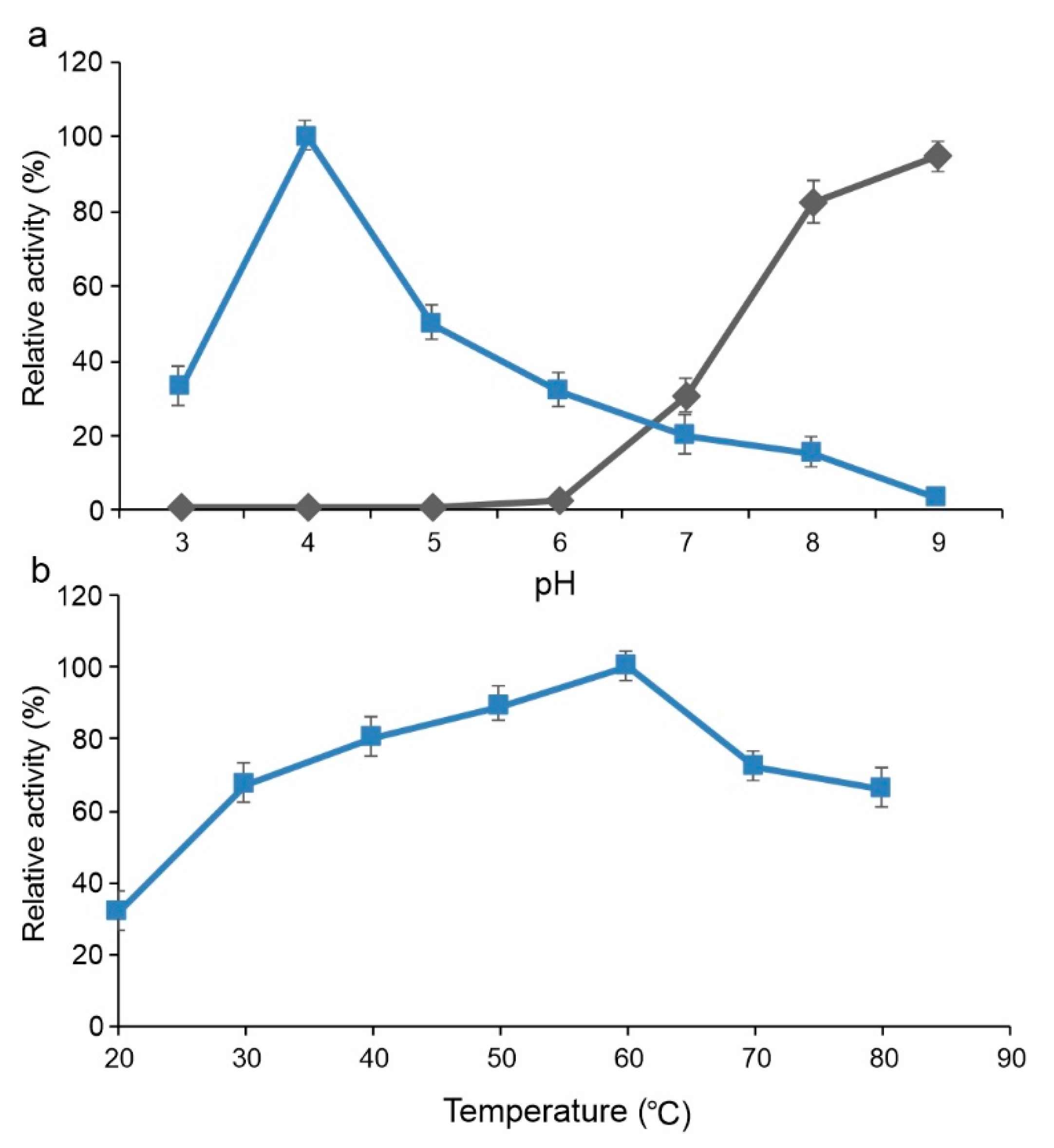
2.3.1. Functional Analysis of Sbv333-TA

The functional characterization of Sbv333-TA was carried out by investigating the influence of different reaction conditions on enzyme activity (Figure 4). The transaminase activity of Sbv333-TA was evaluated by spectrophotometric assays at 245 nm following the formation of the product acetophenone from the benchmark substrates (S)-methyl benzyl amine ((S)-MBA) and pyruvate. The influence of pH on Sbv333-TA activity was evaluated at pH values ranging from 7.0 to 9.5. As shown in Figure 4a, Sbv333-TA, similarly to most of the known (S)-ATAs, shows its maximum activity at alkaline pHs, specifically at pH 9.0 (0.43 U mg−1 pure protein at 30 °C).
The influence of temperature on the enzyme activity was evaluated in the range of temperature between 20 and 90 °C at the optimal pH (9.0) (Figure 4b). Surprisingly, Sbv333-TA showed a thermophilic character with a constant activity increase with temperature up to 90 °C. Accordingly, Sbv333-TA showed remarkable thermostability. In fact, it retains 100% of starting activity after 3 h incubation at temperatures ranging from 30 °C to 70 °C and it maintains 25% of initial activity even when incubated at 80 °C for 3 h (Figure 4c). To support these results, the melting temperature (TM) of Sbv333-TA was evaluated by CD analysis, by monitoring structural changes at 220 nm. CD analysis clearly defined a TM of 85 °C (Figure S3), a value very close to those estimated for the hyperthermophilic enzymes B3-TA (TM 88 °C) and Tr-TA (TM 87 °C) [27,28].
To evaluate Sbv333-TA activity toward a series of (R)- or (S)-aromatic amines, bearing a methyl, ethyl, or propyl side-chain adjacent to the amine function, a spectrophotometric assay was carried out using pyruvate as amino acceptor. Sbv333-TA resulted to be active only toward (S)-MBA, indicating that more sterically hindered aromatic amines are not the substrate of this enzyme and, on the other hand, confirming that the enzyme is strictly (S)-selective, as inferred by sequence analysis. Subsequently, several keto acids, ketones, esters and aldehydes were evaluated as potential amino acceptors in transamination reactions using (S)-MBA as an amino donor (Table 3). The reaction’s outcome was estimated after 24 h by GC-MS analysis and Sbv333-TA demonstrated a broad substrate scope in this regard (see Discussion for details).

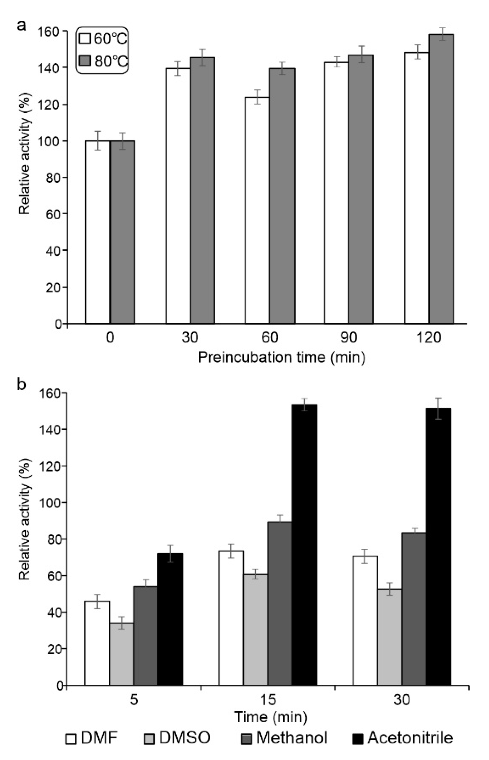
2.3.2. Functional Analysis of Sbv286-LAC

Laccase activity profile at different pHs was determined using ABTS and syringaldazine as the substrate with buffers of different pH (pH 3.0–9.0) and results are presented as relative enzyme activity where maximal activity was set as 100% (Figure 5). Sbv286-LAC reached its maximum activity towards ABTS at pH 4.0 (0.3 U mg−1 pure protein at 30 °C, Figure 5a), and was active from pH 3.0 to pH 8.0, while its activity peaked at alkaline pH using syringaldazione as substrate (0.05 U mg−1 pure protein at 30 °C). The temperature optimum (60 °C) was determined by enzyme assays with ABTS at temperatures ranging from 20 to 80 °C at pH 4.0 (Figure 5b).
Sbv286-LAC exhibited not only a remarkable thermophilicity, but also a high thermostability at 60 °C and 80 °C, as well as a 1.5-fold increase in enzyme activity even after 2 h of incubation at high temperatures in comparison to control (enzyme activity measured before incubation at 60 °C and 80 °C) (Figure 6a). Enzyme activity was affected by the presence of 10% (v/v) of solvents in the reaction buffer (Figure 6b). While DMF, DMSO and methanol reduced the enzyme activity between 20–70% in comparison to no-solvents control, acetonitrile enhanced the Sbv286-LAC by 1.5-fold upon incubation for 15 and 30 min (Figure 6b).

3. Discussion

Following our previous work of screening of a library of Streptomyces spp. rhizosphere isolates to explore their biocatalytic potential [2], in this work, we selected three strains from this library, i.e., Streptomyces sp. BV129, BV286 and BV333, which, according to functional screening, showed the capability to produce interesting enzymatic activities. The genome of the three strains was sequenced, de novo assembled and submitted for genome annotation. It is noteworthy that we found some apparent discrepancies between the results obtained by functional screening [2] and gene annotation by Prokka. For example, no gene was annotated as gelatinases, but strain BV129 and strain BV333 resulted to produce these enzymes. Or, on the other hand, various genes were annotated as lipases in all three strains, but no lipase activity was detected in strain BV286 by functional screening. However, these differences could be due to inaccuracies of the annotation algorithm or to the substrates used during the functional screening that could not be suitable for the enzymes expressed by the selected Streptomyces strains.
With the three new genomes in our hands, we focused our search for novel enzymes on transaminases and laccases, enzymes that are of particular interest for biocatalytic applications. Transaminase and laccase candidates were found in the genomes of all three strains and the most promising candidates (Sbv333-TA and Sbv286-LAC) were selected based on sequence alignment score and presence of key active site residues. It must be pointed out that only homologs to (S)-selective ATAs were identified. Considering that (S)-selective ATAs are mainly produced by bacteria whereas (R)-selective ATAs belong to a completely different fold type class (PLP fold type IV) from that of (S)-ATA (PLP fold type I) and are usually produced by fungi [29,30], the identification of only (S)-ATA homologs in Streptomyces is not surprising. Moreover, it is worth noting that the Prokka algorithm predicted around 30 aminotransferases in each genome, but much fewer ATA candidates have been found by a more specific bioinformatics search. It is important to highlight that our search was restricted to homologues of experimentally confirmed ATAs showing activity toward a wide range of substrates (the so-called high-activity ATAs) [26], and these enzymes are relatively rare when compared to transaminases accepting as amino donors only α-amino acids.
Bioinformatic analysis showed that neither Sbv333-TA nor Sbv286-LAC corresponded to already characterized enzymes and, interestingly, Sbv333-TA shared high sequence identity with ATA sequences from extremophiles, whereas Sbv286-LAC showed high similarity to the so-called small laccases, typical two-domains bacterial laccases [17].
Sbv333-TA and Sbv286-LAC were cloned and overexpressed in E. coli Rosetta, and while Sbv286-LAC resulted to be successfully expressed in this host, Sbv333-TA formed inclusion bodies and its successful production was obtained by co-expression with co-chaperons GroES and GroEL. Activity assays using (S)-MBA and ABTS as substrates, respectively, revealed that, as expected, the two proteins were functionally active transaminase and laccase, thus both proteins were submitted for functional characterization.
Although Streptomyces BV333 and BV286 are mesophilic strains, both enzymes showed a thermophilic character. Sbv333-TA activity increased constantly with temperature up to 90 °C and remarkably, the same behavior is described in literature only for B3-TA [10], the most thermostable wild-type TA reported to date. Indeed, these data are in correlation with the relatively high sequence identity between these two proteins (41% identity). Bacterial laccases and laccase-like multi-copper oxidases are usually described as thermophilic enzymes and Sbv286-LAC, with an optimum temperature of 60 °C, is in line with these findings [19,31]. Furthermore, the two enzymes showed remarkable thermal stability, Sbv333-TA retaining 100% of starting activity after 3 h incubation at temperatures up to 70 °C and Sbv286-LAC resulting even thermoactivated at 60 °C and 80 °C after 2 h incubation. Sbv333-TA thermostability is comparable to that of other thermophilic TAs, such as the TA from Thermomicrobium roseum, which retains 100% of starting activity after 3 h incubation at 70 °C, as well. The exceptional thermostability of Sbv333-TA was confirmed also by determining its melting temperature (TM) which resulted to be of 85 °C, a value only slightly lower than the TM reported for the thermostable B3-TA (88 °C) and the ATA from T. roseum (87 °C) [27]. On the other side, the thermoactivation effect observed with Sbv286-LAC at high temperature was previously shown by Ece and coworkers with a laccase from S. cyaneus [18]. It is worth noting that the thermostability of these enzymes makes these biocatalysts quite attractive for industrial applications. In fact, (thermo)stable enzymes usually result suitable to be used under industrial process harsh conditions, such as at high temperatures and/or in the presence of organic solvents.
The relative activities of the two enzymes were also tested at different pHs and Sbv333-TA showed the best activity at pH 9.0, which is in agreement with the pH optimum of most of the transaminases described to date, whereas Sbv286-LAC reached maximum activity at pH 4.0 or 8.5 according to the tested substrate (ABTS or syringaldazine respectively) like other laccases, such as the one from S. sviceus [20].
In addition, Sbv286-LAC tolerance to organic co-solvents (10% (v/v)) was evaluated and this enzyme resulted to be quite stable in MeOH and DMF, while, surprisingly, acetonitrile enhanced its activity by 1.5 fold. Some bacterial laccases have exhibited high tolerance to different solvents as well, including ethanol, methanol, DMF, acetonitrile, acetone, and DMSO. For example Bacillus pumilus W3 laccase retains > 50% of its activity in solvents such as ethanol, methanol, DMF and DMSO 10% (v/v) [32] while the laccase from Bacillus licheniformis retained 55% of its activity in the presence of 40% DMSO [33]. Similar to our study, Wu and co-workers tested different fungal laccases and they also observed an activity enhancement by 1.5- to 4.0-fold by enzyme pre-incubation in acetone, methanol, ethanol, DMSO, and DMF [34].
Finally, the activity of Sbv333-TA was tested toward three differently substituted amino donors and toward a wide array of amino acceptors. Concerning the amino donors, only (S)-MBA, bearing a small methyl group, was accepted, this result suggesting that the substrate-binding pocket is not large enough to accept bulkier substituents. Moreover, the enzyme did not convert (R)-MBA, demonstrating to be strictly (S)-selective, as expected given the similarity to class I (S)-selective TAs. As far as amino acceptor concerns, this transaminase accepted a broad range of substrates (Table 3). Comparing the amino donor spectra of the new enzyme with the spectra of the closest relative B3-TA and the spectra of the most characterized (S)-ATA from Vibrio fluvialis [10], it is possible to appreciate some analogies and differences. α-Ketoacids, such as pyruvate and 2-oxo butyrate are accepted by all three enzymes, while the di-ketoacid α-ketoglutarate is not accepted by any of them. In the respect of aldehydes, glyoxylic acid was one of the best substrates in all three cases, while propionaldehyde and phenyl propionaldehyde are mostly preferred by Sbv333-TA. Very low conversions were obtained with aliphatic ketones such as methyl isobutyl-ketone (7% conv.). Remarkably, Sbv333-TA showed activity toward β-ketoesters, and in particular, it was active toward methyl acetoacetate (52% conv.) that gave low conversions with B3-TA and Vf-TA and toward ethyl benzoylacetate (47% conv.), a precursor of β-phenylalanine, which is not accepted by most of the described transaminases [35,36]. This result is quite interesting considering that amination of β-keto acids is quite challenging, as they decompose easily in an aqueous solution and amination of more stable β-keto esters would be a valuable alternative.

4. Materials and Methods

4.1. Reagents and Chemicals

Microbiological media and components such as yeast extract, tryptone, casamino acids were from Oxoid (Hampshire, UK). Amino donors, amino acceptors, ABTS, PLP, IPTG, inorganic salts were purchased from Sigma-Aldrich (Munich, Germany). All other reagents were of analytical grade and commercially available. All strains and plasmids used in this study are presented in Table S4.

4.2. Analytical Methods

GC-MS analyses were carried out on an HP-5MS column (30 m × 0.25 mm × 0.25 μm, Agilent) on a Finnigan TRACE DSQ GC/MS instrument (ThermoQuest, San Jose, CA, USA). Acetylation of (S)-MBA before injection was done as described in Ferrandi et al. 2017 [10]. GC-MS analysis was carried out by keeping column temperature at 60 °C for 1 minute, then raising the temperature to 150 °C at 6 °C/min, hold 1 min and finally raising the temperature from 150 °C to 210°C at 12 °C/min. Under these conditions retention times were: acetophenone, 7.6 min; (S)-α-methylbenzylamine ((S)-MBA), 16.3 min.
CD analysis was performed on nitrogen-flushed Jasco J-1100 spectropolarimeter (Easton, MD, USA) interfaced with a thermostatically controlled cell holder. For the determination of apparent TM, spectral scans at increasing temperatures were performed at 210 nm, varying the temperatures as follows: 20 up to 65 °C at 5 °C/min data pitch each 2 °C, hold 30 s; 65 up to 90 °C at 2.5 °C/min, data pitch each 0.5 °C, hold 30 s; 90 up to 95 °C at 5 °C/min, pitch data each 2 °C, hold 30 s. This analysis was carried out using a purified Sbv333-TA sample diluted in degassed water (0.15 mg mL−1 final concentration) in quartz cuvettes with 0.1 cm path length.
Scanning electron micrographs of three Streptomyces strains grown on MSF agar were obtained by a high-resolution field emission Zeiss Ultra Plus-SEM (Carl Zeiss AG, Oberkochen, Germany) using InLens detector with an accelerating voltage of 5 kV at a working distance of 5 mm. Prior to imaging, strains were fixed onto the SEM stubs using carbon tape and sputtered with gold/palladium (80/20 ratio) for 10 s.

4.3. Streptomyces sp. BV129, BV286, BV333 Genome Sequencing, Annotation, and Analysis

Streptomyces spp. genomic DNA (gDNA) was isolated by the method of Nikodinovic et al. [37].
The sequencing library for an Illumina HiSeq2500 was prepared from extracted DNA with the Nextera DNA kit (Illumina) using a standard protocol. Sequencing was performed in a paired-end mode with 2 × 150 cycles for every species. After de-multiplexing with Illumina’s software bcl2fastq-1.8.4 with default settings for adapter trimming (at least 90% match of bases should match) and no mismatches allowed in sequencing barcode, all reads undergo quality-based trimming to remove potential contaminants, low quality reads and sequencing adapters with the help of BBDuk from the BBMap package version 34.41 (https://sourceforge.net/projects/bbmap/). To pass the quality filter, read quality needed to be higher than a Phred score of 20 and achieve a minimal length of 50 bp after quality-based and adapter trimming. Additionally, every sample was quality-controlled before and after trimming with the FastQC (http://www.bioinformatics.babraham.ac.uk/projects/fastqc/). FastQC evaluates per base sequence quality, average base composition, GC content, sequence length distribution and adapter contaminations after trimming. Samples statistics are given in Table S5.
The assembly was carried out on a computational cluster running under CentOS 6.9 by ABySS version 1.5.2 [38] after iterative k-mer length optimization with the k = 85 (BV129, BV333) and k = 92 (BV286). Gene annotation was performed with the Prokka 1.12 [24].
The phylogenetic tree was inferred with the FastME 2.1.6.1 [23] and the tree was estimated from the distance matrix and rooted at the midpoint [39].

4.4. In Silico Screening for Novel Transaminases and Laccases

Bioinformatic search for novel biocatalysts was carried out by performing alignment analysis with known sequences retrieved from the NCBI database as queries. The program LAST (http://lastweb.cbrc.jp/) was used with default settings [40]. Bioinformatic analysis was done for three novel Streptomyces genomes in order to identify novel sequences coding for transaminases (S- and R-selective) and laccases. Sequence alignment was done using ClustalΩ [41,42].

4.5. Gene Cloning and Recombinant Strains

Selected enzyme-coding sequences (BV333_03408 and BV286_03089) were amplified from Streptomyces gDNA under standard PCR conditions using XtraTaq polymerase (Genespin, Milan, Italy) and primers (Table S6). Standard PCR amplifications were carried out in 50 µL reaction mixtures containing 100 ng gDNA, primers (1 µM each), dNTPs (0.2 mM each), 2 U of Xtra.Taq polymerase and 5 µL of buffer containing MgCl2. All PCR reagents were from Genespin. PCR conditions were as follows: 95 °C for 3 min, followed by 35 cycles at 95 °C for 30 s, 65 °C for 30 s, 72 °C for 100s, and then 72 °C for 10 min. Amplified sequences were purified from agarose gel (1.0%, w/v) using the Wizard® SV Gel and PCR Clean-Up System (Promega, Wisconsin, USA) before cloning. Sequences BV333_03408 and BV286_03089 were cloned in the pETite C-His Kan plasmid using the Expresso T7 Cloning and Expression kit from Lucigen (Wisconsin, USA). According to the Expresso T7 Cloning and Expression kit manual, amplified BV333_03408 or BV286_03089 were mixed with the pETite linear plasmid and transformed in chemically competent HI-Control 10 G cells. The resulting plasmids pETite-Sbv333-TA and pETite-Sbv286-LAC were purified using the E.Z.N.A. Plasmid Mini kit II (Omega/VWR). Correct insertion of amplified sequences was confirmed by sequencing on both strands (Biofab Research (Rome, Italy)) using primers T7 promoter and pETite reverse (Table S7). Purified pETite-Sbv333-TA and pETite-Sbv286-LAC were subsequently transformed in E. coli Rosetta for protein expression.

4.5.1. Sbv333-TA Expression and Purification

E. coli Rosetta (DE3)-Sbv333-TA cells were grown overnight at 37 °C, 220 rpm, in LB medium supplemented with 30 μg mL−1 kanamycin and 34 μg mL−1 chloramphenicol (LBkan30cam34, 100 mL). Amounts of 20 mL of precultures were subsequently inoculated in 500 mL LBkan30cam34 medium and maintained at 37 °C, 220 rpm, till the OD600 cell density reached 0.5–1. Recombinant protein expression was induced by the addition of IPTG (1 mL of 1 M solution in water) and the culture was transferred to 30 °C with shaking 220 rpm and grown overnight. Cells were thus recovered by centrifugation (5000 rpm, 30 min, 4 °C), resuspended in 20 mL wash buffer (20 mM KP buffer, pH 7.0, 500 mM NaCl, 20 mM imidazole) and lysed by sonication. Cell extracts were separated from insoluble debris by centrifugation (11,000 rpm, 30 min) and the presence of soluble recombinant protein in clear lysates was assessed by SDS-PAGE (10% T, 2.6% C).
To perform protein purification, clear cell extracts containing the overexpressed protein were incubated with the Ni Sepharose 6 Fast Flow agarose resin (Ni-NTA) (GE Healthcare, Italy) for 90 min at 4 °C under mild shaking. The mixture was then loaded onto a glass column (10 × 110 mm), the resin was washed with 20 mL of wash buffer (20 mM imidazole, 500 mM NaCl, 20 mM potassium phosphate buffer pH 8.0). His-tagged Sbv333-TA was thus eluted using a 3-step gradient (10 mL washing buffer with 100, 200, and 300 mM imidazole, respectively), dialyzed against 20 mM KP buffer, pH 9.0, at 4 °C for 16 h and stored at −80 °C. Protein content was estimated according to the method of Bradford with the Bio-Rad Protein Assay and protein purity was assessed by SDS-PAGE analysis (10% T, 2.6% C). The molecular weight protein standard mixture from Bio-Rad (Karlsruhe, Germany) was used as a reference. Gels were stained for protein detection with Coomassie Brilliant Blue.
Due to unsatisfactory results obtained using E. coli Rosetta (DE3)-Sbv333-TA, pETite-Sbv333-TA was transformed into E. coli BL21(DE3) containing the plasmid pGro7 (Takara Bio Inc., Kyoto, Japan), obtaining the E. coli BL21(DE3)-GroES, GroEL-Sbv333-TA strain. An overnight preculture of E. coli BL21(DE3)-GroES, GroEL-Sbv333-TA grown in LBkan30 at 37 °C was then inoculated in 1 L of LB kan30cam20 and 2 mg mL−1 arabinose was added for induction of chaperone proteins. When cell density reached OD600 0.4–0.6, recombinant expression of Sbv333-TA was induced by the addition of IPTG (1 mL of 1 M solution in water) and the culture was kept at 30 °C overnight. Finally, cells were harvested by centrifugation, lysed by sonication and Sbv333-TA was purified as described above.

4.5.2. Sbv286-LAC Expression and Purification

E. coli Rosetta (DE3)-Sbv286-LAC cells were grown at 37 °C and 180 rpm for 16 h in LBkan30cam34 (100 mL). The following day, 1% overnight inoculum was added to fresh LB, M9 or TB media supplemented with antibiotics (30 μg mL−1 kanamycin and 34 μg mL−1 chloramphenicol) and cultures were induced with 1 mM IPTG at OD600 between 0.4–0.6. Enzyme expression was carried out at 37 °C for 3 h, 28 °C for 16 h and at 17 °C for 72 h with shaking at 180 rpm. Sbv286-LAC was purified using QIAGEN Ni-NTA spin kit following the manufacturer’s instruction (QIAGEN, Hilden, Germany). Purified Sbv286-LAC, as well as whole cells and cell extracts samples collected during recombinant expression optimization, were analyzed by SDS-PAGE (10% T, 2.6% C), as previously described for Sbv333-TA.

4.6. Functional Characterization of Sbv333-TA and Sbv286-LAC

Sbv333-TA activity was determined by spectrophometric assays in quartz cuvettes (assay volume 0.5 mL) containing transaminase assay solution (2.5 mM pyruvate, 2.5 mM (S)-MBA in 0.1 M KP buffer, pH 9.0 and 0.25% (v/v) dimethyl sulfoxide (DMSO)). Formation of acetophenone upon enzyme addition (10–50 μL of purified Sbv333-TA, 70–350 µg) was followed at 245 nm (ε245 = 12 mM−1 cm−1) on a Jasco V-530 UV/VIS spectrophotometer. One unit of activity is defined as the enzyme activity that produces 1 μmol of acetophenone per minute under the assay conditions described above. Sbv286-LAC activity was detected spectrophometrically at 420 nm using ABTS (2,2’-azino-bis(3-ethylbenzothiazoline-6-sulphonic acid) or syringaldazine at 530 nm as substrates (ε420 = 36.0 mM−1cm−1 , ε530 = 65.0 mM−1cm−1 respectively). The assay was performed in acrylic cuvettes (total volume 1 mL) in 20 mM Na-acetate buffer pH 4.0 0.2 mM CuSO4, containing 1 mM ABTS or 20 mM Tris-HCl buffer pH 8.5 0.2 mM CuSO4 containing 20 µM syringaldazine and adding 10–20 µL (28–56 µg) of purified Sbv286-LAC. One unit of enzymatic activity was defined as the amount of enzyme that oxidizes 1 mmol of ABTS or 1 mmol of syringaldazine per minute under the described assay conditions.
The optimum pH of Sbv333-TA was determined within a pH range of 7.0 to 9.5 in 20 mM potassium phosphate buffer (pH 7.0–9.0) or 20 mM Tris-HCl (pH 9.5) at 30 °C, while temperature optimum was determined by heating the assay solutions (20 mM potassium phosphate buffer pH 9.0) in cuvettes in a water bath in the range of 30–90 °C for 15 min before adding the purified enzyme (10–50 μL of purified Sbv333-TA, 70–350 µg). The thermal stability of Sbv333-TA was evaluated by incubating enzyme samples for 3 h at temperatures ranging from 30 to 90 °C and then measuring spectrophotometrically the residual activity following the formation of acetophenone. Experiments were done in duplicate on two independent occasions.
The influence of pH on Sbv286-LAC activity was determined within a pH range of 3.0 to 10.0 using 20 mM acetate buffer (pH 3.0–5.0), 20 mM potassium phosphate buffer (pH 6.0–7.0), 20 mM Tris-HCl (pH 8.0–9.0), respectively. Sbv286-LAC temperature optimum was determined by heating the assay solutions in cuvettes in a water bath in the range of 20–80 °C for 15 minutes before adding the purified enzyme. The thermal stability of purified laccase was assessed by measuring the residual activity after incubation of purified Sbv286-LAC solution at 60 °C and 80 °C. Enzymes samples were taken at 30, 60, 90 and 120 min, quickly cooled on ice and activity was determined by the ABTS assay described above in 20 mM acetate buffer, pH 4.0. Solvent stability of Sbv286-LAC was assessed in 10% (v/v) dimethyl formamide (DMF), dimethyl sulfoxide (DMSO), methanol and acetonitrile by measuring residual enzyme activity using ABTS assay after incubation in solvents at room temperature for 5, 15 and 30 min. Experiments were done at least in duplicate.
The enantioselectivity of Sbv333-TA was evaluated by performing the acetophenone assay in the presence of (R)-α-methylbenzylamine as substrate under the same conditions described for (S)-MBA. The formation of propiophenone and butyrophenone was determined under the same conditions at 242 nm using either (S)- or (R)-ethylbenzylamine and (S)- or (R)-phenylbutylamine, respectively, as substrates. Sbv333-TA amino acceptor spectrum was evaluated at 30 °C in 0.5 mL reaction mixture containing 0.1 M KP buffer, pH 9.0, 10 mM (S)-MBA, 10 mM acceptor, 1 mM PLP, and 0.5 mg of purified enzyme. Conversions of (S)-MBA (after derivatization) into acetophenone were evaluated after 24 h by GC-MS analysis as described in the analytical methods section.

Supplementary Materials

The following are available online at https://www.mdpi.com/article/10.3390/catal11080919/s1, Figure S1: SDS-PAGE E. coli Rosetta expressing aminotransferase from pETite_BV333 TA. 1) before the induction 2) after the induction with IPTG 3) cell lysate 4) purified enzyme M) Marker – SDS-PAGE Standard Broad Range (Bio-Rad, USA), Figure S2: SDS-PAGE gel of purified laccase Sbv286-LAC. 1) before the induction 2) after the induction with IPTG 3) purified enzyme M) Marker – SDS-PAGE Standard Broad Range (Bio-Rad, USA), Figure S3: Sbv333-TA melting curve obtained by circular dichroism spectroscopic analysis at 210 nm Table S1: Bioinformatics tools used to perform phylogenetic classification, Table S2: GenBank accession number of query sequences used in the bioinformatics search for novel ATA. Entry 1-11: (S)-selective ATA, Entry 12–18: (R)-selective ATA, Table S3: GenBank accession number of query sequences used in the bioinformatics search for novel laccases, Table S4: Bacterial strains and plasmids used in this study, Table S5: Sequencing statistics, Table S6: Oligonucleotide primers used in this study.

Author Contributions

Conceptualization, D.M. and J.N.-R. methodology, E.E.F., Y.V., C.G. and R.S.; investigation, J.S., L.D., S.V., and Y.V.; resources, D.M. and J.N.-R.; data curation, E.E.F., J.S., Y.V.; writing—original draft preparation, J.S., L.D., Y.V., R.S. and E.E.F.; writing—review and editing, S.V., E.E.F., D.M. and J.N.-R.; funding acquisition, D.M. and J.N.-R. All authors have read and agreed to the published version of the manuscript.

Funding

This research was funded by Ministry of Education, Science and Technological Development of the Republic of Serbia, 451-03-9/2021-14/ 200042 and S.V. and J.NR. are partially funded from the European Union’s Horizon 2020 research and innovation program under grant agreement No 870292 (BioICEP).

Data Availability Statement

Publicly available datasets were analyzed in this study (https://www.ncbi.nlm.nih.gov/bioproject/739376). The additional data presented in this study are available on request from the corresponding author.

Conflicts of Interest

The authors declare no conflict of interest.

References

  1. Spasic, J.; Mandic, M.; Djokic, L.; Nikodinovic-Runic, J. Streptomyces spp. in the biocatalysis toolbox. Appl. Microbiol. Biotechnol. 2018, 102, 3513–3536. [Google Scholar] [CrossRef]
  2. Spasic, J.; Mandic, M.; Radivojevic, J.; Jeremic, S.; Vasiljevic, B.; Nikodinovic-Runic, J.; Djokic, L. Biocatalytic potential of Streptomyces spp. isolates from rhizosphere of plants and mycorrhizosphere of fungi. Biotechnol. Appl. Biochem. 2018, 65, 822–833. [Google Scholar] [CrossRef]
  3. Lopes, A.M.; Ferreira Filho, E.X.; Moreira, L.R.S. An update on enzymatic cocktails for lignocellulose breakdown. J. Appl. Microbiol. 2018, 125, 632–645. [Google Scholar] [CrossRef] [Green Version]
  4. Pellis, A.; Cantone, S.; Ebert, C.; Gardossi, L. Evolving biocatalysis to meet bioeconomy challenges and opportunities. New Biotechnol. 2018, 40, 154–169. [Google Scholar] [CrossRef]
  5. Markets&Markets. Biocatalysis & Biocatalysts Market by Type, Application, and Geography-Forecast to 2019. 2015. Available online: https://www.marketsandmarkets.com/Market-Reports/biocatalysis-biocatalyst-market-71612080.html (accessed on 12 May 2021).
  6. Kelly, S.A.; Magill, D.J.; Megaw, J.; Skvortsov, T.; Allers, T.; McGrath, J.W.; Allen, C.C.R.; Moody, T.S.; Gilmore, B.F. Characterisation of a solvent-tolerant haloarchaeal (R)-selective transaminase isolated from a Triassic period salt mine. Appl. Microbiol. Biotechnol. 2019, 103, 5727–5737. [Google Scholar] [CrossRef] [PubMed] [Green Version]
  7. Sun, X.; Li, Y.; Tian, Z.; Qian, Y.; Zhang, H.; Wang, L. A novel thermostable chitinolytic machinery of Streptomyces sp. F-3 consisting of chitinases with different action modes. Biotechnol. Biofuels 2019, 12, 136. [Google Scholar] [CrossRef] [PubMed]
  8. Prakash, D.; Nawani, N.; Prakash, M.; Bodas, M.; Mandal, A.; Khetmalas, M.; Kapadnis, B. Actinomycetes: A repertory of green catalysts with a potential revenue resource. BioMed Res. Int. 2013, 2013, 264020. [Google Scholar] [CrossRef] [PubMed]
  9. Cho, S.S.; Park, D.J.; Simkhada, J.R.; Hong, J.H.; Sohng, J.K.; Lee, O.H.; Yoo, J.C. A neutral lipase applicable in biodiesel production from a newly isolated Streptomyces sp. CS326. Bioprocess Biosyst. Eng. 2012, 35, 227–234. [Google Scholar] [CrossRef] [PubMed]
  10. Ferrandi, E.E.; Monti, D. Amine transaminases in chiral amines synthesis: Recent advances and challenges. World J. Microbiol. Biotechnol. 2017, 34, 13. [Google Scholar] [CrossRef]
  11. Savile, C.K.; Janey, J.M.; Mundorff, E.C.; Moore, J.C.; Tam, S.; Jarvis, W.R.; Colbeck, J.C.; Krebber, A.; Fleitz, F.J.; Brands, J.; et al. Biocatalytic asymmetric synthesis of chiral amines from ketones applied to sitagliptin manufacture. Science 2010, 329, 305–309. [Google Scholar] [CrossRef] [Green Version]
  12. Yonaha, K.; Suzuki, K.; Toyama, S. Streptomyces beta-alanine: Alpha-ketoglutarate aminotransferase, a novel omega-amino acid transaminase. Purification, crystallization, and enzymologic properties. J. Biol. Chem. 1985, 260, 3265–3268. [Google Scholar] [CrossRef]
  13. Yonaha, K.; Suzuki, K.; Toyama, S. 4-Aminobutyrate:2-oxoglutarate aminotransferase of Streptomyces griseus: Purification and properties. Eur. J. Biochem. 1985, 146, 101–106. [Google Scholar] [CrossRef]
  14. Burgie, E.S.; Thoden, J.B.; Holden, H.M. Molecular architecture of DesV from Streptomyces venezuelae: A PLP-dependent transaminase involved in the biosynthesis of the unusual sugar desosamine. Protein Sci. 2007, 16, 887–896. [Google Scholar] [CrossRef] [PubMed] [Green Version]
  15. Bassanini, I.; Ferrandi, E.E.; Riva, S.; Monti, D. Biocatalysis with laccases: An updated overview. Catalysts 2021, 11, 26. [Google Scholar] [CrossRef]
  16. Zerva, A.; Simić, S.; Topakas, E.; Nikodinovic-Runic, J. Applications of microbial laccases: Patent review of the past decade (2009–2019). Catalysts 2019, 9, 1023. [Google Scholar] [CrossRef] [Green Version]
  17. Guan, Z.-B.; Luo, Q.; Wang, H.-R.; Chen, Y.; Liao, X.-R. Bacterial laccases: Promising biological green tools for industrial applications. Cell. Molec. Life Sci. 2018, 75, 3569–3592. [Google Scholar] [CrossRef]
  18. Ece, S.; Lambertz, C.; Fischer, R.; Commandeur, U. Heterologous expression of a Streptomyces cyaneus laccase for biomass modification applications. AMB Express 2017, 7, 86. [Google Scholar] [CrossRef]
  19. Sherif, M.; Waung, D.; Korbeci, B.; Mavisakalyan, V.; Flick, R.; Brown, G.; Abou-Zaid, M.; Yakunin, A.F.; Master, E.R. Biochemical studies of the multicopper oxidase (small laccase) from Streptomyces coelicolor using bioactive phytochemicals and site-directed mutagenesis. Microbial Biotechnol. 2013, 6, 588–597. [Google Scholar] [CrossRef]
  20. Gunne, M.; Urlacher, V.B. Characterization of the alkaline laccase Ssl1 from Streptomyces sviceus with unusual properties discovered by genome mining. PLoS ONE 2012, 7, e52360. [Google Scholar] [CrossRef] [PubMed]
  21. Gunne, M.; Höppner, A.; Hagedoorn, P.L.; Urlacher, V.B. Structural and redox properties of the small laccase Ssl1 from Streptomyces sviceus. FEBS J. 2014, 281, 4307–4318. [Google Scholar] [CrossRef]
  22. Meier-Kolthoff, J.P.; Göker, M. TYGS is an automated high-throughput platform for state-of-the-art genome-based taxonomy. Nat. Comm. 2019, 10, 2182. [Google Scholar] [CrossRef]
  23. Lefort, V.; Desper, R.; Gascuel, O. FastME 2.0: A comprehensive, accurate, and fast distance-based phylogeny inference program. Mol. Biol. Evol. 2015, 32, 2798–2800. [Google Scholar] [CrossRef] [Green Version]
  24. Seemann, T. Prokka: Rapid prokaryotic genome annotation. Bioinformatics 2014, 30, 2068. [Google Scholar] [CrossRef] [PubMed]
  25. Hyatt, D.; Chen, G.-L.; LoCascio, P.F.; Land, M.L.; Larimer, F.W.; Hauser, L.J. Prodigal: Prokaryotic gene recognition and translation initiation site identification. BMC Bioinform. 2010, 11, 119. [Google Scholar] [CrossRef] [Green Version]
  26. Steffen-Munsberg, F.; Vickers, C.; Kohls, H.; Land, H.; Mallin, H.; Nobili, A.; Skalden, L.; van den Bergh, T.; Joosten, H.J.; Berglund, P.; et al. Bioinformatic analysis of a PLP-dependent enzyme superfamily suitable for biocatalytic applications. Biotechnol. Advan. 2015, 33, 566–604. [Google Scholar] [CrossRef] [PubMed]
  27. Mathew, S.; Deepankumar, K.; Shin, G.; Hong, E.Y.; Kim, B.-G.; Chung, T.; Yun, H. Identification of novel thermostable ω-transaminase and its application for enzymatic synthesis of chiral amines at high temperature. RSC Adv. 2016, 6, 69257–69260. [Google Scholar] [CrossRef]
  28. Ferrandi, E.E.; Previdi, A.; Bassanini, I.; Riva, S.; Peng, X.; Monti, D. Novel thermostable amine transferases from hot spring metagenomes. Appl. Microbiol. Biotechnol. 2017, 101, 4963–4979. [Google Scholar] [CrossRef] [PubMed]
  29. Höhne, M.; Schätzle, S.; Jochens, H.; Robins, K.; Bornscheuer, U.T. Rational assignment of key motifs for function guides in silico enzyme identification. Nat. Chem. Biol. 2010, 6, 807. [Google Scholar] [CrossRef] [PubMed]
  30. Sayer, C.; Martinez-Torres, R.J.; Richter, N.; Isupov, M.N.; Hailes, H.C.; Littlechild, J.A.; Ward, J.M. The substrate specificity, enantioselectivity and structure of the (R)-selective amine: Pyruvate transaminase from Nectria haematococca. FEBS J. 2014, 281, 2240–2253. [Google Scholar] [CrossRef] [PubMed] [Green Version]
  31. Ihssen, J.; Reiss, R.; Luchsinger, R.; Thöny-Meyer, L.; Richter, M. Biochemical properties and yields of diverse bacterial laccase-like multicopper oxidases expressed in Escherichia coli. Sci. Rep. 2015, 5, 10465. [Google Scholar] [CrossRef] [Green Version]
  32. Guan, Z.-B.; Song, C.-M.; Zhang, N.; Zhou, W.; Xu, C.-W.; Zhou, L.-X.; Zhao, H.; Cai, Y.-J.; Liao, X.-R. Overexpression, characterization, and dye-decolorizing ability of a thermostable, pH-stable, and organic solvent-tolerant laccase from Bacillus pumilus W3. J. Mol. Catal. B Enzym. 2014, 101, 1–6. [Google Scholar] [CrossRef]
  33. Tonin, F.; Melis, R.; Cordes, A.; Sanchez-Amat, A.; Pollegioni, L.; Rosini, E. Comparison of different microbial laccases as tools for industrial uses. New Biotechnol. 2016, 33, 387–398. [Google Scholar] [CrossRef] [PubMed]
  34. Wu, M.-H.; Lin, M.-C.; Lee, C.-C.; Yu, S.-M.; Wang, A.H.J.; Ho, T.-H.D. Enhancement of laccase activity by pre-incubation with organic solvents. Sci. Rep. 2019, 9, 9754. [Google Scholar] [CrossRef] [PubMed]
  35. Buß, O.; Voss, M.; Delavault, A.; Gorenflo, P.; Syldatk, C.; Bornscheuer, U.; Rudat, J. β-Phenylalanine ester synthesis from stable β-keto ester substrate using engineered ω-transaminases. Molecules 2018, 23, 1211. [Google Scholar] [CrossRef] [Green Version]
  36. Midelfort, K.S.; Kumar, R.; Han, S.; Karmilowicz, M.J.; McConnell, K.; Gehlhaar, D.K.; Mistry, A.; Chang, J.S.; Anderson, M.; Villalobos, A.; et al. Redesigning and characterizing the substrate specificity and activity of Vibrio fluvialis aminotransferase for the synthesis of imagabalin. Protein Eng. Des. Sel. 2012, 26, 25–33. [Google Scholar] [CrossRef] [Green Version]
  37. Nikodinovic, J.; Barrow, K.D.; Chuck, J.A. High yield preparation of genomic DNA from Streptomyces. BioTechniques 2003, 35, 932–934. [Google Scholar] [CrossRef] [Green Version]
  38. Simpson, J.T.; Wong, K.; Jackman, S.D.; Schein, J.E.; Jones, S.J.; Birol, I. ABySS: A parallel assembler for short read sequence data. Genome Res. 2009, 19, 1117–1123. [Google Scholar] [CrossRef] [Green Version]
  39. Farris, J.S. Estimating phylogenetic trees from distance matrices. Am. Nat. 1972, 106, 645–668. [Google Scholar] [CrossRef]
  40. Kielbasa, S.M.; Wan, R.; Sato, K.; Horton, P.; Frith, M.C. Adaptive seeds tame genomic sequence comparison. Genome Res. 2011, 21, 487–493. [Google Scholar] [CrossRef] [Green Version]
  41. Sievers, F.; Wilm, A.; Dineen, D.; Gibson, T.J.; Karplus, K.; Li, W.; Lopez, R.; McWilliam, H.; Remmert, M.; Soding, J.; et al. Fast, scalable generation of high-quality protein multiple sequence alignments using Clustal Omega. Mol. Syst. Biol. 2011, 7, 539. [Google Scholar] [CrossRef]
  42. Thompson, J.D.; Higgins, D.G.; Gibson, T.J. CLUSTAL W: Improving the sensitivity of progressive multiple sequence alignment through sequence weighting, position-specific gap penalties and weight matrix choice. Nucleic Acids Res. 1994, 22, 4673–4680. [Google Scholar] [CrossRef] [PubMed] [Green Version]
Figure 1. Streptomyces sp. BV129, BV333 and BV286 morphology upon growth for 7 days on mannitol–soy flower medium (a) agar plates; (b) electron micrographs of material collected from the plates.
Catalysts 11 00919 g001
Figure 2. Phylogenetic tree inferred with the FastME 2.1.6.1. using whole-genome comparison with Streptomyces sp. BV129, BV333 and BV286 highlighted in red.
Catalysts 11 00919 g002
Figure 3. Multiple sequence alignment of Sbv333-TA, Tr-TA and B3-TA.
Catalysts 11 00919 g003
Figure 4. The influence of pH (a) and temperature (b) on Sbv333-TA activity and influence of temperature (c) on Sbv333-TA stability. Values are means of two independent experiments ± standard deviations (SD).
Catalysts 11 00919 g004
Figure 5. pH and temperature optimum of the recombinant Sbv286-LAC. Relative laccase activity at (a) different pH using ABTS (□) and syringaldazione (◊) as substrates and (b) different temperatures. Values are means of two independent experiments ± standard deviations (SD).
Catalysts 11 00919 g005
Figure 6. Thermal and solvent stability of the recombinant Sbv286-LAC. Relative laccase activity after incubation (a) at 60 °C and 80 °C for up to 2 h and (b) in different solvents. Untreated control reactions were set to 100%. Values are means of two independent experiments ± standard deviations (SD).
Catalysts 11 00919 g006
Table 1. Genome assembly statistics of Streptomyces sp. BV129, BV333 and BV286.
Sample IDStreptomyces sp. BV129Streptomyces sp. BV286Streptomyces sp. BV333
Genome size6,475,2429,426,0477,323,588
Genome coverage83.5059.0272.73
Number of contigs82212699
Longest contig704,247515,226151,113
N50 *144,188114,51028,611
L50 **152680
GC content (%)71.8670.7773.2
Predicted genes593383266373
Predicted tRNAs849283
Predicted CDS584882336289
Predicted tmRNA111
*—N50—the minimum contig length among contigs required to cover 50% of the whole genome sequence length; can be used as a measure of a quality of assembled genome. Genomes of comparable size with the higher N50 number will be less fragmented and therefore should have higher assembly quality. **—L50 is a minimum number of contigs to reach half of the assembly size. Together with N50 serves as an assembly quality measure.
Table 2. Prokka-based prediction of selected genes in the de novo assembled genomes of Streptomyces sp. BV129, BV333 and BV286. Enzymes potentially acting in the degradation of different substrates are shown together with the corresponding EC category, other categories for which no genes were predicted by Prokka are not shown.
EC no.Streptomyces sp. BV129Streptomyces sp. BV286Streptomyces sp. BV333
lipase3.1.1.3784
cutinase3.1.1.74010
cellulase3.2.1.43113
protease3.4.-.-124160136
laccase1.10.3.1211
tyrosinase1.14.18.1110
aminotransferase (transaminase)2.6.1.-323538
Predicted genes with EC number 204927632114
Table 3. Amino acceptor spectrum of Sbv333-TA.
Amino AcceptorConversion (%)
Catalysts 11 00919 i001α-ketoglutarate -
Catalysts 11 00919 i002Pyruvate 54
Catalysts 11 00919 i0032-Oxobutyrate 46
Catalysts 11 00919 i004Glioxylate88
Catalysts 11 00919 i005Methylacetoacetate 52
Catalysts 11 00919 i006Ethylacetoacetate 35
Catalysts 11 00919 i007Ethylbenzoylacetate 47
Catalysts 11 00919 i008Methyl isobutyl ketone7
Catalysts 11 00919 i009Propionaldehyde 100
Catalysts 11 00919 i010 Phenylpropionaldehyde64
Publisher’s Note: MDPI stays neutral with regard to jurisdictional claims in published maps and institutional affiliations.

