Article

Promoting Photoelectrochemical Water Oxidation on Ti-Doped Fe2O3 Nanowires Photoanode by O2 Plasma Treatment

1
Key Laboratory of UV-Emitting Materials and Technology of Chinese Ministry of Education, School of Physics, Northeast Normal University, 5268 Renmin Street, Changchun 130024, China
2
Institute for Interdisciplinary Quantum Information Technology, Jilin Engineering Normal University, Changchun 130052, China
*
Authors to whom correspondence should be addressed.
Catalysts 2021, 11(1), 82; https://doi.org/10.3390/catal11010082
Submission received: 14 December 2020 / Revised: 2 January 2021 / Accepted: 7 January 2021 / Published: 9 January 2021
(This article belongs to the Special Issue Commemorative Issue in Honor of Professor Akira Fujishima)

Abstract

:
Surface electron traps on semiconductor photoanodes mediate surface recombination and deteriorate the photoelectrochemical (PEC) water oxidation performance of the photoanode. Developing convenient methods to reduce surface electron traps is therefore essential for high efficiency PEC water oxidation on semiconductor photoanodes, particularly for nanostructured photoanodes with large surface area. Herein, we employ a O2 plasma treatment to boost the PEC water oxidation performance of Ti-doped Fe2O3 (Ti-Fe2O3) nanowires photoanodes, aiming to reduce surface oxygen vacancies, the dominant electron traps on Ti-Fe2O3 surface. X-ray diffraction (XRD), scanning electron microscopy and spectroscopic analyses show that the oxygen plasma treatment changes the structural, morphological and optical properties negligibly, but it does reduce the content of surface oxygen vacancies, as estimated from O1s X-ray photoelectron spectroscopy spectra. An optimal O2 plasma treatment (200 W, 70 s) increases the photocurrent density of the Ti-Fe2O3 nanowire photoanode to 2.14 mA·cm−2 (1.23 V vs. RHE) under air mass 1.5G simulated solar light, which is 1.95 times higher than the pristine Ti-Fe2O3 nanowire photoanode. The surface hole transfer efficiency is also improved by 1.66 times due to the reduced surface recombination. The work suggests that O2 plasma treatment is a convenient but effective method to boost the PEC water oxidation performance of Ti-Fe2O3 photoanode and might be applicable to other semiconducting oxide photoanodes for high efficiency PEC water splitting.

Graphical Abstract

1. Introduction

Photoelectrochemical (PEC) water splitting into hydrogen and oxygen with a semiconductor photoanode is regarded as a promising strategy to provide clean and sustainable energy since it was first reported by Fujishima and Honda in 1972 [1], in which a highly efficient photoanode plays a decisive role. The nanostructured Fe2O3 photoanode has attracted a large number of investigations due to its suitable photon absorption (2.0–2.2 eV), shortened minority diffusion distance, and large contact area with the electrolyte for surface reactions [2,3,4,5]. However, the large surface area of nanostructural Fe2O3 usually introduces many surface states, such as surface oxygen vacancies and Fe2+ sites, which mediate the unwanted surface charge recombination [6,7,8,9]. Thus, it is an essential issue to reduce the surface states of the Fe2O3 photoanode for high efficiency PEC water splitting.
Surface modification of the passivation layer and loading of the cocatalyst are usually adopted to reduce the surface recombination mediated by surface oxygen vacancies and improve the PEC performance of Fe2O3 photoanodes [10,11,12,13,14]. For example, Zou et al. demonstrated that the ion-impermeable Al2O3 layer on Ti-Fe2O3 nanorods passivated the surface states and suppressed the back reaction, which improved the photocurrent onset potential [14]. The result proved the necessity of surface states treatment of Ti-doped Fe2O3 nanowire photoanodes. However, the methods of surface loading the passivation layer and cocatalyst have some drawbacks, introducing a barrier in the interface or reducing the absorption of photons. In addition, Yu et al. reported that the most significant issue in suppressing the surface states of Fe2O3 was to reduce the oxygen vacancies on the surface of the electrode [15]. Therefore, it is still necessary to develop a convenient and effective method to controllably tune the surface oxygen vacancy states of Fe2O3 photoanodes.
Herein, we employed oxygen plasma treatment to reduce the surface oxygen vacancy of Ti-doped Fe2O3 (Ti-Fe2O3) nanowire photoanodes. Oxygen plasma, consisting of highly reactive oxygen ions with large concentration and kinetic energy, is expected to oxidize the surface of Ti-Fe2O3 in a convenient way without introducing impurities. The effect of oxygen plasma on the surface states and PEC performance of Ti- Fe2O3 photoanodes were investigated. At an optimal treatment condition (200 W, 70 s), the PEC performance of Ti-Fe2O3 photoanodes was remarkably improved, exhibiting a 1.95 times higher photocurrent than the pristine Ti-Fe2O3 photoanode at the potential of 1.23 V vs. reversible hydrogen electrode (RHE). The surface hole transfer efficiency of the plasma-treated Ti-Fe2O3 (OPT-Ti-Fe2O3) photoanode reached 78%, 1.66-fold higher than the pristine one. O1s X-ray photoelectron spectroscopy (XPS) analysis showed that the integrated area ratio of oxygen vacancy to lattice oxygen at the surface of the OPT-200W-70s electrode was reduced by 6.7%. The work suggests that O2 plasma treatment is a convenient but effective method for tuning the surface defect states of semiconducting oxides photoanode for enhanced PEC water splitting.

2. Results and Discussion

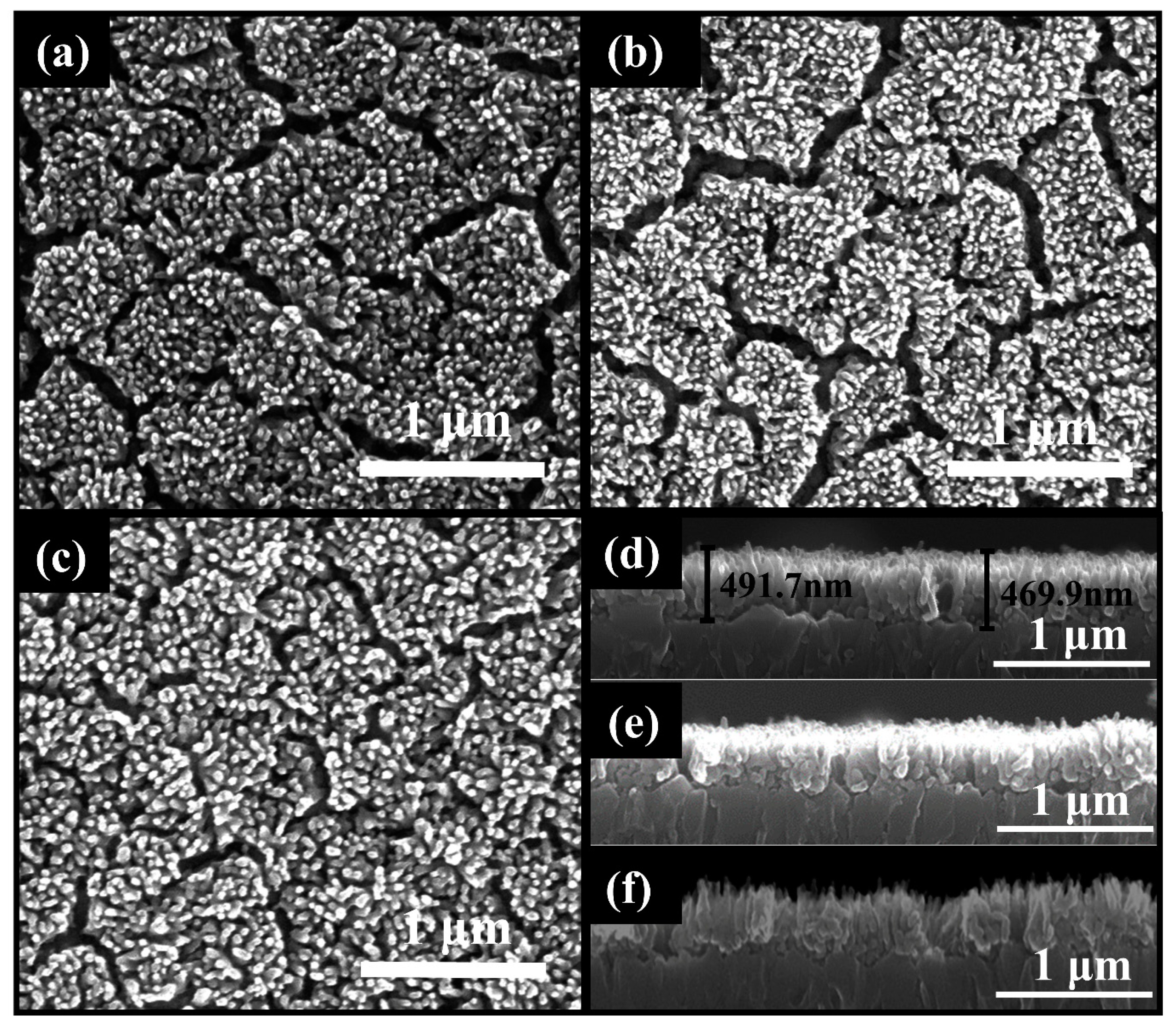
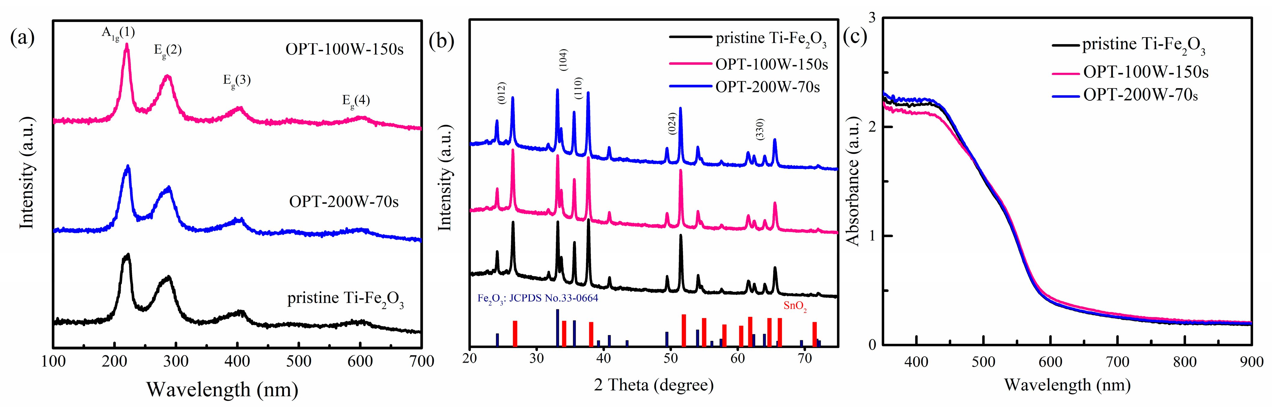
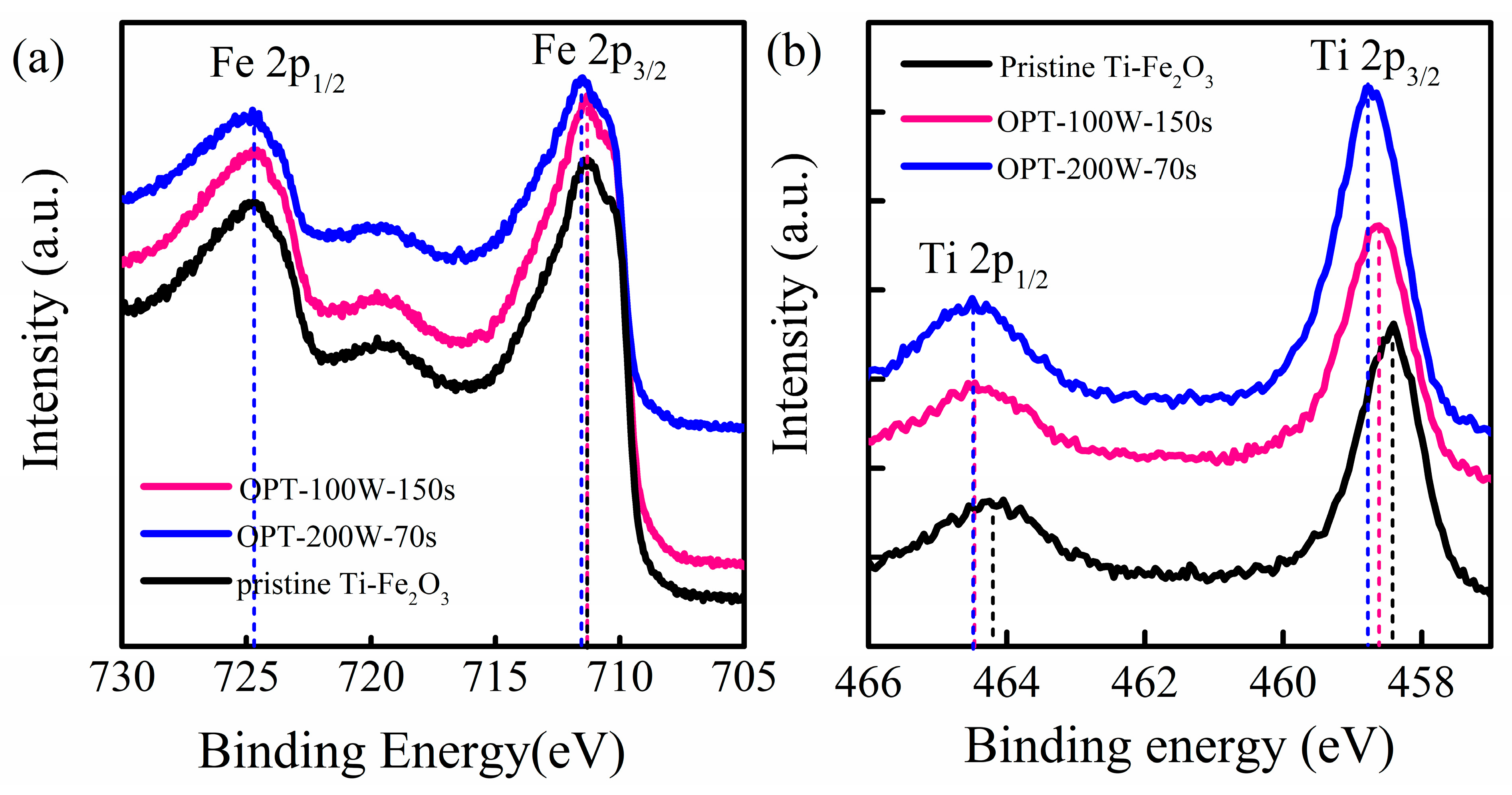
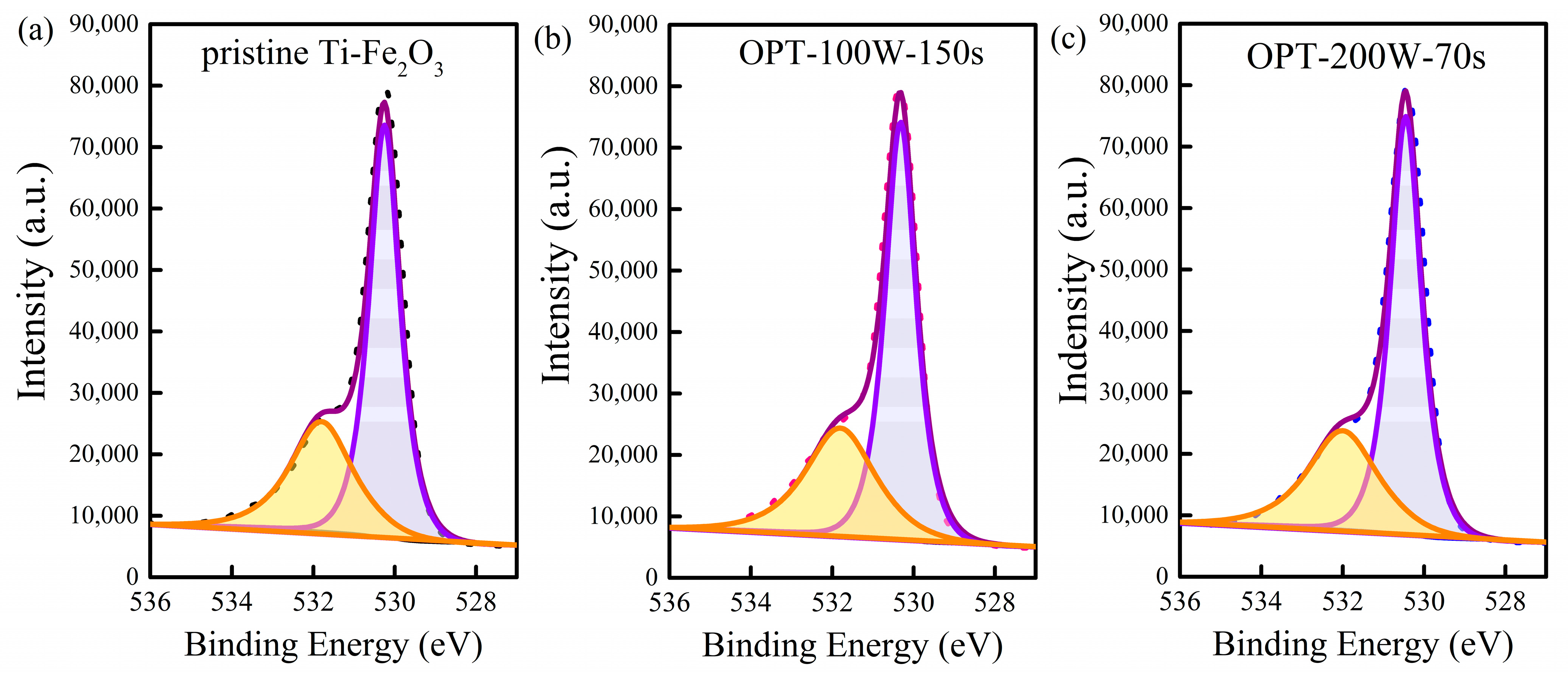
2.1. Effect of O2 Plasma on Morphology, Structure, and Optical Properties of the Electrodes

Ti-Fe2O3 nanowire photoanodes were selected to investigate the effect of O2 plasma treatment on the PEC water oxidation performance. The films were synthesized on F-doped SnO2 conductive glass (FTO) substrates via a hydrothermal method and post-treated with O2 plasma. The scanning electron microscopy (SEM) images in Figure 1 present the surface and cross-sectional images of the pristine Ti-Fe2O3 and oxygen-plasma-treated Ti-Fe2O3 (OPT-Ti-Fe2O3) photoanodes. Figure 1a,d indicate that the diameter of the Fe2O3 nanowire was 40–50 nm while the pristine Ti-Fe2O3 film was about 490 nm thick. After treatment at the power of 100 W for 150 s and 200 W for 70 s, the surface morphology of OPT-100W-150s and OPT-200W-70s had no noticeable change in Figure 1b,c, and the thickness of OPT-Ti-Fe2O3 films was still about 490 nm in Figure 1e,f.
Crystal structure of the photoanodes was characterized by Raman spectroscopy and X-ray diffraction (XRD). The Raman spectra of the photoanodes are depicted in Figure 2a. Four phonon lines at 222, 288, 405, and 604 cm−1 appeared in the three spectra of the photoanodes, which corresponded to the A1g(1), Eg(2), Eg(3), and Eg(4) modes of hematite [16]. There were no obvious differences between the photoanodes with or without O2 plasma treatment. This demonstrated that plasma treatment has little effect on the crystal structure of the films. XRD patterns of the photoanodes are shown in Figure 2b. Five distinct diffraction peaks of hematite could be observed in the spectra, and these peaks corresponded to the (012), (104), (110), (024), and (330) facets of Fe2O3 [17]. There were some other diffraction peaks originated from the Sb:SnO2 substrate, and no peak related to the Ti dopant was observed. The result demonstrated that the diffraction peaks of Ti-Fe2O3 photoanodes had no obvious change after plasma treatment.
We also compared the absorption spectra of the Ti-Fe2O3 photoanodes with and without O2 plasma treatment. The absorption spectra (Figure 2c) of the three films were almost the same. The absorption threshold of the films all appeared at around 600 nm, which corresponded to the bandgap of 2.1 eV [18]. This was consistent with the bandgap of hematite. Based on these results, it can be concluded that the morphology, structure, and optical properties of Ti-doped Fe2O3 electrodes change negligibly after O2 plasma treatment.

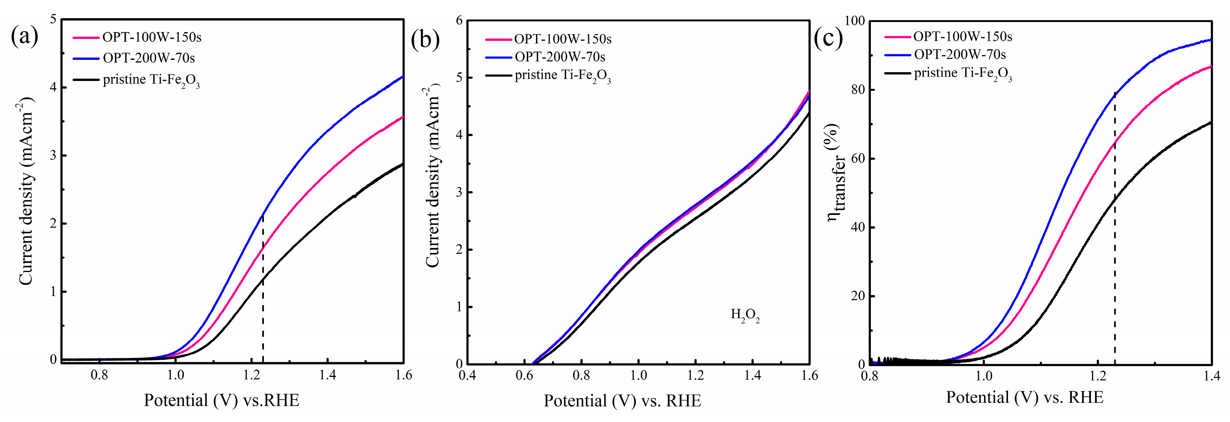
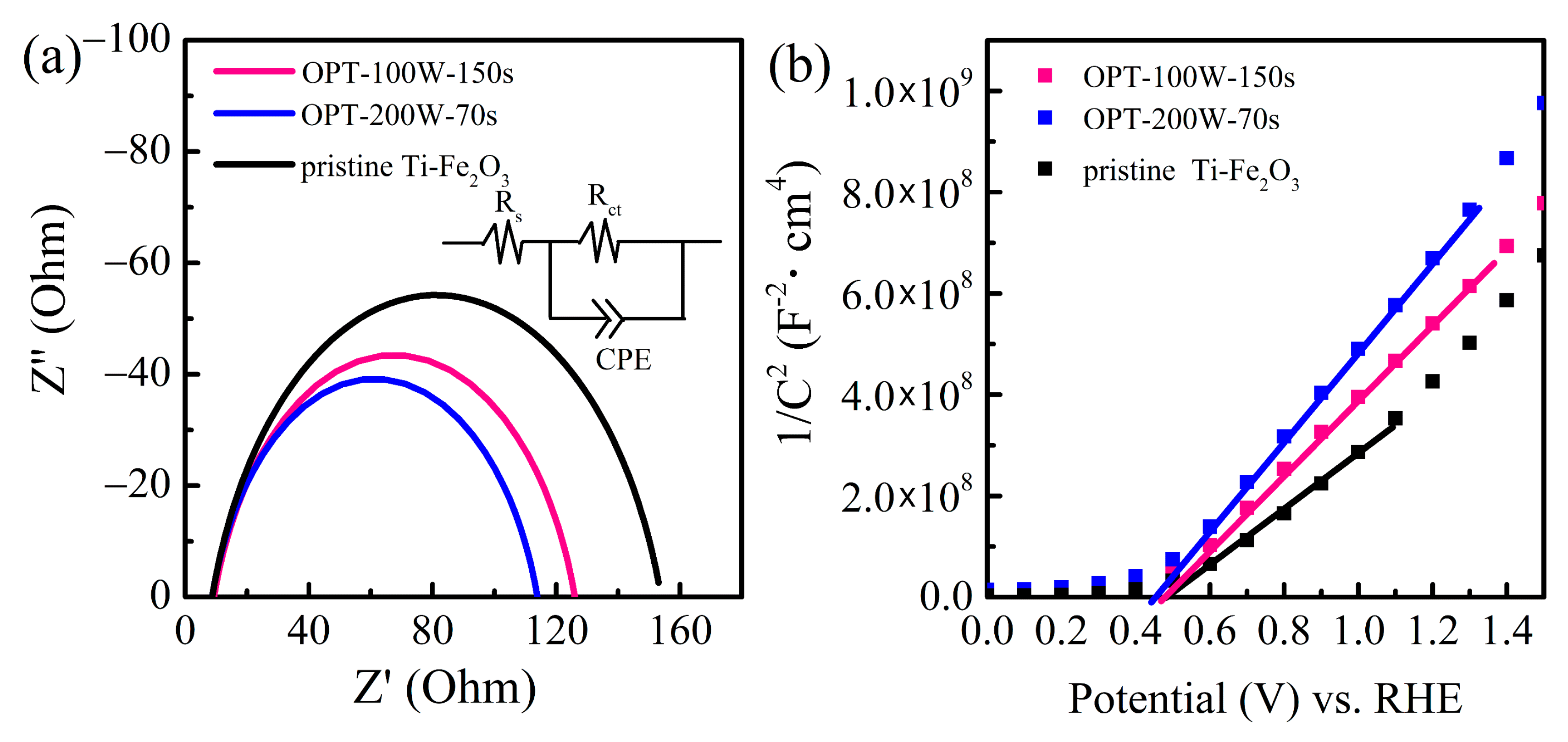
2.2. Effect of O2 Plasma on the PEC Performance of Ti-Fe2O3 Photoanodes

The conditions of O2 plasma treatment were first optimized. The linear scan voltammetry (LSV) curves of pristine Ti-Fe2O3 and OPT-Ti-Fe2O3 photoanodes were performed in 1 M NaOH (PH = 13.6) aqueous solution at a scan rate of 10 mVs−1, and the results are presented in Figure S1. The optimal films of OPT-100W-150s and OPT-200W-70s treated under different power of O2 plasma were both selected to investigate the power effect of O2 plasma treatment on the PEC performance of Ti-Fe2O3 photoanode. The J.-V. curves of pristine Ti-Fe2O3, OPT-200W-70s, and OPT-100W-150s are measured in the electrolyte of 1 M NaOH aqueous solution as shown in Figure 3a. It can be seen that the OPT-200W-70s photoanode showed the highest photocurrent density of 2.14 mA·cm−2 at 1.23 V vs. RHE, which was 1.95 times higher than 1.10 mA·cm−2 of the pristine Ti-Fe2O3. The onset potential of the OPT-200W-70s photoanode exhibited an approximate 50 mV cathodic shift compared with pristine Ti-Fe2O3, which lowered the applied potential to drive the PEC water oxidation reaction. It was also found that the photocurrent of OPT-200W-70s here showed greater superiority for water oxidation than many reported Ti-Fe2O3 nanowires as depicted in Table S1 [5,19,20,21]. This all suggests that O2 plasma treatment is an effective method for promoting the PEC water oxidation kinetics of Ti-Fe2O3 photoanodes.
In addition, the stability of the Ti-Fe2O3 photoanodes was investigated, and the I-t curves of pristine Ti-Fe2O3 and OPT-200W-70s were measured at the constant potential of 1.23 V vs. RHE as depicted in Figure S2. The photocurrent density of the pristine Ti-Fe2O3 photoanode was kept at 1.1 mAcm−2 at the duration of 2000 s at 1.23 V vs. RHE. The OPT-200W-70s showed the photocurrent density of 2 mAcm−2 at the moment of light on, and then the photocurrent rapidly dropped in the first 500 s. After that, the photocurrent dropped slowly and almost stabilized at 1.72 mAcm−2. Although the photocurrent dropped a little during the measurement process, it was still far higher than the pristine Ti-Fe2O3 photoanode at the same constant potential.
To investigate the effect of O2 plasma on the surface charge transfer of Ti-Fe2O3 photoanode, we further calculate the hole transfer efficiency from the I-V curves using H2O2 as hole scavenger. When adding H2O2 in the electrolyte, the photocurrent densities of pristine Ti-Fe2O3, OPT-100W-150s, and OPT-200W-70s tended to be almost the same, as shown in Figure 3b. It can be concluded that the carriers’ generation and bulk charge transport in the three electrodes show little difference, because almost 100% of the photogenerated holes are considered to be extracted from the electrodes in the presence of the hole scavenger H2O2 in the electrolyte. The practical charge transfer efficiencies ( η transfer) of these photoanodes are calculated using the ratio of JH2O/JH2O2, where the JH2O and JH2O2 denote the photocurrent density measured in 1 M NaOH aqueous solution without and with 0.5 M H2O2, respectively. The curves are depicted in Figure 3c [22,23,24]. It can be seen that OPT-200W-70s and OPT-100W-150s showed higher η transfer of 78% and 64% than the 47% of pristine Ti-Fe2O3 in the measured potential range, which were 1.66 and 1.36 times that of pristine Ti-Fe2O3. These were very close to the 1.95 and 1.5 times enhancements of photocurrents, correspondingly. The result reveals that O2 plasma treatment significantly increased the surface charge transfer efficiency of the photoanodes.
Electrochemical impedance spectra (EIS) of pristine and OPT-Ti-Fe2O3 photoanodes were also measured to further study the charge transport and transfer dynamics at a bias of 1.23 V vs. RHE under illumination conditions. The impedance spectra of the photoanodes are illustrated in Figure 4a, in which the impedance of Ti-Fe2O3 treated with plasma is smaller than the pristine one. The impedance of OPT-200W-70s was the smallest. By fitting the EIS data according to the equivalent circuit shown in the inset of Figure 4a, the data of the series resistance (Rs), charge transfer resistance (Rct), the bulk capacitance of hematite (Cbulk), and the capacitance of the space charge layer at the interface of Fe2O3/electrolyte (Csc) for the electrodes were obtained. The relevant data are listed in Table 1. Compared with the pristine Ti-Fe2O3 photoanode, the charge transfer resistance of OPT-200W-70s and OPT-100W-150s photoanodes both decreased, and OPT-200W-70s had the lowest charge transfer resistance. This further indicated that the O2 plasma treatment improved the surface charge transfer dynamics of the Ti-Fe2O3 photoanode. Moreover, the series resistances also decreased a little after O2 plasma treatment.
As the carrier density is an important factor for PEC performance, the Mott-Schottky measurement was taken to understand the effect of O2 plasma treatment on the carrier densities of Ti-Fe2O3 photoanodes. Mott-Schottky curves of the electrodes were all measured at 1 kHz in a dark condition as illustrated in Figure 4b. The effective carrier densities of the electrodes could be calculated according to the equation [25,26]:
1 / C 2 = 2 e ε ε 0 N d × ( V V FB )
where ε, ε0, e, Nd, V, VFB in the equation represented the permittivity of Fe2O3 (ε = 80) [27,28], the permittivity of vacuum (ε0 = 8.8542 × 10⁻¹² F/m), fundamental charge constant (e = 1.602 × 10⁻¹⁹ C), carrier density, applied potential, and flat band potential, respectively [25,29]. The flat band of pristine Ti-Fe2O3, OPT-100W-150s, and OPT-200W-70s are approximate 0.49, 0.48, 0.46 V vs. RHE, respectively. The carrier densities of the photoanodes can be calculated by the slopes of the Mott-Schottky curves. For pristine Ti-Fe2O3, the carrier density was calculated to be 3.16 × 1019 cm−3. The electrodes of OPT-100W-150s and OPT-200W-70s had lower carrier densities of 2.38 × 1019 cm−3 and 2.01 × 1019 cm−3, respectively. The decreased carrier density may be associated with the decreased donor-type states.

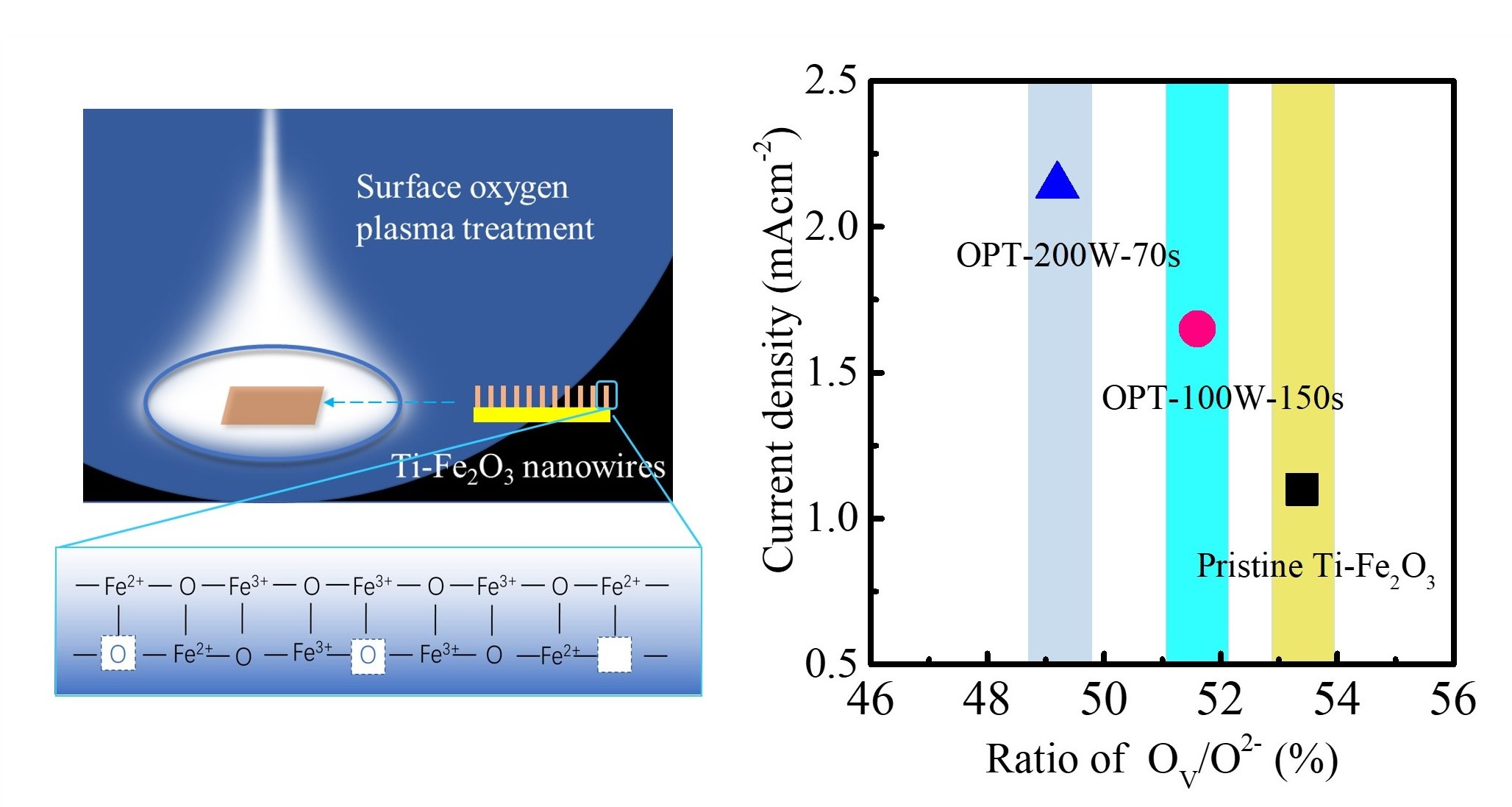
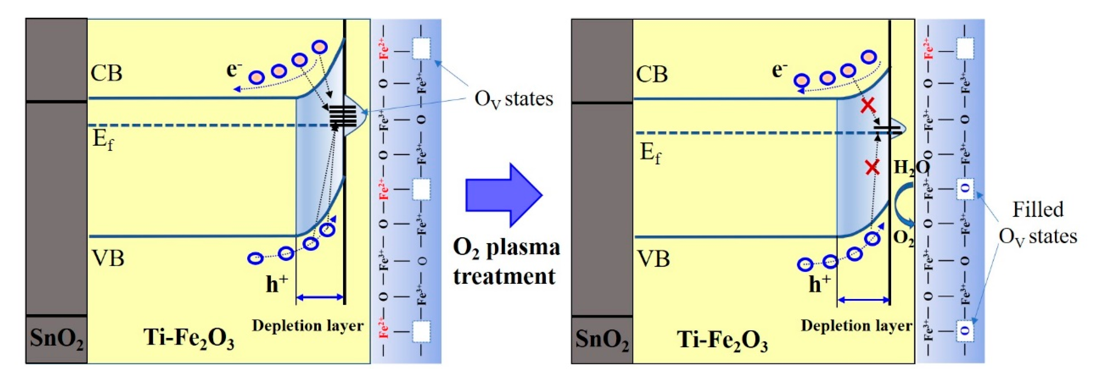
2.3. Mechanism of O2 Plasma Treatment on Promoting PEC Performance

To explore the impact of plasma on the improved PEC performance, we carried out XPS analysis on the films, because the plasma was a surface treatment strategy that could strongly affect the chemical environment near the surface of the electrode. The Fe 2p spectra of the electrodes are summarized in Figure 5a, in which the peaks appear around 724.78 eV and 711.3 eV, indicating a typical spectrum for Fe 2p1/2 and Fe 2p3/2 of Ti-doped Fe2O3, respectively [30,31]. For Fe 2p3/2 peaks, the peak at 711.3 eV and the shoulder at 710.3 eV were attributed to Fe(III)-O and Fe(II)-O, which correspond to Fe3+ and Fe2+ chemical states, respectively. To further clarify the effect of O2 plasma treatment, XPS difference spectra were calculated as shown in Figure S3. The negative peaks at about 710 eV in the difference spectra suggest the decreased intensity of Fe(II)-O peak after O2 plasma treatment. For OPT-200W-70s, the peak of Fe 2p1/2 and Fe 2p3/2 significantly shifted to the higher binding energy of 725 eV and 711.5 eV, respectively, as listed in Table 2. The Ti 2p XPS spectra of pristine Ti-Fe2O3 (Figure 5b) presented the peaks at the binding energy of 458.39 eV and 464.23 eV that were associated with Ti 2p3/2 and Ti 2p1/2, respectively [32]. After O2 plasma treatment, the peaks both gradually shifted to higher binding energy with the increase of the power, and the values are listed in Table 2. OPT-200W-70s exhibited a maximum shift of 0.36 eV for 2p3/2 and 0.3 eV for 2p1/2. The result suggests that the O2 plasma treatment has a great influence on the chemical state of the sample surface.
The deconvoluted peaks for O 1s are depicted in Figure 6. The observed main peak at 530.2 eV and the shoulder peak at approximate 531.8 eV were attributed to the lattice oxygen (denoted as O2-) and the defect sites with low oxygen coordination, i.e., oxygen vacancies on sample surface (denoted as Ov) [33,34,35,36]. The main peak at 530.2 eV also shifted toward higher binding energy after O2 plasma treatment, and the maximum shift was about 0.22 eV, as depicted in Table 1. Furthermore, the integrated area of Ov/O2- of the three films was calculated to understand the change in the oxygen-related surface states under different treatment conditions. For pristine Ti-Fe2O3, the ratio of Ov/O2- was calculated to be 53.4%. After treatment with O2 plasma, the ratios of OPT-100W-150s and OPT-200W-70s were decreased to 51.6% and 49.8%, respectively. The values are illustrated in Table 3. The results strongly suggest that O2 plasma treatment reduced the oxygen vacancies on the surface of Ti-Fe2O3 films. Thus, the electron density around Ti and Fe was decreased due to the increase in lattice oxygen, which resulted in the decreased shielding ability and the binding energy moving towards high energy. Combined with the previous discussion, it is believed that the enhanced PEC performance of Ti-Fe2O3 was attributed to the reduction of oxygen vacancies at the surface of electrode, rather than the variation in properties such as light absorption, morphology, and crystal structure.
Finally, let us discuss the effect of O2 plasma treatment on the enhanced PEC performance of Ti-Fe2O3 photoanodes. The Fe2O3 nanowire photoanode has a large number of oxygen vacancies on the surface, which will generate extra electrons to reduce Fe3+ to Fe2+ [37]. When the concentration of oxygen vacancy is too high, Fe2+ sites may cause the recombination of photogenerated holes [38], which decreases surface charge transfer properties, and therefore retards the kinetics of hole reaction [15,39]. Furthermore, the oxygen vacancy is difficult to remove as an intrinsic defect. However, O2 plasma has high reactivity to fill up the oxygen vacancies at the photoanode surface. Thus, the ratio of Ov/O2- is reduced when highly reactive oxygen atoms collide with the photoanodes’ surface, which in turn causes the reduction in donor density of the Ti-Fe2O3 photoanode. The filling of the oxygen vacancy also reduced the amount of Fe2+ sites, which was consistent with the decreased intensity of Fe(II)-O peaks in XPS spectra. Therefore, the surface recombination mediated by oxygen vacancies was lowered, and the charge transfer properties were enhanced, leading to a remarkably improved PEC performance, as illustrated in Scheme 1.
Furthermore, the PEC performances had little change under different processing time when the plasma treatment was set as 100 W. This might be because the kinetic energy of oxygen plasma is small, and only a small thickness of the surface layer was affected by the plasma. For the power of 200 W with a greater oxygen plasma kinetic energy, the treatment’s depth became deeper. More oxygen vacancies were remedied by extending the treatment time; hence, the PEC performance became better. However, when the time is further increased, the smaller carrier density will result in a smaller surface electric field. This will decrease the charge separation and deteriorate the PEC performance of the photoanodes.

3. Materials and Methods

3.1. Materials

Sodium sulphate (NaSO4, 99%), ferric chloride (FeCl3·6H2O, 99%), and titanium tetrachloride (TiCl4, 99.5%) were purchased from Aladdin Reagent Company (Shanghai, China). Hydrogen peroxide (H2O2, 30%), and sodium hydroxide (NaOH, 95%) were obtained from Beijing Chemical Reagent Company (Beijing, China). The fluorine-doped SnO2 (SnO2: F) transparent conducting glass (FTO, resistance: 17 Ω/eq) substrates were from Yingkou OPV Tech New Energy Co. Ltd. (Yingkou, China).

3.2. Preparation of Ti-Doped Fe2O3 Photoanodes

3.2.1. Preparation of Ti-Fe2O3 Nanowires Array on FTO Substrate

Ti-doped hematite nanowires array films were prepared on FTO conductive glass by the reported hydrothermal method [40]. FTO substrate (1.1 cm × 2.5 cm) was cleaned by the normal method [41]. A 20 mL mixed solution containing 1 M FeCl3·6H2O and 1 M Na2SO4 was prepared first, and then 0.5 mL TiCl4 ice water with a concentration of 40 mM was added. Then FTO substrate was put into a Teflon-lined stainless steel autoclave that was filled with 2.5 mL solution. The reaction was performed at a temperature of 120 °C for 4 h, and Ti-doped FeOOH nanowires array with yellow color were obtained. After rinsing with deionized water, the films were annealed by rapid thermal annealing at 800 °C for 30 s, and Ti-FeOOH films were transformed to Ti-Fe2O3 nanowires.

3.2.2. O2 Plasma Treated Ti-Doped Hematite Nanowires

The oxygen gas plasma treatment was performed using a PE-ALD (Jiaxing Kemin electronic equipment Technology Company, KMT-500s, Jiaxing, China) equipped with a radio frequency coil with adjustable power. The O2 plasma treatment (OPT) was performed with the power of 100 W for different times (90 s, 150 s, and 210 s), and 200 W for 50 s, 70 s, and 90 s at the temperature of 100 °C. O2 OPT-Ti-Fe2O3 film at 150 °C with the power of 150 W for 90 s was also performed for comparative study. The samples were hereafter denoted as pristine Ti-Fe2O3, OPT-100W-90s, OPT-100W-150s, OPT-100W-210s, OPT-200W-50s, OPT-200W-70s, OPT-200W-90s, OPT-150 °C-150W-90s. The characterization and the PEC measurement of the samples are presented in the supporting information.

4. Conclusions

The study demonstrated that the PEC performance of Ti-doped Fe2O3 nanowire photoanodes was remarkably improved after surface treatment with O2 plasma. O2 plasma treatment decreased the oxygen vacancies at the surface of Ti-Fe2O3 nanowire films by 6.7% (from 53.4% to 49.8%). The charge transfer of Ti-Fe2O3 nanowire photoanodes was improved by 1.66 times via reducing the losses caused by the oxygen vacancies’ related recombination. At 1.23 V vs. RHE, the photocurrent density of O2 OPT-Ti-Fe2O3 at the power of 200 W for 70 s reached 2.14 mAcm−2, which was 1.95 times that of the pristine Ti-Fe2O3. O2 plasma treatment is demonstrated to be an effective strategy to improve the charge transfer ability at the surface of Ti-doped Fe2O3 photoanode. We believe that further optimizations of the O2 plasma conditions should lead to even more efficient electrodes, and it can also be applied to tune the surface states and promote the PEC performance of other semiconductor photoelectrodes.

Supplementary Materials

The following are available online at https://www.mdpi.com/2073-4344/11/1/82/s1, Figure S1: Current-potential (J-V) curves of (a) pristine Ti-Fe2O3, OPT-100W-90s, OPT-100W-150s, and OPT-100W-210s photoanodes. (b) Current-potential (J-V) curves of pristine Ti-Fe2O3, OPT-200W-50s, OPT-200W-70s, and OPT-200W-90s photoanodes. The I-V curves are measured with the scan rate of 10 mVs−1 from 0.7 V to 1.6 V vs. RHE under irradiation; Figure S2: I-t curves of pristine Ti-Fe2O3 and OPT-200W-70s photoanodes; Table S1: The structure, fabrication method, and photocurrent density of some reported works on Ti-doped a-Fe2O3 nanowires photoanodes.

Author Contributions

C.L., experimental investigation, writing—original draft; D.W., writing—review and editing, visualization; J.G., investigation; Y.L., Methodology, Supervision; X.Z., validation, supervision, writing—review and editing. All authors have read and agreed to the published version of the manuscript.

Funding

This work was supported by the Natural Science Foundation of China (Grant nos. 91833303, 51872044 and 51972051), the Fund from Department of Science and Technology of Jilin Province (Grant no. 20180101175JC, and 20180520007JH), the Fund from Department of Education of Jilin Province (Grant no. JJKH20200178KJ), and the 111 project (Grant no. B13013).

Institutional Review Board Statement

Not applicable.

Informed Consent Statement

Not applicable.

Data Availability Statement

Data is contained within the article or supplementary material.

Conflicts of Interest

The authors declare no conflict of interest.

References

  1. Fujishima, A.; Honda, K. Electrochemical photolysis of water at a semiconductor electrode. Nature 1972, 238, 37–38. [Google Scholar] [CrossRef] [PubMed]
  2. Shen, S.-H.; Kronawitter, C.X.; Wheeler, D.A.; Guo, P.; Lindley, S.A.; Jiang, J.; Zhang, J.-Z.; Guo, L.; Mao, S.-S. Physical and photoelectrochemical characterization of Ti-doped hematite photoanodes prepared by solution growth. J. Mater.Chem. A 2013, 1, 14498–14506. [Google Scholar] [CrossRef]
  3. Franking, R.; Li, L.; Lukowski, M.A.; Meng, F.; Tan, Y.; Hamers, R.J.; Jin, S. Facile post-growth doping of nanostructured hematite photoanodes for enhanced photoelectrochemical water oxidation. Energy Environ. Sci. 2013, 6, 500–512. [Google Scholar] [CrossRef]
  4. Mazzaro, R.; Bibi, S.B.; Natali, M.; Bergamini, G.; Morandi, V.; Ceroni, P.; Vomiero, A. Hematite nanostructures: An old material for a new story. Simultaneous photoelectrochemical oxidation of benzylamine and hydrogen production through Ti doping. Nano Energy 2019, 61, 36–46. [Google Scholar] [CrossRef]
  5. Feng, F.; Li, C.; Jian, J.; Li, F.; Xu, Y.-X.; Wang, H.-Q.; Jia, L.-C. Gradient Ti-doping in hematite photoanodes for enhanced photoelectrochemical performance. J. Power Sour. 2020, 449, 227473. [Google Scholar] [CrossRef]
  6. Upul Wijayantha, K.G.; Saremi-Yarahmadi, S.; Peter, L.M. Kinetics of oxygen evolution at α-Fe2O3 photoanodes: A study by photoelectrochemical impedance spectroscopy. Phys. Chem. Chem. Phys. 2011, 13, 5264–5270. [Google Scholar] [CrossRef] [Green Version]
  7. Pendlebury, S.R.; Barroso, M.; Cowan, A.J.; Sivula, K.; Tang, J.W.; Grätzel, M.; Klug, D.; Durrant, J.R. Dynamics of photogenerated holes in nanocrystalline α-Fe2O3 electrodes for water oxidation probed by transient absorption spectroscopy. Chem. Commun. 2011, 47, 716–718. [Google Scholar] [CrossRef]
  8. Klahr, B.; Gimenez, S.; Fabregat-Santiago, F.; Hamann, T.; Bisquert, J. Water oxidation at hematite photoelectrodes: The role of surface states. J. Am. Chem. Soc. 2012, 134, 4294–4302. [Google Scholar] [CrossRef] [Green Version]
  9. Hamann, T.W. Splitting water with rust: Hematite photoelectrochemistry. Dalton Trans. 2012, 41, 7830–7834. [Google Scholar] [CrossRef]
  10. McDonald, K.J.; Choi, K.-S. Synthesis and photoelectrochemical properties of Fe2O3/ZnFe2O4 composite photoanodes for use in solar water oxidation. Chem. Mater. 2011, 23, 4863–4869. [Google Scholar] [CrossRef]
  11. Steier, L.; Herraiz-Cardona, I.; Gimenez, S.; Fabregat-Santiago, F.; Bisquert, J.; Tilley, S.D.; Grätzel, M. Understanding the role of underlayers and overlayers in thin film hematite photoanodes. Adv. Funct. Mater. 2014, 24, 7681–7688. [Google Scholar] [CrossRef]
  12. Zhong, D.-K.; Sun, J.-W.; Inumaru, H.; Gamelin, D.R. Solar water oxidation by composite catalyst/α-Fe2O3 photoanodes. J. Am. Chem. Soc. 2009, 131, 6086–6087. [Google Scholar] [CrossRef] [PubMed]
  13. Zhong, D.-K.; Gamelin, D.R. Photoelectrochemical water oxidation by cobalt catalyst (“Co–Pi”)/α-Fe2O3 composite photoanodes: Oxygen evolution and resolution of a kinetic bottleneck. J. Am. Chem. Soc. 2010, 132, 4202–4207. [Google Scholar] [CrossRef] [PubMed]
  14. Fan, Z.-W.; Xu, Z.; Yan, S.-C.; Zou, Z.-G. Tuning the ion permeability of an Al2O3 coating layer on Fe2O3 photoanodes for improved photoelectrochemical water oxidation. J. Mater. Chem. A 2017, 5, 8402–8407. [Google Scholar] [CrossRef]
  15. Hu, Z.-F.; Shen, Z.-R.; Yu, J.C. Covalent fixation of surface oxygen atoms on hematite photoanode for enhanced water oxidation. Chem. Mater. 2016, 28, 564–572. [Google Scholar] [CrossRef]
  16. Zhu, C.-Q.; Li, C.-L.; Zheng, M.-J.; Delaunay, J.J. Plasma-induced oxygen vacancies in ultrathin hematite nanoflakes promoting photoelectrochemical water oxidation. ACS Appl. Mater. Interfaces 2015, 7, 22355–22363. [Google Scholar] [CrossRef]
  17. Li, M.-Y.; Zhang, Z.-Y.; Lyu, F.-Y.; He, X.-J.; Liang, Z.-H.; Balogun, M.S.; Lu, X.-H.; Fang, P.-P.; Tong, Y.-X. Facile hydrothermal synthesis of three dimensional hematite nanostructures with enhanced water splitting performance. Electrochim. Acta 2015, 186, 95–100. [Google Scholar] [CrossRef]
  18. Kyesmen, P.I.; Nombona, N.; Diale, M. Modified annealing approach for preparing multi-layered hematite thin films for photoelectrochemical water splitting. Mater. Res. Bull. 2020, 131, 110964. [Google Scholar] [CrossRef]
  19. Wu, F.-K.; Xie, J.-L.; You, Y.-E.; Zhao, Z.-Y.; Wang, L.-L.; Chen, X.-Y.; Yang, P.-P.; Huang, Y.-L. Cobalt metal–organic framework ultrathin cocatalyst overlayer for improved photoelectrochemical activity of Ti-doped hematite. ACS Appl. Energy Mater. 2020, 3, 4867–4876. [Google Scholar] [CrossRef]
  20. Mo, R.; Liu, Q.; Li, H.-X.; Yang, S.; Zhong, J.-X. Photoelectrochemical water oxidation in α-Fe2O3 thin films enhanced by a controllable wet-chemical Ti-doping strategy and Co–Pi co-catalyst modification. J. Mater. Sci. Mater. El. 2019, 30, 21444–21453. [Google Scholar] [CrossRef]
  21. Cao, D.-P.; Zhang, J.-B.; Wang, A.-C.; Yu, X.-H.; Mi, B.-X. Fabrication of Cr-doped SrTiO3/Ti-doped α-Fe2O3 photoanodes with enhanced photoelectrochemical properties. J. Mater. Sci. Technol. 2020, 56, 189–195. [Google Scholar] [CrossRef]
  22. Wang, G.-M.; Yang, Y.; Ling, Y.-C.; Wang, H.-Y.; Lu, X.-H.; Pu, Y.-C.; Zhang, J.-Z.; Tong, Y.-X.; Li, Y. An electrochemical method to enhance the performance of metal oxides for photoelectrochemical water oxidation. J. Mater. Chem. A 2016, 4, 2849–2855. [Google Scholar] [CrossRef]
  23. Dotan, H.; Sivula, K.; Grätzel, M.; Rothschild, A.; Warren, S.C. Probing the photoelectrochemical properties of hematite (α-Fe2O3) electrodes using hydrogen peroxide as a hole scavenger. Energy Environ. Sci. 2011, 4, 958–964. [Google Scholar] [CrossRef]
  24. Klotz, D.; Grave, D.A.; Rothschild, A. Accurate determination of the charge transfer efficiency of photoanodes for solar water splitting. Phys. Chem. Chem. Phys. 2017, 19, 20383–20392. [Google Scholar] [CrossRef] [PubMed] [Green Version]
  25. Bohn, C.D.; Agrawal, A.K.; Walter, E.C.; Vaudin, M.D.; Herzing, A.A.; Haney, P.M.; Talin, A.A.; Szalai, V.A. Effect of tin doping on α-Fe2O3 photoanodes for water splitting. J. Phys. Chem. C 2012, 116, 15290–15296. [Google Scholar] [CrossRef]
  26. Orazem, M.E.; Tribollet, B. Electrochemical Impedance Spectroscopy; Wiley: New York, NY, USA, 2001; pp. 225–230. [Google Scholar]
  27. Wilhelm, S.M.; Yun, K.S.; Ballenger, L.W.; Hackerman, N. Semiconductor properties of iron oxide electrodes. J. Electrochem. Soc. 1979, 126, 419–424. [Google Scholar] [CrossRef]
  28. Turner, J.E.; Hendewerk, M.; Parmeter, J.; Neiman, D.; Somorjai, G.A. The characterization of doped iron oxide electrodes for the photodissociation of water: Stability, optical, and electronic properties. J. Electrochem. Soc. 1984, 131, 1777–1783. [Google Scholar] [CrossRef]
  29. Gao, S.; Wang, D.; Wang, Y.-L.; Li, C.; Liu, Y.-C.; Suzuki, N.; Terashima, C.; Fujishima, A.; Zhang, X.-T. Activating titanium dopants in hematite photoanode by rapid thermal annealing for enhancing photoelectrochemical water oxidation. Electrochim. Acta 2019, 318, 746–753. [Google Scholar] [CrossRef]
  30. Fujii, T.; de Groot, F.M.F.; Sawatzky, G.A.; Voogt, F.C.; Hibma, T.; Okada, K. In situ XPS analysis of various iron oxide films grown by NO2-assisted molecular-beam epitaxy. Phys. Rev. B Condens. Matter Mater. Phys. 1999, 59, 3195–3202. [Google Scholar] [CrossRef] [Green Version]
  31. Lu, J.; Jiao, X.; Chen, D.; Li, W. Solvothermal synthesis and characterization of Fe3O4 and γ-Fe2O3 nanoplates. J. Phys. Chem. C 2009, 113, 4012–4017. [Google Scholar] [CrossRef]
  32. Chae, S.Y.; Rahman, G.; Joo, O.S. Elucidation of the structural and charge separation properties of titanium-doped hematite films deposited by electrospray method for photoelectrochemical water oxidation. Electrochim. Acta 2019, 297, 784–793. [Google Scholar] [CrossRef]
  33. Kawabe, T.; Tabata, K.; Suzuki, E.; Yamaguchi, Y.; Nagasawa, Y. Electronic states of chemisorbed oxygen species and their mutually related studies on SnO2 thin film. J. Phys. Chem. B 2001, 105, 4239–4244. [Google Scholar] [CrossRef]
  34. Bao, J.; Zhang, X.; Fan, B.; Zhang, J.; Zhou, M.; Yang, W.; Hu, X.; Wang, H.; Pan, B.; Xie, Y. Ultrathin spinel-structured nanosheets rich in oxygen deficiencies for enhanced electrocatalytic water oxidation. Angew. Chem. Int. Ed. 2015, 54, 7399–7404. [Google Scholar] [CrossRef] [PubMed]
  35. Moir, J.; Soheilnia, N.; Liao, K.; O’Brien, P.; Tian, Y.; Burch, K.S.; Ozin, G.A. Activation of ultrathin films of hematite for photoelectrochemical water splitting via H2 treatment. ChemSusChem 2015, 8, 1557–1567. [Google Scholar] [CrossRef]
  36. Janowitz, C.; Scherer, V.; Mohamed, M.; Krapf, A.; Dwelk, H.; Manzke, R.; Galazka, Z.; Uecker, R.; Irmacher, K.; Fornari, R.; et al. Experimental electronic structure of In2O3 and Ga2O3. New J. Phys. 2011, 13, 085014. [Google Scholar] [CrossRef]
  37. Lee, J.; Han, S. Thermodynamics of native point defects in α-Fe2O3: An Ab initio study. Phys. Chem. Chem. Phys. 2013, 15, 18906–18914. [Google Scholar] [CrossRef]
  38. Rioult, M.; Stanescu, D.; Fonda, E.; Barbier, A.; Magnan, H. Oxygen vacancies engineering of iron oxides films for solar water splitting. J. Phys. Chem. C 2016, 120, 7482–7490. [Google Scholar] [CrossRef]
  39. Fu, Z.; Jiang, T.; Zhang, L.; Liu, B.; Wang, D.; Wang, L.; Xie, T. Surface treatment with Al3+ on a Ti-doped α-Fe2O3 nanorod array photoanode for efficient photoelectrochemical water splitting. J. Mater. Chem. A 2014, 2, 13705–13712. [Google Scholar] [CrossRef]
  40. Vayssieres, L.; Beermann, N.; Lindquist, S.E.; Hagfeldt, A. Controlled aqueous chemical growth of oriented three-dimensional crystalline nanorod arrays: Application to iron(III) oxides. Chem. Mater. 2001, 13, 233–235. [Google Scholar] [CrossRef]
  41. Li, C.; Wang, D.; Suzuki, N.; Terashima, C.; Liu, Y.-C.; Fujishima, A.; Zhang, X.-T. A coral-like hematite photoanode on a macroporous SnO2:Sb substrate for enhanced photoelectrochemical water oxidation. Electrochim. Acta 2020, 360, 137012. [Google Scholar] [CrossRef]
Figure 1. The top-view scanning electron microscopy (SEM) images of (a) pristine Ti-Fe2O3, (b) OPT-100W-150s, and (c) OPT-200W-70s photoanodes; cross-sectional images of (d) pristine Ti-Fe2O3, (e) OPT-100W-150s, and (f) OPT-200W-70s photoanodes.
Catalysts 11 00082 g001
Figure 2. (a) Raman, (b) X-ray diffraction (XRD) patterns, and (c) absorption spectra of pristine Ti-Fe2O3, OPT-100W-150s, and OPT-200W-70s photoanodes, respectively.
Catalysts 11 00082 g002
Figure 3. (a) The J-V curves of optimum OPT-Ti-Fe2O3 and pristine Ti-Fe2O3 photoanodes measured in the electrolyte of 1 M NaOH aqueous solution. (b) Linear sweep voltammetry (LSV) scans of OPT-100W-150s, OPT-200W-70s, and pristine Ti-Fe2O3 photoanodes under irradiation in 1 M NaOH aqueous solution with 0.5 M H2O2 from 0.4 V to 1.6 V vs. reversible hydrogen electrode (RHE) with a scan rate of 10 mV s−1. (c) Charge transfer efficiency of OPT-100W-150s, OPT-200W-70s, and pristine Ti-Fe2O3.
Catalysts 11 00082 g003
Figure 4. (a) Nyquist plots and (b) Mott-Schottky curves of pristine Ti-Fe2O3, OPT-100W-150s, and OPT-200W-70s electrodes. The inset is the equivalent circuit.
Catalysts 11 00082 g004
Figure 5. X-ray photoelectron spectroscopy analysis: (a) Fe 2p peaks and (b) Ti 2p peaks of OPT-100W-150s, OPT-200W-70s, and pristine Ti-Fe2O3 samples, respectively.
Catalysts 11 00082 g005
Figure 6. X-ray photoelectron spectroscopy of O 1s peaks of (a) pristine Ti-Fe2O3, (b) OPT-100W-150s, and (c) OPT-200W-70s samples.
Catalysts 11 00082 g006
Scheme 1. Schematic illustration of the effect of O2 plasma treatment on the surface oxygen vacancies of Ti-doped Fe2O3. (OV denotes the surface oxygen vacancies of Ti-doped Fe2O3).
Catalysts 11 00082 sch001
Table 1. The fitted equivalent circuit parameters from electrochemical impedance spectra (EIS) data of pristine Ti-Fe2O3, OPT-100W-150s, and OPT-200W-70s photoanodes.
Circuit ElementRs (Ω)Rct (Ω)Cbulk (F)Csc (F)
Pristine Ti-Fe2O39.87143.498.77 × 10−53.98 × 10⁻⁴
OPT−100W−150s9.66114.259.46 × 10−57.99 × 10⁻⁴
OPT−200W−70s9.41103.679.48 × 10−57.14 × 10⁻⁴
Table 2. The listed binding energy values of Fe, Ti, and O elements in X-ray photoelectron spectroscopy (XPS) spectra for the films of the pristine Ti-Fe2O3, OPT-100W-150s, and OPT-200W-70s.
SampleBinding Energy (eV)
Fe 2p1/2Fe 2p3/2Ti 2p1/2Ti 2p3/2O1s (Main Peak)
Pristine Ti-Fe2O3724.78711.3464.23458.39530.22
OPT-100W-150s724.78711.3464.53458.64530.31
OPT-200W-70s725711.5464.53458.75530.45
Table 3. The ratio of the integrated area of Ov/O2− in the XPS spectra for the pristine Ti-Fe2O3, OPT-100W-150s, and OPT-200W-70s samples.
SampleRatio of Ov/O2−
Pristine Ti-Fe2O353.4%
OPT-100W-150s51.6%
OPT-200W-70s49.8%
Publisher’s Note: MDPI stays neutral with regard to jurisdictional claims in published maps and institutional affiliations.

