Article

Prevention of Inflammation-Driven Colon Carcinogenesis in Human MUC1 Transgenic Mice by Vaccination with MUC1 DNA and Dendritic Cells

1
Laboratory of Cancer Biology and Molecular Immunology, Graduate School of Pharmaceutical Sciences, The University of Tokyo, 7-3-1 Hongo, Bunkyo-ku, Tokyo 113-0033, Japan
2
Faculty of Pharmacy, Universitas Gadjah Mada, Sekip Utara, Yogyakarta 55283, Indonesia
3
Intractable Disease Research Center, Graduate School of Medicine, Juntendo University, 2-1-1 Hongo, Bunkyo-ku, Tokyo 113-8421, Japan
4
Laboratory of Molecular Immunology, Institute for Quantitative Biosciences, The University of Tokyo, Yayoi, Bunkyo-ku, Tokyo 113-0032, Japan
5
Department of Immunology, Mayo Clinic Arizona, 13400 E. Shea Blvd., Scottsdale, AZ 85259, USA
6
Division of Glycobiologics, Graduate School of Medicine, Juntendo University, 2-1-1 Hongo, Bunkyo-ku, Tokyo 113-8421, Japan
*
Authors to whom correspondence should be addressed.
Cancers 2023, 15(6), 1920; https://doi.org/10.3390/cancers15061920
Submission received: 31 January 2023 / Revised: 9 March 2023 / Accepted: 20 March 2023 / Published: 22 March 2023
(This article belongs to the Special Issue Mucins and Cancers)

Abstract

:

Simple Summary

Colorectal cancer is one of the most common cancers worldwide and improved treatments are needed. The present research focused on the use of a DNA vaccine targeted at one of the commonly found tumor-associated antigens, human MUC1. MUC1 transgenic mice were vaccinated with MUC1 DNA mixed with bone-marrow-derived dendritic cells (BMDCs). Then, inflammation-driven colon cancer was induced by injection of a carcinogen, azoxymethane, and administration of dextran-sulfate-sodium-containing drinking water. This mouse model of colon cancer resembles the colon cancer found in humans. The number and size of colonic tumors were compared to mice vaccinated with MUC1 DNA alone or BMDCs alone. The combination of MUC1 DNA and BMDCs reduced the tumor burden more than in the controls, showing an additive preventive effect. These findings demonstrate that in this human MUC1 transgenic mouse model, MUC1-expressing colonic tumors can be partially prevented by a vaccine containing MUC1 DNA and BMDCs.

Abstract

The preventive efficacy of MUC1-specific DNA immunization on inflammation-driven colon carcinogenesis in human MUC1 transgenic (MUC1.Tg) mice was investigated. Mice were vaccinated with MUC1 DNA mixed with autologous bone-marrow-derived dendritic cells (BMDCs), and then colonic tumors were induced by azoxymethane (AOM) injection and oral administration of dextran sulfate sodium (DSS). Two types of tumors, squamous metaplasia and tubular adenoma, were observed. Both expressed high levels of MUC1 as indicated by the binding of anti-MUC1 antibodies with different specificities, whereas MUC1 expression was not detected in normal colonic mucosa. When mice were immunized with MUC1 DNA + BMDCs, tumor incidence, tumor number, and tumor size were significantly reduced. In contrast, vaccination with MUC1 DNA alone or BMDCs alone was ineffective in reducing tumor burden. Inflammation caused by DSS was not suppressed by the MUC1 DNA + BMDCs vaccination. Furthermore, MUC1 protein expression levels, as judged by anti-MUC1 antibody binding in tumors grown after vaccination, did not significantly differ from the control. In conclusion, an inflammation-driven carcinogenesis model was established in MUC1.Tg mice, closely resembling human colon carcinogenesis. In this model, vaccination with MUC1 DNA + BMDCs was effective in overriding MUC1 tolerance and reducing the tumor burden by a mechanism not affecting the level of colonic inflammation.

Graphical Abstract

1. Introduction

Colorectal cancer is one of the most common cancers worldwide, leading to significant morbidity and mortality [1]. Patients with ulcerative colitis and Crohn’s disease are at an increased risk for developing colorectal cancer. Although the genetic basis of colorectal cancer predisposition in these inflammatory bowel diseases has not been clearly elucidated, it is believed that chronic inflammation is one of the pathogenic sequences of colorectal carcinogenesis. The connection between inflammation and carcinogenesis is well established and has received a great body of supporting evidence from genetic, pharmacological, and epidemiological data [2].
Despite efforts to improve conventional therapy, the mortality of colorectal cancer is still high, particularly related to the recurrence of the disease and the occurrence of liver metastases. Novel approaches, such as cancer immunotherapy targeting tumor-associated antigens, which are expected to show fewer side-effects and to provide better prevention of metastasis and recurrence than conventional cancer therapy, represent an attractive therapeutic avenue.
There has been great interest in cancer vaccines, which have the potential for controlling metastatic disease, prolonging the time to recurrence, and ultimately serving as a preventive measure. DNA vaccines have several advantages over other vaccine technologies [3]. DNA vaccines have been reported to be beneficial for maintaining high levels of tumor antigen expression at the vaccination site and for eliciting strong antitumor immunity in hosts [4,5,6] Several studies in experimental mouse models showed that tumor growth was suppressed successfully by vaccination with DNA encoding tumor antigens [7,8,9,10,11,12]. In light of the recent progress regarding the use of mRNA and DNA vaccination technology to stem the COVID-19 pandemic [13,14] and to provide new treatment possibilities for cancer [15], genetic vaccines may soon become an accepted therapeutic modality.
In the present study, we focused on MUC1 as a therapeutic target. MUC1 is a tumor-associated transmembrane antigen expressed at low levels on the apical surface of ductal epithelial cells in several organs, and overexpressed in the majority of adenocarcinomas and their precursor lesion, where its tandem repeat region is reported to have truncated O-glycans [16,17,18,19,20]. As a result, truncated novel glycopeptide epitopes are processed and presented to the immune system [10,21,22,23]. MUC1 localization in tumors has not only been reported to be restricted to the apical surface, but has also been found all around the cell surface and in the cytoplasm. MUC1’s highly increased expression and altered glycosylation in tumors provide a source of predictable disease-associated neo-epitopes that could exert an important influence on the innate immune system and represent targets of adaptive immunity [24,25].
Tumor-associated MUC1 has long been considered as a potential target molecule that might be able to elicit an immune response that then provides protection against MUC1-expressing tumor cells. Various strategies targeting the MUC1 tumor antigen to prevent, stabilize, or eradicate cancerous lesions have been pursued [26,27]. MUC1 peptides [28], dendritic cells (DCs) transfected with MUC1 cDNA [29,30], MUC1-mannan and MUC1-mannose fusion proteins [31,32], and MUC1 plasmid DNA [33,34] have all been reported to have the ability to suppress the growth of MUC1-expressing tumors in wild-type mice. In addition, studies in human MUC1-transgenic (MUC1.Tg) mice have shown that vaccination with MUC1 peptides [35,36] and MUC1 DNA [37,38] can effectively break the tolerance to injected MUC1-expressing tumor cells. In humans, injection of oxidized mannan-MUC1 after breast cancer surgery successfully prevented tumor recurrence for up to 15 years [39,40].
We have previously shown that a MUC1 DNA vaccine can prevent colonic tumors and lung metastasis in C57BL/6 mice [41,42]. In the present study, we performed experiments using MUC1.Tg mice, which express the full-length human MUC1 under its own promoter, thus having a similar MUC1 expression level and pattern as seen in humans [43]. In contrast to previous studies that injected MUC1-expressing tumor cells to induce cancer, here, we optimized the azoxymethane-dextran sulfate sodium (AOM-DSS) model [44,45,46] in MUC1.Tg mice to induce colorectal carcinogenesis. By using this model, we showed the development of MUC1-expressing colorectal tumors in an environment and immune system that recognizes MUC1 as a self (de novo) antigen and closely mimics the colitis-associated colorectal carcinogenesis conditions as seen in humans. Further, we sought to investigate if the anti-tumor immunity can be improved by addition of dendritic cells (DCs). DCs have been reported as professional antigen-presenting cells that play a critical role in the induction and regulation of immune responses [47]. It has been proposed that the manipulation of DCs as a “natural” vaccine adjuvant may prove to be a particularly effective way to stimulate antitumor immunity [48,49].
Here, we demonstrated the preventive efficacy of the MUC1 DNA vaccine when combined with purified autologous bone-marrow-derived DCs (BMDCs) against a colitis-associated colorectal tumor in MUC1.Tg mice. Administration of the MUC1 DNA vaccine alone or BMDCs alone was ineffective. These findings highlight the importance of combining the DNA vaccine with dendritic cells to achieve immune protection against tumor formation.

2. Materials and Methods

2.1. Animals

MUC1.Tg mice with a C57BL/6 background [43] were maintained in The University of Tokyo, Graduate School of Pharmaceutical Sciences’ animal facility under specific-pathogen-free conditions. MUC1-positive transgenic mice were confirmed by genotyping PCR as previously described [43]. Male MUC1.Tg mice aged 6 to 8 weeks were used. All experiments were approved by the Bioscience Committee of the Graduate School of Pharmaceutical Sciences of The University of Tokyo (approval code: PH30-4) and performed according to the guidelines of the Bioscience Committee of The University of Tokyo.

2.2. Antibodies

MUC1-specific Abs were biotin-conjugated chicken anti-MUC1 cytoplasmic tail (CTP) polyclonal antibody (pAb) kindly provided by Dr. Veer P. Bhavanandan (retired from the Pennsylvania State University, Old Main, State College, PA, USA) and biotin-conjugated monoclonal antibody (mAb) MY1E.12, biotin-conjugated mAb HMFG1, as well as biotin-conjugated mAb HMFG2 kindly provided by Dr. Joyce Taylor-Papadimitriou (Guys Hospital, London, UK). Biotin-conjugated IgG2a (BioLegend, Inc., San Diego, CA, USA) and biotin-conjugated IgY (Santa Cruz Biotechnology, Santa Cruz, CA, USA) were used as isotype controls.

2.3. Construction of MUC1 cDNA

Full-length human MUC1 cDNA containing 22 tandem repeats was provided originally in the pDKOF vector by Dr. Olivera J. Finn (University of Pittsburgh, Pittsburgh, PA, USA) [50]. From the pDKOF vector, the cDNA was cut out and re-cloned into the HindIII site of the pcDNA3.1 vector (Invitrogen; Thermo Fisher Scientific, Waltham, MA, USA), which includes the CMV promoter (pcDNA 3.1.-MUC1). A plasmid without MUC1 cDNA (pcDNA3.1) was used as the control. MUC1 cDNA in the pcDNA3.1 vector (pcDNA-MUC1) and pcDNA3.1 plasmid alone were amplified in the Escherichia coli strain DH5 and purified using Qiagen Mega-Plasmid columns (Qiagen, Inc., Germantown, MD, USA) according to the manufacturer’s protocol. This purification kit ensures a residual endotoxin level of 9.3 EU/µg DNA. Purification of DNA plasmid was confirmed by gel electrophoresis. The DNA was dissolved in Hanks’ Balanced Salt Solution (HBSS) at a concentration of 4 mg/mL and stored at −20 °C.

2.4. Preparation of BMDCs

BMDCs were prepared from the femur and tibia of MUC1.Tg mice as previously described [51] with the following modifications. Cells (1 × 10⁶ cells/mL) were cultured in RPMI1640 medium supplemented with 10% FCS, penicillin and streptomycin (1000 IU/mL and 10 mg/mL, respectively), 2 mM glutamine, and 0.05 mM 2-mercaptoethanol in the presence of 1000 U/mL of recombinant murine GM-CSF. On day 6, CD11c+ cells were purified using anti-CD11c MicroBeads and AutoMACS (Miltenyi Biotec, Bergisch Gladbach, Germany) and re-plated into fresh 24-well plates. On day 7, non-adherent cells were collected and suspended in 50 µL of HBSS. Cell viability as checked by the trypan blue exclusion test was routinely higher than 90%. The maturation level was checked by staining with anti-CD11c and anti-MHCII antibodies and flowcytometric analysis. Cells consisted of MHCIIlow-intermediate (50%) and MHCIIhi (20–30%) CD11c+ BMDCs. These cells were used for vaccination.

2.5. Vaccination

Mice were randomly allocated to the treatment groups and received three vaccinations at weekly intervals in the footpad with 100 μg of MUC1 DNA dissolved in 50 μL of HBSS mixed with or without 5 × 10⁵ BMDCs per mouse, per vaccination.

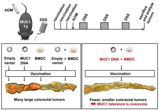
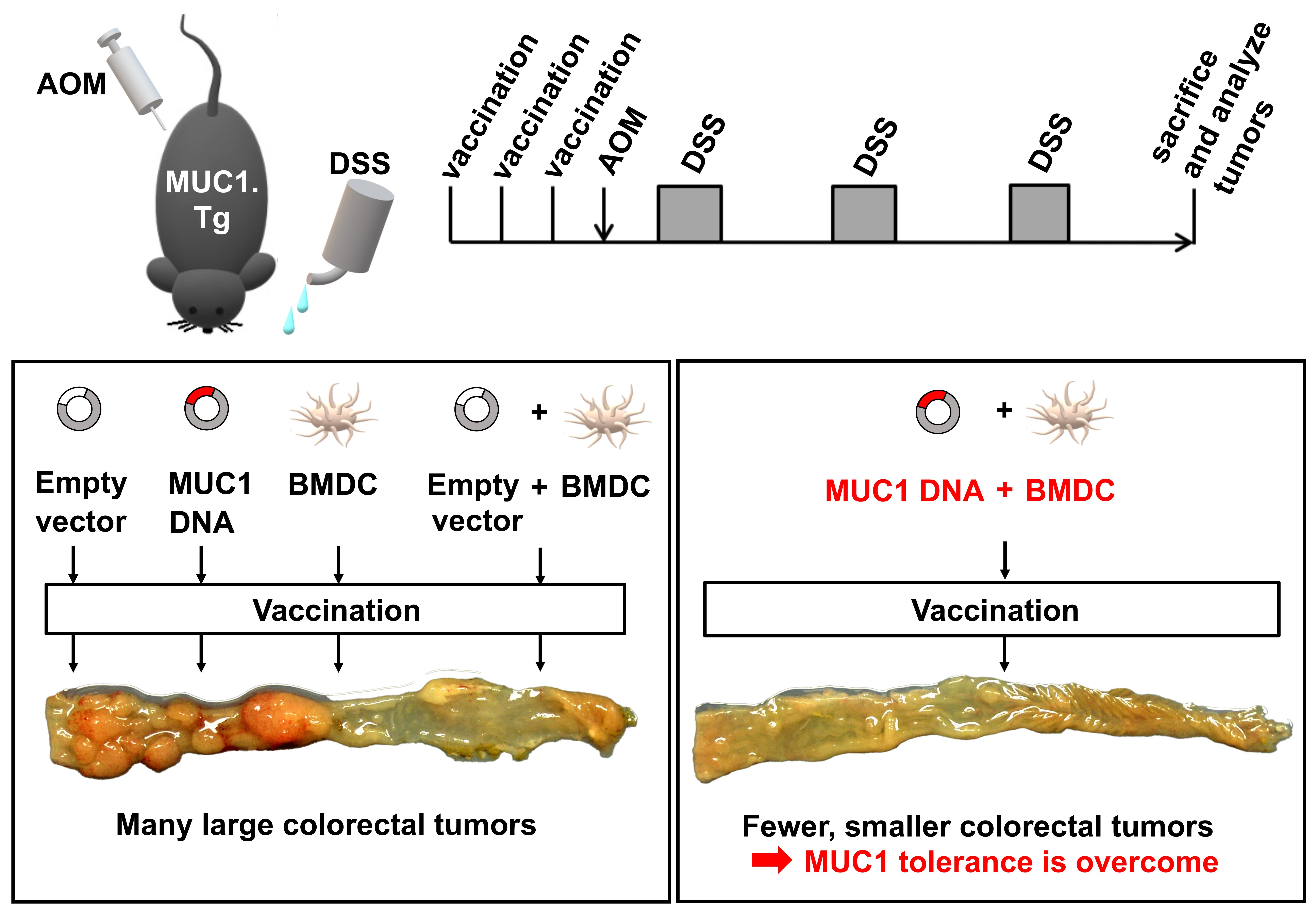
2.6. Experimental Colitis-Associated Colorectal Carcinogenesis

A single dose of 10 mg/kg body weight AOM (Sigma-Aldrich; Merck, Darmstadt, Germany), dissolved in sterile saline before use, was injected intraperitoneally (i.p.) 7 days after the last vaccination. Administration of three cycles of 1% (w/v) DSS (MP Biomedicals, LLC, Aurora, OH, USA) in drinking water was started 7 days after AOM injection for 4 days in the first cycle, and 5 days in the second and third cycle, followed by regular drinking water in between. Mice who died during the DSS treatment were not included into the data analysis. Mice were sacrificed 2 weeks after the last DSS exposure. The experimental protocol is schematically shown in Figure 1A. Colon samples were embedded in Tissue Tek O.C.T. Compound (Sakura Finetek, Tokyo, Japan).

2.7. Gross and Histopathological Examination

General observation was conducted to assess clinical symptoms and mortality during vaccinations and AOM-DSS treatment. Tumor size was measured by measuring the length and width of each tumor with a ruler. Tumor numbers were counted under a microscope. Histopathological alterations in the colon were examined using H&E-stained (Sakura Finetek) 6 μm thick serial cryosections of O.C.T. Compound-embedded colon samples. Colonic tumors were classified according to Boivin [52] and Reeve [53].

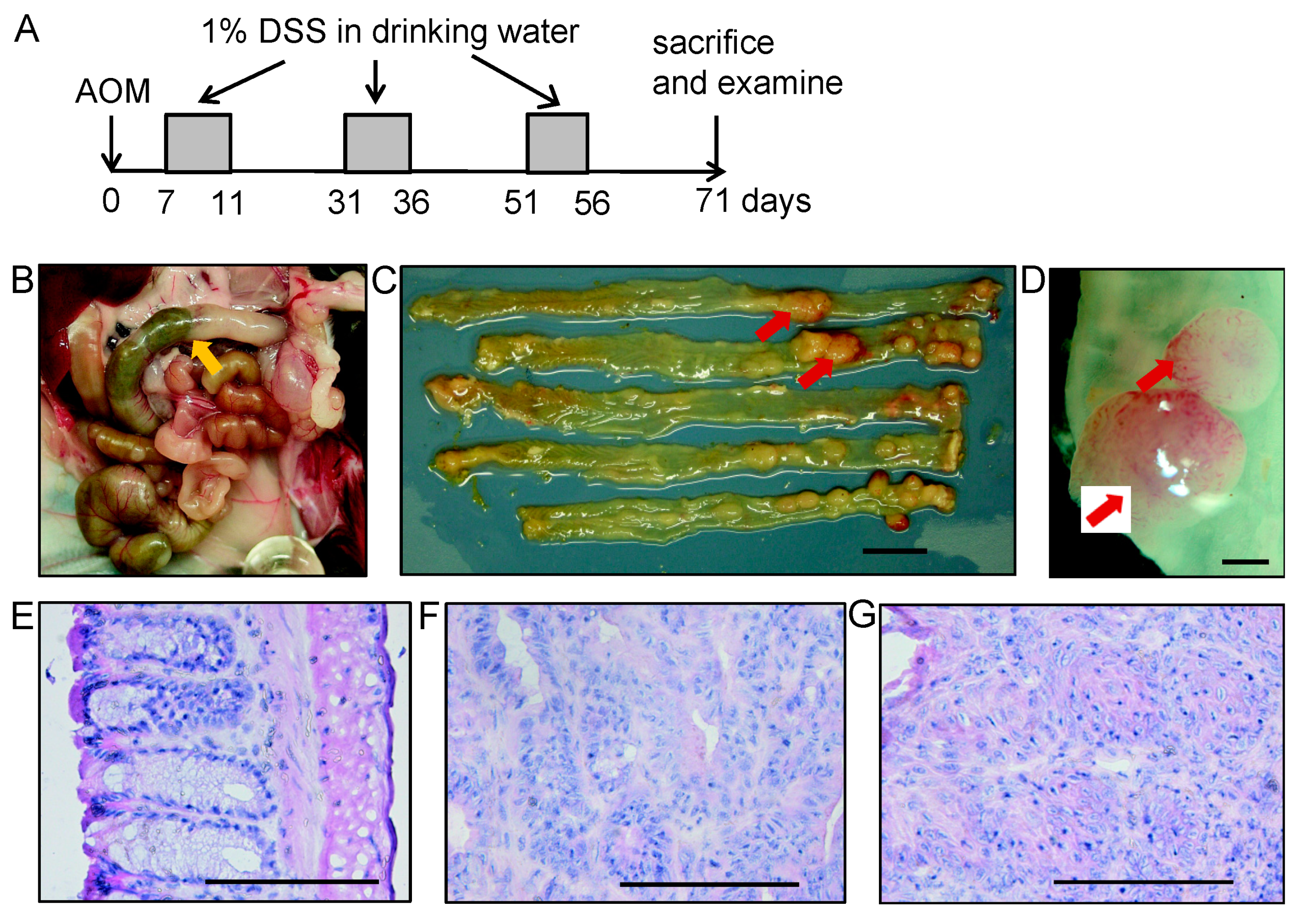
2.8. Immunohistochemical Analysis

For MUC1 detection, 6 μm thick cryostat sections of normal colon as well as colonic tissue with tubular adenoma or squamous metaplasia were used. Sections were pretreated for 30 min with 0.3% hydrogen peroxide at room temperature to eliminate endogenous peroxidase and washed three times with Dulbecco’s PBS. All endogenous biotin, biotin receptors, or avidin binding sites were blocked using the VECTASTAIN ABC HRP Kit (Vector Laboratories, Burlingame, CA, USA), according to the manufacturer’s protocol. The slides were incubated overnight at 4 °C with biotin-conjugated primary Abs or isotype control Abs at 5 μg/mL diluted in 3% BSA in PBS. Subsequently, Streptavidin Horseradish Peroxidase Conjugate (Invitrogen) was used at 1/200 dilution. Sections were counterstained with H&E. Immunoreactivity against MUC1 Ab was assessed under a microscope (Leica Microsystems, Wetzlar, Germany). To measure MUC1-glycoform expression in tumors, semi-quantitative analysis of immunohistochemically stained sections was performed by roughly estimating the percentage of MUC1-positive cells as compared to all epithelial cells.

2.9. Effect of MUC1 DNA Vaccination on Azoxymethane-Dextran Sulfate Sodium (AOM-DSS)-Induced Colorectal Inflammation

Seven days after the last vaccination, 10 mg/kg body weight of AOM diluted in sterile saline was injected i.p. Administration of 1% (w/v) DSS diluted in drinking water was started on day 7 after AOM injection for 1 cycle of 5 days. Mice were sacrificed on day 5 of DSS administration, and the body weight, hemoccult and/or presence of blood, and histological features were analyzed. The experimental setup is depicted in Figure 4A. Body weight change was determined by measuring the body weight on Day 1 and Day 5 of DSS treatment, the time period that had shown the largest body weight loss in a preliminary experiment. Bleeding scores were determined as 0 = no bleeding; 1 = positive Guaiac occult blood test (minimal color change to green); 2 = positive Guaiac occult blood test (maximal color change to blue); 3 = blood visibly present in the stool with no clotting on the anus; 4 = gross bleeding from the anus with clotting present. Inflammation with or without mucosal ulcers in the large bowel was scored on H&E-stained sections. For scoring, large intestinal inflammation was graded according to the following morphological criteria: grade 0 = normal appearance; grade 1 = shortening and loss of the basal 1/3 of the actual crypts with mild inflammation in the mucosa; grade 2 = loss of the basal 2/3 of the crypts with moderate inflammation in the mucosa; grade 3 = loss of entire crypts with severe inflammation in the mucosa and submucosa, but retention of the surface epithelium; grade 4 = presence of mucosal ulcers with severe inflammation (infiltration of neutrophils, lymphocytes, and plasma cells) in the mucosa, submucosa, muscularis propria, and/or subserosa. Two sections of the colon (one colorectal and one anal) were assessed and scored for each mouse and the inflammatory score was expressed as the mean of these scores.

2.10. Statistical Analysis

To analyze differences in tumor numbers and MUC1-expressing cells, the Steel–Dwass test was performed using R [54]. To analyze differences in AOM-DSS-induced intestinal inflammation, the non-parametric Mann–Whitney U test was performed using GraphPad Prism version 6.00 [55]. Results were considered significant at p < 0.05.

3. Results

3.1. MUC1 Expression in Tumors Developed by Colitis-Associated Colorectal Carcinogenesis in MUC1.Tg Mice

The experimental protocol is schematically shown in Figure 1A. AOM-DSS treatment induced the growth of tumor polyps mainly in the distal part of the colon as shown in Figure 1B–D. Figure 1E shows a normal part of the colon.
Figure 1. Gross pathology and histopathology of colitis-associated colorectal tumor induced by azoxymethane-dextran sulfate sodium (AOM-DSS) in MUC1.Tg mice. (A) Experimental protocol. Azoxymethane (AOM) was injected at 10 mg/kg body weight in the intraperitoneal site, followed by periodical administration of 1% (w/v) dextran sulfate sodium (DSS) in drinking water. Mice were sacrificed two weeks after the last DSS administration. (B) Colon position in the mouse abdomen as indicated by the yellow arrow. (C,D) Tumors developed as pedunculated nodules observed in the distal part of the colon as indicated by red arrows. (C) Gross pathological appearance of colorectal tumors showed pale to reddish polypoid nodules in the colorectal site. Histopathological analysis was performed on H&E-stained sections. Representative histopathological images of (E) normal colon, (F) tubular adenoma, and (G) squamous metaplasia of AOM-DSS-treated MUC1.Tg mice. Images in (EG) were captured at ×200 magnification. Scale bars show (C) 10 mm, (D) 1 mm, and (EG) 100 μm.
Cancers 15 01920 g001
Some tumors were tubular adenomas with various grades of dysplasia (Figure 1F). Another type of tumor observed was squamous metaplasia with atypia, which was characterized by a stratified squamous epithelium that developed in the colorectal area (Figure 1G).
MUC1 expression has been reported in human colon adenoma and adenocarcinoma, but not in normal colonic epithelia [16,56]. We hypothesized that AOM-DSS treatment could induce MUC1-expressing colorectal tumor formation in MUC1.Tg mice. Therefore, we performed immunohistochemical analysis in non-vaccinated MUC1.Tg mice to confirm MUC1 expression in AOM-DSS-induced colorectal tumors by using anti-MUC1 antibodies (Abs) with different specificities. Anti-MUC1 CTP Ab recognizes the MUC1 cytoplasmic tail regardless of glycosylation status [57]. Monoclonal Ab MY.1E12 recognizes sialyl-T MUC1 [58,59]. The binding of HMFG1 mAb to the PDTR epitope in MUC1 is partially influenced by certain nearby glycans [59], while the binding of HMFG2 mAb to MUC1 is glycosylation-independent [59]. The results showed that various MUC1 glycoforms were expressed in AOM-DSS induced tumors (Figure 2A). MUC1 was expressed mostly along the apical membrane of adenomatous cells (Figure 2A,f–h). However, in squamous metaplasia, MUC1 was highly expressed, not only in the apical part of tumor cells but also on the whole cell surface and in the cytoplasm (Figure 2A,k–m). The staining intensity of all anti-MUC1 Abs was stronger in squamous metaplasia tumors than in colorectal adenoma tumors (Figure 2A–E).

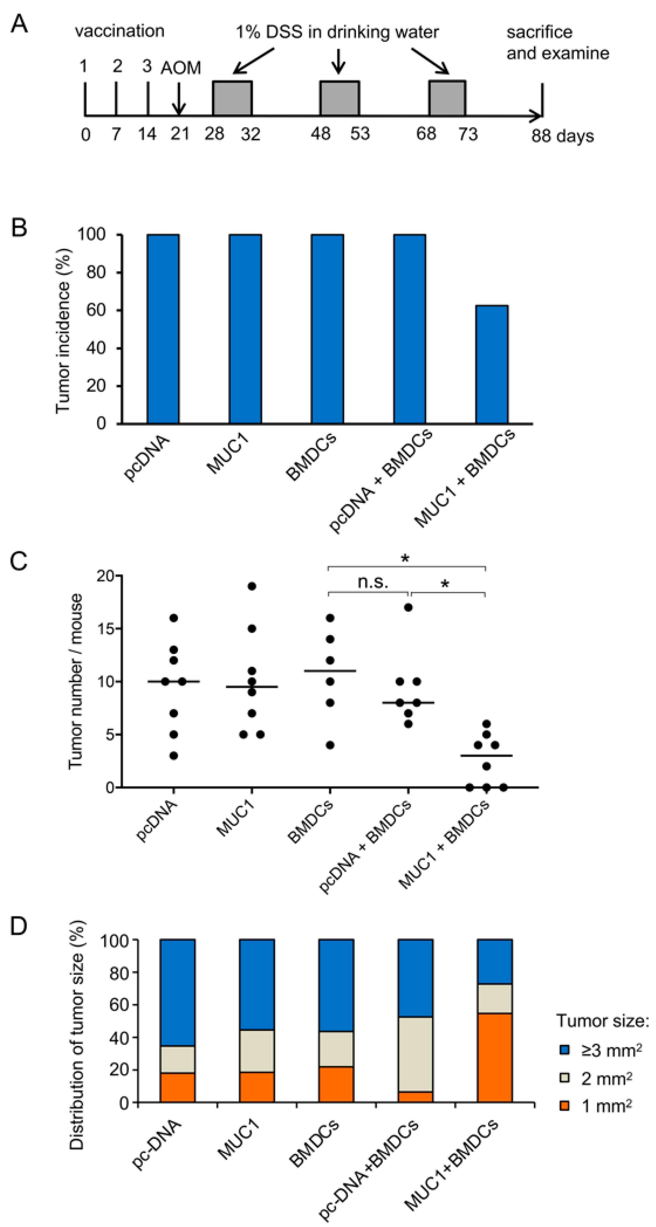
3.2. Preventive Efficacy of MUC1 DNA Vaccine on Experimental Colitis-Associated Colorectal Carcinogenesis

To evaluate the efficacy of MUC1-targeted immunotherapy, we examined the preventive effect of the MUC1 DNA vaccine in an experimental colitis-associated colorectal carcinogenesis model. MUC1.Tg mice were divided into five groups (n = 10 per group) and vaccinated with the following: (1) mock plasmid (pcDNA), (2) MUC1 plasmid (MUC1), (3) BMDCs (BMDCs), (4) pcDNA mixed with BMDCs (pcDNA + BMDCs), and (5) MUC1 + BMDCs, each followed by AOM-DSS treatment. The experimental protocol is schematically shown in Figure 3A.
Vaccinated mice were observed for 2 weeks after the last DSS treatment. Regarding the general observation, no differences in clinical symptoms, body weight, or length of the colon were observed between groups. Severe bleeding was observed during or after DSS administration in all groups. To judge whether the vaccination inhibits tumor formation, tumor multiplicity was evaluated in each treatment group. The results showed that although colorectal tumor formation could not be prevented completely, vaccination with MUC1 + BMDCs reduced the tumor incidence by 37.5% compared to the control (Figure 3B). The tumor multiplicity in MUC1.Tg mice vaccinated with MUC1 + BMDCs was significantly lower compared to mice vaccinated with BMDCs (p < 0.05) and mice vaccinated with pcDNA + BMDCs (p < 0.05) (Figure 3C). In addition, the tumor size distribution showed that in MUC1.Tg mice vaccinated with MUC1 + BMDCs, the frequency of smaller tumors (1 mm2) was highest, while that of larger tumors (ranging from 3 mm2 to 54 mm2) was lowest compared to all other groups (Figure 3D). These results strongly indicate that combining MUC1 DNA and BMDCs is critical to inducing an effective prevention of tumor formation in the colitis-associated colorectal carcinogenesis model.

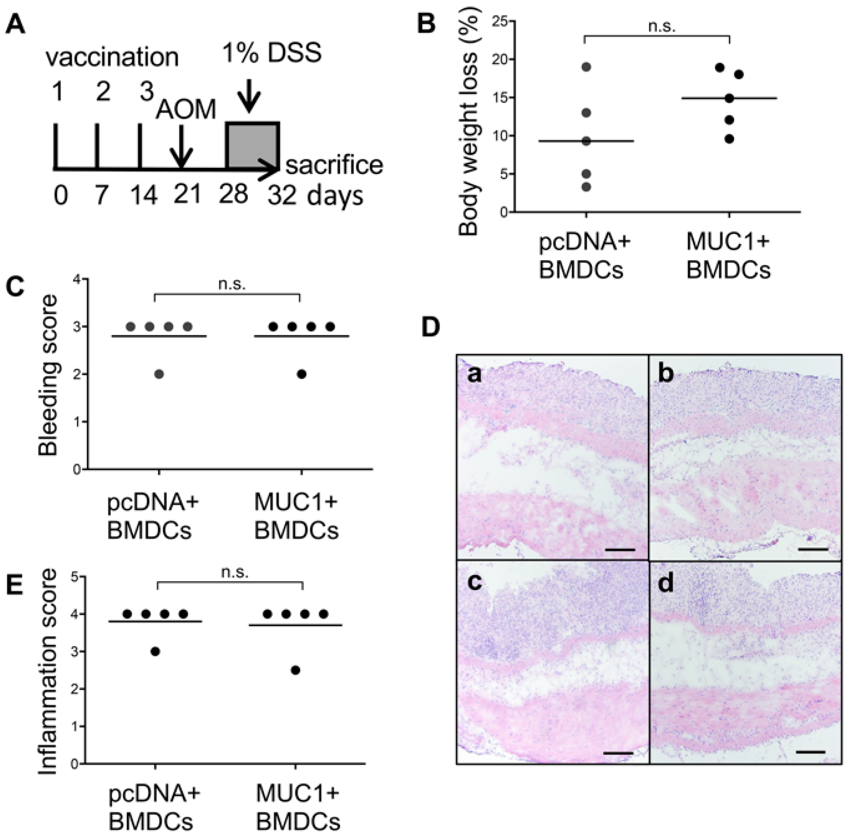
3.3. Effect of MUC1 DNA Vaccination on AOM-DSS Induced Colorectal Inflammation

In the present study, we used a carcinogenesis model in which inflammation is crucial to promoting tumorigenesis. Hence, we evaluated the effect of vaccination on inflammation induced by AOM-DSS administration (Figure 4A). The results showed that there were no significant differences regarding body weight loss (Figure 4B) and bleeding scores (Figure 4C). Colorectal and anal regions of both groups of mice showed a loss of entire crypts with severe inflammation in the mucosa and submucosa (Figure 4D). Inflammation scores between mice vaccinated with pcDNA + BMDCs and mice vaccinated with MUC1 + BMDCs were not different (Figure 4E). These results clearly show that there was no significant difference in the tissue damage due to DSS administrations between mice vaccinated with pcDNA + BMDCs or MUC1 + BMDCs.

4. Discussion

This study was designed to evaluate the efficacy of MUC1 DNA vaccination in the prevention of colitis-associated colorectal carcinogenesis. Our results strongly suggest that the MUC1 vaccine is effective in preventing, although not completely, colitis-associated colorectal tumor formation when combined with purified CD11c+ BMDCs. This study is the first to report the efficacy of a combined MUC1 + BMDC vaccine in the prevention of spontaneous, carcinogen-induced colorectal carcinogenesis in MUC1.Tg mice.
While several previous studies have examined the efficacy of MUC1 DNA vaccination in cancer, these studies were mostly conducted in wild-type mice and/or administered exogenous tumor cells. The significance of the present study lies in (1) the use of MUC1.Tg mice who express de novo MUC1 as a self-antigen and have been reported to be tolerant to MUC1 [43], (2) the use of an inflammation-induced colon cancer model that closely resembles the colon carcinogenesis process seen in humans, and (3) the use of DCs to enhance the anti-tumor immune response. Previously, we have shown that the growth of administered MUC1-expressing tumor cells in MUC1.Tg mice is enhanced, likely due to the presence of MUC1-specific regulatory T cells [60]. The results of the present study show that by the combination of MUC1 DNA and BMDCs, an immune response could be elicited to eliminate MUC1-expressing tumor cells at an early stage of carcinogenesis, providing evidence that MUC1 tolerance can be overcome in this clinically relevant colon cancer model.
The MUC1.Tg AOM-DSS carcinogenesis model provides high reproducibility and a simple mode of application and resembles human colorectal tumors in pathological and molecular aspects. Therefore, this model provides new opportunities to develop MUC1-targeted cancer immunotherapy for humans. MUC1 has been reported to be highly expressed in human colorectal adenoma and adenocarcinoma, particularly at the advanced stage [16]. Therefore, this vaccine might also be effective in the prevention of colorectal cancer recurrence.
In the present study, we showed that vaccination with MUC1 + BMDCs significantly reduced tumor incidence and tumor burden by inhibiting colitis-associated colorectal tumor formation. In addition, this combined vaccine might also inhibit colorectal tumor growth, represented by the smaller tumor size in this group. In contrast, vaccination using MUC1 or BMDCs alone was ineffective. Vaccination with DNA-encoding tumor antigens has been reported to be beneficial for maintaining high levels of tumor antigen expression at the vaccination site and for inducing strong antitumor immunity [61,62]. Regarding MUC1, this hypothesis has been supported by preclinical studies in mice. For instance, Kontani et al. reported that injecting MUC1 DNA and non-primed DCs at the same site, in contrast to injecting at different sites, enhanced MUC1-specific antitumor immunity in wild-type C57BL/6 mice [63]. Their results showed that inoculated DCs were able to process and express MUC1 DNA-encoding antigens at the inoculation site.
Our findings showed that, although this vaccine strategy successfully reduced tumor formation, complete inhibition was not achieved. It has been reported that tumors can escape from the immune system by reducing their tumor-associated antigen [64]. Although the glycoform of MUC1 to which the strong immune response seen in the present study was elicited is not known, the tumors in MUC1.Tg mice, which became a target of the immune response, appeared to express a variety of glycoforms. Further, immunohistochemical analysis of DSS-induced tumors using MY.1E12 mAb revealed that the types of tumors observed showed expression of MUC1 irrespective of whether the mice had been vaccinated or not. These preliminary results suggest that downregulation of MUC1 antigen levels may not be the mechanism used by these tumors to escape from MUC1-specific immune responses. The combination with other therapies should be one strategy to improve the tumor suppression efficacy.
A previous study, which used interleukin 10-deficient mice crossed with MUC1.Tg mice that developed spontaneous inflammatory bowel disease, showed that vaccination with a Tn-MUC1 100-mer peptide could reduce inflammation [24]. In our model, DSS-induced chronic inflammation is critical to the induction of colorectal tumor formation. Therefore, we asked whether the effects of the MUC1 + BMDC vaccine were mediated by an inhibition of inflammation. The results showed that there was no significant difference in the tissue damage due to DSS administrations between pcDNA + BMDCs- and MUC1 + BMDCs-vaccinated mice, suggesting that the tumor suppression effects of the MUC1 + BMDC vaccine in the AOM-DSS model are not mediated by inhibition of inflammation. Taken together with the fact that tumor suppression was only seen when BMDCs were administered together with MUC1 DNA, it is reasonable to assume that a MUC1-specific acquired immune response is involved. Previously, we vaccinated wild-type mice with MUC1 DNA and challenged them by injection of MUC1-expressing tumor cells in an experimental model of colon carcinoma. In that model, CD4+ T cells were found to be responsible for the anti-tumor response both at the orthotopic and the metastatic tumor site [42]. What mechanisms mediate the effect of tumor prevention by vaccination with MUC1 + BMDCs in the AOM-DSS-induced colorectal carcinogenesis model remains to be elucidated.

5. Conclusions

We have reported that vaccination with MUC1 DNA combined with BMDCs reduced the tumor burden in colitis-associated colorectal carcinogenesis in MUC1.Tg mice. The vaccination inhibited not only tumor formation but also tumor growth. This study highlights the crucial contribution of BMDCs to enhancing antitumor immunity of the MUC1 DNA vaccine and provides supporting preclinical evidence for its tolerance-breaking efficacy in a clinically relevant mouse model of human colon cancer.

Author Contributions

Conceptualization, T.I., K.D.-N., and R.M.; Methodology, D.S. and K.M.; Statistical analysis, K.M.; Formal Analysis, R.M.; Resources, S.J.G.; Data Curation, R.M. and D.S.; Writing—Original Draft Preparation, R.M.; Writing—Review and Editing, K.D.-N. and T.I.; Supervision, T.I.; Project Administration, K.D.-N.; Funding Acquisition, T.I. and K.D.-N. All authors have read and agreed to the published version of the manuscript.

Funding

This research was supported by JSPS KAKENHI grant numbers JP23689009, JP21790144, JP20K07163, and JP19790118 to K.D.-N., and KAKENHI grant numbers JP21390018 and JP25293011 to T.I.

Institutional Review Board Statement

All experiments were approved by the Bioscience Committee of the Graduate School of Pharmaceutical Sciences of The University of Tokyo (approval code: PH30-4) and performed according to the guidelines of the Bioscience Committee of The University of Tokyo.

Informed Consent Statement

Not applicable.

Acknowledgments

We would like to thank Olivera J. Finn (University of Pittsburgh, USA) for kindly providing the pDKOF vector and Veer P. Bhavanandan (formerly Pennsylvania State University, Old Main, State College, PA, USA) and Joyce Taylor-Papadimitriou (Guys Hospital, London, UK) for providing MUC1-specific Abs. We are grateful to Toshiro Niki (Jichi Medical University, Shimotsuke, Japan) for valuable advice on the pathology of murine colonic tumors and to Satomi Yoshinaga for taking care of MUC1.Tg mice. We would also like to thank Katrin B. Ishii-Schrade for critical revision and editorial assistance in the preparation of the manuscript and figures.

Conflicts of Interest

The authors declare no conflict of interest. The funders had no role in the design of the study; in the collection, analyses, or interpretation of data; in the writing of the manuscript; or in the decision to publish the results.

References

  1. Arnold, M.; Sierra, M.S.; Laversanne, M.; Soerjomataram, I.; Jemal, A.; Bray, F. Global patterns and trends in colorectal cancer incidence and mortality. Gut 2017, 66, 683–691. [Google Scholar] [CrossRef] [PubMed] [Green Version]
  2. Terzic, J.; Grivennikov, S.; Karin, E.; Karin, M. Inflammation and colon cancer. Gastroenterology 2010, 138, 2101–2114.e2105. [Google Scholar] [CrossRef] [PubMed]
  3. Yang, B.; Jeang, J.; Yang, A.; Wu, T.C.; Hung, C.F. DNA vaccine for cancer immunotherapy. Hum. Vaccines Immunother. 2014, 10, 3153–3164. [Google Scholar] [CrossRef] [PubMed] [Green Version]
  4. Donnelly, J.J.; Ulmer, J.B.; Shiver, J.W.; Liu, M.A. DNA vaccines. Annu. Rev. Immunol. 1997, 15, 617–648. [Google Scholar] [CrossRef] [PubMed]
  5. Gurunathan, S.; Klinman, D.M.; Seder, R.A. DNA vaccines: Immunology, application, and optimization. Annu. Rev. Immunol. 2000, 18, 927–974. [Google Scholar] [CrossRef] [PubMed] [Green Version]
  6. Chen, Y.L.; Chang, M.C.; Chiang, Y.C.; Lin, H.W.; Sun, N.Y.; Chen, C.A.; Sun, W.Z.; Cheng, W.F. Immuno-modulators enhance antigen-specific immunity and anti-tumor effects of mesothelin-specific chimeric DNA vaccine through promoting DC maturation. Cancer Lett. 2018, 425, 152–163. [Google Scholar] [CrossRef]
  7. Iwasaki, A.; Torres, C.A.; Ohashi, P.S.; Robinson, H.L.; Barber, B.H. The dominant role of bone marrow-derived cells in CTL induction following plasmid DNA immunization at different sites. J. Immunol. 1997, 159, 11–14. [Google Scholar] [CrossRef]
  8. Chen, Y.; Hu, D.; Eling, D.J.; Robbins, J.; Kipps, T.J. DNA vaccines encoding full-length or truncated Neu induce protective immunity against Neu-expressing mammary tumors. Cancer Res. 1998, 58, 1965–1971. [Google Scholar]
  9. Chen, C.H.; Wang, T.L.; Hung, C.F.; Yang, Y.; Young, R.A.; Pardoll, D.M.; Wu, T.C. Enhancement of DNA vaccine potency by linkage of antigen gene to an HSP70 gene. Cancer Res. 2000, 60, 1035–1042. [Google Scholar]
  10. Hanisch, F.G.; Muller, S. MUC1: The polymorphic appearance of a human mucin. Glycobiology 2000, 10, 439–449. [Google Scholar] [CrossRef] [Green Version]
  11. Yang, Y.; Shao, Z.; Gao, J. Antitumor Effect of a DNA Vaccine Harboring Prostate Cancer-Specific Antigen with IL-12 as an Intramolecular Adjuvant. J. Mol. Microbiol. Biotechnol. 2017, 27, 168–174. [Google Scholar] [CrossRef] [PubMed]
  12. Baghban Rahimi, S.; Mohebbi, A.; Vakilzadeh, G.; Biglari, P.; Razeghi Jahromi, S.; Mohebi, S.R.; Shirian, S.; Gorji, A.; Ghaemi, A. Enhancement of therapeutic DNA vaccine potency by melatonin through inhibiting VEGF expression and induction of antitumor immunity mediated by CD8+ T cells. Arch. Virol. 2018, 163, 587–597. [Google Scholar] [CrossRef] [PubMed]
  13. Polack, F.P.; Thomas, S.J.; Kitchin, N.; Absalon, J.; Gurtman, A.; Lockhart, S.; Perez, J.L.; Perez Marc, G.; Moreira, E.D.; Zerbini, C.; et al. Safety and Efficacy of the BNT162b2 mRNA COVID-19 Vaccine. N. Engl. J. Med. 2020, 383, 2603–2615. [Google Scholar] [CrossRef] [PubMed]
  14. Xu, Z.; Patel, A.; Tursi, N.J.; Zhu, X.; Muthumani, K.; Kulp, D.W.; Weiner, D.B. Harnessing Recent Advances in Synthetic DNA and Electroporation Technologies for Rapid Vaccine Development Against COVID-19 and Other Emerging Infectious Diseases. Front. Med. Technol. 2020, 2, 571030. [Google Scholar] [CrossRef]
  15. Miao, L.; Zhang, Y.; Huang, L. mRNA vaccine for cancer immunotherapy. Mol. Cancer 2021, 20, 41. [Google Scholar] [CrossRef]
  16. Nakamori, S.; Ota, D.M.; Cleary, K.R.; Shirotani, K.; Irimura, T. MUC1 mucin expression as a marker of progression and metastasis of human colorectal carcinoma. Gastroenterology 1994, 106, 353–361. [Google Scholar] [CrossRef]
  17. Ajioka, Y.; Allison, L.J.; Jass, J.R. Significance of MUC1 and MUC2 mucin expression in colorectal cancer. J. Clin. Pathol. 1996, 49, 560–564. [Google Scholar] [CrossRef] [Green Version]
  18. Vlad, A.M.; Kettel, J.C.; Alajez, N.M.; Carlos, C.A.; Finn, O.J. MUC1 immunobiology: From discovery to clinical applications. Adv. Immunol. 2004, 82, 249–293. [Google Scholar] [CrossRef]
  19. Adsay, N.V.; Merati, K.; Andea, A.; Sarkar, F.; Hruban, R.H.; Wilentz, R.E.; Goggins, M.; Iocobuzio-Donahue, C.; Longnecker, D.S.; Klimstra, D.S. The dichotomy in the preinvasive neoplasia to invasive carcinoma sequence in the pancreas: Differential expression of MUC1 and MUC2 supports the existence of two separate pathways of carcinogenesis. Mod. Pathol. 2002, 15, 1087–1095. [Google Scholar] [CrossRef] [Green Version]
  20. Furr, A.E.; Ranganathan, S.; Finn, O.J. Aberrant expression of MUC1 mucin in pediatric inflammatory bowel disease. Pediatr. Dev. Pathol. 2010, 13, 24–31. [Google Scholar] [CrossRef]
  21. Ryan, S.O.; Vlad, A.M.; Islam, K.; Gariepy, J.; Finn, O.J. Tumor-associated MUC1 glycopeptide epitopes are not subject to self-tolerance and improve responses to MUC1 peptide epitopes in MUC1 transgenic mice. Biol. Chem. 2009, 390, 611–618. [Google Scholar] [CrossRef] [PubMed] [Green Version]
  22. Vlad, A.M.; Muller, S.; Cudic, M.; Paulsen, H.; Otvos, L., Jr.; Hanisch, F.G.; Finn, O.J. Complex carbohydrates are not removed during processing of glycoproteins by dendritic cells: Processing of tumor antigen MUC1 glycopeptides for presentation to major histocompatibility complex class II-restricted T cells. J. Exp. Med. 2002, 196, 1435–1446. [Google Scholar] [CrossRef] [Green Version]
  23. Nath, S.; Mukherjee, P. MUC1: A multifaceted oncoprotein with a key role in cancer progression. Trends Mol. Med. 2014, 20, 332–342. [Google Scholar] [CrossRef] [PubMed] [Green Version]
  24. Beatty, P.L.; Narayanan, S.; Gariepy, J.; Ranganathan, S.; Finn, O.J. Vaccine against MUC1 antigen expressed in inflammatory bowel disease and cancer lessens colonic inflammation and prevents progression to colitis-associated colon cancer. Cancer Prev. Res. 2010, 3, 438–446. [Google Scholar] [CrossRef] [Green Version]
  25. Mukherjee, P.; Basu, G.D.; Tinder, T.L.; Subramani, D.B.; Bradley, J.M.; Arefayene, M.; Skaar, T.; De Petris, G. Progression of pancreatic adenocarcinoma is significantly impeded with a combination of vaccine and COX-2 inhibition. J. Immunol. 2009, 182, 216–224. [Google Scholar] [CrossRef] [PubMed] [Green Version]
  26. Taylor-Papadimitriou, J.; Burchell, J.M.; Graham, R.; Beatson, R. Latest developments in MUC1 immunotherapy. Biochem. Soc. Trans. 2018, 46, 659–668. [Google Scholar] [CrossRef] [PubMed] [Green Version]
  27. Kimura, T.; McKolanis, J.R.; Dzubinski, L.A.; Islam, K.; Potter, D.M.; Salazar, A.M.; Schoen, R.E.; Finn, O.J. MUC1 vaccine for individuals with advanced adenoma of the colon: A cancer immunoprevention feasibility study. Cancer Prev. Res. 2013, 6, 18–26. [Google Scholar] [CrossRef] [Green Version]
  28. Zhang, S.; Graeber, L.A.; Helling, F.; Ragupathi, G.; Adluri, S.; Lloyd, K.O.; Livingston, P.O. Augmenting the immunogenicity of synthetic MUC1 peptide vaccines in mice. Cancer Res. 1996, 56, 3315–3319. [Google Scholar] [PubMed]
  29. Gong, J.; Chen, L.; Chen, D.; Kashiwaba, M.; Manome, Y.; Tanaka, T.; Kufe, D. Induction of antigen-specific antitumor immunity with adenovirus-transduced dendritic cells. Gene Ther. 1997, 4, 1023–1028. [Google Scholar] [CrossRef] [Green Version]
  30. Wang, Q.; Cao, W.; Yang, Z.G.; Zhao, G.F. DC targeting DNA vaccines induce protective and therapeutic antitumor immunity in mice. Int. J. Clin. Exp. Med. 2015, 8, 17565–17577. [Google Scholar]
  31. Glaffig, M.; Stergiou, N.; Hartmann, S.; Schmitt, E.; Kunz, H. A Synthetic MUC1 Anticancer Vaccine Containing Mannose Ligands for Targeting Macrophages and Dendritic Cells. ChemMedChem 2018, 13, 25–29. [Google Scholar] [CrossRef]
  32. Lees, C.J.; Apostolopoulos, V.; Acres, B.; Ong, C.S.; Popovski, V.; McKenzie, I.F. The effect of T1 and T2 cytokines on the cytotoxic T cell response to mannan-MUC1. Cancer Immunol. Immunother. 2000, 48, 644–652. [Google Scholar] [CrossRef]
  33. Graham, R.A.; Burchell, J.M.; Beverley, P.; Taylor-Papadimitriou, J. Intramuscular immunisation with MUC1 cDNA can protect C57 mice challenged with MUC1-expressing syngeneic mouse tumour cells. Int. J. Cancer 1996, 65, 664–670. [Google Scholar] [CrossRef]
  34. Zhang, S.; Zhang, H.; Shi, H.; Yu, X.; Kong, W.; Li, W. Induction of immune response and anti-tumor activities in mice with a DNA vaccine encoding human mucin 1 variable-number tandem repeats. Hum. Immunol. 2008, 69, 250–258. [Google Scholar] [CrossRef]
  35. Mukherjee, P.; Pathangey, L.B.; Bradley, J.B.; Tinder, T.L.; Basu, G.D.; Akporiaye, E.T.; Gendler, S.J. MUC1-specific immune therapy generates a strong anti-tumor response in a MUC1-tolerant colon cancer model. Vaccine 2007, 25, 1607–1618. [Google Scholar] [CrossRef] [Green Version]
  36. Stergiou, N.; Glaffig, M.; Jonuleit, H.; Schmitt, E.; Kunz, H. Immunization with a Synthetic Human MUC1 Glycopeptide Vaccine against Tumor-Associated MUC1 Breaks Tolerance in Human MUC1 Transgenic Mice. ChemMedChem 2017, 12, 1424–1428. [Google Scholar] [CrossRef]
  37. Shi, F.F.; Gunn, G.R.; Snyder, L.A.; Goletz, T.J. Intradermal vaccination of MUC1 transgenic mice with MUC1/IL-18 plasmid DNA suppresses experimental pulmonary metastases. Vaccine 2007, 25, 3338–3346. [Google Scholar] [CrossRef]
  38. Snyder, L.A.; Goletz, T.J.; Gunn, G.R.; Shi, F.F.; Harris, M.C.; Cochlin, K.; McCauley, C.; McCarthy, S.G.; Branigan, P.J.; Knight, D.M. A MUC1/IL-18 DNA vaccine induces anti-tumor immunity and increased survival in MUC1 transgenic mice. Vaccine 2006, 24, 3340–3352. [Google Scholar] [CrossRef]
  39. Apostolopoulos, V.; Pietersz, G.A.; Tsibanis, A.; Tsikkinis, A.; Drakaki, H.; Loveland, B.E.; Piddlesden, S.J.; Plebanski, M.; Pouniotis, D.S.; Alexis, M.N.; et al. Pilot phase III immunotherapy study in early-stage breast cancer patients using oxidized mannan-MUC1 [ISRCTN71711835]. Breast Cancer Res. 2006, 8, R27. [Google Scholar] [CrossRef]
  40. Vassilaros, S.; Tsibanis, A.; Tsikkinis, A.; Pietersz, G.A.; McKenzie, I.F.; Apostolopoulos, V. Up to 15-year clinical follow-up of a pilot Phase III immunotherapy study in stage II breast cancer patients using oxidized mannan-MUC1. Immunotherapy 2013, 5, 1177–1182. [Google Scholar] [CrossRef] [Green Version]
  41. Kamata, M.; Denda-Nagai, K.; Kubota, N.; Aida, S.; Takeda, K.; Irimura, T. Vaccination of mice with MUC1 cDNA suppresses the development of lung metastases. Clin. Exp. Metastasis 2002, 19, 689–696. [Google Scholar] [CrossRef]
  42. Sugiura, D.; Aida, S.; Denda-Nagai, K.; Takeda, K.; Kamata-Sakurai, M.; Yagita, H.; Irimura, T. Differential effector mechanisms induced by vaccination with MUC1 DNA in the rejection of colon carcinoma growth at orthotopic sites and metastases. Cancer Sci. 2008, 99, 2477–2484. [Google Scholar] [CrossRef] [PubMed]
  43. Rowse, G.J.; Tempero, R.M.; VanLith, M.L.; Hollingsworth, M.A.; Gendler, S.J. Tolerance and immunity to MUC1 in a human MUC1 transgenic murine model. Cancer Res. 1998, 58, 315–321. [Google Scholar] [PubMed]
  44. Neufert, C.; Becker, C.; Neurath, M.F. An inducible mouse model of colon carcinogenesis for the analysis of sporadic and inflammation-driven tumor progression. Nat. Protoc. 2007, 2, 1998. [Google Scholar] [CrossRef] [PubMed]
  45. Tanaka, T.; Kohno, H.; Suzuki, R.; Yamada, Y.; Sugie, S.; Mori, H. A novel inflammation-related mouse colon carcinogenesis model induced by azoxymethane and dextran sodium sulfate. Cancer Sci. 2003, 94, 965–973. [Google Scholar] [CrossRef] [PubMed]
  46. Wirtz, S.; Neufert, C.; Weigmann, B.; Neurath, M.F. Chemically induced mouse models of intestinal inflammation. Nat. Protoc. 2007, 2, 541–546. [Google Scholar] [CrossRef]
  47. Banchereau, J.; Steinman, R.M. Dendritic cells and the control of immunity. Nature 1998, 392, 245–252. [Google Scholar] [CrossRef]
  48. Wculek, S.K.; Cueto, F.J.; Mujal, A.M.; Melero, I.; Krummel, M.F.; Sancho, D. Dendritic cells in cancer immunology and immunotherapy. Nat. Rev. Immunol. 2020, 20, 7–24. [Google Scholar] [CrossRef]
  49. Apostolopoulos, V.; Pietersz, G.A.; Tsibanis, A.; Tsikkinis, A.; Stojanovska, L.; McKenzie, I.F.; Vassilaros, S. Dendritic cell immunotherapy: Clinical outcomes. Clin. Transl. Immunol. 2014, 3, e21. [Google Scholar] [CrossRef] [Green Version]
  50. Denda-Nagai, K.; Fujita, K.; Fujime, M.; Nakatsugawa, S.; Ishigaki, T.; Irimura, T. Absence of correlation of MUC1 expression to malignant behavior of renal cell carcinoma in experimental systems. Clin. Exp. Metastasis 2000, 18, 77–81. [Google Scholar] [CrossRef]
  51. Inaba, K.; Inaba, M.; Romani, N.; Aya, H.; Deguchi, M.; Ikehara, S.; Muramatsu, S.; Steinman, R.M. Generation of large numbers of dendritic cells from mouse bone marrow cultures supplemented with granulocyte/macrophage colony-stimulating factor. J. Exp. Med. 1992, 176, 1693–1702. [Google Scholar] [CrossRef]
  52. Boivin, G.P.; Washington, K.; Yang, K.; Ward, J.M.; Pretlow, T.P.; Russell, R.; Besselsen, D.G.; Godfrey, V.L.; Doetschman, T.; Dove, W.F.; et al. Pathology of mouse models of intestinal cancer: Consensus report and recommendations. Gastroenterology 2003, 124, 762–777. [Google Scholar] [CrossRef] [Green Version]
  53. Reeve, D.R. Squamous metaplasia in the healing of chronic colonic ulcers of the rat. J. Pathol. 1975, 117, 15–22. [Google Scholar] [CrossRef]
  54. R Core Team. R: A Language and Environment for Statistical Computing. Available online: http://www.R-project.org/ (accessed on 25 January 2023).
  55. GraphPad Software. Available online: www.graphpad.com (accessed on 25 January 2023).
  56. Cao, Y.; Schlag, P.M.; Karsten, U. Immunodetection of epithelial mucin (MUC1, MUC3) and mucin-associated glycotopes (TF, Tn, and sialosyl-Tn) in benign and malignant lesions of colonic epithelium: Apolar localization corresponds to malignant transformation. Virchows Arch. 1997, 431, 159–166. [Google Scholar] [CrossRef]
  57. Higuchi, T.; Xin, P.; Buckley, M.S.; Erickson, D.R.; Bhavanandan, V.P. Characterization of the rabbit homolog of human MUC1 glycoprotein isolated from bladder by affinity chromatography on immobilized jacalin. Glycobiology 2000, 10, 659–667. [Google Scholar] [CrossRef] [Green Version]
  58. Takeuchi, H.; Kato, K.; Denda-Nagai, K.; Hanisch, F.G.; Clausen, H.; Irimura, T. The epitope recognized by the unique anti-MUC1 monoclonal antibody MY.1E12 involves sialyl alpha 2-3galactosyl beta 1-3N-acetylgalactosaminide linked to a distinct threonine residue in the MUC1 tandem repeat. J. Immunol. Methods 2002, 270, 199–209. [Google Scholar] [CrossRef]
  59. Yoshimura, Y.; Denda-Nagai, K.; Takahashi, Y.; Nagashima, I.; Shimizu, H.; Kishimoto, T.; Noji, M.; Shichino, S.; Chiba, Y.; Irimura, T. Products of Chemoenzymatic Synthesis Representing MUC1 Tandem Repeat Unit with T-, ST- or STn-antigen Revealed Distinct Specificities of Anti-MUC1 Antibodies. Sci. Rep. 2019, 9, 16641. [Google Scholar] [CrossRef] [Green Version]
  60. Sugiura, D.; Denda-Nagai, K.; Takashima, M.; Murakami, R.; Nagai, S.; Takeda, K.; Irimura, T. Local effects of regulatory T cells in MUC1 transgenic mice potentiate growth of MUC1 expressing tumor cells in vivo. PLoS ONE 2012, 7, e44770. [Google Scholar] [CrossRef] [Green Version]
  61. Maecker, H.T.; Umetsu, D.T.; DeKruyff, R.H.; Levy, S. Cytotoxic T cell responses to DNA vaccination: Dependence on antigen presentation via class II MHC. J. Immunol. 1998, 161, 6532–6536. [Google Scholar] [CrossRef]
  62. Liu, M.A. DNA vaccines: A review. J. Intern. Med. 2003, 253, 402–410. [Google Scholar] [CrossRef] [Green Version]
  63. Kontani, K.; Taguchi, O.; Ozaki, Y.; Hanaoka, J.; Tezuka, N.; Sawai, S.; Inoue, S.; Fujino, S.; Maeda, T.; Itoh, Y.; et al. Novel vaccination protocol consisting of injecting MUC1 DNA and nonprimed dendritic cells at the same region greatly enhanced MUC1-specific antitumor immunity in a murine model. Cancer Gene Ther. 2002, 9, 330–337. [Google Scholar] [CrossRef] [PubMed] [Green Version]
  64. Jager, E.; Ringhoffer, M.; Altmannsberger, M.; Arand, M.; Karbach, J.; Jager, D.; Oesch, F.; Knuth, A. Immunoselection in vivo: Independent loss of MHC class I and melanocyte differentiation antigen expression in metastatic melanoma. Int. J. Cancer 1997, 71, 142–147. [Google Scholar] [CrossRef]
Figure 2. MUC1 staining profiles and percentage of MUC1-expressing tumors and cells in colorectal tumors induced by azoxymethane-dextran sulfate sodium treatment in non-vaccinated MUC1.Tg mice as investigated using four MUC1-specific antibodies. (A) Representative images from immunohistochemistry (IHC) using four anti-MUC1 antibodies (Abs) with different specificities. The following antibodies were used: polyclonal Ab (pAb) anti-CTP (a,e,i), monoclonal Ab (mAb) MY.1E12 (b,f,k), mAb HMFG1 (c,g,l), and mAb HMFG2 (d,h,m). Normal colon of MUC1.Tg mice sections showed negative binding with all anti-MUC1 Abs (upper panel). Bindings of various anti-MUC1 Abs were observed in tubular adenoma (n = 36, fh) and squamous metaplasia (n = 10, km). All images were captured at ×200 magnification. Scale bar shows 100 μm. Semi-quantitative analysis of IHC staining results was performed to evaluate MUC1 glycoform expression in tumors. (B,C) Percentage of MUC1 glycoform-expressing tumors among (B) adenoma and (C) squamous metaplasia. (D,E) Percentage of MUC1-positive cells as compared to all epithelial cells in (D) adenoma tumors and (E) squamous metaplasia tumors. Each dot represents one tumor. Horizontal bar represents median. For statistical analysis, the Steel–Dwass test was performed. *** p < 0.001.
Cancers 15 01920 g002
Figure 3. Efficacy evaluation of vaccination in MUC1.Tg mice after azoxymethane-dextran sulfate sodium (AOM-DSS) treatment. (A) Experimental protocol. MUC1.Tg mice were divided into five groups and vaccinated with the following: (1) mock plasmid (pcDNA), (2) MUC1 plasmid (MUC1), (3) BMDCs (BMDCs), (4) pcDNA mixed with BMDCs (pcDNA + BMDCs), and (5) MUC1 + BMDCs, each followed by AOM-DSS treatment. Mice received three vaccinations at weekly intervals in the footpad with 100 μg of MUC1 DNA dissolved in 50 μL of Hanks’ Balanced Salt Solution mixed with or without 5 × 10⁵ BMDCs per mouse, per vaccination. One week following the third vaccination, azoxymethane (AOM) was injected at 10 mg/kg body weight in the intraperitoneal site, followed by periodical administration of 1% (w/v) dextran sulfate sodium (DSS) in drinking water. Mice were sacrificed two weeks after the last DSS administration. (B) Tumor incidence in AOM-DSS-treated MUC1.Tg mice after vaccination, all groups are n = 10. (C) Number of tumors per mouse in vaccinated MUC1.Tg mice. Each dot represents one mouse. The horizontal bar represents median. (D) Distribution of tumor size in vaccinated mice. MUC1.Tg mice vaccinated with MUC1 + BMDCs showed a higher percentage of small (1 mm2) tumors and a lower percentage of large (≥3 mm2) tumors compared to all other groups. For statistical analysis in (C), the Steel–Dwass test was performed. * p < 0.05, n.s.: not significant.
Cancers 15 01920 g003
Figure 4. Effect of vaccination with MUC1 + BMDCs on the degree of colonic inflammation induced by azoxymethane-dextran sulfate sodium (AOM-DSS) treatment in MUC1.Tg mice. Colonic inflammation was evaluated in pcDNA + BMDCs and MUC1 + BMDCs vaccinated mice, after injection of AOM (10 mg/kg) and 5 days of 1% DSS administration. (A) Experimental setup. (B) Body weight loss. (C) Bleeding score. (D) Representative images of Hematoxylin/Eosin-stained cryosections of the colorectal (a,c) and anal (b,d) region in pcDNA + BMDCs (top) and MUC1 + BMDCs (bottom) vaccinated mice. Scale bar shows 100 μm. (E) Inflammation score. In (B,C), and (E), each dot represents one mouse. The horizontal bar represents median. Mann–Whitney U test. n.s.: not significant.
Cancers 15 01920 g004
Disclaimer/Publisher’s Note: The statements, opinions and data contained in all publications are solely those of the individual author(s) and contributor(s) and not of MDPI and/or the editor(s). MDPI and/or the editor(s) disclaim responsibility for any injury to people or property resulting from any ideas, methods, instructions or products referred to in the content.

