Next Article in Journal
Cancer-Associated Fibroblasts Modulate Transcriptional Signatures Involved in Proliferation, Differentiation and Metastasis in Head and Neck Squamous Cell Carcinoma
Next Article in Special Issue
In Uveal Melanoma, Angiopoietin-2 but Not Angiopoietin-1 Is Increased in High-Risk Tumors, Providing a Potential Druggable Target
Previous Article in Journal
Comprehensive Profiling of Hypoxia-Related miRNAs Identifies miR-23a-3p Overexpression as a Marker of Platinum Resistance and Poor Prognosis in High-Grade Serous Ovarian Cancer
Previous Article in Special Issue
Prognostic Implications of MRI Melanin Quantification and Cytogenetic Abnormalities in Liver Metastases of Uveal Melanoma
 
 
cancers-logo
Article Menu

Article Menu

Article

Immune Checkpoint Blockade for Metastatic Uveal Melanoma: Patterns of Response and Survival According to the Presence of Hepatic and Extrahepatic Metastasis

by
Elias A. T. Koch
1,2,
Anne Petzold
1,2,
Anja Wessely
1,2,
Edgar Dippel
3,
Anja Gesierich
4,
Ralf Gutzmer
5,
Jessica C. Hassel
6,
Sebastian Haferkamp
7,
Bettina Hohberger
8,
Katharina C. Kähler
9,
Harald Knorr
8,
Nicole Kreuzberg
10,
Ulrike Leiter
11,
Carmen Loquai
12,
Friedegund Meier
13,
Markus Meissner
14,
Peter Mohr
15,
Claudia Pföhler
16,
Farnaz Rahimi
17,
Dirk Schadendorf
18,19,
Beatrice Schell
20,
Max Schlaak
21,
Patrick Terheyden
22,
Kai-Martin Thoms
23,
Beatrice Schuler-Thurner
1,2,
Selma Ugurel
18,
Jens Ulrich
24,
Jochen Utikal
25,26,
Michael Weichenthal
9,
Fabian Ziller
27,
Carola Berking
1,2,
Markus V. Heppt
1,2,* and
on behalf of the German Dermatologic Cooperative Oncology Group (DeCOG, Committee Ocular Melanoma)
add Show full author list remove Hide full author list
1
Department of Dermatology, Universitätsklinikum Erlangen, Friedrich-Alexander-University Erlangen-Nürnberg (FAU), 91054 Erlangen, Germany
2
Comprehensive Cancer Center Erlangen-European Metropolitan Area of Nuremberg (CCC ER-EMN), 91054 Erlangen, Germany
3
Department of Dermatology, Ludwigshafen Medical Center, 67059 Ludwigshafen, Germany
4
Department of Dermatology, University Hospital Würzburg, 97080 Würzburg, Germany
5
Skin Cancer Center Minden, Department of Dermatology, Mühlenkreiskliniken AöR, Ruhr University Bochum Campus Minden, 32423 Minden, Germany
6
Skin Cancer Center, Department of Dermatology and National Center for Tumor Diseases (NCT), University Hospital Heidelberg, 69120 Heidelberg, Germany
7
Department of Dermatology, University Hospital Regensburg, 93053 Regensburg, Germany
8
Department of Ophthalmology, Universitätsklinikum Erlangen, Friedrich-Alexander-University Erlangen-Nürnberg (FAU), 91054 Erlangen, Germany
9
Department of Dermatology, University Hospital Schleswig-Holstein, Campus Kiel, 24105 Kiel, Germany
10
Department of Dermatology and Venereology, Skin Cancer Center at the Center of Integrated Oncology (CIO) Köln Bonn, University Hospital of Cologne, 50937 Cologne, Germany
11
Department of Dermatology, Center for Dermatooncology, University Hospital Tübingen, 72056 Tübingen, Germany
12
Department of Dermatology, University Medical Center Mainz, 55131 Mainz, Germany
13
Skin Cancer Center at the University Cancer Centre Dresden and National Center for Tumor Diseases & Department of Dermatology, University Hospital Carl Gustav Carus, 01307 Dresden, Germany
14
Department of Dermatology, Venereology and Allergology, Goethe University, 60590 Frankfurt am Main, Germany
15
Department of Dermatology, Elbeklinikum, 21614 Buxtehude, Germany
16
Department of Dermatology, Saarland University Medical School, 66421 Homburg/Saar, Germany
17
Department of Dermatology and Allergy, Munich University Hospital (LMU), 81377 Munich, Germany
18
Department of Dermatology, University Hospital Essen, University Duisburg-Essen, 45147 Essen, Germany
19
German Cancer Consortium, Partner Site Essen, 45147 Essen, Germany
20
Department of Dermatology, SRH Wald-Klinikum Gera, 07548 Gera, Germany
21
Department of Dermatology, Venerology and Allergology, Charité—Universitätsmedizin Berlin, Corporate Member of Freie Universität Berlin, Humboldt-Universität zu Berlin and Berlin Institute of Health, 10117 Berlin, Germany
22
Department of Dermatology, University of Lübeck, 23562 Lübeck, Germany
23
Department of Dermatology, University Medical Center Goettingen, 37075 Goettingen, Germany
24
Department of Dermatology, Harzklinikum Dorothea Christiane Erxleben, 06484 Quedlinburg, Germany
25
Skin Cancer Unit, German Cancer Research Center (DKFZ), 68167 Heidelberg, Germany
26
Department of Dermatology, Venereology and Allergology, University Medical Center Mannheim, Ruprecht-Karl University of Heidelberg, 68167 Mannheim, Germany
27
Department of Dermatology, DRK Krankenhaus Rabenstein, 09117 Chemnitz, Germany
*
Author to whom correspondence should be addressed.
Cancers 2021, 13(13), 3359; https://doi.org/10.3390/cancers13133359
Submission received: 7 June 2021 / Revised: 27 June 2021 / Accepted: 29 June 2021 / Published: 4 July 2021
(This article belongs to the Special Issue Metastatic Uveal Melanoma)

Abstract

:

Simple Summary

This retrospective multicenter study examines the influence of hepatic and extrahepatic metastases on the response of immune checkpoint blockade (ICB) in patients with metastatic uveal melanoma. A better response to dual ICB was observed in the presence of extrahepatic metastases in two recently published phase II trials. Therefore, we investigated two cohorts with and without extrahepatic metastasis and have assembled a population of 178 patients treated with ICB. The survival of this large cohort of patients with advanced UM was more favorable than that reported in previous benchmark studies. Patients with both hepatic and extrahepatic metastasis showed more favorable survival and higher response to dual ICB than those with hepatic metastasis only.

Abstract

Background: Since there is no standardized and effective treatment for advanced uveal melanoma (UM), the prognosis is dismal once metastases develop. Due to the availability of immune checkpoint blockade (ICB) in the real-world setting, the prognosis of metastatic UM has improved. However, it is unclear how the presence of hepatic and extrahepatic metastasis impacts the response and survival after ICB. Methods: A total of 178 patients with metastatic UM treated with ICB were included in this analysis. Patients were recruited from German skin cancer centers and the German national skin cancer registry (ADOReg). To investigate the impact of hepatic metastasis, two cohorts were compared: patients with liver metastasis only (cohort A, n = 55) versus those with both liver and extra-hepatic metastasis (cohort B, n = 123). Data were analyzed in both cohorts for response to treatment, progression-free survival (PFS), and overall survival (OS). The survival and progression probabilities were calculated with the Kaplan–Meier method. Log-rank tests, χ2 tests, and t-tests were performed to detect significant differences between both cohorts. Results: The median OS of the overall population was 16 months (95% CI 13.4–23.7) and the median PFS, 2.8 months (95% CI 2.5–3.0). The median OS was longer in cohort B than in cohort A (18.2 vs. 6.1 months; p = 0.071). The best objective response rate to dual ICB was 13.8% and to anti-PD-1 monotherapy 8.9% in the entire population. Patients with liver metastases only had a lower response to dual ICB, yet without significance (cohort A 8.7% vs. cohort B 16.7%; p = 0.45). Adverse events (AE) occurred in 41.6%. Severe AE were observed in 26.3% and evenly distributed between both cohorts. Conclusion: The survival of this large cohort of patients with advanced UM was more favorable than reported in previous benchmark studies. Patients with both hepatic and extrahepatic metastasis showed more favorable survival and higher response to dual ICB than those with hepatic metastasis only.

1. Introduction

At least 40–50% of patients with uveal melanoma (UM), depending on the genetic background of the primary tumor, develop metastases, which spread predominantly to the liver [1]. Since there is no standardized and effective treatment for advanced UM, the prognosis remains poor once metastasis develops [2]. A meta-analysis of studies published between 1980 and 2017 including 2494 patients calculated a median OS across all treatment modalities of 1.07 years [3]. However, the population of this meta-analysis was treated mainly in the time before immune checkpoint blockade (ICB) was available and some patients show a more favorable disease course with longer OS. In analogy to the use in cutaneous melanoma (CM), ICB includes the antibodies anti-CTLA-4 (ipilimumab), anti-PD-1 (nivolumab, pembrolizumab), and the combination of anti-PD-1 with anti-CTLA-4 (dual ICB). Two recent phase II trials investigated the value of dual ICB in metastatic UM. Piulats et al. reported that the OS in patients with exclusive liver metastases was shorter than that in patients with metastases in locations other than the liver and those with both liver and other metastases [4]. Pelster et al. achieved a response in 6 patients of whom 5 had both liver and extrahepatic metastasis [5]. These results imply that the presence of liver metastasis only may represent an unfavorable prognostic factor for response to ICB. To further dissect the role of hepatic metastasis on the response of ICB we aimed to compare two cohorts of patients with and without extrahepatic metastasis from UM in a real-world setting.

2. Materials and Methods

2.1. Patient Population and Study Design

We performed a retrospective multi-center explorative analysis. Patients with metastatic UM receiving any ICB (ipilimumab, nivolumab, pembrolizumab, dual ICB) were eligible. A total of 178 patients were included and divided into two cohorts. Cohort A comprised patients with liver metastases only (n = 55, cohort A) while cohort B included those with several metastatic sites (i.e., hepatic and extra-hepatic, n = 123, cohort B). Patients without liver metastases were excluded. Clinical data and the treatment outcomes of interest were extracted from the original patient records from 15 German skin cancer centers (Erlangen n = 55, Tübingen n = 19, München n = 18, Mainz n = 7, Mannheim n = 5, Frankfurt n = 4, Kiel n = 4, Dresden n = 3, Köln n = 3, Göttingen n = 2, Heidelberg n = 2, Homburg n = 2, Ludwigshafen n = 2, Lübeck n = 2, Würzburg n = 2), as well as from the prospective multicentric skin cancer registry ADOReg of the German Dermatologic Cooperative Oncology Group (DeCOG) (n = 48). The ADOReg collects data for high-quality real-world evidence studies; all ADOReg patient IDs included in this study were checked for duplicates. The data were collected and merged into a central database before analysis. This study was approved by the institutional review board of the medical faculty of the Munich University Hospital (approval number 413-16 UE) and was conducted following the principles of the Helsinki Declaration in its current version.

2.2. Data Collection and Treatment Outcomes

The recorded clinical data at baseline comprised demographics with sex, age, number of organ systems affected by metastasis, and date of death or last documented patient contact. At the date of ICB start, the Eastern Cooperative Oncology Group (ECOG) performance status and serum lactate dehydrogenase (LDH) levels were collected from patient charts and analyzed for their prognostic value. Regarding the treatment, we recorded the number and type of therapies, ICB start date, date of progression during ICB, the best response to ICB (based on the RECIST criteria version 1.1), adverse event assessment, and grading based on the CTCAE criteria (version 5) and if the patients received radiation or liver-directed treatment. We summarized any metastases besides liver, bone, pulmonary, CNS, lymph node, connective tissue, and skin metastases as a category “other metastases.”
OS was calculated as the time from ICB start until melanoma-specific or treatment-related death. PFS was determined as the time from treatment start until disease progression confirmed by radiologic imaging or clinically evident (if radiologic imaging lacking because of decline in clinical condition). Complete (CR) and partial (PR) response were summarized as objective response rate (ORR). Time-to-event analyses were calculated where death or disease progression was considered as events. If neither occurred or if patients were lost to follow-up, the date of the last documented presentation was used as a censored observation.

2.3. Statistical Analyses

The survival and progression probabilities were calculated with the Kaplan–Meier method. Log-rank tests were performed to compare the survival and progression probabilities of the two cohorts. Furthermore, χ2 tests and t-tests were conducted (i) to test the comparability of the two cohorts, i.e., concerning possible different baseline characteristics, and (ii) to compare the response to ICB of both cohorts. In all cases, two-tailed p-values were calculated and considered significant with values p < 0.05. Patients with missing values for a given variable were excluded. No imputation of missing data was performed. All analyses were carried out with the software R (https://www.r-project.org/ (accessed on 1 March 2021) using the packages “survival” and “survminer”.

3. Results

3.1. Baseline Patient Characteristics

A total of 178 patients with metastatic UM were included. Eighty-two percent (n = 146) were naïve to systemic treatment and received ICB as the first-line therapy; 49.4% of patients had an ECOG status of 0 (n = 88). The serum LDH was elevated in 50% of cases (n = 89) at baseline. Both parameters were evenly distributed among both cohorts (54.5% vs. 44.7% and 50.9% vs. 49.6%, respectively). The patients had predominantly metastases to the liver (100%), lung (46.1%), bone (26.4%), lymph node (23%), CNS (14%), skin (13.5%), connective tissue (4.5%) and in 28.7% “other metastases.” Other baseline characteristics are listed in detail in Table 1.

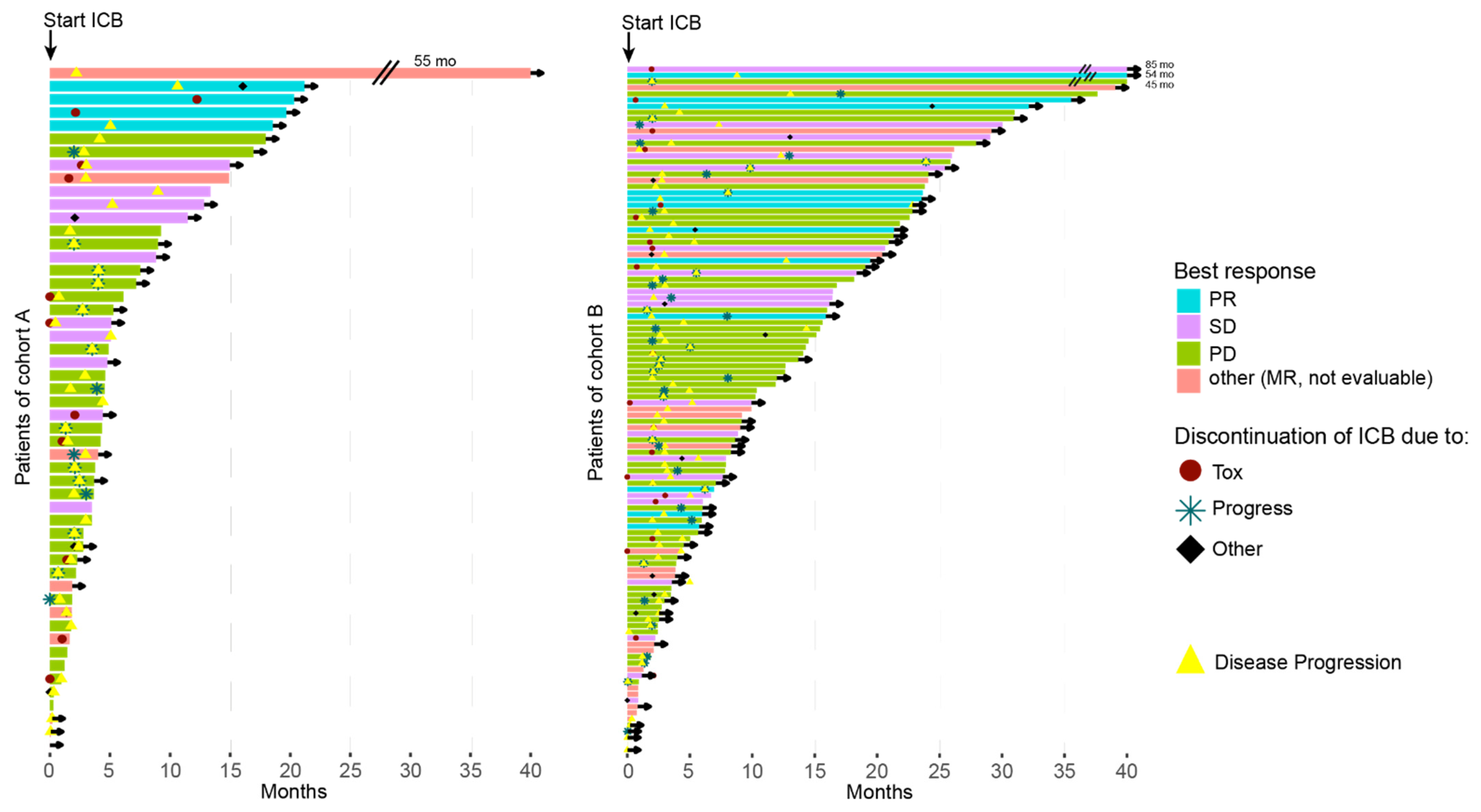
3.2. Response Rates to ICB
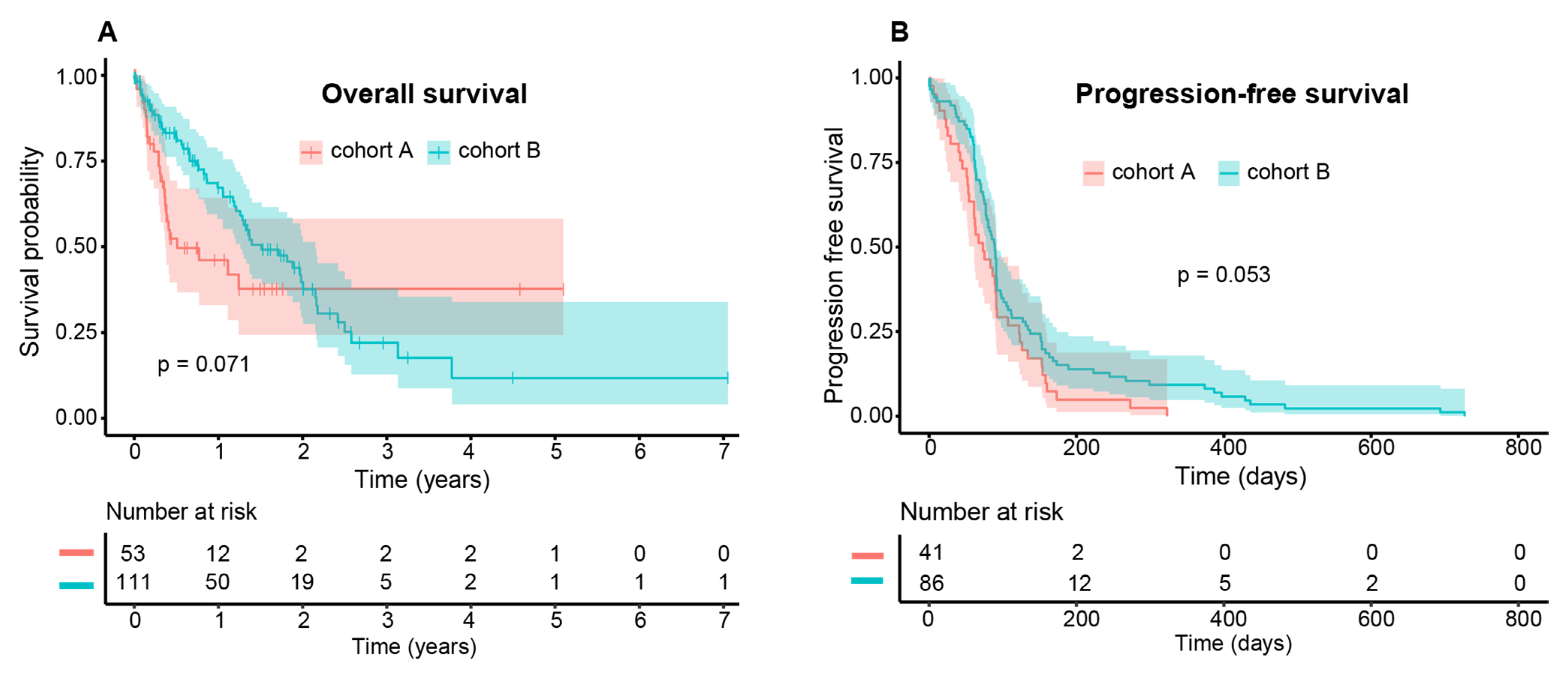
Dual ICB was applied in 109 patients (61.2%; cohort A 69.1% vs. cohort B 57.7%), while 15 patients received ipilimumab monotherapy (8.4%; cohort A 0% vs. cohort B 12.2%). PD-1 inhibitors were given as monotherapy in 53 patients (29.8%; cohort A 30.9% vs. cohort B 29.3%). The best ORR to dual ICB was 13.8% and to anti-PD-1 monotherapy 8.9% in the entire population (27 patients were not evaluable for radiologic response). No patients achieved a CR, while 17 patients had a PR. Patients with liver metastases only (cohort A) showed a numerically worse response to dual ICB, yet without significance (cohort A 8.7% vs. cohort B 16.7%; p = 0.45). In contrast, the ORR to single PD-1 inhibition was numerically higher in cohort A, albeit not significantly (cohort A 14.3% vs. cohort B 6.5%; p = 0.77). Details of the patterns of response to ICB are summarized in Table 2. ICB was given on average for 2.0 months (95% CI 0–13.0) and 2.1 months (95% CI 0–24.4) in cohorts A and B, respectively (p = 0.14).

3.3. Survival Data

The entire cohort showed a median OS of 16 months (95% CI 13.4–23.7) and a median PFS of 2.8 months (95% CI 2.5–3.0) to any ICB. There was a statistical trend in OS (p = 0.071) and PFS (p = 0.053) for both cohorts. The median values differed conspicuously for OS (cohort A 6.1 months (95% CI 4.4-not available) vs. cohort B 18.2 months (95% CI 15.1–25.9)). In contrast, the median PFS was similar in both cohorts (cohort A 2.4 months (95% CI 2.0–3.0) vs. cohort B 2.9 (95% CI 2.5–3.0)) (Figure 1). The survival was also more favorable in cohort B evident in a swimmer’s plot comparing both cohorts (Figure 2).

3.4. Adverse Events (AE)

A total of 133 AE were reported in 74 (41.6%) patients. Of all events, 72 AE (54.1%) were graded as severe (grade 3–5), with no difference between both cohorts (p = 0.93). These 72 events were observed in 47 patients (26.3%; cohort A 14 patients (29.1%) vs. cohort B 31 patients (25.2%); p = 0.72). The treatment was discontinued in 36 cases due to unacceptable toxicity. One death occurred in cohort A during treatment but was most likely due to disease progression. The most common events were colitis (n = 30), hepatitis (n = 19), thyroiditis (n = 13), hypophysitis (n = 8), pancreatitis (n = 6), myalgia with myositis (n = 5), and cutaneous toxicity (n = 6). No significant differences were detected between both cohorts (Table 3; Supplementary Materials Table S1).

4. Discussion

Here, we present to our knowledge the hitherto largest published cohort of patients with metastatic UM who were treated with ICB. We detected a median OS of 16 months (95% CI 13.4–23.7) and a median PFS of 2.8 months (95% CI 2.5–3.0) to any ICB. There was considerably better OS and PFS in patients with both hepatic and extra-hepatic metastatic sites (cohort B), albeit without reaching statistical significance (p = 0.071 and p = 0.053, respectively). The median OS of 16 months is higher compared to the previous benchmark survival studies done before the ICB era [3,6]. We conclude that the prognosis of patients with metastatic UM has improved due to the availability and more frequent use of ICB [7,8]. The ORR to dual ICB of 13.8% remains low compared to the ORR in cutaneous and mucosal melanoma. There were no significant differences between the ORR values of the cohorts and only a slight tendency toward higher ORR to dual ICB in patients with both hepatic and extrahepatic metastasis. The ORR is in line with another previously published retrospective study reporting 11.6% to dual ICB [9] as well as to our previous report of 15.6% [10]. Two recently published prospective trials of the combination of ipilimumab with nivolumab in patients with metastatic UM showed results that slightly deviate [4,5]. Piulats et al. enrolled only treatment-naïve patients and reported a lower OS (12.7 vs. 19.1 months), PFS (3 vs. 5.5 months), and ORR (11.5% vs. 18%) compared to Pelster et al., who enrolled patients with any number of prior treatments. Interestingly, Piulats et al. enrolled also more patients with ECOG 0 (84.6% vs. 71%) and fewer patients with elevated LDH (32% vs. 43%), resulting in an unfavorable ORR in a prognostically favorable population [11]. Furthermore, Piulats et al. reported a median number of two liver metastases (range 1–25) and a median size of the biggest liver metastases of 25 mm (range 10–90 mm); 78.8% of the patients had liver metastases and 57.7% presented with extrahepatic disease. The number of patients with exclusive liver metastases was not presented [4]. In comparison, Pelster et al. reported that 49% of patients presented with hepatic and extrahepatic metastases, 31% with liver metastases only, and 20% with extrahepatic metastasis only [5]. The comparison of both cohorts of this population revealed that patients with exclusive liver metastases had a poorer OS and PFS after ICB. In contrast, patients with both hepatic and extrahepatic metastases had higher ORR to dual ICB (8.7% vs. 16.7%) although this difference was not significant. Thus, it remains unclear if the survival benefits observed in cohort B with both hepatic and extrahepatic metastatic sites are specifically due to ICB treatment or if this cohort is prognostically favorable regardless of the treatment with ICB.
In CM, liver metastases are the least responsive metastatic site to dual ICB with a median of 3% tumor regression compared to other metastatic sites with a median of 77% [12]. Mechanistically, macrophages induce apoptosis of CD8+ T cells in the immunosuppressive microenvironment of the liver through fas-ligand binding. This results in an elimination of CD8+ T cells possibly explaining ineffective tumor control and poor response to immunotherapy [13,14,15]. The comparison of CM to UM liver metastases has demonstrated that there is no difference in the extent of immune infiltration, but UM showed a higher ratio of exhausted CD8+ T cells to cytotoxic T cells, total CD8+ T cells, and Th1 cells. In addition, a higher and more frequent PD-L1 expression on CM liver metastases, as well as higher TMB was found compared to those from UM [16]. This may also contribute to the worse treatment response in UM as a low TMB is associated with poor response to ICB while higher PD-L1 expression is predictive for PD-1 inhibitor response [17,18]. It was further shown that PD-1 and PD-L1 expression is generally very low in UM [19]. However, PD-L1 expression was not considered in this study.
AE occurred in 20.8% of patients with anti-PD-1 monotherapy, in 55% with dual ICB, and severe AE in 7.5% with anti-PD-1 monotherapy and 31.2% of patients with dual ICB with no difference between cohorts A and B (Table 3). The rate of severe AE is in line but on the lower range of previously published studies where immune-related grade 3/4 toxicities in dual ICB occurred in about 30–60% of patients [4,5,9,10].
The major limitation of this study is its retrospective design. In particular, the quantification of the exact extent of metastases and tumor burden in the liver was difficult based on chart reviews. According to our data, we could only assess whether liver metastases were present or not. Furthermore, the quality and completeness of the data, in particular the reporting of AE, is highly dependent on the participating cancer centers. Thus, we cannot exclude that AE were underreported in this study.

5. Conclusions

Our data of 178 patients with advanced UM treated with ICB demonstrates an improved OS compared to studies conducted before the ICB era. Counterintuitively, patients with several metastatic sites seem to have a favorable prognosis compared to patients with hepatic metastasis only. If this phenomenon is related to ICB response warrants further investigation. Nevertheless, our results imply that exclusive hepatic metastases are a major unfavorable prognostic factor.

Supplementary Materials

The following are available online at https://www.mdpi.com/article/10.3390/cancers13133359/s1, Table S1: Baseline characteristics between single PD1 and dual ICB therapy.

Author Contributions

E.A.T.K. collected and merged the data into a central database before analysis. A.P. conducted the statistical analyses. A.W. and A.P. designed the figures. E.A.T.K. and M.V.H. wrote the draft of the manuscript. All other authors were involved in data collection and analysis on-site. E.A.T.K., A.P., A.W., E.D., A.G., R.G., J.C.H., S.H., B.H., K.C.K., H.K., N.K., U.L., C.L., F.M., M.M., P.M., C.P., F.R., D.S., B.S., M.S., P.T., K.-M.T., B.S.-T., S.U., J.U. (Jens Ulrich), J.U. (Jochen Utikal), M.W., F.Z., C.B. and M.V.H. were involved in revising the manuscript. All authors have read and agreed to the published version of the manuscript.

Funding

M.V.H. was supported by the clinician scientist program awarded by the German Society of Dermatology (DDG) and the Arbeitsgemeinschaft Dermatologische Forschung (ADF).

Institutional Review Board Statement

This study was approved by the institutional review board of the medical faculty of the Munich University Hospital (approval number 413-16 UE) and was conducted following the principles of the Helsinki Declaration in its current version.

Informed Consent Statement

Patient consent was waived due to retrospective design involving anonymized data from several centers.

Data Availability Statement

Data are contained within the article or Supplementary Materials.

Acknowledgments

We thank all investigators of the German Dermatologic Cooperative Oncology Group (DeCOG) for their general commitment to the research consortium, in particular for the Committee Ocular Melanoma and participation in the ADOReg registry.

Conflicts of Interest

F.M. has received travel support or/and speaker’s fees or/and advisor’s honoraria from Novartis, Roche, BMS, MSD and Pierre Fabre and research funding from Novartis and Roche. C.P. received honoraria (speaker honoraria or honoraria as a consultant) and travel support from: Novartis, BMS, Roche, Merck Serono, MSD, Celgene, AbbVie, SUNPHARMA, UCB, Allergy Therapeutics and LEO. S.U. declares research support from Bristol Myers Squibb and Merck Serono; speakers and advisory board honoraria from Bristol Myers Squibb, Merck Sharp & Dohme, Merck Serono, Novartis and Roche, and travel support from Bristol Myers Squibb, and Merck Sharp & Dohme. J.C.H. declares research support from BMS, advisory board honoraria from Pierre Fabre, Sanofi, Sunpharma and MSD, speakers honoraria from BMS, MSD, Novartis, Roche, Sanofi and Almirall and travel support from Pierre Fabre. F.Z. declares speakers and advisory board honoraria and/or travel support from BMS, MSD, Roche, Novartis, Pierre-Fabre and Sanofi-Aventis outside the submitted work. RG Research support: Pfizer, Johnson&Johnson, Novartis, Amgen, MerckSerono, SUN Pharma, Sanofi. Honoraria for lectures: Roche Pharma, Bristol-MyersSquibb, Novartis, MSD, Almirall-Hermal, Amgen, Merck-Serono, SUN, Pierre-Fabre, Sanofi, SUN Pharma, Bayer Honoraria for advisory boards: Roche Pharma, Bristol-MyersSquibb, Novartis, MSD, Almirall-Hermal, Amgen, Pierre-Fabre, Merck-Serono, 4SC, Immunocore, SUN Pharma, Sanofi, Pfizer. P.T.: BMS, Novartis, MSD, Pierre-Fabre, CureVac, Roche, Kyowa Kirin, Biofrontera, Invited Speaker, Personal BMS, Novartis, Pierre-Fabre, Merck Serono, Sanofi, Roche, Kyowa Kirin, Advisory Board, Personal BMS, Pierre-Fabre, Other, Personal, Travel support. B.S.T. declares advisory board honoraria from Iovance. M.S. reports receiving honoraria and participation in advisory boards of Bristol-Myers Squibb, Novartis, MSD, Roche, Pierre Fabre, Kyowa Kirin, Immunocore and Sanofi-Genzyme. M.S. received travel accommodation and expenses by Novartis, Pierre Fabre, and Sun Pharma. AG speaker’s honoraria from Bristol-Myers Squibb, MSD Sharp & Dohme and Roche; intermittent advisory board relationships with Amgen, Bristol-Myers Squibb, Novartis, MSD Sharp & Dohme, Pierre Fabre Pharmaceuticals, Pfizer, Roche and Sanofi Genzyme; travel and congress fee support from Bristol-Myers Squibb, MSD Sharp & Dohme, Novartis, Pierre Fabre Pharmaceuticals and Roche. Clinical studies: Amgen, Array, Bristol-Myers Squibb, GSK, Novartis, Merck, MSD Sharp & Dohme, Pfizer and Roche. K.M.T. received speaker or consultant honoraria and travel support from Bristol-Myers Squibb, Merck Sharp and Dohme, Roche, Novartis, Pierre Fabre, Sanofi Genzyme, LEO, Galderma, Almirall, La Roche-Posay and Candela.

References

  1. Gonzales, C.A.; Ladas, J.G.; Davis, J.L.; Feuer, W.J.; Holland, G.N. Collaborative ocular melanoma study group. Assessment of metastatic disease status at death in 435 patients with large choroidal melanoma in the collaborative ocular melanoma study (COMS). Arch. Ophthalmol. 2001, 119, 670–676. [Google Scholar] [CrossRef]
  2. Heppt, M.V.; Steeb, T.; Schlager, J.G.; Rosumeck, S.; Dressler, C.; Ruzicka, T.; Nast, A.; Berking, C. Immune checkpoint blockade for unresectable or metastatic uveal melanoma: A systematic review. Cancer Treat. Rev. 2017, 60, 44–52. [Google Scholar] [CrossRef] [PubMed]
  3. Rantala, E.S.; Hernberg, M.; Kivelä, T.T. Overall survival after treatment for metastatic uveal melanoma: A systematic review and meta-analysis. Melanoma Res. 2019, 29, 561–568. [Google Scholar] [CrossRef] [PubMed]
  4. Piulats, J.M.; Espinosa, E.; Merino, L.D.L.C.; Varela, M.; Carrión, L.A.; Martín-Algarra, S.; Castro, R.L.; Curiel, T.; Rodríguez-Abreu, D.; Redrado, M.; et al. Nivolumab plus ipilimumab for treatment-naïve metastatic uveal melanoma: An open-label, multicenter, phase II trial by the Spanish multidisciplinary melanoma group (GEM-1402). J. Clin. Oncol. 2021, 39, 586–598. [Google Scholar] [CrossRef] [PubMed]
  5. Pelster, M.S.; Gruschkus, S.K.; Bassett, R.; Gombos, D.S.; Shephard, M.; Liberty, P.; Glover, M.S.; Simien, R.; Diab, A.; Hwu, P.; et al. Nivolumab and ipilimumab in metastatic uveal melanoma: Results from a single-arm phase II study. J. Clin. Oncol. 2021, 39, 599–607. [Google Scholar] [CrossRef]
  6. Khoja, L.; Atenafu, E.; Suciu, S.; Leyvraz, S.; Sato, T.; Marshall, E.; Keilholz, U.; Zimmer, L.; Patel, S.; Piperno-Neumann, S.; et al. Meta-analysis in metastatic uveal melanoma to determine progression free and overall survival benchmarks: An international rare cancers initiative (IRCI) ocular melanoma study. Ann. Oncol. 2019, 30, 1370–1380. [Google Scholar] [CrossRef]
  7. Heppt, M.V.; Heinzerling, L.; Kähler, K.C.; Forschner, A.; Kirchberger, M.C.; Loquai, C.; Meissner, M.; Meier, F.; Terheyden, P.; Schell, B.; et al. Prognostic factors and outcomes in metastatic uveal melanoma treated with programmed cell death-1 or combined PD-1/cytotoxic T-lymphocyte antigen-4 inhibition. Eur. J. Cancer 2017, 82, 56–65. [Google Scholar] [CrossRef]
  8. Wessely, A.; Steeb, T.; Erdmann, M.; Heinzerling, L.; Vera, J.; Schlaak, M.; Berking, C.; Heppt, M.V. The Role of Immune Checkpoint Blockade in Uveal Melanoma. Int. J. Mol. Sci. 2020, 21, 879. [Google Scholar] [CrossRef] [PubMed] [Green Version]
  9. Najjar, Y.G.; Navrazhina, K.; Ding, F.; Bhatia, R.; Tsai, K.; Abbate, K.; Durden, B.; Eroglu, Z.; Bhatia, S.; Park, S.; et al. Ipilimumab plus nivolumab for patients with metastatic uveal melanoma: A multicenter, retrospective study. J. Immunother. Cancer 2020, 8, e000331. [Google Scholar] [CrossRef] [PubMed]
  10. Heppt, M.V.; Amaral, T.; Kähler, K.C.; Heinzerling, L.; Hassel, J.C.; Meissner, M.; Kreuzberg, N.; Loquai, C.; Reinhardt, L.; Utikal, J.; et al. Combined immune checkpoint blockade for metastatic uveal melanoma: A retrospective, multi-center study. J. Immunother. Cancer 2019, 7, 299. [Google Scholar] [CrossRef] [PubMed]
  11. Khan, S.; Carvajal, R.D. Dual Immunological Checkpoint Blockade for Uveal Melanoma. J. Clin. Oncol. 2021, 39, 554–556. [Google Scholar] [CrossRef] [PubMed]
  12. Da Silva, I.P.; Long, G.; Quek, C.; Gonzalez, M.; Carlino, M.S.; Long, G.V.; Menzies, A.M. Site-specific response patterns, pseudoprogression, and acquired resistance in patients with melanoma treated with ipilimumab combined with anti–PD-1 therapy. Cancer 2020, 126, 86–97. [Google Scholar] [CrossRef] [PubMed] [Green Version]
  13. Somasundaram, R.; Zhang, G.; Fukunaga-Kalabis, M.; Perego, M.; Krepler, C.; Xu, X.; Wagner, C.; Hristova, D.; Zhang, J.; Tian, T.; et al. Tumor-associated B-cells induce tumor heterogeneity and therapy resistance. Nat. Commun. 2017, 8, 607. [Google Scholar] [CrossRef] [Green Version]
  14. Somasundaram, R.; Connelly, T.; Choi, R.; Choi, H.; Samarkina, A.; Li, L.; Gregorio, E.; Chen, Y.; Thakur, R.; Abdel-Mohsen, M.; et al. Tumor-infiltrating mast cells are associated with resistance to anti-PD-1 therapy. Nat. Commun. 2021, 12, 346. [Google Scholar] [CrossRef]
  15. Lindblad, K.E.; Lujambio, A. Liver metastases inhibit immunotherapy efficacy. Nat. Med. 2021, 27, 25–27. [Google Scholar] [CrossRef]
  16. Hoefsmit, E.P.; A Rozeman, E.; Van, T.M.; Dimitriadis, P.; Krijgsman, O.; Conway, J.W.; Da Silva, I.P.; E Van Der Wal, J.; Ketelaars, S.L.C.; Bresser, K.; et al. Comprehensive analysis of cutaneous and uveal melanoma liver metastases. J. Immunother. Cancer 2020, 8, e001501. [Google Scholar] [CrossRef]
  17. Zou, W.; Wolchok, J.D.; Chen, L. PD-L1 (B7-H1) and PD-1 pathway blockade for cancer therapy: Mechanisms, response biomarkers, and combinations. Sci. Transl. Med. 2016, 8, 328rv4. [Google Scholar] [CrossRef] [PubMed] [Green Version]
  18. Schumacher, T.N.; Schreiber, R.D. Neoantigens in cancer immunotherapy. Science 2015, 348, 69–74. [Google Scholar] [CrossRef] [Green Version]
  19. Qin, Y.; Bollin, K.; De Macedo, M.P.; Carapeto, F.; Kim, K.B.; Roszik, J.; Wani, K.M.; Reuben, A.; Reddy, S.T.; Williams, M.D.; et al. Immune profiling of uveal melanoma identifies a potential signature associated with response to immunotherapy. J. Immunother. Cancer 2020, 8, e000960. [Google Scholar] [CrossRef] [PubMed]
Figure 1. Kaplan–Meier estimates of the patient population for (A) OS and (B) PFS to ICB comparing cohort A (red) vs. B (turquoise). Although there was no significant difference in OS and PFS (p = 0.071 and p = 0.053, resp.), the median survival differed considerably (cohort A 6.1 months (95%-CI: 4.4-NA) vs. cohort B 18.2 months (95%-CI: 15.1–25.9)). In contrast, the median PFS only differed slightly (cohort A 2.4 months (95%-CI: 2.0–3.0) vs. cohort B 2.9 months (95%-CI 2.5–3.0)).
Cancers 13 03359 g001
Figure 2. Swimmer plots for cohort A (left) and B (right) demonstrating the OS for each patient. The color shows the best response to ICB, the symbols depict the reason for treatment discontinuation and the yellow triangle shows the time of tumor progression. If a patient was censored, an arrow is drawn.
Cancers 13 03359 g002
Table 1. Characteristics of the study population. Abbreviations: NA = not available, ICB = immune checkpoint blockade.
TotalCohort ACohort BA vs. B
SexWomen89 (50.0%)25 (45.5%)64 (52.0%)p = 0.52
Men89 (50.0%)30 (54.5%)59 (48.0%)
AgeMedian in years
(range)
65.6
(17.7–87.6)
63.4
(32.4–87.6)
65.8
(17.7–85.4)
p = 0.79
LDHNot elevated42 (23.6%)16 (29.1%)26 (21.1%)p = 0.34
Elevated89 (50.0%)28 (50.9%)61 (49.6%)
NA47 (26.4%)11 (20.0%)36 (29.3%)
ECOGECOG 088 (49.4%)30 (54.5%)58 (44.7%)p = 0.44
ECOG 120 (11.2%)3 (5.5%)17 (13.8%
ECOG 24 (2.2%)1 (1.8%)3 (2.4%)
ECOG 32 (1.1%)1 (1.8%)1 (0.8%)
ECOG 40 (0.0%)0 (0.0%)0 (0.0%)
ECOG 50 (0.0%)0 (0.0%)0 (0.0%)
NA64 (36.0%)20 (36.4%)44 (35.8%)
Number of affected organ systemsMedian (range)2 (1–7)13 (2–7)p < 0.001
Affected organ systemsLiver178 (100%)55 (100%)123 (100%)
Pulmonary82 (46.1%)0 (0.0%)82 (66.7%)
Bone47 (26.4%)0 (0.0%)47 (38.2%)
CNS25 (14.0%)0 (0.0%)25 (20.3%)
Lymph node41 (23.0%)0 (0.0%)41 (33.3%)
Connective tissue8 (4.5%)0 (0.0%)8 (6.5%)
Skin24 (13.5%)0 (0.0%)24 (19.5%)
Disseminated10 (5.6%)0 (0.0%)10 (8.1%)
Other51 (28.7%)0 (0.0%)51 (41.5%)
ICB as first-line therapy 146 (82.0%)49 (89.1%)97 (78.9%)p = 0.15
Other therapiesGemcitabine & Treosulfan34 (19.1%)4 (7.2%)30 (24.3%)p = 0.013
Nivolumab38 (21.3%)10 (18.1%)28 (22.7%)p = 0.623
Pembrolizumab25 (14%)6 (10.9%)19 (15.4%)p = 0.568
Sorafenib18 (10.1%)1 (1.8%)17 (13.8%)p = 0.029
DC vaccine14 (7.8%)2 (3.6%)12 (9.7%)p = 0.26
Dacarbazine9 (5%)0 (0%)9 (7.3%)p = 0.091
Trametinib10 (5.6%)2 (3.6%)8 (6.5%)p = 0.678
Fotemustine9 (5%)2 (3.6%)7 (5.6%)p = 0.835
ICB substanceAny177 (99.4%)55 (100%)122 (99.2%)p = 1
anti-PD-1 (pembrolizumab, nivolumab)53 (29.8%)17 (30.9%)36 (29.3%)p = 0.97
anti-CTLA-4 (ipilimumab)15 (8.4%)0 (0.0%)15 (12.2%)p = 0.016
Dual109 (61.2%)38 (69.1%)71 (57.7%)p = 0.20
NA1 (0.6%)0 (0.0%)1 (0.8%)
Table 2. Response rates to ICB according to ICB substance. Abbreviations: CR = complete response, PR = partial response, SD = stable disease, PD = progressive disease, ORR = objective response rate, DCR = disease control rate.
ICB—Any TypeTotalCohort ACohort BTest (Cohorts A vs. B)
CR0/150 (0.0%)0/48 (0.0%)0/102 (0.0%)
PR17/150 (11.3%)5/48 (10.4%)12/102 (11.8%)p = 1
SD36/150 (24.0%)11/48 (22.9%)25/102 (24.5%)p = 0.99
PD91/150 (60.7%)30/48 (62.5%)61/102 (59.8%)p = 0.89
ORR17/150 (11.3%)5/48 (10.4%)12/102 (11.8%)p = 1
DCR53/150 (35.3%)16/48 (33.3%)37/102 (36.3%)p = 0.87
anti-PD-1TotalCohort ACohort BTest (Cohorts A vs. B)
CR0/45 (0.0%)0/14 (0.0%)0/31 (0.0%)
PR4/45 (8.9%)2/14 (14.3%)2/31 (6.5%)p = 0.77
SD9/45 (20%)2/14 (14.3%)7/31 (22.6%)p = 0.81
PD30/45 (66.7%)10/14 (71.4%)20/31 (64.5%)p = 0.91
ORR4/45 (8.9%)2/14 (14.3%)2/31 (6.5%)p = 0.77
DCR13/45 (28.9%)4/14 (28.6%)9/31 (29.0%)p = 1
Dual ICBTotalCohort ACohort BTest (Cohorts A vs. B)
CR0/94 (0.0%)0/34 (0.0%)0/60 (0.0%)
PR13/94 (13.8%)3/34 (8.7%)10/60 (16.7%)p = 0.45
SD25/94 (26.6%)9/34 (26.5%)16/60 (26.7%)p = 1
PD52/94 (55.3%)20/34 (58.8%)32/60 (53.3%)p = 0.77
ORR13/94 (13.8%)3/34 (8.7%)10/60 (16.7%)p = 0.45
DCR38/94 (40.4%)12/34 (35.3%)26/60 (43.3%)p = 0.59
anti-CTLA-4TotalCohort ACohort BTest (cohorts A vs. B)
CR0/11 (0.0%)0/00/11 (0.0%)Not possible
PR0/11 (0.0%)0/00/11 (0.0%)
SD2/11 (18.2%)0/02/11 (18.2%)
PD9/11 (81.8%)0/09/11 (81.8%)
ORR0/11 (0.0%)0/00/11 (0.0%)
DCR2/11 (18.2%)0/02/11 (18.2%)
Table 3. Occurrence of adverse events. Abbreviations: AE = adverse events.
TotalCohort ACohort BTest (Cohorts A vs. B)
Number of AE1334390
Number of severe AE 72 (54.1%)24 (55.8%)48 (53.3%)p = 0.93
Number of patients with AE74 (41.6%)26 (47.3%)48 (39.0%)p = 0.39
Number of patients with severe AE47 (26.3%)16 (29.1%)31 (25.2%)p = 0.72
anti-PD1Dual ICBTest (Cohorts A vs. B)
Number of AE25103
Number of severe AE (grade 3 + 4)6 (24.0%)60 (58.3%)p = 0.0044
Number of patients with AE11 (20.8%)60 (55.0%)p < 0.001
Number of patients with severe AE (grade 3 + 4)4 (7.5%)34 (31.2%)p = 0.0017
Publisher’s Note: MDPI stays neutral with regard to jurisdictional claims in published maps and institutional affiliations.

