
Biopolymers Used for Receptor Immobilization for Nickel-Detection Biosensors in Food

Abstract
:1. Introduction
2. Materials and Methods
2.1. Reagents
2.2. Voltammetric Measurements
2.3. Procedures
2.4. Immobilization of Receptor on Screen Printed Electrodes (SPEs)
2.5. Characterization of Biosensor Performance
2.5.1. Sensitivity
2.5.2. Limit of Detection (LoD)
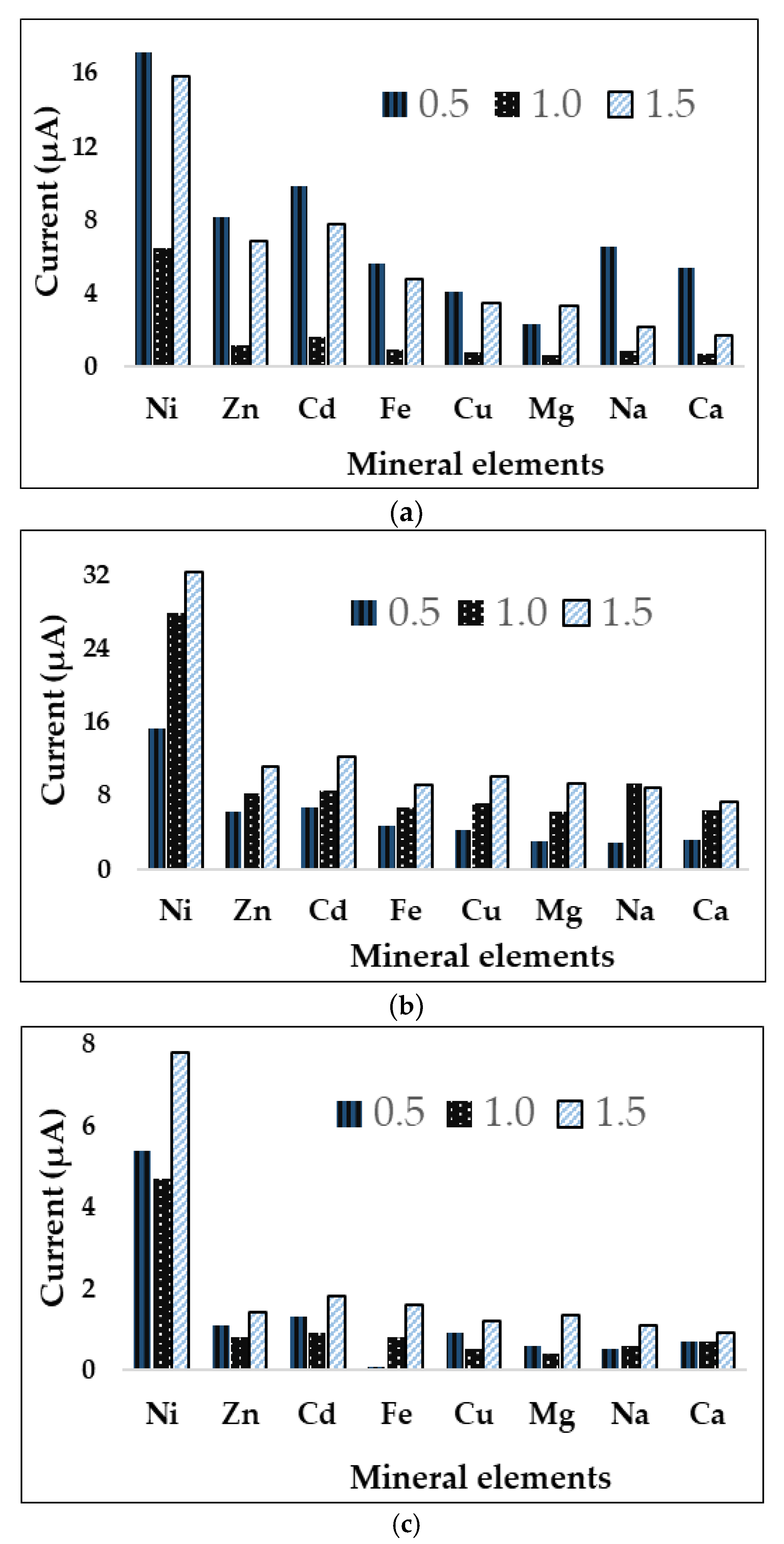
2.5.3. Selectivity
3. Results
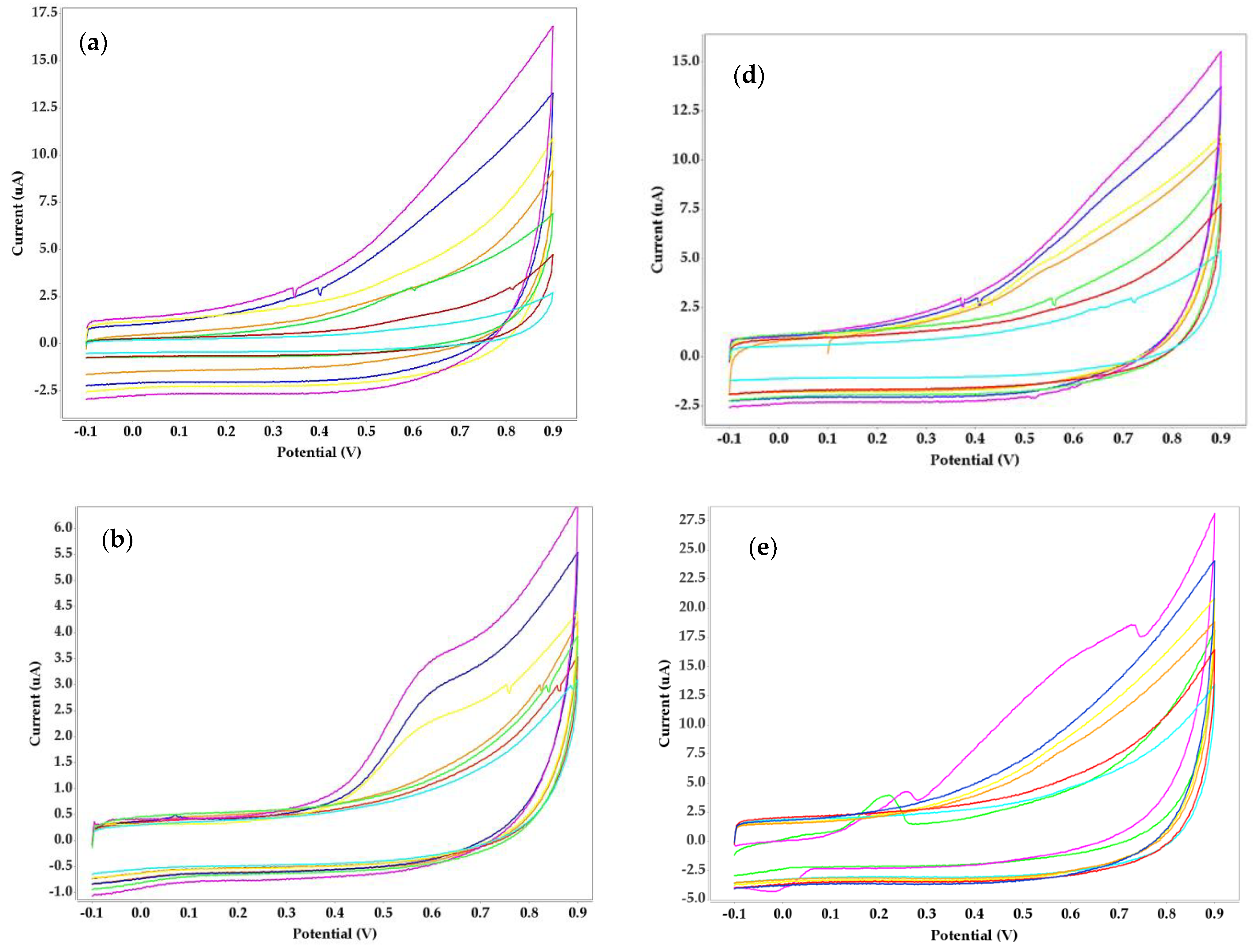
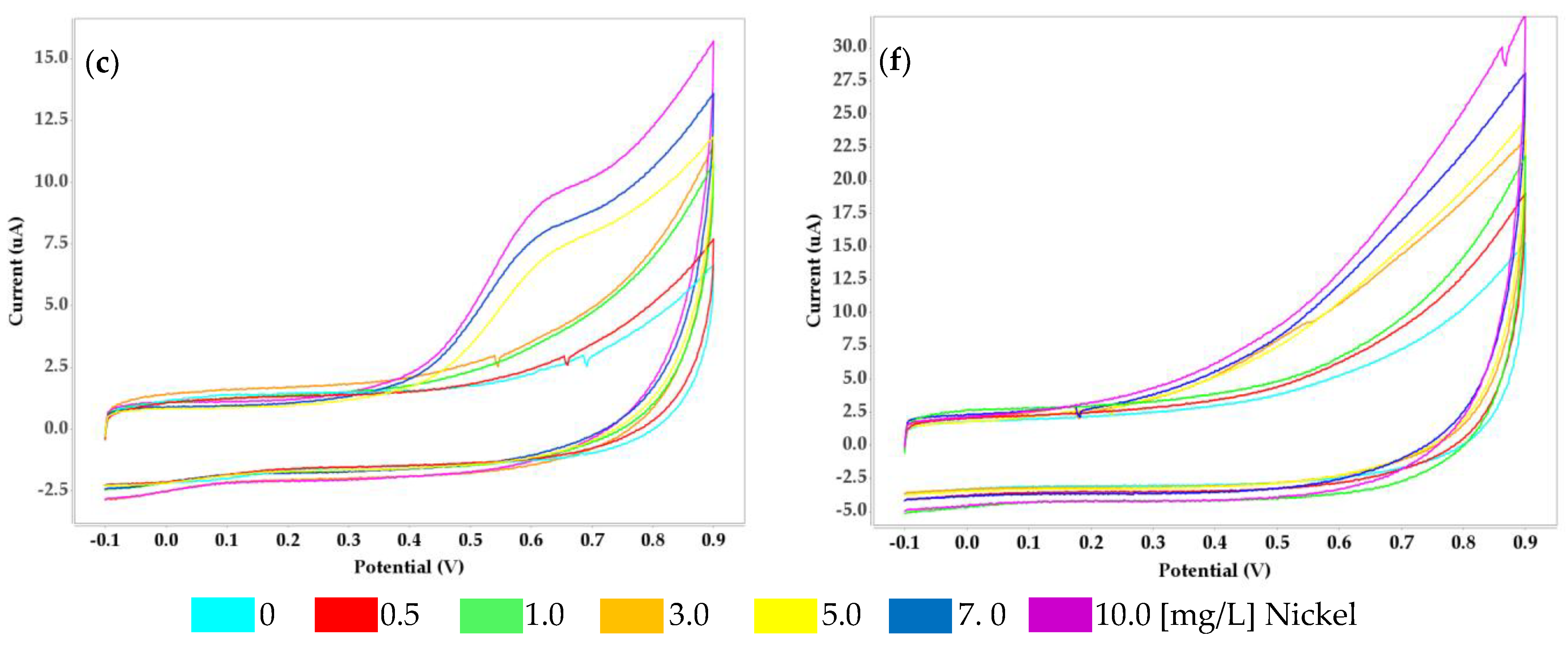
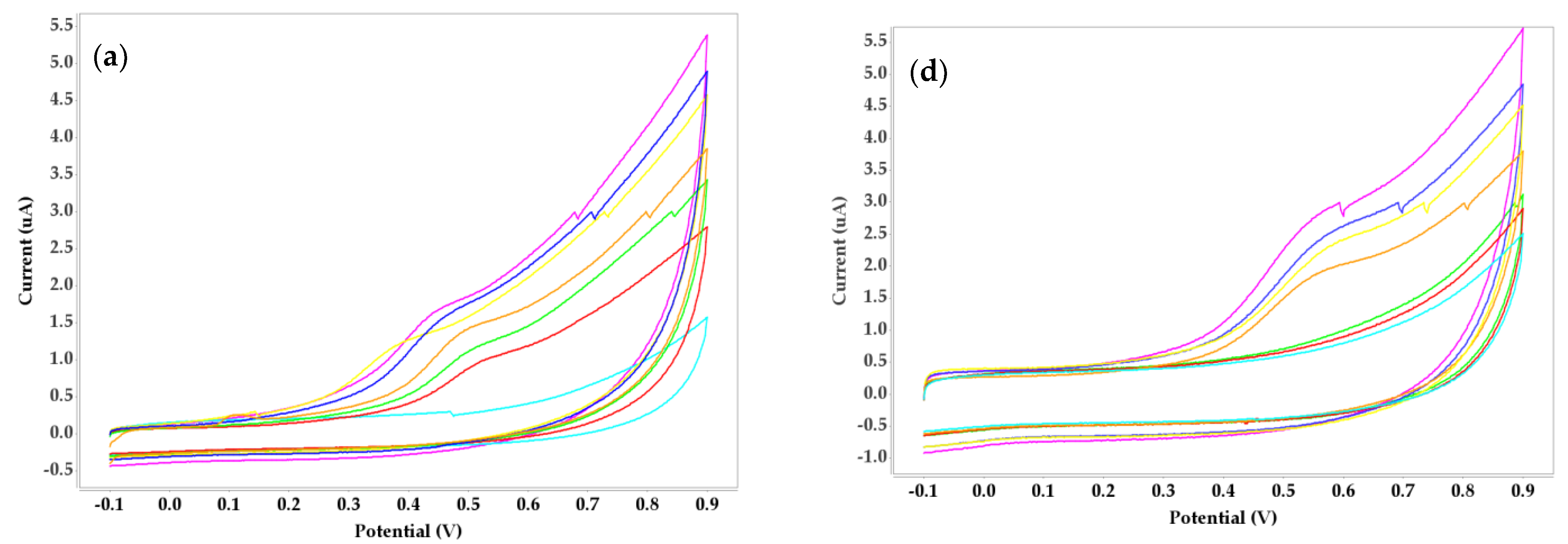
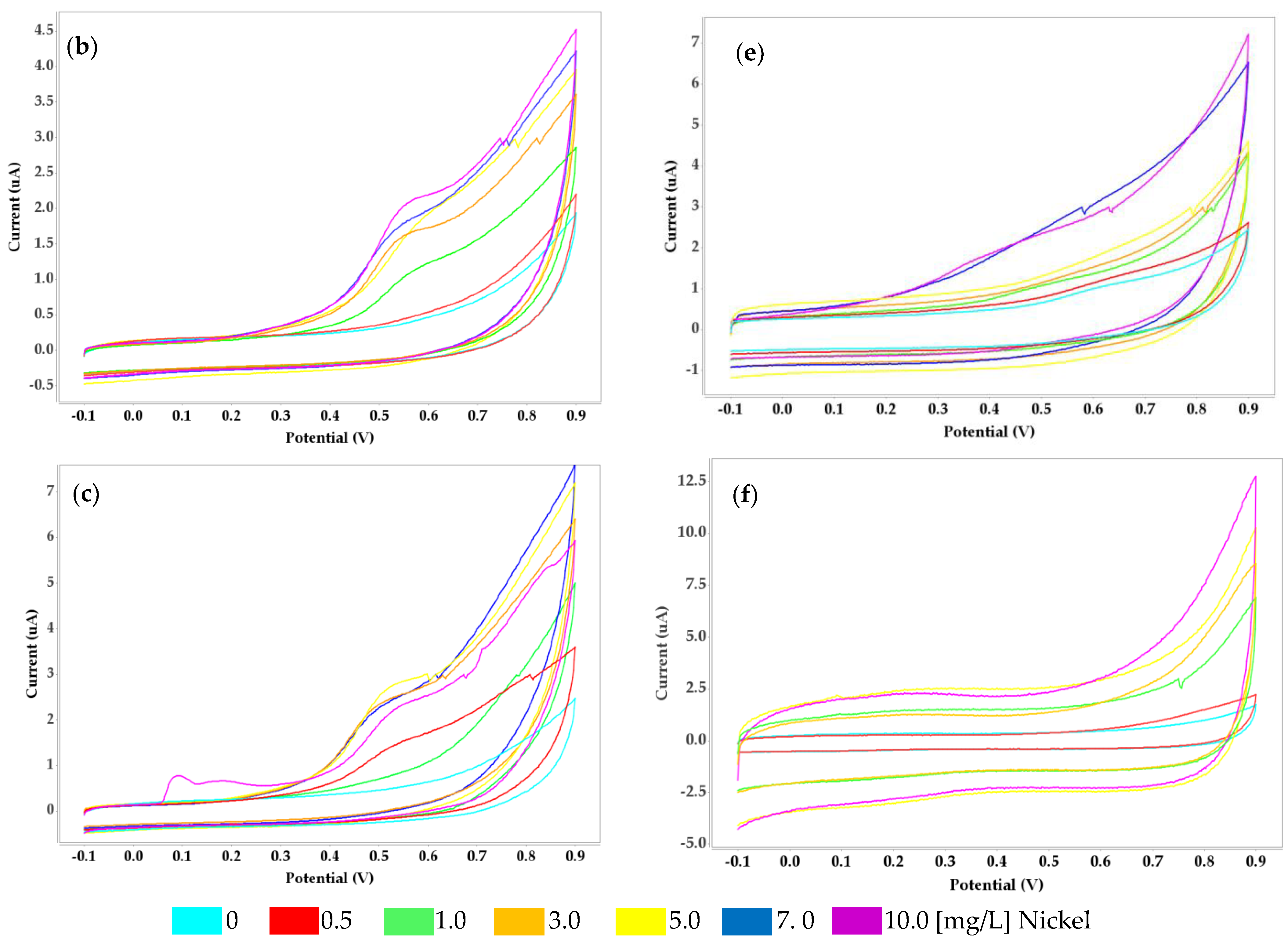
3.1. Voltammetric Behavior of Receptors with Four Biopolymers
3.2. Characteristics of the Biosensor’s Performance
3.3. Optimization of Four Biopolymers and Concentrations
4. Discussion
5. Conclusions
Supplementary Materials
Author Contributions
Funding
Data Availability Statement
Acknowledgments
Conflicts of Interest
References
- Samson, R.; Navale, G.R.; Dharne, M.S. Biosensors: Frontiers in rapid detection of COVID-19. 3 Biotech 2020, 10, 385. [Google Scholar] [CrossRef]
- Kotru, S.; Klimuntowski, M.; Ridha, H.; Uddin, Z.; Askhar, A.A.; Singh, G.; Howlader, M.M. Electrochemical sensing: A prognostic tool in the fight against COVID-19. TrAC 2021, 136, 116198. [Google Scholar] [CrossRef]
- Kaushik, A.K.; Dhau, J.S.; Gohel, H.; Mishra, Y.K.; Kateb, B.; Kim, N.Y.; Goswami, D.Y. Electrochemical SARS-CoV-2 sensing at point-of-care and artificial intelligence for intelligent COVID-19 management. ACS Appl. Bio. Mater. 2020, 3, 7306–7325. [Google Scholar] [CrossRef] [PubMed]
- Wang, X.; Uchiyama, S. Polymers for biosensors construction. State Art Biosens.—Gen. Asp. 2013, 3, 67–84. [Google Scholar]
- Grieshaber, D.; MacKenzie, R.; Vörös, J.; Reimhult, E. Electrochemical biosensors-sensor principles and architectures. Sensors 2008, 8, 1400–1458. [Google Scholar] [CrossRef] [PubMed] [Green Version]
- Teepoo, S.; Dawan, P.; Barnthip, N. Electrospun chitosan-gelatin biopolymer composite nanofibers for horseradish peroxidase immobilization in a hydrogen peroxide biosensor. Biosensors 2017, 7, 47. [Google Scholar] [CrossRef] [Green Version]
- Turner, A.P. Biosensors: Sense and sensibility. Chem. Soc. Rev. 2013, 42, 3184–3196. [Google Scholar] [CrossRef] [Green Version]
- Hossain, S.Z.; Mansour, N. Biosensors for on-line water quality monitoring—A review. Arab J. Basic Appl. Sci. 2019, 26, 502–518. [Google Scholar] [CrossRef]
- Lakard, B. Electrochemical biosensors based on conducting polymers: A review. Appl. Sci. 2020, 10, 6614. [Google Scholar] [CrossRef]
- Alberti, G.; Zanoni, C.; Losi, V.; Magnaghi, L.R.; Biesuz, R. Current Trends in Polymer Based Sensors. Biosensors 2021, 9, 108. [Google Scholar] [CrossRef]
- Sharma, M.; Singh, S.P. Enzyme entrapment approaches and their applications. In Biomass, Biofuels, Biochemicals; Elsevier: Amsterdam, The Netherlands, 2020; pp. 191–216. [Google Scholar]
- Imam, H.T.; Marr, P.C.; Marr, A.C. Enzyme entrapment, biocatalyst immobilization without covalent attachment. Green Chem. 2021, 23, 4980–5005. [Google Scholar] [CrossRef]
- Elnashar, M.M. The Art of Immobilization Using Biopolymers, Biomaterials and Nanobiotechnology, Biotechnology of Biopolymers; IntechOpen: London, UK, 2011. [Google Scholar]
- Jiang, G.; Wang, G.; Zhu, Y.; Cheng, W.; Cao, K.; Xu, G.; Zhao, D.; Yu, H. A scalable bacterial cellulose ionogel for multisensory electronic skin. Research 2022, 2022, 9814767. [Google Scholar] [CrossRef] [PubMed]
- Bannov, A.G.; Popov, M.V.; Brester, A.E.; Kurmashov, P.B. Recent advances in ammonia gas sensors based on carbon nanomaterials. Micromachines 2021, 12, 186. [Google Scholar] [CrossRef] [PubMed]
- Sawant, S.N. Development of biosensors from biopolymer composites. In Biopolymer Composites in Electronics; Elsevier: Amsterdam, The Netherlands, 2017; pp. 353–383. [Google Scholar]
- Abhilash, M.; Thomas, D. Biopolymers for biocomposites and chemical sensor applications. In Biopolymer Composites in Electronics; Elsevier: Amsterdam, The Netherlands, 2017; pp. 405–435. [Google Scholar]
- Pestov, A.; Bratskaya, S. Chitosan and its derivatives as highly efficient polymer ligands. Molecules 2016, 21, 330. [Google Scholar] [CrossRef] [PubMed] [Green Version]
- Varma, A.J.; Deshpande, S.V.; Kennedy, J.F. Metal complexation by chitosan and its derivatives: A review. Carbohydr. Polym. 2004, 55, 77–93. [Google Scholar] [CrossRef]
- Zouaoui, F.; Bourouina-Bacha, S.; Bourouina, M.; Jaffrezic-Renault, N.; Zine, N.; Errachid, A. Electrochemical sensors based on molecularly imprinted chitosan: A review. TrAC 2020, 130, 115982. [Google Scholar] [CrossRef]
- Garcia-Rey, S.; Ojeda, E.; Gunatilake, U.B.; Basabe-Desmonts, L.; Benito-Lopez, F. Alginate Bead Biosystem for the Determination of Lactate in Sweat Using Image Analysis. Biosensors 2021, 11, 379. [Google Scholar] [CrossRef]
- Esmaeili, C.; Heng, L.Y.; Ling, Y.P.; Norouzi, P.; Ling, T.L. Potentiometric urea biosensor based on immobilization of urease in Kappa-Carrageenan biopolymer. Sens. Lett. 2017, 15, 851–857. [Google Scholar] [CrossRef]
- Bedi, N.; Srivastava, D.K.; Srivastava, A.; Mahapatra, S.; Dkhar, D.S.; Chandra, P.; Srivastava, A. Marine biological macromolecules as matrix material for biosensor fabrication. Biotechnol. Bioeng. 2022, 119, 2046–2063. [Google Scholar] [CrossRef]
- Prakash, O.; Jaiswal, N. Immobilization of a thermostable-amylase on agarose and agar matrices and its application in starch stain removal. World Appl. Sci. J. 2011, 13, 572–577. [Google Scholar]
- Sudhakar, P.; Singh, A. Agars: Properties and applications. In Polysaccharides: Properties and Applications; Scrivener Publishing: Beverly, MA, USA, 2021; pp. 75–93. Available online: https://onlinelibrary.wiley.com/doi/abs/10.1002/9781119711414.ch5 (accessed on 27 February 2023).
- Zhang, C.; An, D.; Xiao, Q.; Chen, F.Q.; Zhang, Y.H.; Weng, H.F.; Xiao, A.F. Convenient Agarose Preparation with Hydrogen Peroxide and Desulfation Process Analysis. Mar. Drugs 2021, 19, 297. [Google Scholar] [CrossRef]
- Zucca, P.; Fernandez-Lafuente, R.; Sanjust, E. Agarose and its derivatives as supports for enzyme immobilization. Molecules 2016, 21, 1577. [Google Scholar] [CrossRef] [PubMed] [Green Version]
- Goldenberg, A.; Jacob, S.E. Update on systemic nickel allergy syndrome and diet. Eur. Ann. Allergy Clin. Immunol. 2015, 47, 25–26. [Google Scholar] [PubMed]
- Matiz, C.; Jacob, S.E. Systemic contact dermatitis in children: How an avoidance diet can make a difference. Pediatr. Dermatol. 2011, 28, 368–374. [Google Scholar] [CrossRef]
- Schram, S.E.; Warshaw, E.M.; Laumann, A. Nickel hypersensitivity: A clinical review and call to action. Int. J. Dermatol. 2010, 49, 115–125. [Google Scholar] [CrossRef]
- Mazzei, L.; Musiani, F.; Ciurli, S. The structure-based reaction mechanism of urease, a nickel dependent enzyme: Tale of a long debate. JBIC J. Biol. Inorg. Chem. 2020, 25, 829–845. [Google Scholar] [CrossRef] [PubMed]
- Carlsson, H.; Nordlander, E. Computational modeling of the mechanism of urease. Bioinorg. Chem. Appl. 2010, 2010, 364891. [Google Scholar] [CrossRef] [Green Version]
- Shen, F.; Arshi, S.; Magner, E.; Ulstrup, J.; Xiao, X. One-step electrochemical approach of enzyme immobilization for bioelectrochemical applications. Synth. Met. 2022, 291, 117205. [Google Scholar] [CrossRef]
- Lu, W.; Xie, X.; Lan, X.; Wu, P.; Peng, H.; He, J.; Zhong, L.; Liu, X.; Deng, Z.; Tan, Z.; et al. An electrochemical immunosensor for the detection of Glypican-3 based on enzymatic ferrocene-tyramine deposition reaction. Biosens. Bioelectron. 2023, 225, 115081. [Google Scholar] [CrossRef]
- Ferlazzo, A.; Espro, C.; Iannazzo, D.; Neri, G. Development of a novel potentiometric PHD/SPE biosensor for the determination of phenylalanine. In Proceedings of the 2022 IEEE International Symposium on Medical Measurements and Applications (MeMeA), Messina, Italy, 22–24 June 2022; IEEE: New York, NY, USA, 2022; pp. 1–5. [Google Scholar]
- Anchidin-Norocel, L.; Savage, W.K.; Gutt, G.; Amariei, S. Development, Optimization, Characterization, and Application of Electrochemical Biosensors for Detecting Nickel Ions in Food. Biosensors 2021, 11, 519. [Google Scholar] [CrossRef]
- Norocel, L.; Gutt, G. Screen-Printed Voltammetric Biosensors for the Determination of Copper in Wine. Sensors 2019, 19, 4618. [Google Scholar] [CrossRef] [Green Version]
- Norocel, L.; Gutt, G. Development and performance testing of an electrochemical sensor for determination of iron ions in wine. Aust. J. Grape Wine Res. 2019, 25, 161–164. [Google Scholar] [CrossRef]
- Ferrari, E. Gold nanoparticle-based plasmonic biosensors. Biosensors 2023, 13, 411. [Google Scholar] [CrossRef] [PubMed]
- Jiménez-Pérez, R.; Iniesta, J.; Baeza-Romero, M.T.; Valero, E. On the performance of carbon-based screen-printed electrodes for (in) organic hydroperoxides sensing in rainwater. Talanta 2021, 234, 122699. [Google Scholar] [CrossRef] [PubMed]
- Tavares, M.C.; Oliveira, K.A.; de Fátima, A.; Coltro, W.K.; Santos, J.C.C. Paper-based analytical device with colorimetric detection for urease activity determination in soils and evaluation of potential inhibitors. Talanta 2021, 230, 122301. [Google Scholar] [CrossRef] [PubMed]
- Singh, M.; Verma, N.; Garg, A.K.; Redhu, N. Urea biosensors. Sens. Actuators B Chem. 2008, 134, 345–351. [Google Scholar] [CrossRef]
- Song, M.; Lin, X.; Peng, Z.; Xu, S.; Jin, L.; Zheng, X.; Luo, H. Materials and methods of biosensor interfaces with stability. Front. Mater. 2021, 7, 583739. [Google Scholar] [CrossRef]
- Zhang, L.; Yang, Y.; Tan, J.; Yuan, Q. Chemically modified nucleic acid biopolymers used in biosensing. Mater. Chem. Front. 2020, 4, 1315–1327. [Google Scholar] [CrossRef]
- Lanzalaco, S.; Molina, B.G. Polymers and plastics modified electrodes for biosensors: A review. Molecules 2020, 25, 2446. [Google Scholar] [CrossRef]
- Kuralay, F.; Vural, T.; Bayram, C.; Denkbas, E.B.; Abaci, S. Carbon nanotube-chitosan modified disposable pencil graphite electrode for Vitamin B 12 analysis. Colloids Surf. B Biointerfaces 2011, 87, 18–22. [Google Scholar] [CrossRef]
- Zhang, J.; Wang, Z.; He, C.; Liu, X.; Zhao, W.; Sun, S.; Zhao, C. Safe and effective removal of urea by urease-immobilized, carboxyl-functionalized PES beads with good reusability and storage stability. ACS Omega 2019, 4, 2853–2862. [Google Scholar] [CrossRef] [Green Version]
- Pithawala, K.; Mishra, N.; Bahadur, A. Immobilization of urease in alginate, paraffin and lac. J. Serbian Chem. Soc. 2010, 75, 175–183. [Google Scholar] [CrossRef]
- Kara, F.; Demirel, G.; Tümtürk, H. Immobilization of urease by using chitosan–alginate and poly (acrylamide-co-acrylic acid)/κ-carrageenan supports. Bioprocess Biosyst. Eng. 2006, 29, 207–211. [Google Scholar] [CrossRef] [PubMed]
- Mulagalapalli, S.; Kumar, S.; Kalathur, R.C.R.; Kayastha, A.M. Immobilization of urease from pigeonpea (Cajanus cajan) on agar tablets and its application in urea assay. Appl. Biochem. Biotechnol. 2007, 142, 291–297. [Google Scholar] [CrossRef]
- Krajewska, B.; Piwowarska, Z. Free vs. chitosan-immobilized urease: Microenvironmental effects on enzyme inhibitions. Biocatal. Biotransform. 2005, 23, 225–232. [Google Scholar] [CrossRef]


| Biopolymer | Concentration [%] | R2 | Sensitivity [µA Mm−1 cm−2] | Limit of Detection (LoD) [mg/L] | Stability (Weeks) |
|---|---|---|---|---|---|
| Agar | 0.5 | 0.9495 | 0.5375 | 0.068 | 3 |
| 1 | 0.9512 | 2.38 | 0.02 | ||
| 1.5 | 0.9002 | 0.861 | 0.026 | ||
| Alginate | 0.5 | 0.9222 | 0.8102 | 0.094 | 5 |
| 1 | 0.9809 | 0.5719 | 0.098 | ||
| 1.5 | 0.9376 | 0.4951 | 0.099 | ||
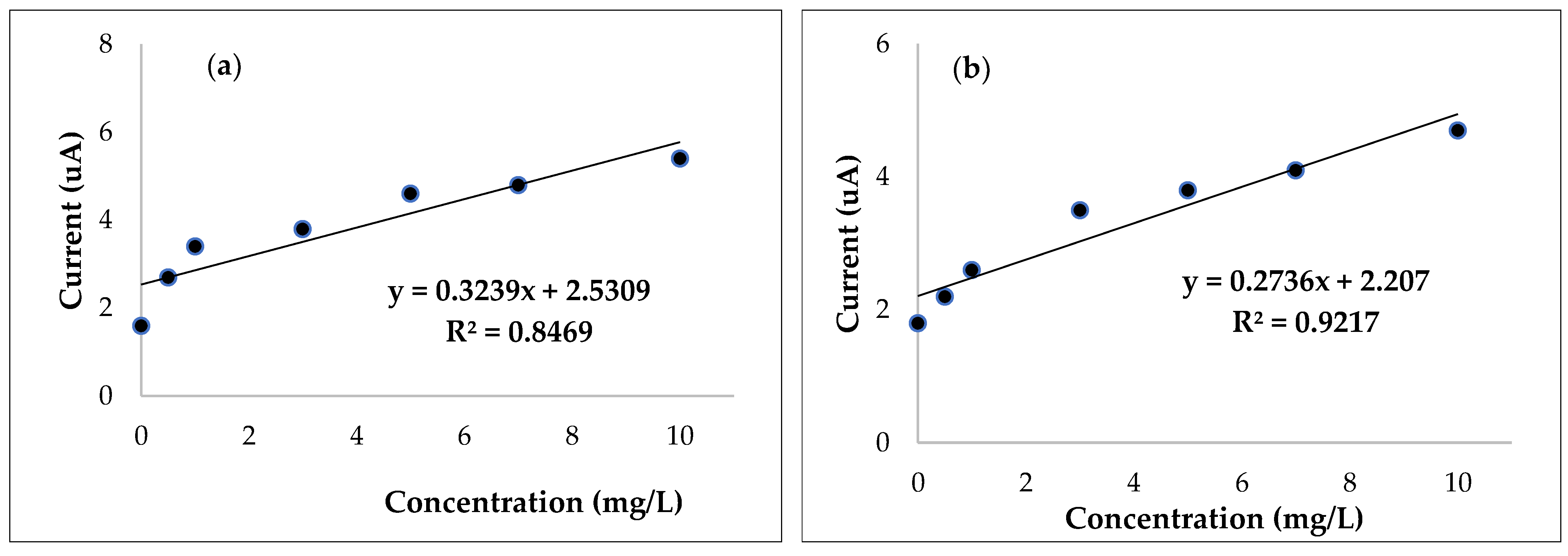
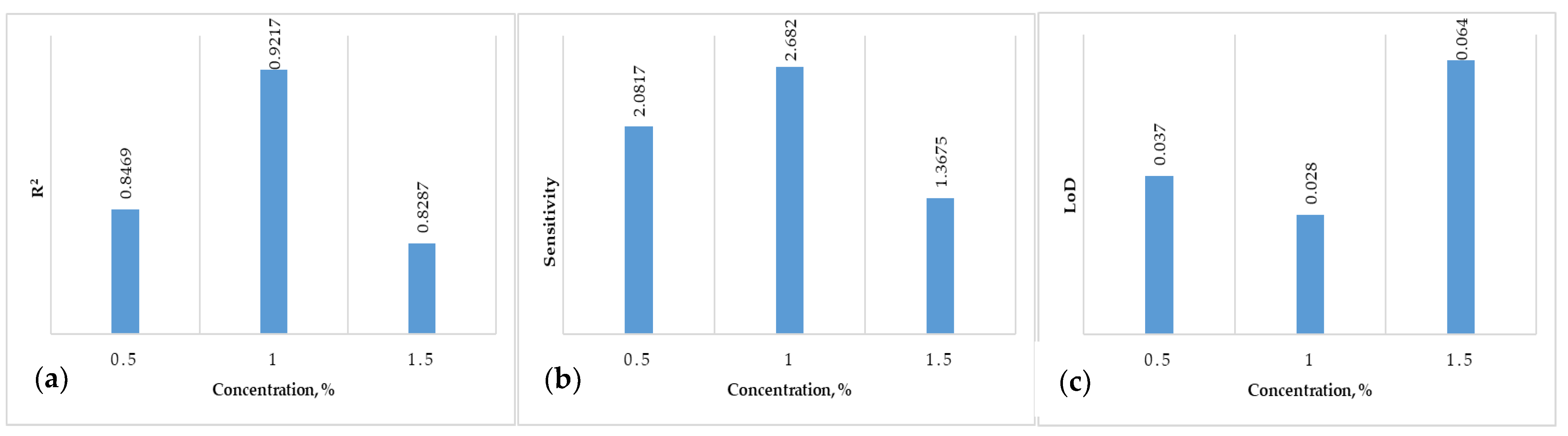
| Carrageenan | 0.5 | 0.8469 | 2.0817 | 0.037 | 6 |
| 1 | 0.9217 | 2.682 | 0.028 | ||
| 1.5 | 0.8287 | 1.3675 | 0.064 | ||
| Chitosan | 0.5 | 0.9778 | 2.5389 | 0.052 | 3 |
| 1 | 0.9096 | 1.5611 | 0.074 | ||
| 1.5 | 0.8411 | 0.6836 | 0.086 |
Disclaimer/Publisher’s Note: The statements, opinions and data contained in all publications are solely those of the individual author(s) and contributor(s) and not of MDPI and/or the editor(s). MDPI and/or the editor(s) disclaim responsibility for any injury to people or property resulting from any ideas, methods, instructions or products referred to in the content. |
© 2023 by the authors. Licensee MDPI, Basel, Switzerland. This article is an open access article distributed under the terms and conditions of the Creative Commons Attribution (CC BY) license (https://creativecommons.org/licenses/by/4.0/).
Share and Cite
Anchidin-Norocel, L.; Savage, W.K.; Gheorghita, R.; Amariei, S. Biopolymers Used for Receptor Immobilization for Nickel-Detection Biosensors in Food. Micromachines 2023, 14, 1529. https://doi.org/10.3390/mi14081529
Anchidin-Norocel L, Savage WK, Gheorghita R, Amariei S. Biopolymers Used for Receptor Immobilization for Nickel-Detection Biosensors in Food. Micromachines. 2023; 14(8):1529. https://doi.org/10.3390/mi14081529
Chicago/Turabian StyleAnchidin-Norocel, Liliana, Wesley K. Savage, Roxana Gheorghita, and Sonia Amariei. 2023. "Biopolymers Used for Receptor Immobilization for Nickel-Detection Biosensors in Food" Micromachines 14, no. 8: 1529. https://doi.org/10.3390/mi14081529