
Epidermal Wearable Biosensors for the Continuous Monitoring of Biomarkers of Chronic Disease in Interstitial Fluid


 ,
,
Abstract
:1. Introduction
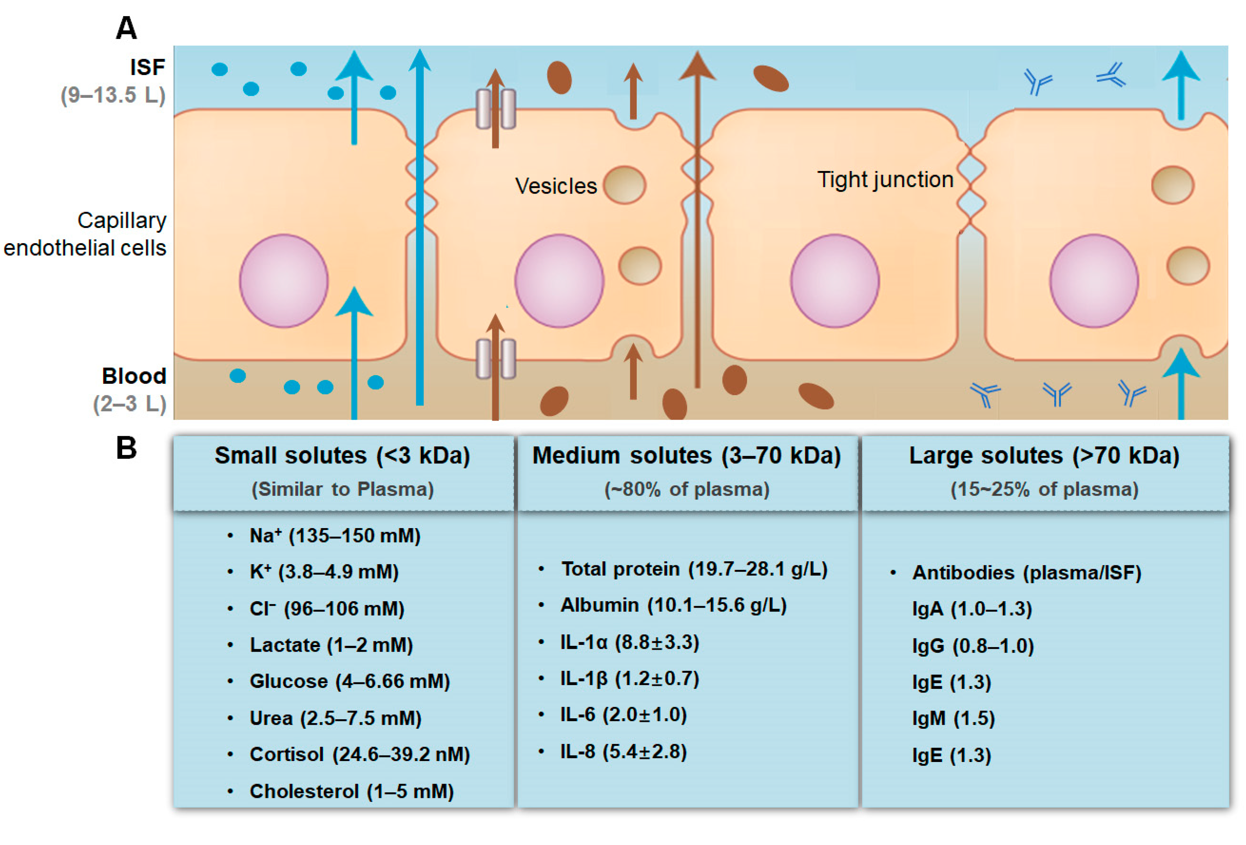
2. ISF Characteristics
3. ISF Extraction Methods
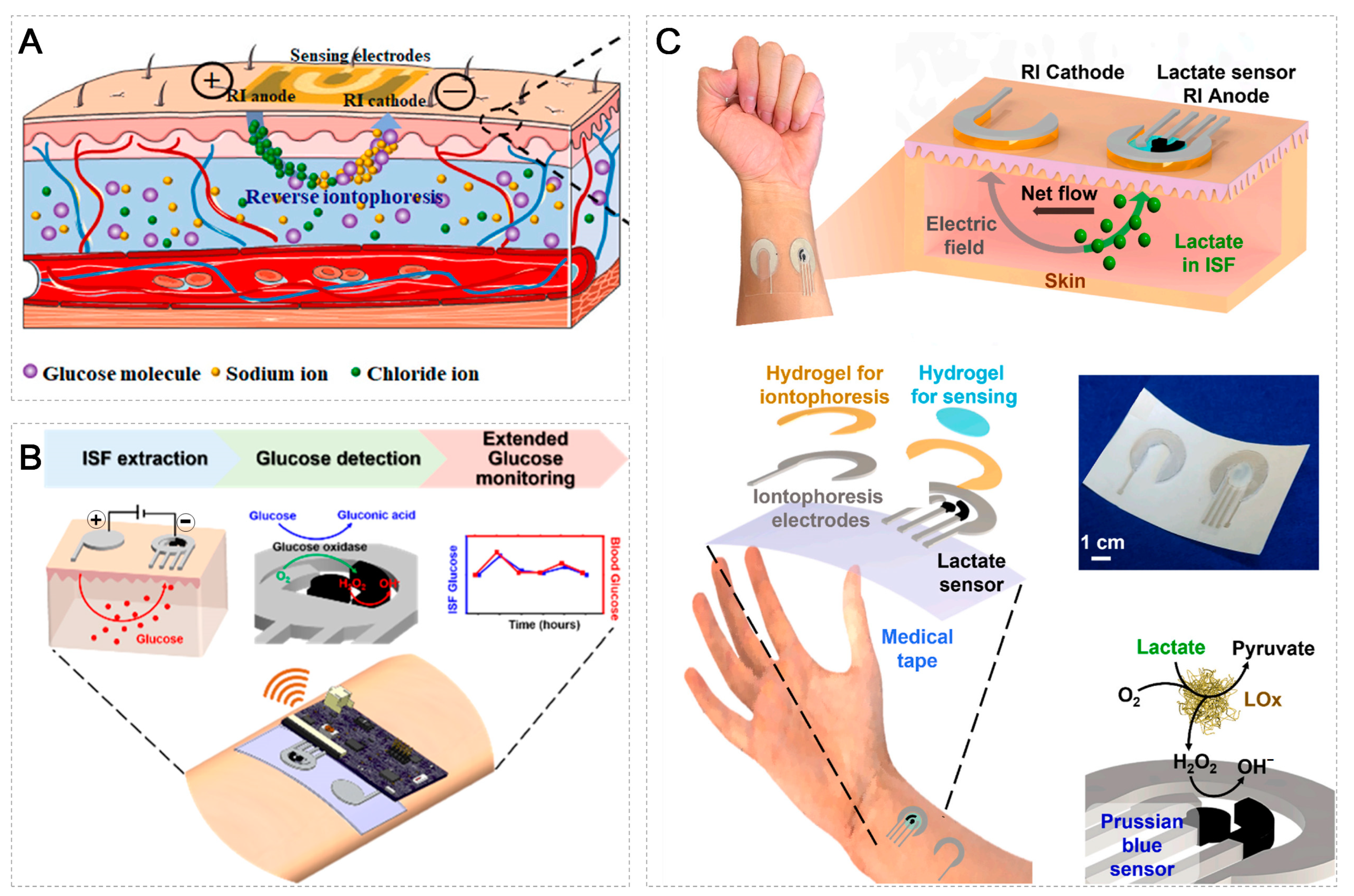
3.1. Reverse Iontophoresis
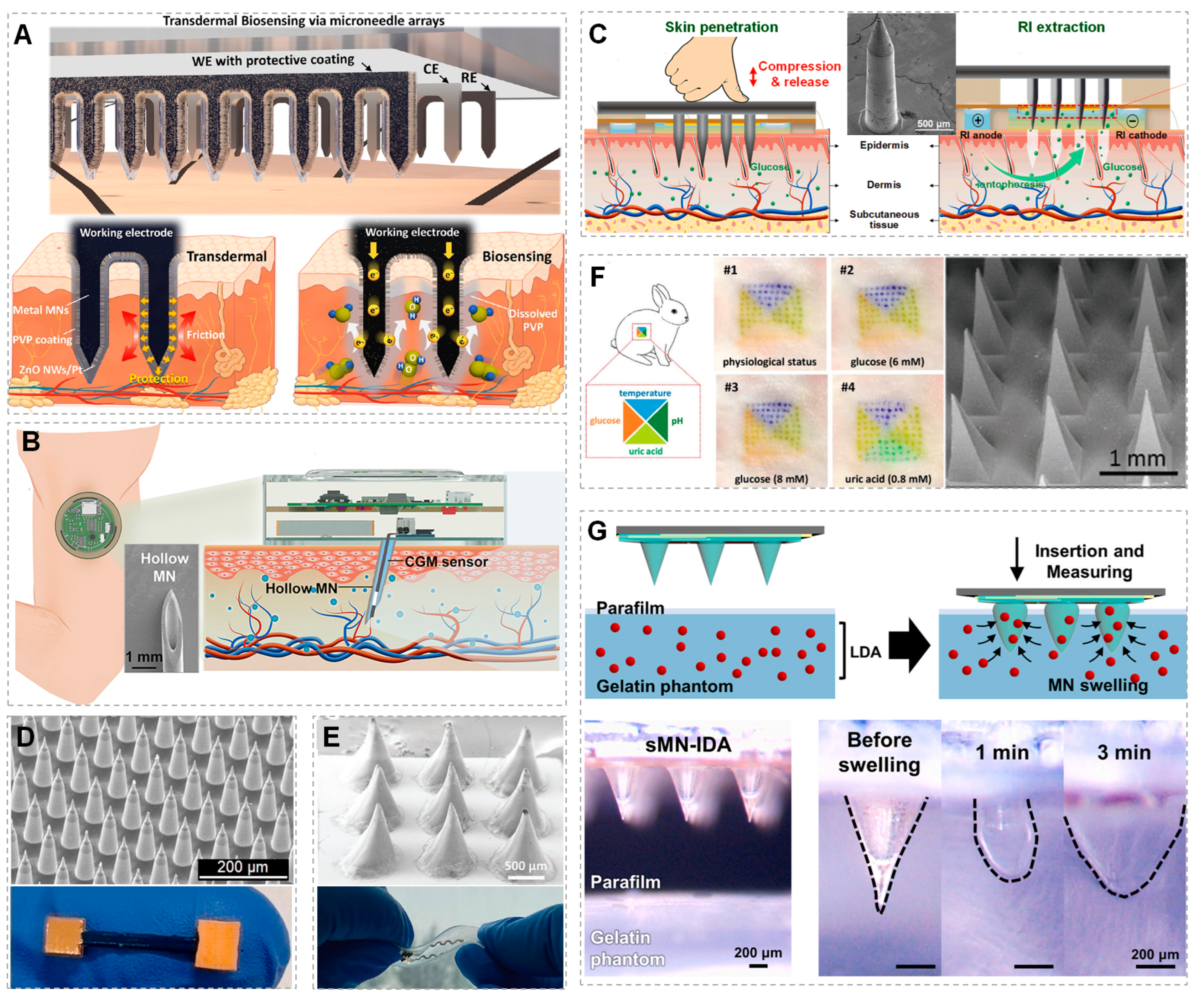
3.2. Microneedle Arrays

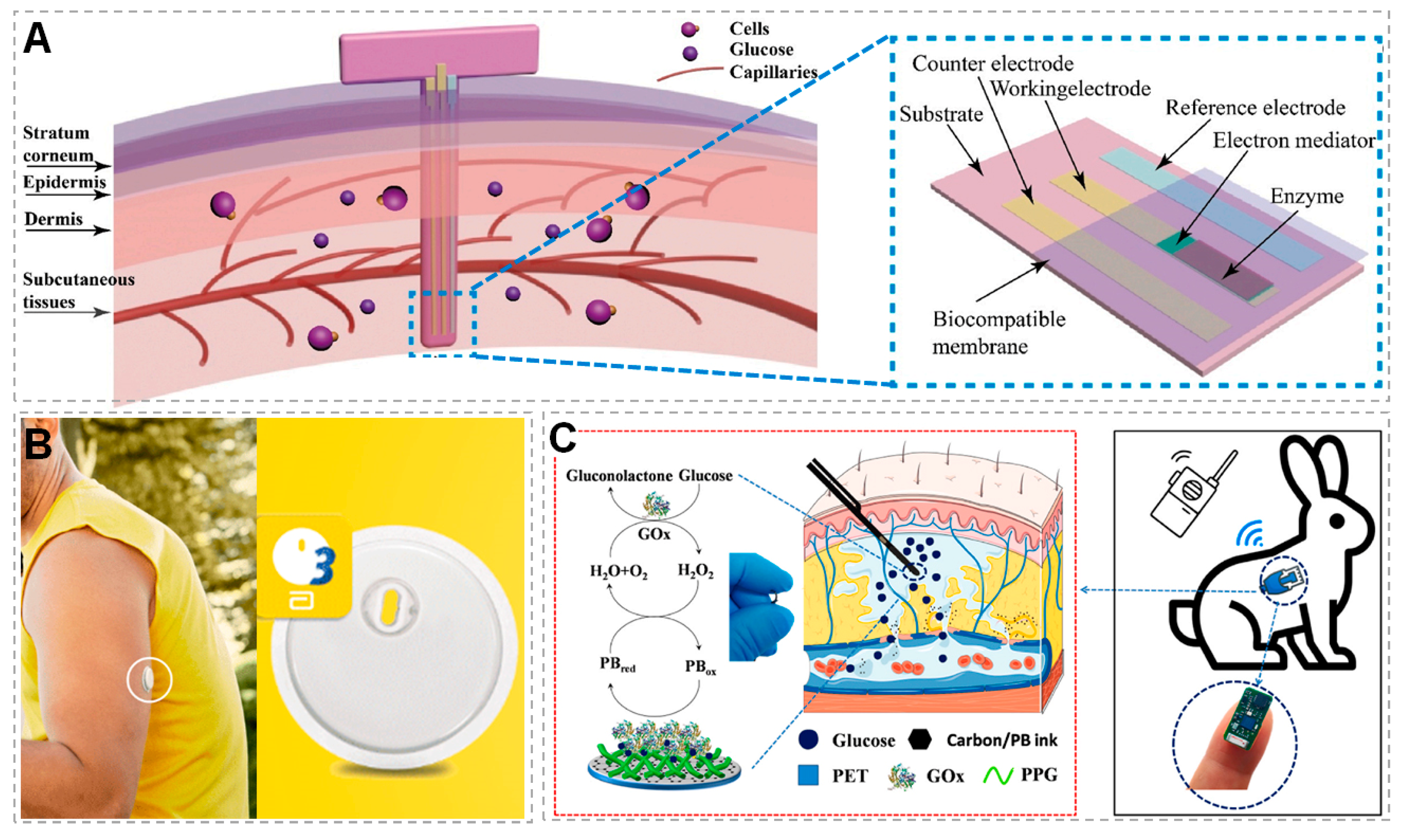
3.3. Indwelling ISF Sensors


| ISF Extraction Methods | Parameters/Related materials | Lasted Time | Detected Biomarkers | Reference |
|---|---|---|---|---|
| RI | Current density = 0.4 mA/cm2 | 10 min | Lactate | [126] |
| Current density = 0.3 mA/cm2 | 5 min | Glucose | [115] | |
| Potential = −3 V | 10 min | Glucose | [126] | |
| Current density = 2 mA/cm2 Potential = 2~5 V | 5 min | Glucose | [125] | |
| Current density = 0.27 mA/cm2 | 3 min | Glucose | [37] | |
| Current density = 0.5 mA/cm2 | 5 min | Glucose | [114] | |
| Current density = 0.3 mA/cm2 | 15 min | Glucose, Lactate | [131] | |
| MNAs | Hydrogel MNA (MeHA 1)
| 3 min | Levodopa, Dopamine | [22] |
Hydrogel MNA (MeHA 1)
| 5 min | Glucose, uric acid, insulin, serotonin | [21] | |
Metal MN
| 2.7 min | Glucose | [144] | |
Metal MNA
| 10 min | Glucose, Na+, K+ | [146] | |
Polymer MNA coated with gold
| 30–40 min | Lactate | [143] | |
Silicon MNA
| 60 min | ErbB2 2 | [58] | |
| Indwelling ISF sensors | Implanted electromagnetic sensor
| Continuous | Glucose | [168] |
Implanted circuit
| Continuous | Glucose | [164] | |
Implanted electrochemical biosensor
| Continuous for 30 days | Glucose | [165] |
4. ISF Sensing Platform for Continuous Monitoring
5. Clinical and Preclinical Applications
6. Summary and Prospect
Author Contributions
Funding
Data Availability Statement
Conflicts of Interest
References
- Kim, J.; Campbell, A.S.; de Avila, B.E.F.; Wang, J. Wearable biosensors for healthcare monitoring. Nat. Biotechnol. 2019, 37, 389–406. [Google Scholar] [CrossRef]
- Yang, Y.; Gao, W. Wearable and flexible electronics for continuous molecular monitoring. Chem. Soc. Rev. 2019, 48, 1465–1491. [Google Scholar] [CrossRef]
- Sun, H.; Zheng, Y.; Shi, G.; Haick, H.; Zhang, M. Wearable Clinic: From Microneedle-Based Sensors to Next-Generation Healthcare Platforms. Small 2023, 2207539. [Google Scholar] [CrossRef] [PubMed]
- Dervisevic, M.; Alba, M.; Prieto-Simon, B.; Voelcker, N.H. Skin in the diagnostics game: Wearable biosensor nano- and microsystems for medical diagnostics. Nano Today 2020, 30, 100828. [Google Scholar] [CrossRef]
- Kukkar, D.; Zhang, D.; Jeon, B.H.; Kim, K.-H. Recent advances in wearable biosensors for non-invasive monitoring of specific metabolites and electrolytes associated with chronic kidney disease: Performance evaluation and future challenges. TrAC Trends Anal. Chem. 2022, 150, 116570. [Google Scholar] [CrossRef]
- Chen, Q.; Zhao, Y.; Liu, Y. Current development in wearable glucose meters. Chin. Chem. Lett. 2021, 32, 3705–3717. [Google Scholar] [CrossRef]
- Yang, X.; Cheng, H. Recent Developments of Flexible and Stretchable Electrochemical Biosensors. Micromachines 2020, 11, 243. [Google Scholar] [CrossRef] [Green Version]
- Heikenfeld, J.; Jajack, A.; Feldman, B.; Granger, S.W.; Gaitonde, S.; Begtrup, G.; Katchman, B.A. Accessing analytes in biofluids for peripheral biochemical monitoring. Nat. Biotechnol. 2019, 37, 407–419. [Google Scholar] [CrossRef]
- Zhao, J.; Guo, H.; Li, J.; Bandodkar, A.J.; Rogers, J.A. Body-Interfaced Chemical Sensors for Noninvasive Monitoring and Analysis of Biofluids. Trends Chem. 2019, 1, 559–571. [Google Scholar] [CrossRef]
- Teymourian, H.; Barfidokht, A.; Wang, J. Electrochemical glucose sensors in diabetes management: An updated review (2010–2020). Chem. Soc. Rev. 2020, 49, 7671–7709. [Google Scholar] [CrossRef]
- Park, H.; Park, W.; Lee, C.H. Electrochemically active materials and wearable biosensors for the in situ analysis of body fluids for human healthcare. NPG Asia Mater. 2021, 13, 23. [Google Scholar] [CrossRef]
- Yuan, X.; Li, C.; Yin, X.; Yang, Y.; Ji, B.; Niu, Y.; Ren, L. Epidermal Wearable Biosensors for Monitoring Biomarkers of Chronic Disease in Sweat. Biosensors 2023, 13, 313. [Google Scholar] [CrossRef]
- Yang, Y.-L.; Chuang, M.-C.; Lou, S.-L.; Wang, J. Thick-film textile-based amperometric sensors and biosensors. Analyst 2010, 135, 1230–1234. [Google Scholar] [CrossRef] [PubMed]
- Zhu, X.; Ju, Y.; Chen, J.; Liu, D.; Liu, H. Nonenzymatic wearable sensor for electrochemical analysis of perspiration glucose. ACS Sens. 2018, 3, 1135–1141. [Google Scholar] [CrossRef]
- Kassal, P.; Kim, J.; Kumar, R.; de Araujo, W.R.; Steinberg, I.M.; Steinberg, M.D.; Wang, J. Smart bandage with wireless connectivity for uric acid biosensing as an indicator of wound status. Electrochem. Commun. 2015, 56, 6–10. [Google Scholar] [CrossRef]
- Kim, J.; de Araujo, W.R.; Samek, I.A.; Bandodkar, A.J.; Jia, W.; Brunetti, B.; Paixao, T.R.; Wang, J. Wearable temporary tattoo sensor for real-time trace metal monitoring in human sweat. Electrochem. Commun. 2015, 51, 41–45. [Google Scholar] [CrossRef]
- Madden, J.; O’Mahony, C.; Thompson, M.; O’Riordan, A.; Galvin, P. Biosensing in dermal interstitial fluid using microneedle based electrochemical devices. Sens. Bio-Sens. Res. 2020, 29, 100348. [Google Scholar] [CrossRef]
- Dervisevic, M.; Dervisevic, E.; Esser, L.; Easton, C.D.; Cadarso, V.J.; Voelcker, N.H. Wearable microneedle array-based sensor for transdermal monitoring of pH levels in interstitial fluid. Biosens. Bioelectron. 2023, 222, 114955. [Google Scholar] [CrossRef] [PubMed]
- Shukla, S.; Machekposhti, S.A.; Joshi, N.; Joshi, P.; Narayan, R.J. Microneedle-Integrated Device for Transdermal Sampling and Analyses of Targeted Biomarkers. Small Sci. 2023, 3, 2200087. [Google Scholar] [CrossRef]
- De la Paz, E.; Saha, T.; Del Cano, R.; Seker, S.; Kshirsagar, N.; Wang, J. Non-invasive monitoring of interstitial fluid lactate through an epidermal iontophoretic device. Talanta 2023, 254, 124122. [Google Scholar] [CrossRef] [PubMed]
- Keyvani, F.; Zheng, H.; Kaysir, M.R.; Mantaila, D.F.; Ghavami Nejad, P.; Rahman, F.A.; Quadrilatero, J.; Ban, D.; Poudineh, M. A Hydrogel Microneedle Assay Combined with Nucleic Acid Probes for On-Site Detection of Small Molecules and Proteins. Angew. Chem. Int. Ed. Engl. 2023, 62, e202301624. [Google Scholar] [CrossRef] [PubMed]
- Park, S.; Kim, Y.J.; Kostal, E.; Matylitskaya, V.; Partel, S.; Ryu, W. Highly-sensitive single-step sensing of levodopa by swellable microneedle-mounted nanogap sensors. Biosens. Bioelectron. 2023, 220, 114912. [Google Scholar] [CrossRef]
- Fogh-Andersen, N.; Altura, B.M.; Altura, B.T.; Siggaard-Andersen, O. Composition of interstitial fluid. Clin. Chem. 1995, 41, 1522–1525. [Google Scholar] [CrossRef]
- Friedel, M.; Thompson, I.A.P.; Kasting, G.; Polsky, R.; Cunningham, D.; Soh, H.T.; Heikenfeld, J. Opportunities and challenges in the diagnostic utility of dermal interstitial fluid. Nat. Biomed. Eng. 2023, 2207539. [Google Scholar] [CrossRef]
- Volden, G.; Thorsrud, A.K.; Bjornson, I.; Jellum, E. Biochemical-composition of suction blister fluid determined by high-resolution multicomponent analysis (capillary gas-chromatography—Mass-spectrometry and two-dimensional electrophoresis). J. Investig. Dermatol. 1980, 75, 421–424. [Google Scholar] [CrossRef] [PubMed] [Green Version]
- Baker, L.B.; Wolfe, A.S. Physiological mechanisms determining eccrine sweat composition. Eur. J. Appl. Physiol. 2020, 120, 719–752. [Google Scholar] [CrossRef] [Green Version]
- Vanhaeringen, N.J. Clinical biochemistry of tears. Surv. Ophthalmol. 1981, 26, 84–96. [Google Scholar] [CrossRef]
- Gonçalves, A.C.; Marson, F.A.L.; Mendonça, R.M.H.; Bertuzzo, C.S.; Paschoal, I.A.; Ribeiro, J.D.; Ribeiro, A.F.; Levy, C.E. Chloride and sodium ion concentrations in saliva and sweat as a method to diagnose cystic fibrosis. J. De Pediatr. 2019, 95, 443–450. [Google Scholar] [CrossRef]
- Kunnel, B.P.; Demuru, S. An epidermal wearable microfluidic patch for simultaneous sampling, storage, and analysis of biofluids with counterion monitoring. Lab. A Chip 2022, 22, 1793–1804. [Google Scholar] [CrossRef] [PubMed]
- Ray, T.R.; Ivanovic, M.; Curtis, P.M.; Franklin, D.; Guventurk, K.; Jeang, W.J.; Chafetz, J.; Gaertner, H.; Young, G.; Rebollo, S.; et al. Soft, skin-interfaced sweat stickers for cystic fibrosis diagnosis and management. Sci. Transl. Med. 2021, 13, eabd8109. [Google Scholar] [CrossRef] [PubMed]
- Badugu, R.; Jeng, B.H.; Reece, E.A.; Lakowicz, J.R. Contact lens to measure individual ion concentrations in tears and applications to dry eye disease. Anal. Biochem. 2018, 542, 84–94. [Google Scholar] [CrossRef]
- Nery, E.W.; Kundys, M.; Jelen, P.S.; Jonsson-Niedziolka, M. Electrochemical Glucose Sensing: Is There Still Room for Improvement? Anal. Chem. 2016, 88, 11271–11282. [Google Scholar] [CrossRef] [PubMed] [Green Version]
- Moyer, J.; Wilson, D.; Finkelshtein, I.; Wong, B.; Potts, R. Correlation between sweat glucose and blood glucose in subjects with diabetes. Diabetes Technol. Ther. 2012, 14, 398–402. [Google Scholar] [CrossRef] [PubMed]
- Yao, H.; Shum, A.J.; Cowan, M.; Laehdesmaeki, I.; Parviz, B.A. A contact lens with embedded sensor for monitoring tear glucose level. Biosens. Bioelectron. 2011, 26, 3290–3296. [Google Scholar] [CrossRef] [PubMed] [Green Version]
- Ye, D.; Liang, G.; Li, H.; Luo, J.; Zhang, S.; Chen, H.; Kong, J. A novel nonenzymatic sensor based on CuO nanoneedle/graphene/carbon nanofiber modified electrode for probing glucose in saliva. Talanta 2013, 116, 223–230. [Google Scholar] [CrossRef] [PubMed]
- Panchbhai, A.S. Correlation of salivary glucose level with blood glucose level in diabetes mellitus. J. Oral. Maxillofac. Res. 2012, 3, e3. [Google Scholar] [CrossRef] [Green Version]
- De la Paz, E.; Barfidokht, A.; Rios, S.; Brown, C.; Chao, E.; Wang, J. Extended Noninvasive Glucose Monitoring in the Interstitial Fluid Using an Epidermal Biosensing Patch. Anal. Chem. 2021, 93, 12767–12775. [Google Scholar] [CrossRef]
- Asaduzzaman, M.; Zahed, M.A.; Sharifuzzaman, M.; Reza, M.S.; Hui, X.; Sharma, S.; Shin, Y.D.; Park, J.Y. A hybridized nano-porous carbon reinforced 3D graphene-based epidermal patch for precise sweat glucose and lactate analysis. Biosens. Bioelectron. 2022, 219, 114846. [Google Scholar] [CrossRef] [PubMed]
- Li, Z.K.; Yun, J.H.; Li, X.Y.; Kim, M.; Li, J.; Lee, D.H.; Wu, A.Y.; Lee, S.W. Power-Free Contact Lens for Glucose Sensing. Adv. Funct. Mater. 2023, 2304647. [Google Scholar] [CrossRef]
- Arakawa, T.; Tomoto, K.; Nitta, H.; Toma, K.; Takeuchi, S.; Sekita, T.; Minakuchi, S.; Mitsubayashi, K. A Wearable Cellulose Acetate-Coated Mouthguard Biosensor for In Vivo Salivary Glucose Measurement. Anal. Chem. 2020, 92, 12201–12207. [Google Scholar] [CrossRef]
- Messenger, B.; Clifford, M.N.; Morgan, L.M. Glucose-dependent insulinotropic polypeptide and insulin-like immunoreactivity in saliva following sham-fed and swallowed meals. J. Endocrinol. 2003, 177, 407–412. [Google Scholar] [CrossRef] [Green Version]
- Mork, T.A.; Killeen, C.T.; Patel, N.K.; Dohnal, J.M.; Karydes, H.C.; Leikin, J.B. Massive Insulin Overdose Managed by Monitoring Daily Insulin Levels. Am. J. Ther. 2011, 18, E162–E166. [Google Scholar] [CrossRef]
- Tékus, E.; Kaj, M.; Szabó, E.; Szénási, N.L.; Kerepesi, I.; Figler, M.; Gábriel, R.; Wilhelm, M. Comparison of blood and saliva lactate level after maximum intensity exercise. Acta Biol. Hung. 2012, 63, 89–98. [Google Scholar] [CrossRef] [PubMed]
- Bollella, P.; Sharma, S.; Cass, A.E.G.; Antiochia, R. Minimally-invasive microneedle-based biosensor array for simultaneous lactate and glucose monitoring in artificial interstitial fluid. Electroanalysis 2019, 31, 374–382. [Google Scholar] [CrossRef] [Green Version]
- Xuan, X.; Perez-Rafols, C.; Chen, C.; Cuartero, M.; Crespo, G.A. Lactate Biosensing for Reliable On-Body Sweat Analysis. ACS Sens. 2021, 6, 2763–2771. [Google Scholar] [CrossRef] [PubMed]
- Takamatsu, T.; Sijie, Y.; Miyake, T. Wearable, Implantable, Parity-Time Symmetric Bioresonators for Extremely Small Biological Signal Monitoring. Adv. Mater. Technol. 2023, 8, 2201704. [Google Scholar] [CrossRef]
- Xu, Z.; Song, J.; Liu, B.; Lv, S.; Gao, F.; Luo, X.; Wang, P. A conducting polymer PEDOT:PSS hydrogel based wearable sensor for accurate uric acid detection in human sweat. Sens. Actuators B Chem. 2021, 348, 130674. [Google Scholar] [CrossRef]
- Mendelsohn, M.E.; Abramson, D.H.; Senft, S.; Servodidio, C.A.; Gamache, P.H. Uric acid in the aqueous humor and tears of retinoblastoma patients. J. Am. Assoc. Pediatr. Ophthalmol. Strabismus 1998, 2, 369–371. [Google Scholar] [CrossRef]
- Zhao, J.; Huang, Y. Salivary uric acid as a noninvasive biomarker for monitoring the efficacy of urate-lowering therapy in a patient with chronic gouty arthropathy. Clin. Chim. Acta 2015, 450, 115–120. [Google Scholar] [CrossRef]
- Das, M.; Borah, N.C.; Ghose, M.; Choudhury, N. Reference ranges for serum uric acid among healthy Assamese people. Biochem. Res. Int. 2014, 2014, 171053. [Google Scholar] [CrossRef]
- Wang, Q.; Oyarzabal, E.A.; Song, S.; Wilson, B.; Santos, J.H.; Hong, J.-S. Locus coeruleus neurons are most sensitive to chronic neuroinflammation-induced neurodegeneration. Brain Behav. Immun. 2020, 87, 359–368. [Google Scholar] [CrossRef] [PubMed]
- Zhang, P.; Wu, X.; Xue, H.; Wang, Y.; Luo, X.; Wang, L. Wearable transdermal colorimetric microneedle patch for Uric acid monitoring based on peroxidase-like polypyrrole nanoparticles. Anal. Chim. Acta 2022, 1212, 339911. [Google Scholar] [CrossRef] [PubMed]
- Bi, Y.; Sun, M.; Wang, J.; Zhu, Z.; Bai, J.; Emran, M.Y.; Kotb, A.; Bo, X.; Zhou, M. Universal Fully Integrated Wearable Sensor Arrays for the Multiple Electrolyte and Metabolite Monitoring in Raw Sweat, Saliva, or Urine. Anal. Chem. 2023, 95, 6690–6699. [Google Scholar] [CrossRef] [PubMed]
- Moreddu, R.; Nasrollahi, V.; Kassanos, P.; Dimov, S.; Vigolo, D.; Yetisen, A.K. Lab-on-a-Contact Lens Platforms Fabricated by Multi-Axis Femtosecond Laser Ablation. SMALL 2021, 17, 2102008. [Google Scholar] [CrossRef]
- Kim, J.; Imani, S.; de Araujo, W.R.; Warchall, J.; Valdes-Ramirez, G.; Paixao, T.; Mercier, P.P.; Wang, J. Wearable salivary uric acid mouthguard biosensor with integrated wireless electronics. Biosens. Bioelectron. 2015, 74, 1061–1068. [Google Scholar] [CrossRef] [PubMed] [Green Version]
- Honarmand, M.H.; Farhad-Mollashahi, L.; Nakhaee, A.; Nehi, M. Salivary Levels of ErbB2 and CEA in Oral Squamous Cell Carcinoma Patients. Asian Pac. J. Cancer Prev. APJCP 2016, 17, 77–80. [Google Scholar] [CrossRef] [Green Version]
- Capobianco, J.A.; Shih, W.Y.; Yuan, Q.A.; Adams, G.P.; Shih, W.H. Label-free, all-electrical, in situ human epidermal growth receptor 2 detection. Rev. Sci. Instrum. 2008, 79, 076101. [Google Scholar] [CrossRef] [Green Version]
- Dervisevic, M.; Alba, M.; Adams, T.E.; Prieto-Simon, B.; Voelcker, N.H. Electrochemical immunosensor for breast cancer biomarker detection using high-density silicon microneedle array. Biosens. Bioelectron. 2021, 192, 113496. [Google Scholar] [CrossRef]
- Gunther, V.; Bauer, I.; Hedderich, J.; Mettler, L.; Schubert, M.; Mackelenbergh, M.T.; Maass, N.; Alkatout, I. Changes of salivary estrogen levels for detecting the fertile period. Eur. J. Obs. Gynecol. Reprod. Biol. 2015, 194, 38–42. [Google Scholar] [CrossRef]
- Shin, Y.Y.; Jeong, J.S.; Park, M.-N.; Lee, J.-E.; An, S.-M.; Cho, W.-S.; Kim, S.C.; An, B.-S.; Lee, K.-S. Regulation of steroid hormones in the placenta and serum of women with preeclampsia. Mol. Med. Rep. 2018, 17, 2681–2688. [Google Scholar] [CrossRef] [Green Version]
- Kang, Y.E.; Seong, K.Y.; Yim, S.G.; Lee, Y.; An, S.M.; Kim, S.C.; Kim, K.; An, B.S.; Lee, K.S.; Yang, S.Y. Nanochannel-driven rapid capture of sub-nanogram level biomarkers for painless preeclampsia diagnosis. Biosens. Bioelectron. 2020, 163, 112281. [Google Scholar] [CrossRef] [PubMed]
- Forman, H.J.; Bernardo, A.; Davies, K.J. What is the concentration of hydrogen peroxide in blood and plasma? Arch. Biochem. Biophys. 2016, 603, 48–53. [Google Scholar] [CrossRef]
- Jin, Q.; Chen, H.-J.; Li, X.; Huang, X.; Wu, Q.; He, G.; Hang, T.; Yang, C.; Jiang, Z.; Li, E.; et al. Reduced Graphene Oxide Nanohybrid-Assembled Microneedles as Mini-Invasive Electrodes for Real-Time Transdermal Biosensing. Small 2019, 15, 1804298. [Google Scholar] [CrossRef]
- Barber, R.; Davis, J.; Papakonstantinou, P. Stable Chitosan and Prussian Blue-Coated Laser-Induced Graphene Skin Sensor for the Electrochemical Detection of Hydrogen Peroxide in Sweat. Acs Appl. Nano Mater. 2023, 6, 10290–10302. [Google Scholar] [CrossRef]
- Venugopal, M.; Arya, S.K.; Chornokur, G.; Bhansali, S. A Realtime and Continuous Assessment of Cortisol in ISF Using Electrochemical Impedance Spectroscopy. Sens. Actuators A Phys. 2011, 172, 154–160. [Google Scholar] [CrossRef] [PubMed] [Green Version]
- Jia, M.; Chew, W.M.; Feinstein, Y.; Skeath, P.; Sternberg, E.M. Quantification of cortisol in human eccrine sweat by liquid chromatography—Tandem mass spectrometry. Analyst 2016, 141, 2053–2060. [Google Scholar] [CrossRef] [Green Version]
- Ku, M.; Kim, J.; Won, J.E.; Kang, W.; Park, Y.G.; Park, J.; Lee, J.H.; Cheon, J.; Lee, H.H.; Park, J.U. Smart, soft contact lens for wireless immunosensing of cortisol. Sci. Adv. 2020, 6, eabb2891. [Google Scholar] [CrossRef]
- Cohen, J.; Deans, R.; Dalley, A.; Lipman, J.; Roberts, M.S.; Venkatesh, B. Measurement of tissue cortisol levels in patients with severe burns: A preliminary investigation. Crit. Care 2009, 13, R189. [Google Scholar] [CrossRef] [Green Version]
- Lee, H.-B.; Meeseepong, M.; Tran Quang, T.; Kim, B.-Y.; Lee, N.-E. A wearable lab-on-a-patch platform with stretchable nanostructured biosensor for non-invasive immunodetection of biomarker in sweat. Biosens. Bioelectron. 2020, 156, 112133. [Google Scholar] [CrossRef]
- Wang, B.; Zhao, C.Z.; Wang, Z.Q.; Yang, K.A.; Cheng, X.B.; Liu, W.F.; Yu, W.Z.; Lin, S.Y.; Zhao, Y.C.; Cheung, K.M.; et al. Wearable aptamer-field-effect transistor sensing system for noninvasive cortisol monitoring. Sci. Adv. 2022, 8, eabk0967. [Google Scholar] [CrossRef]
- Martin, X.D.; Brennan, M.C. Serotonin in human tears. Eur. J. Ophthalmol. 1994, 4, 159–165. [Google Scholar] [CrossRef] [PubMed]
- Karbownik, M.S.; Hicks, S.D. The Association of Salivary Serotonin With Mood and Cardio-Autonomic Function: A Preliminary Report. Front. Psychiatry 2022, 13, 788153. [Google Scholar] [CrossRef] [PubMed]
- Carling, R.S.; Degg, T.J.; Allen, K.R.; Bax, N.D.S.; Barth, J.H. Evaluation of Whole Blood Serotonin and Plasma and Urine 5-Hydroxyindole Acetic Acid in Diagnosis of Carcinoid Disease. Ann. Clin. Biochem. 2002, 39, 577–582. [Google Scholar] [CrossRef] [PubMed]
- Tyano, S.; Zalsman, G.; Ofek, H.; Blum, I.; Apter, A.; Wolovik, L.; Sher, L.; Sommerfeld, E.; Harell, D.; Weizman, A. Plasma serotonin levels and suicidal behavior in adolescents. Eur. Neuropsychopharmacol. 2006, 16, 49–57. [Google Scholar] [CrossRef] [PubMed]
- Parlak, O.; Curto, V.F.; Ojeda, E.; Basabe-Desmonts, L.; Benito-Lopez, F.; Salleo, A. Wearable biosensors and sample handling strategies. In Wearable Bioelectronics; Elsevier: Amsterdam, The Netherlands, 2020; pp. 65–88. [Google Scholar]
- Thennadil, S.N.; Rennert, J.L.; Wenzel, B.J.; Hazen, K.H.; Ruchti, T.L.; Block, M.B. Comparison of glucose concentration in interstitial fluid, and capillary and venous blood during rapid changes in blood glucose levels. Diabetes Technol. Ther. 2001, 3, 357–365. [Google Scholar] [CrossRef] [PubMed] [Green Version]
- Cooke, I.; Bevill, R.; Koritz, G. Pharmacokinetics of penicillin G in plasma and interstitial fluid collected with dialysis fiber bundles in sheep. Vet. Res. 1996, 27, 147–160. [Google Scholar]
- Zeitlinger, M.A.; Derendorf, H.; Mouton, J.W.; Cars, O.; Craig, W.A.; Andes, D.; Theuretzbacher, U. Protein binding: Do we ever learn? Antimicrob. Agents Chemother. 2011, 55, 3067–3074. [Google Scholar] [CrossRef] [Green Version]
- Vermeer, B.J.; Reman, F.C.; van Gent, C.M. The determination of lipids and proteins in suction blister fluid. J. Investig. Dermatol. 1979, 73, 303–305. [Google Scholar] [CrossRef] [Green Version]
- Haaverstad, R.; Romslo, I.; Larsen, S.; Myhre, H.O. Protein concentration of subcutaneous interstitial fluid in the human leg—A comparison between the wick technique and the blister suction technique. Int. J. Microcirc.-Clin. Exp. 1996, 16, 111–117. [Google Scholar] [CrossRef]
- Clough, G.F.; Jackson, C.L.; Lee, J.J.; Jamal, S.C.; Church, M.K. What can microdialysis tell us about the temporal and spatial generation of cytokines in allergen-induced responses in human skin in vivo? J. Investig. Dermatol. 2007, 127, 2799–2806. [Google Scholar] [CrossRef] [Green Version]
- Arevalo, M.T.; Rizzo, G.M.; Polsky, R.; Glaros, T.; Mach, P.M. Proteomic Characterization of Immunoglobulin Content in Dermal Interstitial Fluid. J. Proteome Res. 2019, 18, 2381–2384. [Google Scholar] [CrossRef]
- Sharma, A.K.; Badea, M.; Tiwari, S.; Marty, J.-L. Wearable Biosensors: An Alternative and Practical Approach in Healthcare and Disease Monitoring. Molecules 2021, 26, 748. [Google Scholar] [CrossRef] [PubMed]
- Smith, A.A.; Li, R.; Tse, Z.T.H. Reshaping healthcare with wearable biosensors. Sci. Rep. 2023, 13, 4998. [Google Scholar] [CrossRef]
- He, R.; Liu, H.; Fang, T.; Niu, Y.; Zhang, H.; Han, F.; Gao, B.; Li, F.; Xu, F. A Colorimetric Dermal Tattoo Biosensor Fabricated by Microneedle Patch for Multiplexed Detection of Health-Related Biomarkers. Adv. Sci. 2021, 8, e2103030. [Google Scholar] [CrossRef] [PubMed]
- Parrilla, M.; Detamornrat, U.; Dominguez-Robles, J.; Donnelly, R.F.; De Wael, K. Wearable hollow microneedle sensing patches for the transdermal electrochemical monitoring of glucose. Talanta 2022, 249, 123695. [Google Scholar] [CrossRef]
- Zhu, D.D.; Zheng, L.W.; Duong, P.K.; Cheah, R.H.; Liu, X.Y.; Wong, J.R.; Wang, W.J.; Tien Guan, S.T.; Zheng, X.T.; Chen, P. Colorimetric microneedle patches for multiplexed transdermal detection of metabolites. Biosens. Bioelectron. 2022, 212, 114412. [Google Scholar] [CrossRef] [PubMed]
- Goud, K.Y.; Moonla, C.; Mishra, R.K.; Yu, C.M.; Narayan, R.; Litvan, I.; Wang, J. Wearable Electrochemical Microneedle Sensor for Continuous Monitoring of Levodopa: Toward Parkinson Management. ACS Sens. 2019, 4, 2196–2204. [Google Scholar] [CrossRef]
- Ventura, S.A.; Heikenfeld, J.; Brooks, T.; Esfandiari, L.; Boyce, S.; Park, Y.; Kasting, G.B. Cortisol extraction through human skin by reverse iontophoresis. Bioelectrochemistry 2017, 114, 54–60. [Google Scholar] [CrossRef]
- Wascotte, V.; Rozet, E.; Salvaterra, A.; Hubert, P.; Jadoul, M.; Guy, R.H.; Préat, V. Non-invasive diagnosis and monitoring of chronic kidney disease by reverse iontophoresis of urea in vivo. Eur. J. Pharm. Biopharm. 2008, 69, 1077–1082. [Google Scholar] [CrossRef]
- Senel, M.; Dervisevic, M.; Voelcker, N.H. Gold microneedles fabricated by casting of gold ink used for urea sensing. Mater. Lett. 2019, 243, 50–53. [Google Scholar] [CrossRef]
- Bollella, P.; Sharma, S.; Cass, A.E.G.; Antiochia, R. Microneedle-based biosensor for minimally-invasive lactate detection. Biosens. Bioelectron. 2019, 123, 152–159. [Google Scholar] [CrossRef] [PubMed] [Green Version]
- Ming, D.K.; Jangam, S.; Gowers, S.A.N.; Wilson, R.; Freeman, D.M.E.; Boutelle, M.G.; Cass, A.E.G.; O’Hare, D.; Holmes, A.H. Real-time continuous measurement of lactate through a minimally invasive microneedle patch: A phase I clinical study. BMJ Innov. 2022, 8, 87–94. [Google Scholar] [CrossRef]
- Sekar, M.; Sriramprabha, R.; Sekhar, P.K.; Bhansali, S.; Ponpandian, N.; Pandiaraj, M.; Viswanathan, C. Review—Towards Wearable Sensor Platforms for the Electrochemical Detection of Cortisol. J. Electrochem. Soc. 2020, 167, 067508. [Google Scholar] [CrossRef]
- Guyton, A.C.; Frank, M.; Abernathy, B. A concept of negative interstitial pressure based on pressures in implanted perforated capsules. Circ. Res. 1963, 12, 399–414. [Google Scholar] [CrossRef] [PubMed]
- Zhang, J.; Hao, N.; Liu, W.; Lu, M.; Sun, L.; Chen, N.; Wu, M.; Zhao, X.; Xing, B.; Sun, W. In-depth proteomic analysis of tissue interstitial fluid for hepatocellular carcinoma serum biomarker discovery. Br. J. Cancer 2017, 117, 1676–1684. [Google Scholar] [CrossRef] [Green Version]
- Ling, J.; Ng, J.K.; Chan, J.C.; Chow, E. Use of Continuous Glucose Monitoring in the Assessment and Management of Patients With Diabetes and Chronic Kidney Disease. Front. Endocrinol. 2022, 13, 869899. [Google Scholar] [CrossRef] [PubMed]
- Zhang, B.L.; Zhang, X.P.; Chen, B.Z.; Fei, W.M.; Cui, Y.; Guo, X.D. Microneedle-assisted technology for minimally invasive medical sensing. Microchem. J. 2021, 162, 105830. [Google Scholar] [CrossRef]
- Chinnadayyala, S.R.; Park, J.; Satti, A.T.; Kim, D.; Cho, S. Minimally invasive and continuous glucose monitoring sensor based on non-enzymatic porous platinum black-coated gold microneedles. Electrochim. Acta 2021, 369, 137691. [Google Scholar] [CrossRef]
- Zheng, H.; Pu, Z.; Wu, H.; Li, C.; Zhang, X.; Li, D. Reverse iontophoresis with the development of flexible electronics: A review. Biosens. Bioelectron. 2023, 223, 115036. [Google Scholar] [CrossRef]
- Wiig, H.; Heir, S.; Aukland, K. Colloid osmotic pressure of interstitial fluid in rat subcutis and skeletal muscle: Comparison of various wick sampling techniques. Acta Physiol. Scand. 1988, 133, 167–175. [Google Scholar] [CrossRef]
- Baldini, F. Microdialysis-based sensing in clinical applications. Anal. Bioanal. Chem. 2010, 397, 909–916. [Google Scholar] [CrossRef] [PubMed]
- Bhake, R.; Russell, G.M.; Kershaw, Y.; Stevens, K.; Zaccardi, F.; Warburton, V.E.C.; Linthorst, A.C.E.; Lightman, S.L. Continuous Free Cortisol Profiles in Healthy Men Validation of Microdialysis Method. J. Clin. Endocrinol. Metab. 2020, 105, e1749–e1761. [Google Scholar] [CrossRef] [PubMed]
- Pan, M.; Guo, X.; Cai, Q.; Li, G.; Chen, Y. A novel glucose sensor system with Au nanoparticles based on microdialysis and coenzymes for continuous glucose monitoring. Sens. Actuators A Phys. 2003, 108, 258–262. [Google Scholar] [CrossRef]
- Bungay, P.M.; Morrison, P.F.; Dedrick, R.L. Steady-state theory for quantitative microdialysis of solutes and water in vivo and in vitro. Life Sci. 1990, 46, 105–119. [Google Scholar] [CrossRef]
- Li, T.; Li, S.; Shi, J.; Li, X.; Liu, J.; Yang, H.; Wu, W.; Zhao, L.; Zhao, Z. Real-time analysis of metabolites in vivo by online extraction electrospray ionization mass spectrometry coupled to microdialysis. Anal. Chim. Acta 2022, 1205, 339760. [Google Scholar] [CrossRef] [PubMed]
- Basu, A.; Dube, S.; Slama, M.; Errazuriz, I.; Amezcua, J.C.; Kudva, Y.C.; Peyser, T.; Carter, R.E.; Cobelli, C.; Basu, R. Time lag of glucose from intravascular to interstitial compartment in humans. Diabetes 2013, 62, 4083–4087. [Google Scholar] [CrossRef] [Green Version]
- Sjögren, F.; Svensson, C.; Anderson, C. Technical prerequisites for in vivo microdialysis determination of interleukin-6 in human dermis. Br. J. Dermatol. 2002, 146, 375–382. [Google Scholar]
- Ungerstedt, U. Microdialysis--a new technique for monitoring local tissue events in the clinic. Acta Anaesthesiol. Scandinavica. Suppl. 1997, 110, 123. [Google Scholar] [CrossRef]
- Fellows, P.; Noble, M.; Clough, G. Effect of perfusion rate on the recovery of albumin by microdialysis. J. Vasc. Res. 2003, 40, 304. [Google Scholar]
- Heinemann, L. Continuous glucose monitoring by means of the microdialysis technique: Underlying fundamental aspects. Diabetes Technol. Ther. 2003, 5, 545–561. [Google Scholar] [CrossRef]
- Pickl, K.E.; Magnes, C.; Bodenlenz, M.; Pieber, T.R.; Sinner, F.M. Rapid online-SPE-MS/MS method for ketoprofen determination in dermal interstitial fluid samples from rats obtained by microdialysis or open-flow microperfusion. J. Chromatogr. B 2007, 850, 432–439. [Google Scholar] [CrossRef]
- Vering, T.; Adam, S.; Drewer, H.; Dumschat, C.; Steinkuhl, R.; Schulze, A.; Siegel, E.G.; Knoll, M. Wearable microdialysis system for continuous in vivo monitoring of glucose. Analyst 1998, 123, 1605–1609. [Google Scholar] [CrossRef] [PubMed]
- Lipani, L.; Dupont, B.G.R.; Doungmene, F.; Marken, F.; Tyrrell, R.M.; Guy, R.H.; Ilie, A. Non-invasive, transdermal, path-selective and specific glucose monitoring via a graphene-based platform. Nat. Nanotechnol. 2018, 13, 504–511. [Google Scholar] [CrossRef]
- Zhu, W.; Yu, H.; Pu, Z.; Guo, Z.; Zheng, H.; Li, C.; Zhang, X.; Li, J.; Li, D. Effect of interstitial fluid pH on transdermal glucose extraction by reverse iontophoresis. Biosens. Bioelectron. 2023, 235, 115406. [Google Scholar] [CrossRef] [PubMed]
- Jina, A.; Tierney, M.J.; Tamada, J.A.; McGill, S.; Desai, S.; Chua, B.; Chang, A.; Christiansen, M. Design, development, and evaluation of a novel microneedle array-based continuous glucose monitor. J. Diabetes Sci. Technol. 2014, 8, 483–487. [Google Scholar] [CrossRef] [PubMed] [Green Version]
- Kim, Y.-C.; Park, J.-H.; Prausnitz, M.R. Microneedles for drug and vaccine delivery. Adv. Drug Deliv. Rev. 2012, 64, 1547–1568. [Google Scholar] [CrossRef] [PubMed] [Green Version]
- Babity, S.; Laszlo, E.; Brambilla, D. Polymer-Based Microneedles for Decentralized Diagnostics and Monitoring: Concepts, Potentials, and Challenges. Chem. Mater. 2021, 33, 7148–7159. [Google Scholar] [CrossRef]
- Teymourian, H.; Tehrani, F.; Mahato, K.; Wang, J. Lab under the Skin: Microneedle Based Wearable Devices. Adv. Healthc. Mater. 2021, 10, e2002255. [Google Scholar] [CrossRef]
- Kemp, E.; Palomaki, T.; Ruuth, I.A.; Boeva, Z.A.; Nurminen, T.A.; Vanska, R.T.; Zschaechner, L.K.; Perez, A.G.; Hakala, T.A.; Wardale, M.; et al. Influence of enzyme immobilization and skin-sensor interface on non-invasive glucose determination from interstitial fluid obtained by magnetohydrodynamic extraction. Biosens. Bioelectron. 2022, 206, 114123. [Google Scholar] [CrossRef]
- Rao, G.; Glikfeld, P.; Guy, R.H. Reverse iontophoresis: Development of a noninvasive approach for glucose monitoring. Pharm. Res. 1993, 10, 1751–1755. [Google Scholar] [CrossRef]
- Tiwari, N.; Chatterjee, S.; Kaswan, K.; Chung, J.-H.; Fan, K.-P.; Lin, Z.-H. Recent advancements in sampling, power management strategies and development in applications for non-invasive wearable electrochemical sensors. J. Electroanal. Chem. 2022, 907, 116064. [Google Scholar] [CrossRef]
- Min, J.; Sempionatto, J.R.; Teymourian, H.; Wang, J.; Gao, W. Wearable electrochemical biosensors in North America. Biosens. Bioelectron. 2021, 172, 112750. [Google Scholar] [CrossRef] [PubMed]
- Kim, K.O.; Kim, G.J.; Kim, J.H. A cellulose/beta-cyclodextrin nanofiber patch as a wearable epidermal glucose sensor. RSC Adv. 2019, 9, 22790–22794. [Google Scholar] [CrossRef]
- Yao, Y.; Chen, J.; Guo, Y.; Lv, T.; Chen, Z.; Li, N.; Cao, S.; Chen, B.; Chen, T. Integration of interstitial fluid extraction and glucose detection in one device for wearable non-invasive blood glucose sensors. Biosens. Bioelectron. 2021, 179, 113078. [Google Scholar] [CrossRef]
- Xu, C.; Jiang, D.; Ge, Y.; Huang, L.; Xiao, Y.; Ren, X.; Liu, X.; Zhang, Q.; Wang, Y. A PEDOT:PSS conductive hydrogel incorporated with Prussian blue nanoparticles for wearable and noninvasive monitoring of glucose. Chem. Eng. J. 2022, 431, 134109. [Google Scholar] [CrossRef]
- Tierney, M.J.; Tamada, J.A.; Potts, R.O.; Eastman, R.C.; Pitzer, K.; Ackerman, N.R.; Fermi, S.J. The GlucoWatch (R) biographer: A frequent, automatic and noninvasive glucose monitor. Ann. Med. 2000, 32, 632–641. [Google Scholar] [CrossRef] [PubMed]
- Potts, R.O.; Tamada, J.A.; Tierney, M.J. Glucose monitoring by reverse iontophoresis. Diabetes Metab. Res. Rev. 2002, 18 (Suppl. S1), S49–S53. [Google Scholar] [CrossRef] [PubMed]
- Garg, S.K.; Potts, R.O.; Ackerman, N.R.; Fermi, S.J.; Tamada, J.A.; Chase, H.P. Correlation at fingerstick blood glucose measurements with GlucoWatch biographer glucose results in young subjects with type 1 diabetes. Diabetes Care 1999, 22, 1708–1714. [Google Scholar] [CrossRef]
- Giri, T.K.; Chakrabarty, S.; Ghosh, B. Transdermal reverse iontophoresis: A novel technique for therapeutic drug monitoring. J. Control. Release 2017, 246, 30–38. [Google Scholar] [CrossRef]
- Ching, T.S.; Connolly, P. Simultaneous transdermal extraction of glucose and lactate from human subjects by reverse iontophoresis. Int. J. Nanomed. 2008, 3, 211–223. [Google Scholar]
- Ching, C.T.S.; Buisson, Y.; Connolly, P. The effect of pulsed bipolar dc on the simultaneous extraction of glucose and lactate by reverse iontophoresis. Sens. Actuators B-Chem. 2008, 129, 504–509. [Google Scholar] [CrossRef]
- Sieg, A.; Jeanneret, F.; Fathi, M.; Hochstrasser, D.; Rudaz, S.; Veuthey, J.-L.; Guy, R.H.; Delgado-Charro, M.B. Extraction of amino acids by reverse iontophoresis in vivo. Eur. J. Pharm. Biopharm. 2009, 72, 226–231. [Google Scholar] [CrossRef] [PubMed]
- Longo, N.; Li, S.K.; Yan, G.; Kochambilli, R.P.; Papangkorn, K.; Berglund, D.; Ghanem, A.H.; Ashurst, C.L.; Ernst, S.L.; Pasquali, M.; et al. Noninvasive measurement of phenylalanine by iontophoretic extraction in patients with phenylketonuria. J. Inherit. Metab. Dis. 2007, 30, 910–915. [Google Scholar] [CrossRef]
- Delgado-Charro, M.B.; Guy, R.H. Transdermal reverse iontophoresis of valproate: A noninvasive method for therapeutic drug monitoring. Pharm. Res. 2003, 20, 1508–1513. [Google Scholar] [CrossRef] [PubMed]
- Polsky, R.; Narayan, R.; Miller, P. Microneedle-Based Sensors for medical Diagnosis. J. Mater. Chem. B 2016, 4, 1379–1383. [Google Scholar]
- Cheng, Y.; Gong, X.; Yang, J.; Zheng, G.; Zheng, Y.; Li, Y.; Xu, Y.; Nie, G.; Xie, X.; Chen, M.; et al. A touch-actuated glucose sensor fully integrated with microneedle array and reverse iontophoresis for diabetes monitoring. Biosens. Bioelectron. 2022, 203, 114026. [Google Scholar] [CrossRef]
- Giannos, S. Skin microporation: Strategies to enhance and expand transdermal drug delivery. J. Drug Deliv. Sci. Technol. 2014, 24, 293–299. [Google Scholar] [CrossRef]
- Henry, S.; McAllister, D.V.; Allen, M.G.; Prausnitz, M.R. Microfabricated microneedles: A novel approach to transdermal drug delivery. J. Pharm. Sci. 1998, 87, 922–925. [Google Scholar] [CrossRef]
- Zhu, J.; Zhou, X.; Kim, H.J.; Qu, M.; Jiang, X.; Lee, K.; Ren, L.; Wu, Q.; Wang, C.; Zhu, X.; et al. Gelatin Methacryloyl Microneedle Patches for Minimally Invasive Extraction of Skin Interstitial Fluid. Small 2020, 16, e1905910. [Google Scholar] [CrossRef]
- Yang, B.; Fang, X.; Kong, J. Engineered Microneedles for Interstitial Fluid Cell-Free DNA Capture and Sensing Using Iontophoretic Dual-Extraction Wearable Patch. Adv. Funct. Mater. 2020, 30, 2000591. [Google Scholar] [CrossRef]
- Mukerjee, E.; Collins, S.; Isseroff, R.; Smith, R. Microneedle array for transdermal biological fluid extraction and in situ analysis. Sens. Actuators A Phys. 2004, 114, 267–275. [Google Scholar] [CrossRef]
- Freeman, D.M.E.; Ming, D.K.; Wilson, R.; Herzog, P.L.; Schulz, C.; Felice, A.K.G.; Chen, Y.C.; O’Hare, D.; Holmes, A.H.; Cass, A.E.G. Continuous Measurement of Lactate Concentration in Human Subjects through Direct Electron Transfer from Enzymes to Microneedle Electrodes. ACS Sens. 2023, 8, 1639–1647. [Google Scholar] [CrossRef] [PubMed]
- Yang, J.; Gong, X.; Chen, S.; Zheng, Y.; Peng, L.; Liu, B.; Chen, Z.; Xie, X.; Yi, C.; Jiang, L. Development of Smartphone-Controlled and Microneedle-Based Wearable Continuous Glucose Monitoring System for Home-Care Diabetes Management. ACS Sens. 2023, 8, 1241–1251. [Google Scholar] [CrossRef] [PubMed]
- Ma, S.; Li, J.; Pei, L.; Feng, N.; Zhang, Y. Microneedle-based interstitial fluid extraction for drug analysis: Advances, challenges, and prospects. J. Pharm. Anal. 2023, 13, 111–126. [Google Scholar] [CrossRef] [PubMed]
- Yang, J.B.; Zheng, S.T.; Ma, D.Y.; Zhang, T.; Huang, X.S.; Huang, S.; Chen, H.J.; Wang, J.; Jiang, L.L.; Xie, X. Masticatory system-inspired microneedle theranostic platform for intelligent and precise diabetic management. Sci. Adv. 2022, 8, 18. [Google Scholar] [CrossRef]
- Liu, F.; Lin, Z.; Jin, Q.; Wu, Q.; Yang, C.; Chen, H.J.; Cao, Z.; Lin, D.A.; Zhou, L.; Hang, T.; et al. Protection of Nanostructures-Integrated Microneedle Biosensor Using Dissolvable Polymer Coating. ACS Appl. Mater. Interfaces 2019, 11, 4809–4819. [Google Scholar] [CrossRef]
- Teymourian, H.; Moonla, C.; Tehrani, F.; Vargas, E.; Aghavali, R.; Barfidokht, A.; Tangkuaram, T.; Mercier, P.P.; Dassau, E.; Wang, J. Microneedle-Based Detection of Ketone Bodies along with Glucose and Lactate: Toward Real-Time Continuous Interstitial Fluid Monitoring of Diabetic Ketosis and Ketoacidosis. Anal. Chem. 2020, 92, 2291–2300. [Google Scholar] [CrossRef]
- Parrilla, M.; Cuartero, M.; Padrell Sanchez, S.; Rajabi, M.; Roxhed, N.; Niklaus, F.; Crespo, G.A. Wearable All-Solid-State Potentiometric Microneedle Patch for Intradermal Potassium Detection. Anal. Chem. 2019, 91, 1578–1586. [Google Scholar] [CrossRef] [Green Version]
- Liu, G.S.; Kong, Y.; Wang, Y.; Luo, Y.; Fan, X.; Xie, X.; Yang, B.R.; Wu, M.X. Microneedles for transdermal diagnostics: Recent advances and new horizons. Biomaterials 2020, 232, 119740. [Google Scholar] [CrossRef]
- Liu, Y.; Luo, X.; Yu, Q.; Ye, L.; Yang, L.; Cui, Y. Review of point-of-care platforms for diabetes: (1) sensing. Sens. Actuators Rep. 2022, 4, 100113. [Google Scholar] [CrossRef]
- Heifler, O.; Borberg, E.; Harpak, N.; Zverzhinetsky, M.; Krivitsky, V.; Gabriel, I.; Fourman, V.; Sherman, D.; Patolsky, F. Clinic-on-a-Needle Array toward Future Minimally Invasive Wearable Artificial Pancreas Applications. ACS Nano 2021, 15, 12019–12033. [Google Scholar] [CrossRef] [PubMed]
- Kim, Y.; Prausnitz, M.R. Sensitive sensing of biomarkers in interstitial fluid. Nat. Biomed. Eng. 2021, 5, 3–5. [Google Scholar] [CrossRef] [PubMed]
- Wang, Z.; Luan, J.; Seth, A.; Liu, L.; You, M.; Gupta, P.; Rathi, P.; Wang, Y.; Cao, S.; Jiang, Q.; et al. Microneedle patch for the ultrasensitive quantification of protein biomarkers in interstitial fluid. Nat. Biomed. Eng. 2021, 5, 64–76. [Google Scholar] [CrossRef] [PubMed]
- Kim, S.; Lee, M.S.; Yang, H.S.; Jung, J.H. Enhanced extraction of skin interstitial fluid using a 3D printed device enabling tilted microneedle penetration. Sci. Rep. 2021, 11, 14018. [Google Scholar] [CrossRef] [PubMed]
- Yoon, Y.; Lee, G.S.; Yoo, K.; Lee, J.-B. Fabrication of a microneedle/CNT hierarchical micro/nano surface electrochemical sensor and its in-vitro glucose sensing characterization. Sensors 2013, 13, 16672–16681. [Google Scholar] [CrossRef] [Green Version]
- Zheng, Y.; Omar, R.; Zhang, R.; Tang, N.; Khatib, M.; Xu, Q.; Milyutin, Y.; Saliba, W.; Broza, Y.Y.; Wu, W.; et al. A Wearable Microneedle-Based Extended Gate Transistor for Real-Time Detection of Sodium in Interstitial Fluids. Adv. Mater. 2022, 34, e2108607. [Google Scholar] [CrossRef]
- Al Hayek, A.A.; Al Dawish, M.A. The potential impact of the FreeStyle Libre flash glucose monitoring system on mental well-being and treatment satisfaction in patients with type 1 diabetes: A prospective study. Diabetes Ther. 2019, 10, 1239–1248. [Google Scholar] [CrossRef] [Green Version]
- Zhu, B.; Li, X.; Zhou, L.; Su, B. An Overview of Wearable and Implantable Electrochemical Glucose Sensors. Electroanalysis 2021, 34, 237–245. [Google Scholar] [CrossRef]
- Bailey, T.; Bode, B.W.; Christiansen, M.P.; Klaff, L.J.; Alva, S. The performance and usability of a factory-calibrated flash glucose monitoring system. Diabetes Technol. Ther. 2015, 17, 787–794. [Google Scholar] [CrossRef]
- Krakauer, M.; Botero, J.F.; Lavalle-Gonzalez, F.J.; Proietti, A.; Barbieri, D.E. A review of flash glucose monitoring in type 2 diabetes. Diabetol. Metab. Syndr. 2021, 13, 42. [Google Scholar] [CrossRef]
- Rodbard, D. Continuous glucose monitoring: A review of successes, challenges, and opportunities. Diabetes Technol. Ther. 2016, 18 (Suppl. S2), S3–S13. [Google Scholar] [CrossRef] [PubMed] [Green Version]
- Liu, Y.; Yang, L.; Cui, Y. Transdermal amperometric biosensors for continuous glucose monitoring in diabetes. Talanta 2023, 253, 124033. [Google Scholar] [CrossRef]
- Hassan, R.S.; Lee, J.; Kim, S. A Minimally Invasive Implantable Sensor for Continuous Wireless Glucose Monitoring Based on a Passive Resonator. IEEE Antennas Wirel. Propag. Lett. 2020, 19, 124–128. [Google Scholar] [CrossRef]
- Jin, X.; Li, G.; Xu, T.; Su, L.; Yan, D.; Zhang, X. Fully integrated flexible biosensor for wearable continuous glucose monitoring. Biosens. Bioelectron. 2022, 196, 113760. [Google Scholar] [CrossRef] [PubMed]
- Tan, J.; Chang, Y.-C.; Tang, Q.; Yang, Y.-L.; Tseng, T.-F. Radio Frequency-Based Implantable Glucose Biosensor. Int. J. Electrochem. Sci. 2022, 17, 22015. [Google Scholar] [CrossRef]
- Freestyle Libre Abbott. 2023. Available online: https://www.freestyle.abbott/uk-en/products/freestyle-libre-3.html (accessed on 30 June 2023).
- Malik, J.; Kim, S.; Seo, J.M.; Cho, Y.M.; Bien, F. Minimally Invasive Implant Type Electromagnetic Biosensor for Continuous Glucose Monitoring System: In Vivo Evaluation. IEEE Trans. Biomed. Eng. 2023, 70, 1000–1011. [Google Scholar] [CrossRef]
- Bandodkar, A.J.; Wang, J. Non-invasive wearable electrochemical sensors: A review. Trends Biotechnol. 2014, 32, 363–371. [Google Scholar] [CrossRef]
- Mian, Z.; Hermayer, K.L.; Jenkins, A. Continuous glucose monitoring: Review of an innovation in diabetes management. Am. J. Med. Sci. 2019, 358, 332–339. [Google Scholar] [CrossRef]
- Kim, E.R.; Joe, C.; Mitchell, R.J.; Gu, M.B. Biosensors for healthcare: Current and future perspectives. Trends Biotechnol. 2023, 41, 374–395. [Google Scholar] [CrossRef]
- Tierney, M.; Tamada, J.; Potts, R.; Jovanovic, L.; Garg, S.; Team, C.R. Clinical evaluation of the GlucoWatch® biographer: A continual, non-invasive glucose monitor for patients with diabetes. Biosens. Bioelectron. 2001, 16, 621–629. [Google Scholar] [CrossRef]
- Ling, Y.; An, T.; Yap, L.W.; Zhu, B.; Gong, S.; Cheng, W. Disruptive, Soft, Wearable Sensors. Adv. Mater. 2019, 32, 1904664. [Google Scholar] [CrossRef] [PubMed]
- Yokus, M.A.; Daniele, M.A. Integrated non-invasive biochemical and biophysical sensing systems for health and performance monitoring: A systems perspective. Biosens. Bioelectron. 2021, 184, 113249. [Google Scholar] [CrossRef]
- Heo, Y.J.; Kim, S.-H. Toward Long-Term Implantable Glucose Biosensors for Clinical Use. Appl. Sci. 2019, 9, 2158. [Google Scholar] [CrossRef] [Green Version]
- Bobrowski, T.; Schuhmann, W. Long-term implantable glucose biosensors. Curr. Opin. Electrochem. 2018, 10, 112–119. [Google Scholar] [CrossRef]
- Christiansen, M.P.; Garg, S.K.; Brazg, R.; Bode, B.W.; Bailey, T.S.; Slover, R.H.; Sullivan, A.; Huang, S.; Shin, J.; Lee, S.W. Accuracy of a fourth-generation subcutaneous continuous glucose sensor. Diabetes Technol. Ther. 2017, 19, 446–456. [Google Scholar] [CrossRef] [PubMed]
- Chen, Y.; Lu, S.; Zhang, S.; Li, Y.; Qu, Z.; Chen, Y.; Lu, B.; Wang, X.; Feng, X. Skin-like biosensor system via electrochemical channels for noninvasive blood glucose monitoring. Sci. Adv. 2017, 3, e1701629. [Google Scholar] [CrossRef] [Green Version]
- Kubiak, T.; Wörle, B.; Kuhr, B.; Nied, I.; Gläsner, G.; Hermanns, N.; Kulzer, B.; Haak, T. Microdialysis-Based 48-Hour Continuous Glucose Monitoring with GlucoDay™: Clinical Performance and Patients’ Acceptance. Diabetes Technol. Ther. 2006, 8, 570–575. [Google Scholar] [CrossRef]
- Donnelly, R.F.; Mooney, K.; Caffarel-Salvador, E.; Torrisi, B.M.; Eltayib, E.; McElnay, J.C. Microneedle-mediated minimally invasive patient monitoring. Ther. Drug Monit. 2014, 36, 10–17. [Google Scholar] [CrossRef]
- Zimmermann, S.; Fienbork, D.; Stoeber, B.; Flounders, A.W.; Liepmann, D. A microneedle-based glucose monitor: Fabricated on a wafer-level using in-device enzyme immobilization. In Proceedings of the TRANSDUCERS’03. 12th International Conference on Solid-State Sensors, Actuators and Microsystems, Orlando, FL, USA, 20–24 June 2021; pp. 99–102. [Google Scholar]
- Coffey, J.W.; Corrie, S.R.; Kendall, M.A. Early circulating biomarker detection using a wearable microprojection array skin patch. Biomaterials 2013, 34, 9572–9583. [Google Scholar] [CrossRef]
- Labuz, J.M.; Takayama, S. Elevating sampling. Lab. A Chip 2014, 14, 3165–3171. [Google Scholar] [CrossRef] [Green Version]
- Pu, Z.; Wang, R.; Xu, K.; Li, D.; Yu, H. A flexible electrochemical sensor modified by graphene and AuNPs for continuous glucose monitoring. In Proceedings of the 2015 IEEE SENSORS, Busan, Republic of Korea, 1–4 November 2015; pp. 1–4. [Google Scholar]
- Pazos, M.D.; Hu, Y.; Elani, Y.; Browning, K.L.; Jiang, N.; Yetisen, A.K. Tattoo inks for optical biosensing in interstitial fluid. Adv. Healthc. Mater. 2021, 10, 2101238. [Google Scholar] [CrossRef] [PubMed]
- Yuen, J.M.; Shah, N.C.; Walsh Jr, J.T.; Glucksberg, M.R.; Van Duyne, R.P. Transcutaneous glucose sensing by surface-enhanced spatially offset Raman spectroscopy in a rat model. Anal. Chem. 2010, 82, 8382–8385. [Google Scholar] [CrossRef] [PubMed] [Green Version]
- Freckmann, G. Basics and use of continuous glucose monitoring (CGM) in diabetes therapy. J. Lab. Med. 2020, 44, 71–79. [Google Scholar] [CrossRef]
- Bruttomesso, D.; Laviola, L.; Avogaro, A.; Bonora, E.; Del Prato, S.; Frontoni, S.; Orsi, E.; Rabbone, I.; Sesti, G.; Purrello, F. The use of real time continuous glucose monitoring or flash glucose monitoring in the management of diabetes: A consensus view of Italian diabetes experts using the Delphi method. Nutr. Metab. Cardiovasc. Dis. 2019, 29, 421–431. [Google Scholar] [CrossRef] [PubMed] [Green Version]
- Arya, S.K.; Dey, A.; Bhansali, S. Polyaniline protected gold nanoparticles based mediator and label free electrochemical cortisol biosensor. Biosens. Bioelectron. 2011, 28, 166–173. [Google Scholar] [CrossRef] [PubMed]
- Tehrani, F.; Teymourian, H.; Wuerstle, B.; Kavner, J.; Patel, R.; Furmidge, A.; Aghavali, R.; Hosseini-Toudeshki, H.; Brown, C.; Zhang, F. An integrated wearable microneedle array for the continuous monitoring of multiple biomarkers in interstitial fluid. Nat. Biomed. Eng. 2022, 6, 1214–1224. [Google Scholar] [CrossRef]
- Kim, G.J.; Kim, K.O. Novel glucose-responsive of the transparent nanofiber hydrogel patches as a wearable biosensor via electrospinning. Sci. Rep. 2020, 10, 18858. [Google Scholar] [CrossRef]
- Nicholas, D.; Logan, K.A.; Sheng, Y.; Gao, J.; Farrell, S.; Dixon, D.; Callan, B.; McHale, A.P.; Callan, J.F. Rapid paper based colorimetric detection of glucose using a hollow microneedle device. Int. J. Pharm. 2018, 547, 244–249. [Google Scholar] [CrossRef]
- Chinnadayyala, S.R.; Cho, S. Porous platinum black-coated minimally invasive microneedles for non-enzymatic continuous glucose monitoring in interstitial fluid. Nanomaterials 2020, 11, 37. [Google Scholar] [CrossRef]
- Pu, Z.; Zou, C.; Wang, R.; Lai, X.; Yu, H.; Xu, K.; Li, D. A continuous glucose monitoring device by graphene modified electrochemical sensor in microfluidic system. Biomicrofluidics 2016, 10, 011910. [Google Scholar] [CrossRef] [Green Version]
- Razzaghi, M.; Seyfoori, A.; Pagan, E.; Askari, E.; Hassani Najafabadi, A.; Akbari, M. 3D Printed Hydrogel Microneedle Arrays for Interstitial Fluid Biomarker Extraction and Colorimetric Detection. Polymers 2023, 15, 1389. [Google Scholar] [CrossRef] [PubMed]
- Taylor, R.M.; Baca, J.T. Feasibility of Interstitial Fluid Ketone Monitoring with Microneedles. Metabolites 2022, 12, 424. [Google Scholar] [CrossRef] [PubMed]
- Wang, Q.; Molinero-Fernandez, A.; Casanova, A.; Titulaer, J.; Campillo-Brocal, J.C.; Konradsson-Geuken, A.; Crespo, G.A.; Cuartero, M. Intradermal Glycine Detection with a Wearable Microneedle Biosensor: The First In Vivo Assay. Anal. Chem. 2022, 94, 11856–11864. [Google Scholar] [CrossRef]
- Xiang, L.; Wang, Y.; Xia, F.; Liu, F.; He, D.; Long, G.; Zeng, X.; Liang, X.; Jin, C.; Wang, Y. An epidermal electronic system for physiological information acquisition, processing, and storage with an integrated flash memory array. Sci. Adv. 2022, 8, eabp8075. [Google Scholar] [CrossRef] [PubMed]
- Bhalla, N.; Pan, Y.; Yang, Z.; Payam, A.F. Opportunities and challenges for biosensors and nanoscale analytical tools for pandemics: COVID-19. ACS Nano 2020, 14, 7783–7807. [Google Scholar] [CrossRef] [PubMed]
- Heikenfeld, J.; Jajack, A.; Rogers, J.; Gutruf, P.; Tian, L.; Pan, T.; Li, R.; Khine, M.; Kim, J.; Wang, J. Wearable sensors: Modalities, challenges, and prospects. Lab. A Chip 2018, 18, 217–248. [Google Scholar] [CrossRef] [PubMed] [Green Version]
- Sia, S.K.; Kricka, L.J. Microfluidics and point-of-care testing. Lab. A Chip 2008, 8, 1982–1983. [Google Scholar] [CrossRef]
- Janghorban, M.; Aradanas, I.; Kazemi, S.; Ngaju, P.; Pandey, R. Recent Advances, Opportunities, and Challenges in Developing Nucleic Acid Integrated Wearable Biosensors for Expanding the Capabilities of Wearable Technologies in Health Monitoring. Biosensors 2022, 12, 986. [Google Scholar] [CrossRef]
- Song, Y.; Mukasa, D.; Zhang, H.; Gao, W. Self-Powered Wearable Biosensors. Acc. Mater. Res. 2021, 2, 184–197. [Google Scholar] [CrossRef]
- Zhang, Y.; Hu, Y.; Jiang, N.; Yetisen, A.K. Wearable artificial intelligence biosensor networks. Biosens. Bioelectron. 2022, 219, 114825. [Google Scholar] [CrossRef]


| Chronic Disease | Biomarkers | Biofluids | |||||
|---|---|---|---|---|---|---|---|
| Characteristics | ISF | Sweat | Tear | Saliva | Serum | ||
| Cystic Fibrosis | Cl− | Con. 1 (mM) | 96–106 [17,25] | 10–90 [26] | 120–135 [27] | 6–35 [28] | 96–106 [25] |
| WP. 2 | Epidermal patch [29] | Epidermal sticker [30] | Contact lens [31] | – | – | ||
| Diabetes | Glucose | Con. (mM) | 4–6.66 [8] | 0.02–0.6 [32,33] | 0.05–0.5 [32,34] | 0.03–0.08 [35,36] | 4.44–6.66 [8] |
| WP. | Epidermal patch [37] | Epidermal patch [38] | Contact lens [39] | Mouthguard sensor [40] | – | ||
| Insulin | Con. | – | – | – | 22–28 4 78–114 5 pM [41] | 2.6–31.1 μU/mL [42] | |
| WP. | Epidermal MNA patch [21] | – | – | – | – | ||
| Sepsis | Lactate | Con. (mM) | 1–2 [17] | 5–40 [5] | 1–5 [27] | 0.11 ± 0.02 [43] | 0.5–1 [8] |
| WP. | Epidermal MNA patch [44] | Bandage [45] | Wireless sensing system [46] | – | – | ||
| Gout | Uric acid | Con. (μM) | – | 30–80 [47] | 0.03–0.42 [48] | 10–30 [49] | 100–500 [50,51] |
| WP. | Epidermal MNA patch [52] | Epidermal patch [53] | Contact lens [54] | Mouthguard sensor [55] | – | ||
| Breast cancer | ErbB2 3 | Con. (ng/mL) | – | – | – | 0.5–44.7 [56] | 2–15 [57] |
| WP. | Epidermal MNA patch [58] | – | – | – | – | ||
| Preeclampsia | Estrogen | Con. (nM) | – | – | – | Positive [59] | 31.5–44.6 [60] |
| WP. | Epidermal MNA patch [61] | – | – | – | – | ||
| Neurodegeneration | H2O2 | Con. (μM) | – | – | <200 | – | 1–5 [62] |
| WP. | Epidermal MNA patch [63] | Epidermal patch [64] | – | – | – | ||
| Anxiety | Cortisol | Con. (nM) | 24.6–39.2 [65] | 0.66–7.73 [66] | 2.76–110 [67] | 7.7–14.0 [65] | 2.76–8.28 [68] |
| WP. | In vitro immunosensor [65] | Epidermal patch [69,70] | Contact lens [67] | – | – | ||
| Mood, Stress | Serotonin | Con. (nM) | – | – | 3.4–21.5 [71] | 7175–9804 [72] | 30–170 [73,74] |
| WP. | Epidermal MNA patch [21] | – | – | – | – | ||
| Bio- Markers | Related Disease | ISF Extraction Strategies | Related Materials | Sensing Techniques | Detection Range | Detection Limit | Application | Reference |
|---|---|---|---|---|---|---|---|---|
| Glucose | Diabetes | RI | Ag-G/CNTs 1 textile | Electrochemical | 0–0.1 mM 1–30 mM | 0.06 μM | Preclinical | [125] |
| RI | PVA/BTCA/β-CD/GOx/AuNPs NF 2 hydrogels | Electrochemical | 0–0.5 mM | 0.01 mM | Preclinical | [191] | ||
| MNA | Au-MWCNTs/pMB 3 | Electrochemical | 0.05–5 mM | 7 μM | Preclinical | [44] | ||
| MNA | Ag/AgCl | Electrochemical | 2.5–22.5 mM | – | Preclinical | [86] | ||
| MNA | Photopolymer | Colorimetric | 0–10 mM | – | Preclinical | [192] | ||
| MNA | Au/Pt-black/Nf | Electrochemical | 1–30 mM | 22 µM | Preclinical | [193] | ||
| MD | AuNPs/Ag/AgCl | Electrochemical | 0–9 mM | 0.08 mM | Preclinical | [194] | ||
| RI | Ag/AgCl | Electrochemical | 0–22 mM | – | Preclinical | [37] | ||
| MNA | PEGDA 4 | Colorimetric | 0–12 mM | – | Preclinical | [195] | ||
| Insulin | Diabetes | MNA | MeHA 5 | Aptamer-based assay | 0.1–3 nM | 1.3 μM | Preclinical | [21] |
| Serotonin | Mood, sleep, digestion, wound healing, bone health, blood clotting | MNA | MeHA | Aptamer-based assay | 0.5–4 μM | 0.1 μM | Preclinical | [21] |
| Ketone bodies | Diabetic ketoacidosis | MNA | – | Electrochemical | 1–10 mM | 50 μM | Preclinical | [148] |
| MNA | – | Electrochemical | 0.1–2.4 mM | – | Preclinical | [196] | ||
| pH | Acute respiratory distress, peripheral artery disease, etc. | MNA | OrmoComp® (Polymer) | Electrochemical | 4.0–8.6 | – | Preclinical | [18] |
| MNA | PEGDA | Colorimetric | 7.0–10.0 | − | Preclinical | [195] | ||
| Lactate | Sepsis, malaria, dengue | MNA | Au-MWCNTs/pMB | Electrochemical | 10–100 μM | 3 μM | Preclinical | [44] |
| MNA | Poly(carbonate) | Electrochemical | 0–30 mM | − | Preclinical | [143] | ||
| RI | Ag | Electrochemical | 0–5 mM | 0.15 mM | Preclinical | [20] | ||
| Uric acid | Gout | MNA | Poly(vinyl alcohol) | Colorimetric | 200–1000 μM | 65 μM | Preclinical | [52] |
| MNA | Hyaluronic acid | Colorimetric | − | − | Preclinical | [85] | ||
| ErbB2 | Breast cancer | MNA | Silicon | Electrochemical | 10–250 ng/mL | 4.8 ng/mL | Preclinical | [58] |
| Estrogen | Preeclampsia | MNA | Aluminum | Immunoassay | 0.5–1000 ng mL−1 | 50 pg mL−1 | Preclinical | [61] |
| Glycine | Multiple physiological functions | MNA | Stainless steel | Electrochemical | 25–600 μM | 7.9 μM | Preclinical | [197] |
| Levodopa | Parkinson management | MNA | MeHA | Electrochemical | 10 nM–10 μM | 100 nM | Preclinical | [22] |
| MNA | Carbonpaste | Electrochemical | 0.25–3 μM | 0.25 μM | Preclinical | [88] | ||
| H2O2 | Senescence, neurodegeneration, cancer | MNA | Steel | Electrochemical | 0–6 mM | 0.1 mM | Preclinical | [63] |
Disclaimer/Publisher’s Note: The statements, opinions and data contained in all publications are solely those of the individual author(s) and contributor(s) and not of MDPI and/or the editor(s). MDPI and/or the editor(s) disclaim responsibility for any injury to people or property resulting from any ideas, methods, instructions or products referred to in the content. |
© 2023 by the authors. Licensee MDPI, Basel, Switzerland. This article is an open access article distributed under the terms and conditions of the Creative Commons Attribution (CC BY) license (https://creativecommons.org/licenses/by/4.0/).
Share and Cite
Yuan, X.; Ouaskioud, O.; Yin, X.; Li, C.; Ma, P.; Yang, Y.; Yang, P.-F.; Xie, L.; Ren, L. Epidermal Wearable Biosensors for the Continuous Monitoring of Biomarkers of Chronic Disease in Interstitial Fluid. Micromachines 2023, 14, 1452. https://doi.org/10.3390/mi14071452
Yuan X, Ouaskioud O, Yin X, Li C, Ma P, Yang Y, Yang P-F, Xie L, Ren L. Epidermal Wearable Biosensors for the Continuous Monitoring of Biomarkers of Chronic Disease in Interstitial Fluid. Micromachines. 2023; 14(7):1452. https://doi.org/10.3390/mi14071452
Chicago/Turabian StyleYuan, Xichen, Oumaima Ouaskioud, Xu Yin, Chen Li, Pengyi Ma, Yang Yang, Peng-Fei Yang, Li Xie, and Li Ren. 2023. "Epidermal Wearable Biosensors for the Continuous Monitoring of Biomarkers of Chronic Disease in Interstitial Fluid" Micromachines 14, no. 7: 1452. https://doi.org/10.3390/mi14071452