Article

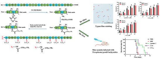
Anti-Toxoplasma gondii Effects of Lipopeptide Derivatives of Lycosin-I

1
Department of Parasitology, Xiangya School of Medicine, Central South University, Changsha 410013, China
2
The National & Local Joint Engineering Laboratory of Animal Peptide Drug Development, College of Life Sciences, Hunan Normal University, Changsha 410081, China
3
China-Africa Research Center of Infectious Diseases, Xiangya School of Medicine, Central South University, Changsha 410013, China
*
Author to whom correspondence should be addressed.
Toxins 2023, 15(8), 477; https://doi.org/10.3390/toxins15080477
Submission received: 12 June 2023 / Revised: 21 July 2023 / Accepted: 24 July 2023 / Published: 26 July 2023

Abstract

:
Toxoplasmosis, caused by Toxoplasma gondii (T. gondii), is a serious zoonotic parasitic disease. We previously found that Lycosin-I exhibited anti-T. gondii activity, but its serum stability was not good enough. In this study, we aimed to improve the stability and activity of Lycosin-I through fatty acid chain modification, so as to find a better anti-T. gondii drug candidate. The α/ε-amino residues of different lysine residues of Lycosin-I were covalently coupled with lauric acid to obtain eight lipopeptides, namely L-C12, L-C12-1, L-C12-2, L-C12-3, L-C12-4, L-C12-5, L-C12-6, and L-C12-7. Among these eight lipopeptides, L-C12 showed the best activity against T. gondii in vitro in a trypan blue assay. We then conjugated a shorter length fatty chain, aminocaproic acid, at the same modification site of L-C12, namely L-an. The anti-T. gondii effects of Lycosin-I, L-C12 and L-an were evaluated via an invasion assay, proliferation assay and plaque assay in vitro. A mouse model acutely infected with T. gondii tachyzoites was established to evaluate their efficacy in vivo. The serum stability of L-C12 and L-an was improved, and they showed comparable or even better activity than Lycosin-I did in inhibiting the invasion and proliferation of tachyzoites. L-an effectively prolonged the survival time of mice acutely infected with T. gondii. These results suggest that appropriate fatty acid chain modification can improve serum stability and enhance anti-T. gondii effect of Lycosin-I. The lipopeptide derivatives of Lycosin-I have potential as a novel anti-T. gondii drug candidate.
Key Contribution: In this study, we modified the peptide Lycosin-I with fatty acid chains, which resulted in an increase in the serum stability of anti-T. gondii activity in vitro of lipopeptide derivatives of Lycosin-I such as L-C12 and L-an, while L-an effectively prolonged the survival time of mice acutely infected with T. gondii tachyzoites.

Graphical Abstract

1. Introduction

Toxoplasma gondii (T. gondii), a unicellular eukaryote, is a serious pathogenic organism that parasitizes the nucleated cells of humans and various warm-blooded animals [1]. Toxoplasmosis caused by T. gondii is a zoonotic parasitic disease. Approximately one-third of people worldwide are infected with T. gondii [2]. Domestic and wild animals are also at risk of toxoplasmosis. Approximately one-third of domestic and non-domestic cats have been exposed to T. gondii [3]. Although acute infection with T. gondii is usually asymptomatic in immunocompetent individuals, or in rare cases may cause a mild flu-like illness, the parasite typically remains active in immunocompromised individuals, leading to persistent host cell infection and acute disease [4,5,6,7].
Early research into anti-T. gondii drugs began in the 1940s. In the early 1950s, Eyles and Coleman observed the synergistic effect of the sulfadiazine–pyrimethamine combination in treating toxoplasmosis in mice [8]. More than 60 years later, the sulfadiazine–pyrimethamine (SDZ/PYR) combination remains the gold standard for treating toxoplasmosis in humans. However, these drugs have several drawbacks, including a long treatment duration, a high relapse rate and significant adverse effects. In addition, there are currently no drugs available to remove the cysts and destroy the bradyzoites within cysts [9,10]. Researchers have made unremitting efforts to find novel anti-T. gondii drugs with better activity, higher stability, and fewer side effects, focusing mainly on the following areas: (1) To discover the new function of anti-T. gondii from the current clinical drugs. For example, NVP-AEW541, GSK-J4, pravastatin, simvastatin and the clinical antibiotic enrofloxacin have been found to have anti-T. gondii infection [11,12,13]. (2) Some compounds extracted from plants have been found to have anti-T. gondii activity. For example, 3-deoxyanthocyanidins extracted from sorghum bicolor shows anti-T. gondii activity in vitro [14]. The aqueous extracts of Astragalus membranaceus and Scutellaria baicalensis also show potent anti-T. gondii activity [15]. Ursolic acid, found in various medicinal plants, can induce the direct inhibition of T. gondii or increase the survival of mice by effectively blocking and inhibiting the viability of T. gondii [16]. (3) Compounds extracted from animal natural products [17], such as Bplec, a C-type lectin, and BNSP-7, a phospholipase analogue, isolated from the crude venom of Bothrops Pauloensis, have anti-T. gondii activity [18,19]. (4) Peptides. In recent years, peptides have played an important role in key research areas such as pharmacology, physiology and neurobiology [20]. To date, only a few peptides with anti-T. gondii activity have been reported in the literature. The 17-amino acid peptide, cal14.1a, produced by Conus californicus, reduces the invasion and proliferation of T. gondii [21]. Longicin P4, a 21 amino acid basic peptide from Haemaphysalis longicornis, has an inhibitory effect on T. gondii, Gram-negative and Gram-positive bacteria [22]. In addition, our previous studies have shown that the peptide Lycosin-I from the spider Lycosa singoriensis, the crude venoms from the spiders Ornitoctonus huwena and Chilobrachys jingzhao, and the peptide XYP1 from the spider Lycosa coelestis have significant anti-T. gondii activity in vitro and in vivo [23,24,25].
Cationic peptides with an α-helical structure found in venoms constitute a class of antimicrobial peptides (AMPs). In this class, amphiphilic peptides with net positive charges tend to adopt an α-helix structure when in contact with a hydrophilic/hydrophobic interface (such as the membrane of microorganisms) [26], which helps the AMPs to insert into the cell membrane and disrupt the integrity of the cell membrane [27]. Phospholipids on cell membranes are one of the targets of transmembrane peptides with an α -helical structure [28]. As a member of the AMP family, Lycosin-I plays a bactericidal role by combining with the bacterial cell membrane through electrostatic action to disrupt the membrane structure and cause the contents to leak out [29,30]. It is also a cell-penetrating peptide (CPP) that can carry coupled spherical gold nanoparticles into cancer cells [31]. However, the serum stability of Lycosin-I is not good enough, and it is completely degraded into smaller peptides after incubation in mouse serum for 24 h [32].
To improve the structural stability and activity of the peptide, its structure is modified via various methods including acetylation, amidation, amino acid replacement, D-amino acid substitution, the deletion of inactive amino acids, cyclization into a cyclic peptide, and fatty acid modification into a lipopeptide. Recent studies have shown that cyclic peptides and lipopeptides can enhance the biological activity and stability of peptides [32,33,34,35]. Fatty acids, a class of essential nutrients, regulate a wide range of vital processes and disorders including diabetes, cancer, and cardiovascular disease [36,37]. The fatty acid modification of peptides has high hydrophobicty and durability, making it a very promising method for improving peptide function. Glucagon-like peptide-1 analogs from Xenopus laevis modified with mycophenolic acid analogs containing fatty acids of different lengths (aminocaproic acid and dodecanoic acid) significantly improved the efficacy and duration of anti-diabetes treatment [38].
Fatty acids are carboxylic acids with long aliphatic chains that bind to the N-terminus or lysine side chains in peptides. In addition, cysteine residues can also be modified by fatty acids to obtain corresponding lipopeptide derivatives. Peptides modified with fatty acids are mainly used for the following aspects: (1) To improve the transmission of the molecular peptides mediated [39,40]. PepFect14 combined with acyl chains ranging in length from 2 to 22 carbons can optimize the minimal toxicity of low-nucleotide complexes and maintain delivery efficiency [41]. (2) To increase peptide stability. Myristic acid binds to lysine side chains on the insulin alpha chain to provide sufficient stability [42]. In addition, the antibacterial peptide analogs modified with fatty acids at the N-terminus showed high stability in the presence of protease or serum [43]. (3) Enhancing the activity of peptides. For example, B1 coupled with different lengths of fatty acids results in better anti-tumor activity [44]. The N-terminus of Lycosin-I was coupled with fatty acids of different lengths, demonstrating how fatty acids can increase the activity of peptides and how the activity of the peptides that do so differ, with lauric acid showing the most improved activity [45]. Subsequently, the lipopeptides L-C12, L-C12-1, L-C12-2, L-C12-3, L-C12-4, L-C12-5, L-C12-6 and L-C12-7 were obtained via the covalent coupling of lauric acid with α/ε -amino groups of lysine residues at different positions. Their biological properties, such as cytotoxicity and anti-tumor cell metastasis, as well as their particle size, zeta potential, secondary structure, hydrophobicity, and serum stability were identified. L-C12 performed the best among these lipopeptides and its serum stability was significantly increased compared to that of Lycosin-I [32].
Our previous studies confirmed that the spider peptide Lycosin-I has anti-T. gondii activity [23]. In this study, we believe that the position of the fatty acid chain modification and the length of the fatty chain may be the key factors for peptide optimization, so we will further investigate the effect of fatty acid modification with different fatty chain lengths (lauric acid and aminocaproic acid) on the anti-T. gondii activity of Lycosin-I.

2. Results

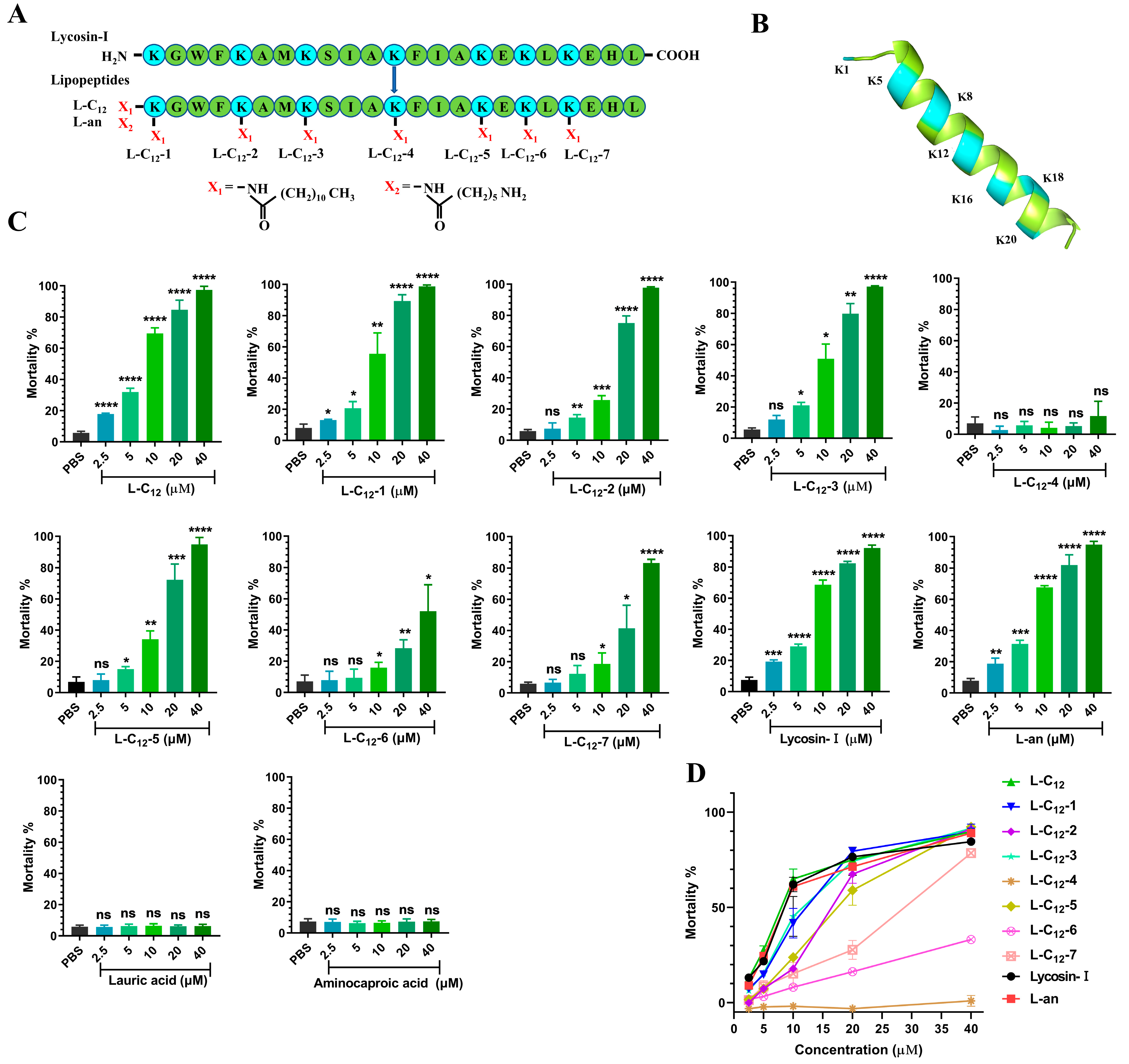
2.1. The Anti-T. gondii Efficacy of Lipopeptides

Eight lipopeptides, covalent couplings of lauric acid with α/ε-amino groups of lysine residues at different sites of Lycosin-I, were synthesized according to the preparation route [32] and designated as L-C12, L-C12-1, L-C12-2, L-C12-3, L-C12-4, L-C12-5, L-C12-6, and L-C12-7 (Figure 1A). The secondary structure of Lycosin-I shows that all the lysines are in an α-helical structure except for the first site where the lysine is located on the linear structure (Figure 1B). As our previous studies confirmed that Lycosin-I has concentration-dependent anti-T. gondii activity, we set up different concentration gradients (2.5 μM, 5 μM, 10 μM, 20 μM, and 40 μM) to evaluate the activity of the lipopeptides against T. gondii in vitro. We estimated the mortality of tachyzoites treated with lipopeptides at different concentrations using the trypan blue assay (Figure S1A,C). Each had a different level of activity against T. gondii. Among them, L-C12 showed the best efficacy against T. gondii in vitro. There was no significant difference in anti-T. gondii activity between L-C12 and Lycosin-I (Figure S1B,D). We then coupled another fatty chain length (aminocaproic acid) to the same modification site of L-C12, namely L-an (Figure 1A), which also showed no statistical difference from Lycosin-I in anti-T. gondii activity (Figure S1B,D). The 50% mortality rates of Lycosin-I, L-C12 and L-an against T. gondii were 7.20 μM, 6.54 μM and 7.10 μM, respectively. In addition, lauric acid and aminocaproic acid alone had no effect on T. gondii in vitro.

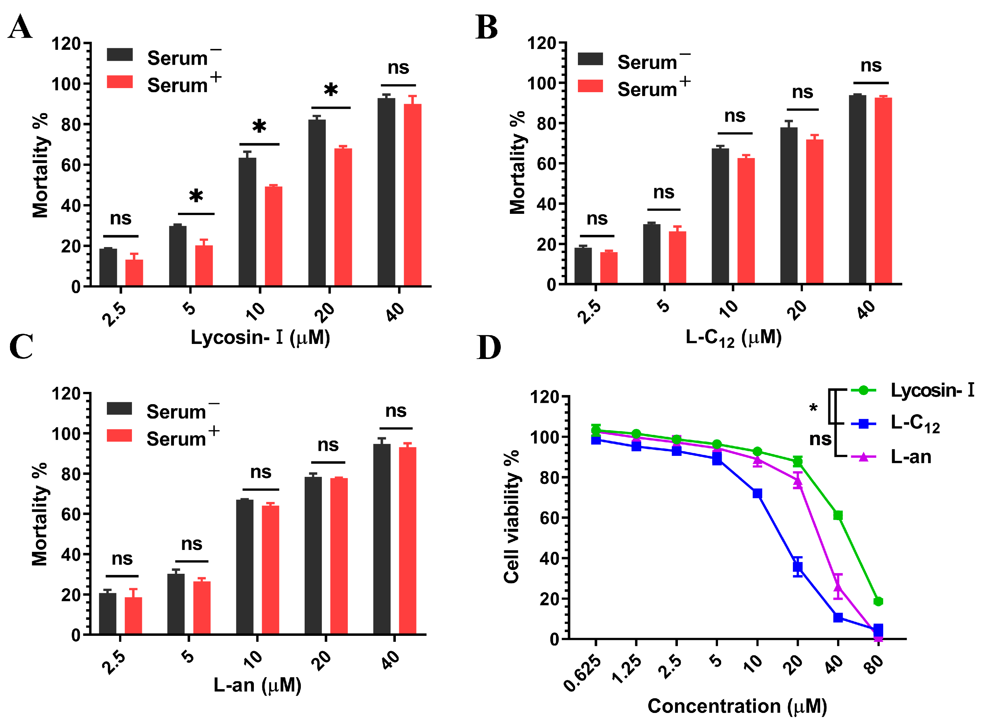
2.2. L-C12 and L-an Increased the Serum Stability of Anti-T. gondii In Vitro

A combination of serum incubation and a trypan blue assay was performed to determine whether or not the anti-T. gondii activity of lipopeptides was affected by serum. Compared to the control group (untreated with serum), Lycosin-I treated with serum significantly had reduced anti-T. gondii activity, whereas L-C12 and L-an showed no significant difference (Figure 2A–C).
To exclude the effect of lipopeptide toxicity on T. gondii activity, the effect of lipopeptides on the host cell human foreskin fibroblast (HFF) viability was evaluated using the CCK-8 assay. Lycosin-I, L-C12, and L-an showed concentration-dependent cytotoxicity to cells. Compared to Lycosin-I, L-C12 showed higher cytotoxicity after modification, while L-an showed no significant difference (Figure 2D). The half-inhibitory concentrations (IC50) of Lycosin-I, L-C12 and L-an were 46.16 μM, 15.62 μM and 30.95 μM, respectively.

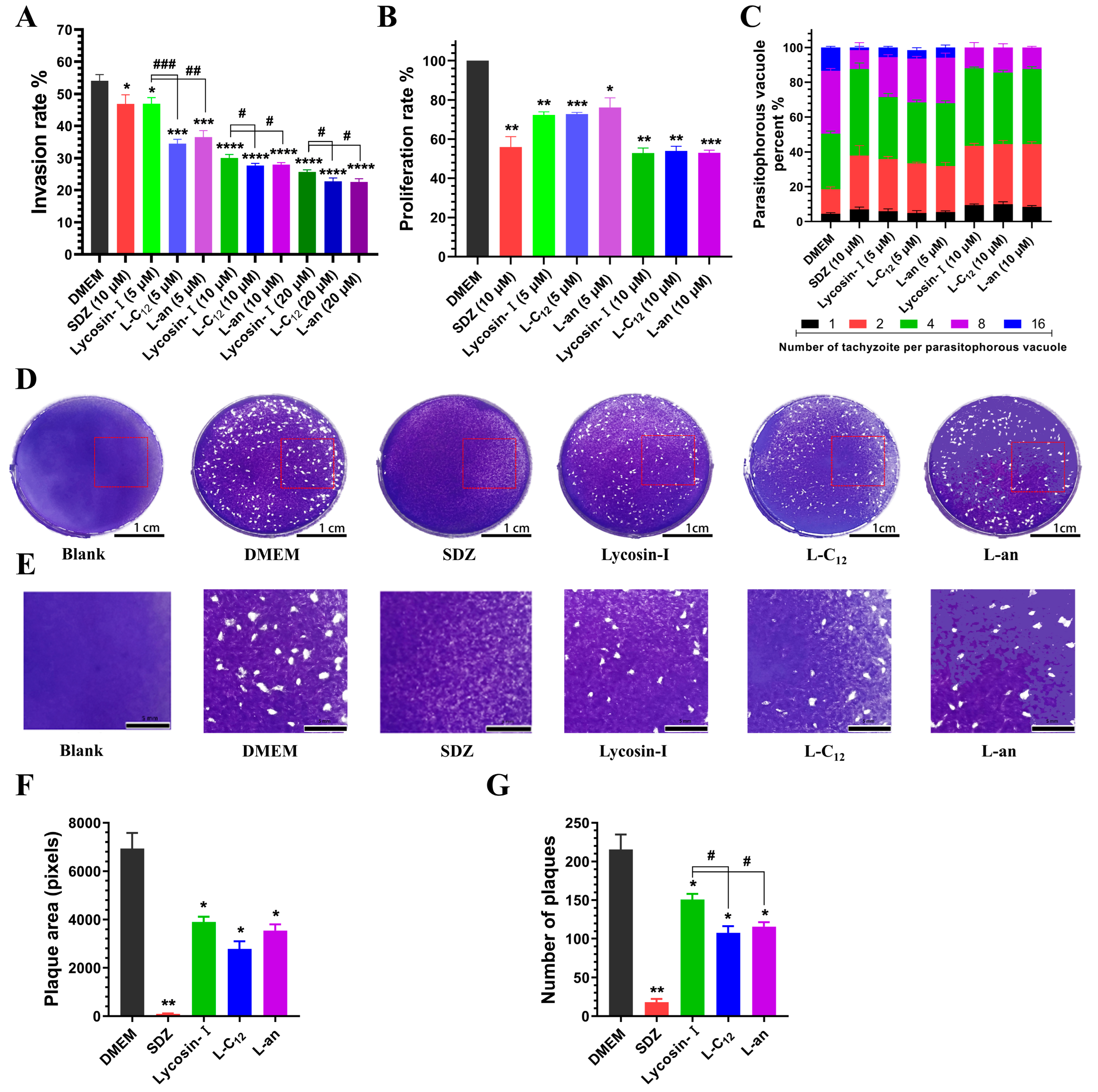
2.3. L-C12 and L-an Inhibit the Invasion and Proliferation of Tachyzoites into Host Cells

The tachyzoites of T. gondii infect cells in a relatively rapid process. They can invade host cells after 30 min of contact, forming PVs, and a host cell can be infected with multiple tachyzoites simultaneously (Figure S2). By calculating the invasion rate of tachyzoites, we found that in the DMEM group (negative control), the invasion rate of tachyzoites on host cells reached more than 50% after 2 h. L-C12 and L-an had a better inhibition effect on tachyzoite invasion than Lycosin-I did especially at a low concentration of 5 μM (Figure 3A). It was not unexpected that Lycosin-I, L-C12 and L-an showed more pronounced inhibition than SDZ (a clinical drug for toxoplasmosis) did at the concentration of 10 μM, because SDZ targets dihydropteroate synthase, affecting T. gondii proliferation rather than directly killing T. gondii.
After invading host cells, tachyzoites proliferate exponentially in the PV and can be arranged in a typical rose shape when they multiply to a certain extent. HFFs infected with tachyzoites were incubated with Lycosin-I, L-C12 and L-an at concentrations of 5 μM and 10 μM for 24 h. After Giemsa staining, the PVs in HFFs were observed under a light microscope (Figure S3). By counting the proportion of PVs with different numbers of tachyzoites in 100 PVs and calculating the proliferation rate of tachyzoites, we found that after incubation for 24 h, the proliferation of tachyzoites in the host cells was significantly inhibited in all drug treatment groups compared to that in the DMEM group. The proliferation rate of tachyzoites in the HFFs treated with Lycosin-I, L-C12 and L-an at 10 μM decreased to below 55% (Figure 3B). In the DMEM group, the number of tachyzoites in the PV was mostly 4 or 8, and some PVs with 16 tachyzoites could be seen. In contrast to the DMEM group, all treatment groups showed some inhibition of tachyzoite proliferation in PV. The number of PVs with 8 or 16 tachyzoites decreased significantly, while the proportion of PVs with 1, 2 and 4 tachyzoites increased. It was hard to see a PV with 16 tachyzoites in Lycosin-I, L-C12 and L-an at the concentration of 10 μM (Figure 3C).
A plaque assay can provide a thorough representation of the ability of tachyzoites to invade, proliferate and migrate. Based on the above results, a concentration of 10 μM was chosen for the plaque assay. HFFs infected with tachyzoites were incubated with DMEM (negative control, DMEM), SDZ (a clinical drug for toxoplasmosis, 10 μM), Lycosin-I (10 μM), L-C12 (10 μM) and L-an (10 μM), respectively, and HFFs not infected with tachyzoites were incubated with DMEM alone (blank control group). After 7 days of incubation, there was no obvious plaque in the drug-positive SDZ group compared to the DMEM group. The plaques of the Lycosin-I, L-C12 and L-an groups were also reduced (Figure 3D,E). In contrast with those in the DMEM group, the number and area of the plaques in SDZ, Lycosin-I, L-C12 and L-an were significantly reduced (Figure 3F,G), indicating that these treatment groups effectively inhibited the proliferation and re-invasion of tachyzoites in HFFs at the concentration of 10 μM. Compared Lycosin-I, L-C12 and L-an showed much better anti-T. gondii efficacy in vitro.

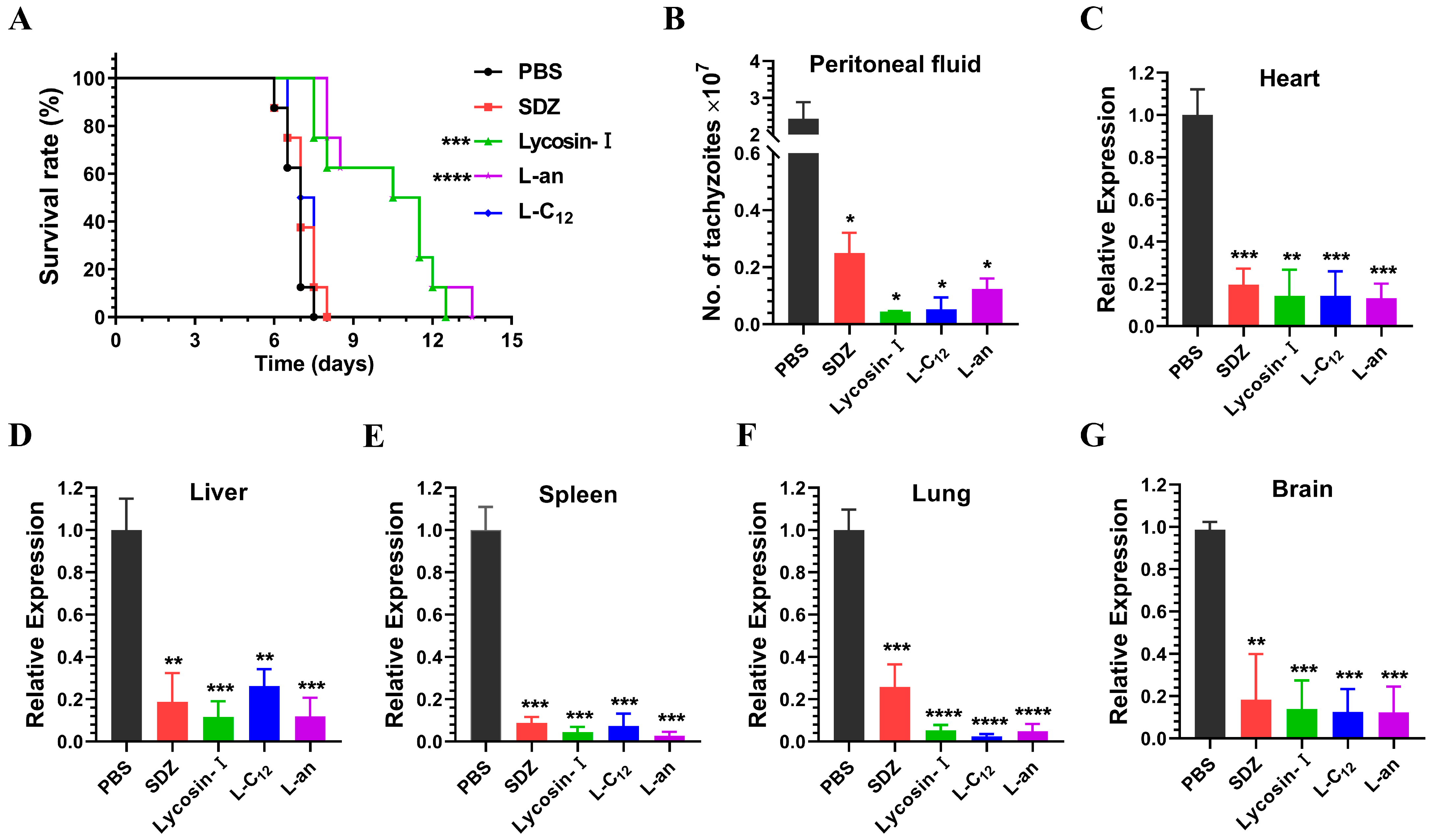
2.4. The Efficacy of Lipopeptides on Anti-T. gondii In Vivo

We have shown that L-C12 and L-an exhibited the same significant anti-T. gondii activity as Lycosin-I did in vitro. To evaluate the efficacy of the lipopeptides in vivo, we established a mouse model of acute T. gondii infection. Although SDZ exerts its anti-Toxoplasma effect at high concentrations in other papers, and its anti-Toxoplasma effect was also observed at 400 µM in vitro and 100 mg/kg in vivo in our previous study [25], in this study, we wanted to explore whether or not the anti-Toxoplasma effect of the peptides was better at the same low concentration as that of the SDZ, so we set the SDZ at the same concentration as that of the peptides in both the in vitro and in vivo experiments. Therefore, the mice were treated with 4 mg/kg/day of SDZ or lipopetides for 7 days, and the survival time of the mice was closely monitored for 15 days. Compared to the control group, SDZ and L-C12 could not prolong the survival time of the mice, while Lycosin-I and L-an significantly prolonged the survival time of the mice (Figure 4A). We also determined the parasite load in the peritoneal fluid and tissues of mice acutely infected with T. gondii after 5 days of treatment. The number of tachyzoites in the peritoneal fluid of the mice was directly counted using a blood cell counting plate (Figure 4B), while the expression of the surface antigen SAG1, a membrane surface protein specific to tachyzoites, was detected via qRT-PCR in the heart, liver, spleen, lung and brain of the mice (Figure 4C–G). Compared to that in the PBS group (negative control), the parasite load was significantly reduced in all treated groups.

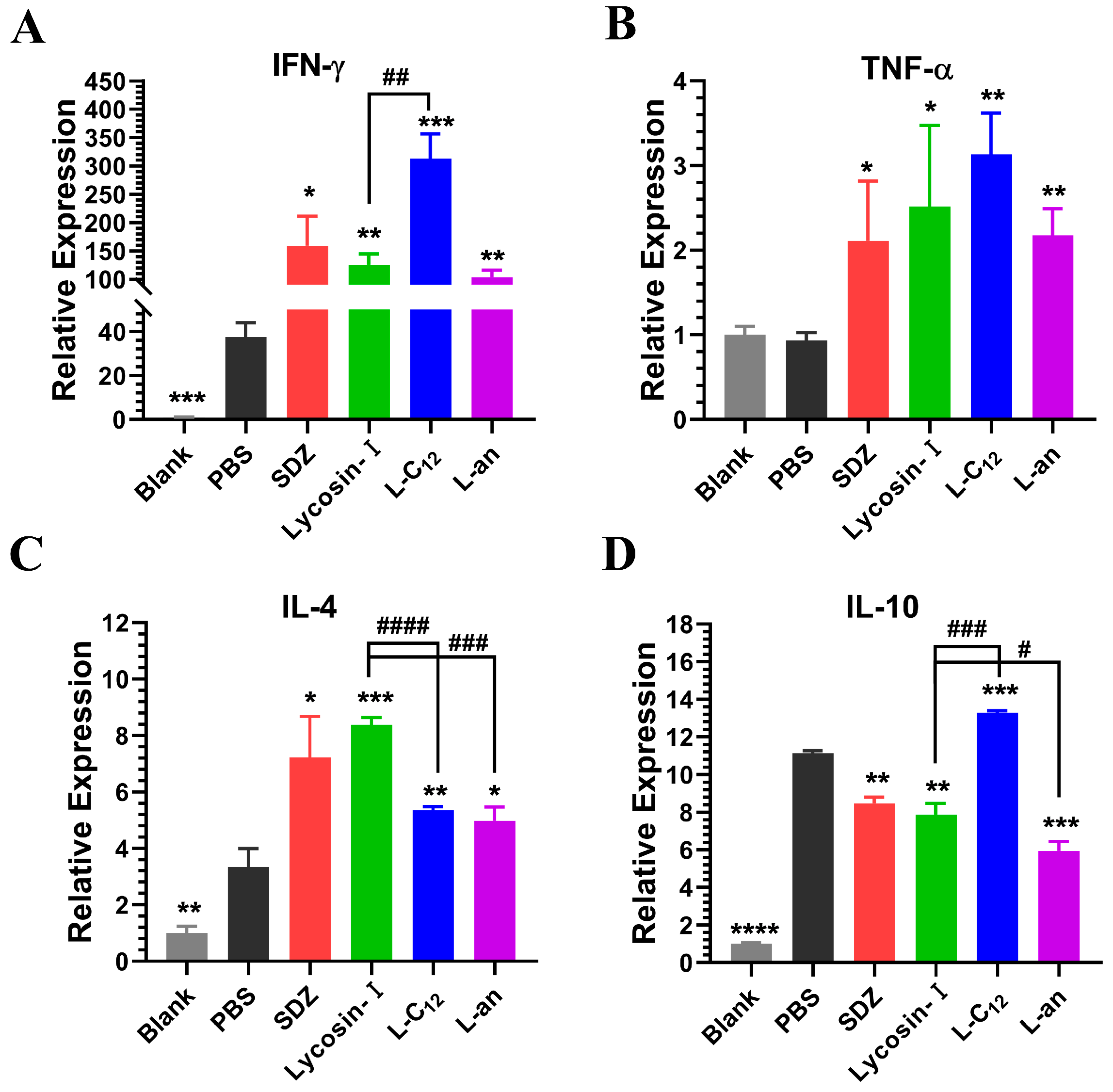
2.5. The Expression of Inflammatory Factors in Mice

Once T. gondii has infected a host, a number of inflammatory responses can be induced in the host to resist T. gondii invasion and proliferation. Therefore, we wanted to investigate the expression levels of anti-inflammatory and pro-inflammatory factors in the mice treated with lipopeptides. The expression levels of IFN-γ, TNF-α, IL-4 and IL-10 in the spleen tissues of mice after 5 days of treatment were determined via qRT-PCR (Figure 5). Compared to the blank control group (mice not infected with T. gondii), the pro-inflammatory cytokines IFN-γ and the anti-inflammatory cytokines IL-4 and IL-10 were significantly upregulated in PBS (negative control; mice infected with T. gondii without drug treatment). Compared to the PBS group, all treatment groups promoted the upregulation of the pro-inflammatory cytokines IFN-γ and TNF-α and the anti-inflammatory cytokine IL-4. SDZ, Lycosin-I and L-an inhibited the upregulation of IL-10, whereas L-C12 promoted it.

3. Discussion

The combination of sulfadiazine and pyrimethamine remains the gold standard for the treatment of toxoplasmosis. However, pyrimethamine is potentially teratogenic and causes reversible bone marrow suppression [9,46,47,48]. In addition, various serious complications of clinical drugs for toxoplasmosis have been reported, such as agranulocytosis, Stevens–Johnson syndrome, toxic epidermal necrolysis and hepatic necrosis, etc. [49,50,51,52,53]. Current treatment for toxoplasmosis is limited, with multiple side effects and long treatment durations (ranging from 4–6 weeks to more than 1 year) [9,54]. In addition, some intrinsic factors of T. gondii, such as increased drug resistance and differential drug susceptibility among different strains, also play an important role in disease progression and treatment failure [55,56,57]. Therefore, there is an urgent need to develop safer and more effective therapeutic alternatives for toxoplasmosis with fewer side effects, which depends on the growing knowledge of Toxoplasma pathophysiology and the discovery of promising drug targets.
In this study, the lipopeptides L-C12, L-C12-1, L-C12-2, L-C12-3, L-C12-4, L-C12-5, L-C12-6 and L-C12-7 were preliminarily evaluated for their direct anti-T. gondii activity in vitro using the trypan blue assay. It was found that the L-C12-4 and L-C12-6 lipopeptides completely lost their anti-T. gondii activity in vitro. We suggested that the lysine at these two sites was the key site for Lycosin-I to exert its anti-T. gondii activity, and that the lauric acid modified at these two sites altered its original physical and chemical properties. Although L-C12, L-C12-1, L-C12-2, L-C12-3, L-C12-5 and L-C12-7 all showed concentration-dependent anti-T. gondii activity in vitro, only L-C12 showed similar efficacy to that of Lycosin-I at 10 μM and a lower concentration; the other lipopeptides were obviously weaker than Lycosin-I in anti-T. gondii activity. The secondary structure of Lycosin-I showed that all the lysines are in the α-helical structure except for the first site where the lysine is located on the linear structure. Therefore, we speculated that the α-amino terminus of the first lysine site of Lycosin-I (the N-terminus of Lycosin-I) was the better site for modification compared to the other lysine sites, which was consistent with the previous research findings [32].
Since fatty acids of different lengths have different activities on peptides [45], we obtained the lipopeptide L-an by coupling another shorter fatty chain (aminocaproic acid) to the N-terminus of Lycosin-I. The trypan blue assay showed that the anti-T. gondii activity of the two lipopeptides in vitro was similar to that of Lycosin-I. However, the cytotoxicity of L-C12 was significantly higher than that of Lycosin-I. L-an showed no significant difference from Lycosin-I, indicating that the longer the fatty chain was, the more toxic it was to the cells. The serum stability of Lycosin-I was increased after fatty acid modification, as shown via mass spectrometry, which showed that Lycosin-I was degraded into smaller peptides after incubation with 10% serum for 24 h, whereas L-C12 was not degraded [32]. Interestingly, we found that Lycosin-I only slightly decreased, rather than completely lost its anti-T. gondii activity in vitro after 24 h of serum incubation. which suggests that although Lycosin-I was degraded into smaller peptides, the key amino acid sequences of anti-T. gondii were still retained in the small peptide; maybe we could optimize the specific activity of peptides by truncating them. After incubation in serum for 24 h, L-C12 and L-an retained substantial anti-T. gondii activity in vitro, consistent with the results of L-C12 mass spectrometry. Although L-an was not shown via mass spectrometry to be resistant to enzymatic degradation in serum, it was confirmed via trypan blue assay that L-an improved the stability of the anti-T. gondii activity of Lycosin-I in serum.
The invasion and pathogenicity of T. gondii to a host is a complex process involving parasite movement and penetration, as well as interaction and attachment with host cells [58]. The proliferation of T. gondii in host cells is mainly influenced by the immune response of the host cells [59,60]. SDZ targets dihydropteroate synthase and thus affects T. gondii proliferation rather than directly killing T. gondii, so the choice of SDZ as a positive control in the invasion assay was inappropriate. However, considering that these lipopeptides were obtained by modifying Lycosin-I, which has been shown to have anti-Toxoplasma activity in our previous studies, and we mainly wanted to compare the anti-Toxoplasma activity of these modified peptides with the parent peptide Lycosin-I, no additional other positive control was performed. In vitro, Lycosin-I, L-C12 and L-an showed significant anti-T. gondii activity, which was consistent with our previous results for Lycosin-I [23]. We found that two lipopeptides, L-C12 and L-an, showed slightly better inhibition efficiency of tachyzoites than Lycosin-I did, especially at the low concentration of 5 μM. Although the parasitophorous vacuole membrane (PVM) was mainly derived from the plasma membrane of host cells, T. gondii also secreted some proteins to participate in the formation of PVM. Furthermore, some proteins of T. gondii are even secreted and released into host cells through the PVM, mediating the host cell’s immune response and achieving the purpose of evading host immunity [61,62,63]. In the plaque assay, Lycosin-I, L-C12, and L-an showed no advantages over the positive drug SDZ at the same concentration, which may have been related to the time of onset and the stability of drug efficacy. In the invasion and proliferation assays, there were 5 × 10⁵ tachyzoites (MOI = 5, tachyzoite number/cell number) in each well, and the incubation time was relatively short (only 2 h or 24 h). Fast-acting peptides therefore have an advantage over slow-acting SDZ. However, only 500 tachyzoites were used in each well in the plaque assay, and the incubation time was longer than 7 days. Peptides may be degraded by serum, resulting in a less good effect than that of positive drugs. The inhibitory effect of L-C12 and L-an was slightly better than that of Lycosin-I, which may have beeb related to the improved serum stability of these two lipopeptides.
Experiments on animals are the most important indicator of drug efficacy. We established a mouse model acutely infected with tachyzoites, recorded the survival time of the mice and determined the parasite load in the tissues of the mice. Lycosin-I and L-an can effectively prolong the survival time of mice. Although SDZ and L-C12 can effectively inhibit the proliferation of tachyzoites in mice, they could not prolong the survival time of mice at the same dose. It is a fact that the clinical dosage of SDZ is 400 mg/kg, and a low dose of SDZ/PYR (20/0.5 mg/kg) failed to protect mice from dying [64], not to mention the fact that only 4 mg/kg was used in the study. As for L-C12, this have may been due to its excessive toxicity to mice, which resulted in the early death of the mice. The cytotoxicity of L-C12 was significantly higher than that of Lycosin-I in vitro, with an IC50 of 15.62 μM compared to that of 46.16 μM for Lycosin-I.
Toxoplasma infection can induce a host immune response that is primarily mediated by T helper cell 1 (Th1) and requires components of both the innate and adaptive immune response [65]. In the initial stage of infection, T. gondii is recognized by innate immune response cells, stimulating dendritic cells, macrophages and neutrophils to produce interleukin-12 (IL-12) and inducing natural killer (NK) cells to produce interferon-gamma (IFN-γ). In addition, tumor necrosis factor (TNF) can interact synergistically with IL-12 to optimize IFN-γ production in these cells [66,67]. These pro-inflammatory cytokines are produced when the adaptive immune response, mediated by CD4+ and CD8+ T lymphocytes, is activated. These lymphocytes produce and secrete several inflammatory mediators, such as nitric oxide (NO), and also cause a greater increase in IL-12 and IFN-γ levels [68,69]. IFN-γ can activate dendritic cells, macrophages, and neutrophils to promote a reduction in or the elimination of T. gondii. When macrophages are activated by IFN-γ, the production of NO increases, contributing to the toxicity of T. gondii [70]. In this study, we found that IFN-γ, but not TNF-α, was significantly increased in mice acutely infected with tachyzoites compared to with normal mice. SDZ, Lycosin-I, L-C12 and L-an promoted the expression of IFN-γ and TNF-α in mice. However, an excessive inflammatory response can lead to host death [66,71]. Therefore, it is important to strike a balance between Th1- and Th2-mediated immune responses. This balance can be mediated by the production of anti-inflammatory cytokines, such as IL-4, IL-10, and transforming growth factor (TGF-β1), which play a role by reducing the production of NO in macrophages and the cytotoxic activity of NK cells [72,73]. Additionally, Toxoplasma seeks mechanisms to evade the strong immune response of a host, such as the induction of anti-inflammatory cytokines. The expression of IL-10 and TGF-β1 increased the susceptibility of BeWo cells to T. gondii infection. Toxoplasma infection can induce the production of IL-4, IL-10 and TGF-β1 in host cells [74]. In this study, we found that the expression of IL-4 and IL-10 was higher in mice acutely infected with tachyzoites than in normal mice. Interestingly, SDZ, Lycosin-I, L-C12 and L-an promoted IL-4 expression in mice compared to that in the negative control mice. However, SDZ, Lycosin-I and L-an inhibited the expression of IL-10, while L-C12 promoted the expression of IL-10. We hypothesized that the increase in IL-4 may have regulated the inflammation caused by the expression of the pro-inflammatory factors IFN-γ and TNF-α, and prevented excessive inflammation leading to the death of the mice. The reduction in IL-10 may have been related to the inhibition of tachyzoites to evade host immunity by inducing the expression of anti-inflammatory factors. As for the promotion of IL-10 expression by L-C12, this may have also been the reason why L-C12 did not prolong the survival time of the mice in the animal survival experiments, because it only focuses on promoting the expression of anti-inflammatory factors to regulate the inflammatory response, which is precisely what helps the tachyzoites to escape the host’s immune response.

4. Conclusions

We identified a better modification site to improve the anti-T. gondii activity of Lycosin-I, namely, the α-amino terminus of the first lysine site of Lycosin-I (the N-terminus of Lycosin-I). L-C12 and L-an obtained via the fatty acid chain modification of Lycosin-I improved serum stability. L-C12 and L-an showed comparable or even better activity than Lycosin-I did in inhibiting tachyzoite invasion, proliferation and migration. L-an effectively prolonged the survival time of mice acutely infected with tachyzoites. These results suggest that the lipopeptide derivative of Lycosin-I has the potential to be a novel drug candidate of anti-T. gondii, which expands the understanding of the effects of peptides and further enriches the research on anti-T. gondii drugs.

5. Materials and Methods

5.1. Animals, Cells and Parasites

Specific pathogen-free (SPF) 8-week-old female BALB/c mice and Kunming (KM) mice were obtained from the Department of Laboratory Animals, Central South University. Human foreskin fibroblasts (HFFs) were cultured in Dulbecco’s modified Eagle’s medium (DMEM) containing 10% fetal bovine serum (FBS) and 1% antibiotics (10,000 U/mL penicillin and 10 mg/mL streptomycin solution). The tachyzoites used in this study were type I of the RH strain of T. gondii.

5.2. Lipopeptide Synthesis

The secondary structure of Lycosin-I was predicted using the I-TASSER server [75] and visualized using pymol software. Lipopetides were synthesized via solid-phase peptide synthesis (SPPS) methods and purified via reversed-phase high-performance liquid chromatography (RP-HPLC) as previously described [32]. The acetonitrile gradient of RP-HPLC can be seen in Table 1. Lycosin-I and L-C12 were synthesized by the Jing Peptide Biotechnology Co., Ltd., Hefei, China, and Pepmic Co., Ltd., Suzhou, China, respectively. Sulfadiazine (SDZ) was purchased from Sangong Biotech, Shanghai, China. Lycosin-I and lipopeptides were dissolved in phosphate-buffered saline (PBS).

5.3. Lipopeptide Screening

To evaluate the difference of anti-T. gondii in the vitro between lipopeptides and Lycosin-I, we set up different concentration gradients, including 2.5 μM, 5 μM, 10 μM, 20 μM and 40 μM. The freshly obtained tachyzoites (3 × 10⁶) were incubated with different concentrations of lipopeptides and PBS at room temperature for 2 h, then centrifuged at 3000 rbp for 8 min and the supernatant was discarded. The tachyzoite pellet was resuspended in PBS and mixed well. Then, the tachyzoite suspension was stained with 0.4% trypan blue staining solution for 5 min. Tachyzoite viability was observed under a light microscope (Motic China Group Co., Ltd., Xiamen, China), and five fields were randomly selected to calculate tachyzoite mortality. Three independent experiments were carried out.

5.4. Serum Stability Assay

It has been shown via mass spectrometry that Lycosin-I but not L-C12 is degraded into smaller peptides after incubation in 10% serum for 24 h, indicating that Lycosin-I improves serum stability after fatty acid modification [32]. A combination of serum incubation and a trypan blue assay was used to investigate whether or not the anti-Toxoplasma activity of lipopeptides and Lycosin-I was affected by serum. Briefly, stock solutions (1 mM) of Lycosin-I, L-C12 and L-an were diluted into 100 μM solutions in PBS containing 10% fetal bovine serum, and incubated at 37 °C for 24 h. Fresh tachyzoites were then treated with these solutions as described in the method in Section 5.3 for lipopeptide screening. Three independent experiments were carried out.

5.5. Cell Viability Assay

HFF cells were seeded in 96-well plates (1 × 10⁴ cells per well). After 24 h of incubation, the previous complete medium was removed and replaced with the new complete medium containing different concentrations (80–1.25 μM, twofold serial dilution) of lipopeptides. Cells were treated with the complete medium in one well as the control group (each group was set up with three wells). After 24 h, 10 μL of CCK-8 reagent was added to each well and then incubated for 2 h. The absorbance value was measured at 450 nm using a microplate reader. Three independent experiments were carried out.

5.6. Invasion Assay

The HFF cells were seeded in 24-well plates with 14 mm coverslips (1 × 10⁵ cells per well) and incubated for 24 h. Fresh tachyzoites were obtained aseptically from the peritoneal fluid of the KM mouse acutely infected with T. gondii. The tachyzoites were treated with Lycosin-I (5 μM, 10 μM), L-C12 (5 μM, 10 μM), L-an (5 μM, 10 μM), SDZ (10 μM, positive control) and DMEM (negative control) for 2 h each. The HFFs were then infected with pre-treated tachyzoites at a multiplicity of infection (MOI) of 5 (tachyzoites/cells = 5). After 2 h, the previous complete medium was removed and the HFFs were washed twice with PBS to remove extracellular tachyzoites. They were then fixed with methanol for 5 min, and then stained with Giemsa for 15 min. The coverslips were examined under a light microscope. The invasion rate of tachyzoites was calculated by counting the number of cells invaded by tachyzoites and the total number of cells in 5 randomly selected fields. Three independent experiments were carried out.

5.7. Intracellular Proliferation Assay

HFFs were seeded in 24-well plates (1 × 10⁵ cells per well) with 14 mm coverslips and incubated for 24 h. The HFFs were infected with fresh tachyzoites at a MOI of 5 for 2 h. The HFFs were then washed twice with PBS to remove extracellular tachyzoites and then treated with Lycosin-I (5 μM, 10 μM), L-C12 (5 μM, 10 μM), L-an (5 μM, 10 μM), SDZ (10 μM, positive control) and DMEM (negative control) for 24 h each. The HFFs were then washed twice with PBS, fixed with methanol for 5 min, and then stained with Giemsa for 15 min. The coverslips were observed under a light microscope to count the number of tachyzoites in 100 parasitophorous vacuoles (PVs). Three independent experiments were carried out.

5.8. Plaque Assay

The HFF cells were seeded in 6-well plates (1 × 10⁶ cells per well) and incubated for 48–72 h. Cells in the 6-well plates were infected with 500 fresh tachyzoites per well for 2 h. The well in which cells were not infected with tachyzoites was considered the blank control group. Cells infected with tachyzoites were then treated with Lycosin-I (10 μM), L-C12 (10 μM), L-an (10 μM), SDZ (10 μM, positive control) and DMEM (negative control), respectively. After 7 days, HFFs in the plates were washed twice with PBS, fixed with methanol for 5 min, and then stained with crystal violet for 20 min. Six-well plates were washed with running water, dried, photographed, and then the number and area of plaques were counted using Photoshop version 2020. Three independent experiments were carried out.

5.9. Survival Assay

To evaluate the anti-T. gondii effect of lipopeptides in vivo, we established a mouse model acutely infected with the RH strain of T. gondii. Forty 8-week-old female BALB/c mice weighing 17–19 g were randomly divided into 5 groups (n = 8 per group) and intraperitoneally injected with 1 × 10³ tachyzoites per mouse. Lycosin-I, L-C12, L-an and sulfadiazine (SDZ) were dissolved in PBS, respectively. As these peptides are small linear peptides that are easily degraded by pepsin; mice were treated with these peptides via intraperitoneal injection (the same way that the mice were infected with T. gondii) rather than oral gavage. After 4 h of infection, mice were traperitoneally injected with 4 mg/kg of Lycosin-I, L-C12, L-an and sulfadiazine solution (once a day) for 7 days. Mice in the negative control group were treated with PBS. Mice in this condition observed daily and the time of death was recorded. The experiment was approved by the Ethics Committee of the School of Basic Medicine of Central South University (approval number: 2021-KT24).

5.10. RNA Extraction and Quantitative Real-Time PCR (qRT-PCR)

Total RNA was extracted from each sample using Total RNA Isolation Kit (ENOVA BIO Co., Ltd., Wuhan, China), in accordance with the manufacturer’s instructions. Reverse transcription of RNA into cDNA was performed using the PerfectStart Uni RT&qPCR Kit (TransGen Biotech Co., Ltd., Beijing, China) in accordance with the instructions. cDNA was used as a template for qRT-PCR using PerfectStart Uni RT&qPCR Kit (TransGen Biotech Co., Ltd., Beijing, China). Primers, listed in Table 2, were designed and synthesized by a biotechnology company (Sangon Biotech Co., Ltd., Shanghai, China). Analysis was performed using the comparative threshold cycle method (2−∆∆Ct). The gene expression levels of genes were normalized to those of housekeeping genes (β-actin or GAPDH).

5.11. Statistical Analysis

Data were analyzed and graphs were constructed using Photoshop version 2020 (Adobe Systems Inc., SAN Jose, CA, USA) and GraphPad Prism version 8.0 (GraphPad Software, San Diego, CA, USA). Difference statistics were calculated using the two-tailed Student’s t-test and p < 0.05 was considered statistically significant.

Supplementary Materials

The following are available online at https://www.mdpi.com/article/10.3390/toxins15080477/s1. Figure S1: The effect of lipopeptides against T. gondii evaluated via the trypan blue assay. Figure S2: Effect of lipopeptides on tachyzoite invasion. Figure S3: Effect of lipopeptides on tachyzoite proliferation.

Author Contributions

Conceptualization, X.L. and L.J.; methodology, P.Z. and Y.L.; software, X.L.; validation, X.L.; formal analysis, X.L.; writing—original draft preparation, X.L.; writing—review and editing, P.Z., Z.L. and L.J.; funding acquisition, J.L., X.L., D.Y. and L.J. All authors have read and agreed to the published version of the manuscript.

Funding

This work was supported by the National Natural Science Foundation of China (grant number 32170510), the Natural Science Foundation of Hunan Province, China (grant number 2020JJ4765), the Open Sharing Fund for the Large-Scale Instruments and Equipment of Central South University (grant number CSUZC202236), Science Popularization project of Hunan Province (project number 2021ZK4058), Graduate Case base construction project of Central South University (project number 2020ALK91), Graduate Research and Innovation Project of Hunan Province (project number CX20220361), Graduate Research and Innovation Project of Central South University (project number 2023ZZTS0858) and Graduate Research and Innovation Project of Hunan Province (project number QL20230021).

Institutional Review Board Statement

The study was approved by the Ethics Committee of the School of Basic Medicine of Central South University on 8 August 2021. (approval number: 2021-KT24).

Informed Consent Statement

Not applicable.

Data Availability Statement

The datasets used and/or analyzed during the current study are available from the corresponding author on reasonable request.

Conflicts of Interest

The authors declare no conflict of interest.

Abbreviations

T. gondiiToxoplasma gondii
AMPsantimicrobial peptides
CPPcell penetrating peptide
SPFspecific pathogen-free
KM miceKunming mice
HFFshuman foreskin fibroblasts
DMEMDulbecco’s modified Eagle’s medium
FBSfetal bovine serum
SPPSsolid phase peptide synthesis
RP-HPLCreversed phase-high performance liquid chromatography
SDZsulfadiazine
PYRpyrimethamine
PBSphosphate-buffered saline
MOImultiplicity of infection
PVparasitophorous vacuole
qRT-PCRquantitative real-time PCR
PVMparasitophorous vacuole membrane
IL-12interleukin-12
NK cellnatural killer cell
IFN-γinterferon gamma
TNFtumor necrosis factor
NOnitric oxide
TGFtransforming growth factor

References

  1. Halonen, S.K.; Weiss, L.M. Toxoplasmosis. Handb. Clin. Neurol. 2013, 114, 125–145. [Google Scholar]
  2. Montoya, J.G.; Liesenfeld, O. Toxoplasmosis. Lancet 2004, 363, 1965–1976. [Google Scholar] [CrossRef]
  3. Hatam-Nahavandi, K.; Calero-Bernal, R.; Rahimi, M.T.; Pagheh, A.S.; Zarean, M.; Dezhkam, A.; Ahmadpour, E. Toxoplasma gondii infection in domestic and wild felids as public health concerns: A systematic review and meta-analysis. Sci. Rep. 2021, 11, 9509. [Google Scholar] [CrossRef]
  4. Pittman, K.J.; Knoll, L.J. Long-Term Relationships: The Complicated Interplay between the Host and the Developmental Stages of Toxoplasma gondii during Acute and Chronic Infections. Microbiol. Mol. Biol. Rev. 2015, 79, 387–401. [Google Scholar] [CrossRef] [Green Version]
  5. Flegr, J.; Escudero, D.Q. Impaired health status and increased incidence of diseases in Toxoplasma-seropositive subjects—An explorative cross-sectional study. Parasitology 2016, 143, 1974–1989. [Google Scholar] [CrossRef] [Green Version]
  6. Wang, Z.-D.; Liu, H.-H.; Ma, Z.-X.; Ma, H.-Y.; Li, Z.-Y.; Yang, Z.-B.; Zhu, X.-Q.; Xu, B.; Wei, F.; Liu, Q. Toxoplasma gondii Infection in Immunocompromised Patients: A Systematic Review and Meta-Analysis. Front. Microbiol. 2017, 8, 389. [Google Scholar] [CrossRef] [Green Version]
  7. Pan, M.; Lyu, C.; Zhao, J.; Shen, B. Sixty Years (1957–2017) of Research on Toxoplasmosis in China-An Overview. Front. Microbiol. 2017, 8, 1825. [Google Scholar] [CrossRef] [Green Version]
  8. Eyles, D.E.; Coleman, N. Synergistic effect of sulfadiazine and daraprim against experimental toxoplasmosis in the mouse. Antibiot. Chemother. 1953, 3, 483–490. [Google Scholar]
  9. Alday, P.H.; Doggett, J.S. Drugs in development for toxoplasmosis: Advances, challenges, and current status. Drug Des. Dev. Ther. 2017, 11, 273–293. [Google Scholar] [CrossRef] [Green Version]
  10. Antczak, M.; Dzitko, K.; Długońska, H. Human toxoplasmosis–Searching for novel chemotherapeutics. Biomed. Pharmacother. 2016, 82, 677–684. [Google Scholar] [CrossRef]
  11. Sanfelice, R.A.; da Silva, S.S.; Bosqui, L.R.; Miranda-Sapla, M.M.; Barbosa, B.F.; Silva, R.J.; Ferro, E.A.V.; Panagio, L.A.; Navarro, I.T.; Bordignon, J.; et al. Pravastatin and simvastatin inhibit the adhesion, replication and proliferation of Toxoplasma gondii (RH strain) in HeLa cells. Acta Trop. 2017, 167, 208–215. [Google Scholar] [CrossRef]
  12. Barbosa, B.F.; Gomes, A.O.; Ferro, E.A.V.; Napolitano, D.R.; Mineo, J.R.; Silva, N.M. Enrofloxacin is able to control Toxoplasma gondii infection in both in vitro and in vivo experimental models. Veter. Parasitol. 2012, 187, 44–52. [Google Scholar] [CrossRef]
  13. Liu, S.; Wu, M.; Hua, Q.; Lu, D.; Tian, Y.; Yu, H.; Cheng, L.; Chen, Y.; Cao, J.; Hu, X.; et al. Two old drugs, NVP-AEW541 and GSK-J4, repurposed against the Toxoplasma gondii RH strain. Parasites Vectors 2020, 13, 242. [Google Scholar] [CrossRef]
  14. Abugri, D.A.; Witola, W.H.; Jaynes, J.M.; Toufic, N. In vitro activity of Sorghum bicolor extracts, 3-deoxyanthocyanidins, against Toxoplasma gondii. Exp. Parasitol. 2016, 164, 12–19. [Google Scholar] [CrossRef] [Green Version]
  15. Yang, X.; Huang, B.; Chen, J.; Huang, S.; Zheng, H.; Lun, Z.-R.; Shen, J.; Wang, Y.; Lu, F. In vitro effects of aqueous extracts of Astragalus membranaceus and Scutellaria baicalensis GEORGI on Toxoplasma gondii. Parasitol. Res. 2012, 110, 2221–2227. [Google Scholar] [CrossRef]
  16. Choi, W.H.; Lee, I.A. Evaluation of Anti-Toxoplasma gondii Effect of Ursolic Acid as a Novel Toxoplasmosis Inhibitor. Pharmaceuticals 2018, 11, 43. [Google Scholar] [CrossRef] [Green Version]
  17. Júnior, E.F.; Menezes, L.F.S.; de Araújo, I.F.S.; Schwartz, E.F. Natural Occurrence in Venomous Arthropods of Antimicrobial Peptides Active against Protozoan Parasites. Toxins 2019, 11, 563. [Google Scholar] [CrossRef] [Green Version]
  18. Castanheira, L.; de Souza, D.L.N.; Silva, R.J.; Barbosa, B.; Mineo, J.R.; Tudini, K.A.; Rodrigues, R.; Ferro, E.V.; Rodrigues, V.D.M. Insights into anti-parasitism induced by a C-type lectin from Bothrops pauloensis venom on Toxoplasma gondii. Int. J. Biol. Macromol. 2015, 74, 568–574. [Google Scholar] [CrossRef]
  19. Borges, I.P.; Castanheira, L.E.; Barbosa, B.F.; de Souza, D.L.N.; da Silva, R.J.; Mineo, J.R.; Tudini, K.A.Y.; Rodrigues, R.S.; Ferro, E.A.V.; Rodrigues, V.D.M. Anti-parasitic effect on Toxoplasma gondii induced by BnSP-7, a Lys49-phospholipase A2 homologue from Bothrops pauloensis venom. Toxicon 2016, 119, 84–91. [Google Scholar] [CrossRef]
  20. Pineda, S.S.; Undheim, E.A.; Rupasinghe, D.B.; Ikonomopoulou, M.P.; King, G.F. Spider venomics: Implications for drug discovery. Future Med. Chem. 2014, 6, 1699–1714. [Google Scholar] [CrossRef]
  21. De Leon-Nava, M.A.; Romero-Nunez, E.; Luna-Nophal, A.; Bernaldez-Sarabia, J.; Sanchez-Campos, L.N.; Licea-Navarro, A.F.; Morales-Montor, J.; Muniz-Hernandez, S. In Vitro Effect of the Synthetic cal14.1a Conotoxin, Derived from Conus californicus, on the Human Parasite Toxoplasma gondii. Mar. Drugs 2016, 14, 66. [Google Scholar] [CrossRef] [Green Version]
  22. Tanaka, T.; Maeda, H.; Matsuo, T.; Boldbattar, D.; Umemiya-Shirafuji, R.; Kume, A.; Suzuki, H.; Xuan, X.; Tsuji, N.; Fujisaki, K. Parasiticidal activity of Haemaphysalis longicornis longicin P4 peptide against Toxoplasma gondii. Peptides 2012, 34, 242–250. [Google Scholar] [CrossRef]
  23. Tang, Y.; Hou, S.; Li, X.; Wu, M.; Ma, B.; Wang, Z.; Jiang, J.; Deng, M.; Duan, Z.; Tang, X.; et al. Anti-parasitic effect on Toxoplasma gondii induced by a spider peptide lycosin-I. Exp. Parasitol. 2019, 198, 17–25. [Google Scholar] [CrossRef]
  24. Hou, S.; Liu, Y.; Tang, Y.; Wu, M.; Guan, J.; Li, X.; Wang, Z.; Jiang, J.; Deng, M.; Duan, Z.; et al. Anti-Toxoplasma gondii effect of two spider venoms in vitro and in vivo. Toxicon 2019, 166, 9–14. [Google Scholar] [CrossRef]
  25. Liu, Y.; Tang, Y.; Tang, X.; Wu, M.; Hou, S.; Liu, X.; Li, J.; Deng, M.; Huang, S.; Jiang, L. Anti-Toxoplasma gondii Effects of a Novel Spider Peptide XYP1 In Vitro and In Vivo. Biomedicines 2021, 9, 934. [Google Scholar] [CrossRef]
  26. Torres, M.D.T.; Sothiselvam, S.; Lu, T.K.; De La Fuente-Nunez, C. Peptide Design Principles for Antimicrobial Applications. J. Mol. Biol. 2019, 431, 3547–3567. [Google Scholar] [CrossRef]
  27. Langenegger, N.; Nentwig, W.; Kuhn-Nentwig, L. Spider Venom: Components, Modes of Action, and Novel Strategies in Transcriptomic and Proteomic Analyses. Toxins 2019, 11, 611. [Google Scholar] [CrossRef] [Green Version]
  28. Bolhassani, A.; Jafarzade, B.S.; Mardani, G. In vitro and in vivo delivery of therapeutic proteins using cell penetrating peptides. Peptides 2017, 87, 50–63. [Google Scholar] [CrossRef]
  29. Tan, H.; Ding, X.; Meng, S.; Liu, C.; Wang, H.; Xia, L.; Liu, Z.; Liang, S. Antimicrobial potential of lycosin-I, a cationic and amphiphilic peptide from the venom of the spider Lycosa singorensis. Curr. Mol. Med. 2013, 13, 900–910. [Google Scholar] [CrossRef]
  30. Wang, L.; Wang, Y.-J.; Liu, Y.-Y.; Li, H.; Guo, L.-X.; Liu, Z.-H.; Shi, X.-L.; Hu, M. In Vitro Potential of Lycosin-I as an Alternative Antimicrobial Drug for Treatment of Multidrug-Resistant Acinetobacter baumannii Infections. Antimicrob. Agents Chemother. 2014, 58, 6999–7002. [Google Scholar] [CrossRef] [Green Version]
  31. Tan, H.; Huang, Y.; Xu, J.; Chen, B.; Zhang, P.; Ye, Z.; Liang, S.; Xiao, L.; Liu, Z. Spider Toxin Peptide Lycosin-I Functionalized Gold Nanoparticles for in vivo Tumor Targeting and Therapy. Theranostics 2017, 7, 3168–3178. [Google Scholar] [CrossRef]
  32. Zhang, P.; Jian, C.; Jian, S.; Zhang, Q.; Sun, X.; Nie, L.; Liu, B.; Li, F.; Li, J.; Liu, M.; et al. Position Effect of Fatty Acid Modification on the Cytotoxicity and Antimetastasis Potential of the Cytotoxic Peptide Lycosin-I. J. Med. Chem. 2019, 62, 11108–11118. [Google Scholar] [CrossRef]
  33. Mwangi, J.; Yin, Y.; Wang, G.; Yang, M.; Li, Y.; Zhang, Z.; Lai, R. The antimicrobial peptide ZY4 combats multidrug-resistant Pseudomonas aeruginosa and Acinetobacter baumannii infection. Proc. Natl. Acad. Sci. USA 2019, 116, 26516–26522. [Google Scholar] [CrossRef] [Green Version]
  34. Henriques, S.T.; Lawrence, N.; Chaousis, S.; Ravipati, A.S.; Cheneval, O.; Benfield, A.H.; Elliott, A.G.; Kavanagh, A.M.; Cooper, M.A.; Chan, L.Y.; et al. Redesigned Spider Peptide with Improved Antimicrobial and Anticancer Properties. ACS Chem. Biol. 2017, 12, 2324–2334. [Google Scholar] [CrossRef]
  35. Yang, G.; Wang, J.; Lu, S.; Chen, Z.; Fan, S.; Chen, D.; Xue, H.; Shi, W.; He, J. Short lipopeptides specifically inhibit the growth of Propionibacterium acnes with a dual antibacterial and anti-inflammatory action. Br. J. Pharmacol. 2019, 176, 2321–2335. [Google Scholar] [CrossRef]
  36. Simopoulos, A.P. Genetic variants in the metabolism of omega-6 and omega-3 fatty acids: Their role in the determination of nutritional requirements and chronic disease risk. Exp. Biol. Med. 2010, 235, 785–795. [Google Scholar] [CrossRef] [PubMed]
  37. Lee, J.M.; Lee, H.; Kang, S.; Park, W.J. Fatty Acid Desaturases, Polyunsaturated Fatty Acid Regulation, and Biotechnological Advances. Nutrients 2016, 8, 23. [Google Scholar] [CrossRef] [Green Version]
  38. Han, J.; Fu, J.; Sun, L.; Han, Y.; Mao, Q.; Liao, F.; Zheng, X.; Zhu, K. Synthesis and pharmaceutical characterization of site specific mycophenolic acid-modified Xenopus glucagon-like peptide-1 analogs. Medchemcomm 2018, 9, 67–80. [Google Scholar] [CrossRef] [PubMed]
  39. Mäe, M.; EL Andaloussi, S.; Lundin, P.; Oskolkov, N.; Johansson, H.J.; Guterstam, P.; Langel, U. A stearylated CPP for delivery of splice correcting oligonucleotides using a non-covalent co-incubation strategy. J. Control. Release 2009, 134, 221–227. [Google Scholar] [CrossRef] [PubMed]
  40. Arukuusk, P.; Pärnaste, L.; Hällbrink, M.; Langel, Ü. PepFects and NickFects for the Intracellular Delivery of Nucleic Acids. Methods Mol. Biol. 2015, 1324, 303–315. [Google Scholar] [CrossRef]
  41. Lehto, T.; Vasconcelos, L.; Margus, H.; Figueroa, R.; Pooga, M.; Hällbrink, M.; Langel, U. Saturated Fatty Acid Analogues of Cell-Penetrating Peptide PepFect14: Role of Fatty Acid Modification in Complexation and Delivery of Splice-Correcting Oligonucleotides. Bioconjugate Chem. 2017, 28, 782–792. [Google Scholar] [CrossRef] [PubMed]
  42. Kurtzhals, P.; Havelund, S.; Jonassen, I.; Markussen, J. Effect of Fatty Acids and Selected Drugs on the Albumin Binding of a Long-Acting, Acylated Insulin Analogue. J. Pharm. Sci. 1997, 86, 1365–1368. [Google Scholar] [CrossRef] [PubMed]
  43. Zhong, C.; Liu, T.; Gou, S.; He, Y.; Zhu, N.; Zhu, Y.; Wang, L.; Liu, H.; Zhang, Y.; Yao, J.; et al. Design and synthesis of new N-terminal fatty acid modified-antimicrobial peptide analogues with potent in vitro biological activity. Eur. J. Med. Chem. 2019, 182, 111636. [Google Scholar] [CrossRef] [PubMed]
  44. Deng, X.; Qiu, Q.; Ma, K.; Wang, X.; Huang, W.; Qian, H. Aliphatic acid-conjugated antimicrobial peptides--potential agents with anti-tumor, multidrug resistance-reversing activity and enhanced stability. Org. Biomol. Chem. 2015, 13, 7673–7680. [Google Scholar] [CrossRef]
  45. Jian, C.; Zhang, P.; Ma, J.; Jian, S.; Zhang, Q.; Liu, B.; Liang, S.; Liu, M.; Zeng, Y.; Liu, Z. The Roles of Fatty-Acid Modification in the Activity of the Anticancer Peptide R-Lycosin-I. Mol. Pharm. 2018, 15, 4612–4620. [Google Scholar] [CrossRef]
  46. Dunay, I.R.; Gajurel, K.; Dhakal, R.; Liesenfeld, O.; Montoya, J.G. Treatment of Toxoplasmosis: Historical Perspective, Animal Models, and Current Clinical Practice. Clin. Microbiol. Rev. 2018, 31, e00057-17. [Google Scholar] [CrossRef] [Green Version]
  47. Montoya, J.G.; Remington, J.S. Management of Toxoplasma gondii infection during pregnancy. Clin. Infect. Dis. 2008, 47, 554–566. [Google Scholar] [CrossRef] [Green Version]
  48. Paquet, C.; Yudin, M.H. No. 285-Toxoplasmosis in Pregnancy: Prevention, Screening, and Treatment. J. Obstet. Gynaecol. Can. 2018, 40, e687–e693. [Google Scholar] [CrossRef]
  49. Ardabili, S.; Kohl, J.; Gül, G.; Hodel, M. What obstetricians should be aware of: Serious side effects of antibiotic toxoplasmosis treatment in pregnancy. BMJ Case Rep. 2021, 14, e240809. [Google Scholar] [CrossRef]
  50. Ben-Harari, R.R.; Goodwin, E.; Casoy, J. Adverse Event Profile of Pyrimethamine-Based Therapy in Toxoplasmosis: A Systematic Review. Drugs R&D 2017, 17, 523–544. [Google Scholar] [CrossRef] [Green Version]
  51. Borkowski, P.K.; Brydak-Godowska, J.; Basiak, W.; Olszyńska-Krowicka, M.; Rabczenko, D. Adverse Reactions in Antifolate-Treated Toxoplasmic Retinochoroiditis. Adv. Exp. Med. Biol. 2018, 1108, 37–48. [Google Scholar] [CrossRef] [PubMed]
  52. Guaraldo, L.; Villar, B.B.D.L.F.; Durão, N.M.G.; Louro, V.C.; Quintana, M.D.S.B.; Curi, A.L.L.; Neves, E.S. Ocular toxoplasmosis: Adverse reactions to treatment in a Brazilian cohort. Trans. R. Soc. Trop. Med. Hyg. 2018, 112, 188–192. [Google Scholar] [CrossRef] [PubMed]
  53. Shammaa, A.M.; Powell, T.G.; Benmerzouga, I. Adverse outcomes associated with the treatment of Toxoplasma infections. Sci. Rep. 2021, 11, 1035. [Google Scholar] [CrossRef]
  54. Konstantinovic, N.; Guegan, H.; Stäjner, T.; Belaz, S.; Robert-Gangneux, F. Treatment of toxoplasmosis: Current options and future perspectives. Food Waterborne Parasitol. 2019, 15, e00036. [Google Scholar] [CrossRef]
  55. Doliwa, C.; Escotte-Binet, S.; Aubert, D.; Velard, F.; Schmid, A.; Geers, R.; Villena, I. Induction of sulfadiazine resistance in vitro in Toxoplasma gondii. Exp. Parasitol. 2013, 133, 131–136. [Google Scholar] [CrossRef]
  56. Meneceur, P.; Bouldouyre, M.A.; Aubert, D.; Villena, I.; Menotti, J.; Sauvage, V.; Garin, J.F.; Derouin, F. In vitro susceptibility of various genotypic strains of Toxoplasma gondii to pyrimethamine, sulfadiazine, and atovaquone. Antimicrob. Agents Chemother. 2008, 52, 1269–1277. [Google Scholar] [CrossRef] [PubMed] [Green Version]
  57. Silva, L.A.; Reis-Cunha, J.L.; Bartholomeu, D.C.; Vítor, R.W. Genetic Polymorphisms and Phenotypic Profiles of Sulfadiazine-Resistant and Sensitive Toxoplasma gondii Isolates Obtained from Newborns with Congenital Toxoplasmosis in Minas Gerais, Brazil. PLoS ONE 2017, 12, e0170689. [Google Scholar] [CrossRef] [PubMed] [Green Version]
  58. Jones, E.J.; Korcsmaros, T.; Carding, S.R. Mechanisms and pathways of Toxoplasma gondii transepithelial migration. Tissue Barriers 2017, 5, e1273865. [Google Scholar] [CrossRef] [Green Version]
  59. Sasai, M.; Yamamoto, M. Pathogen Recognition Receptors: Ligands and Signaling Pathways by Toll-Like Receptors. Int. Rev. Immunol. 2013, 32, 116–133. [Google Scholar] [CrossRef]
  60. Quinn, S.R.; O’Neill, L.A. A trio of microRNAs that control Toll-like receptor signalling. Int. Immunol. 2011, 23, 421–425. [Google Scholar] [CrossRef] [Green Version]
  61. Talevich, E.; Kannan, N. Structural and evolutionary adaptation of rhoptry kinases and pseudokinases, a family of coccidian virulence factors. BMC Evol. Biol. 2013, 13, 117. [Google Scholar] [CrossRef] [Green Version]
  62. Behnke, M.S.; Fentress, S.J.; Mashayekhi, M.; Li, L.X.; Taylor, G.A.; Sibley, L.D. The Polymorphic Pseudokinase ROP5 Controls Virulence in Toxoplasma gondii by Regulating the Active Kinase ROP18. PLoS Pathog. 2012, 8, e1002992. [Google Scholar] [CrossRef] [PubMed]
  63. Qiu, W.; Wernimont, A.; Tang, K.; Taylor, S.; Lunin, V.; Schapira, M.; Fentress, S.; Hui, R.; Sibley, L.D. Novel structural and regulatory features of rhoptry secretory kinases in Toxoplasma gondii. EMBO J. 2009, 28, 969–979. [Google Scholar] [CrossRef] [Green Version]
  64. Martins-Duarte, S.; de Souza, W.; Vommaro, R.C. Toxoplasma gondii: The effect of fluconazole combined with sulfadiazine and pyrimethamine against acute toxoplasmosis in murine model. Exp. Parasitol. 2013, 133, 294–299. [Google Scholar] [CrossRef] [Green Version]
  65. Carruthers, V.B. Host cell invasion by the opportunistic pathogen Toxoplasma gondii. Acta Trop. 2002, 81, 111–122. [Google Scholar] [CrossRef]
  66. Gazzinelli, R.T.; Wysocka, M.; Hayashi, S.; Denkers, E.Y.; Hieny, S.; Caspar, P.; Trinchieri, G.; Sher, A. Parasite-induced IL-12 stimulates early IFN-gamma synthesis and resistance during acute infection with Toxoplasma gondii. J. Immunol. 1994, 153, 2533–2543. [Google Scholar] [CrossRef]
  67. Scharton-Kersten, T.; Denkers, E.Y.; Gazzinelli, R.; Sher, A. Role of IL 12 in induction of cell-mediated immunity to Toxoplasma gondii. Res. Immunol. 1995, 146, 539–545. [Google Scholar] [CrossRef] [PubMed]
  68. Melo, M.B.; Jensen, K.D.; Saeij, J.P. Toxoplasma gondii effectors are master regulators of the inflammatory response. Trends Parasitol. 2011, 27, 487–495. [Google Scholar] [CrossRef] [Green Version]
  69. Miller, C.M.; Boulter, N.R.; Ikin, R.J.; Smith, N.C. The immunobiology of the innate response to Toxoplasma gondii. Int. J. Parasitol. 2009, 39, 23–39. [Google Scholar] [CrossRef]
  70. Buzoni-Gatel, D.; Schulthess, J.; Menard, L.C.; Kasper, L.H. Mucosal defences against orally acquired protozoan parasites, emphasis on Toxoplasma gondii infections. Cell. Microbiol. 2006, 8, 535–544. [Google Scholar] [CrossRef]
  71. Lang, C.; Groß, U.; Lüder, C.G.K. Subversion of innate and adaptive immune responses by Toxoplasma Gondii. Parasitol. Res. 2007, 100, 191–203. [Google Scholar] [CrossRef]
  72. Wille, U.; Villegas, E.N.; Striepen, B.; Roos, D.S.; Hunter, C.A. Interleukin-10 does not contribute to the pathogenesis of a virulent strain of Toxoplasma gondii. Parasite Immunol. 2001, 23, 291–296. [Google Scholar] [CrossRef]
  73. Sakaguchi, S.; Yamaguchi, T.; Nomura, T.; Ono, M. Regulatory T Cells and Immune Tolerance. Cell 2008, 133, 775–787. [Google Scholar] [CrossRef] [PubMed] [Green Version]
  74. de Souza, G.; Silva, R.J.; Milián, I.C.B.; Rosini, A.M.; de Araújo, T.E.; Teixeira, S.C.; Oliveira, M.C.; Franco, P.S.; da Silva, C.V.; Mineo, J.R.; et al. Cyclooxygenase (COX)-2 modulates Toxoplasma gondii infection, immune response and lipid droplets formation in human trophoblast cells and villous explants. Sci. Rep. 2021, 11, 12709. [Google Scholar] [CrossRef] [PubMed]
  75. Zheng, W.; Zhang, C.; Li, Y.; Pearce, R.; Bell, E.W.; Zhang, Y. Folding non-homologous proteins by coupling deep-learning contact maps with I-TASSER assembly simulations. Cell Rep. Methods 2021, 1, 100014. [Google Scholar] [CrossRef]
Figure 1. The effect of lipopeptides against T. gondii was evaluated using the trypan blue assay. (A) The modification site of the lipopeptides. Label X1 indicates that lauric acid was coupled to the α-amino or ε-amino group of Lys. Label X2 indicates that aminocaproic acid was coupled to the α-amino group of Lys. (B) Visualization of the secondary structure of Lycosin-I; blue indicates the lysine. (C) Tachyzoites treated with the control group (PBS) and different concentrations of lipopeptides. Tachyzoite viability was observed under a light microscope, and five fields were randomly selected to calculate tachyzoite mortality (ns > 0.05, * p < 0.05, ** p < 0.01, *** p < 0.001 and **** p < 0.0001 in comparison with PBS). (D) Figure of normalized concentration–response curves (mortality as a percentage of PBS control) constructed from the bar graphs in C.
Toxins 15 00477 g001
Figure 2. (AC) Mortality of lipopeptides treated with serum on tachyzoites. There was a significant difference for Lycosin-I but not for L-C12 and L-an (* p < 0.05, ns > 0.05). (D) A CCK-8 assay was used to evaluate the toxicity of lipopeptides on HFFs (* p < 0.05, ns > 0.05).
Toxins 15 00477 g002
Figure 3. Effect of lipopeptides on invasion and proliferation of tachyzoites into host cells. (A) Statistical analysis of the invasion rate of tachyzoites. Tachyzoites were pretreated with DMEM (negative control), SDZ (a clinical drug for toxoplasmosis, 10 μM), Lycosin-I (5 μM and 10 μM), L-C12 (5 μM and 10 μM), and L-an (5 μM and 10 μM) before exposure to HFFs. (B) The statistics of the proliferation rate of tachyzoites (the proliferation rate of the tachyzoites in the negative group was determined as 100%). HFFs infected with tachyzoites were treated with DMEM (negative control), SDZ (a clinical drug for toxoplasmosis, 10 μM), Lycosin-I (5 μM and 10 μM), L-C12 (5 μM and 10 μM), and L-an (5 μM and 10 μM) for 24 h. (C) The proportion of PVs with different numbers of tachyzoites in 100 PVs. (D) Photograph of a representative well from each group of plaque assays (scale bars = 1 cm). HFFs not infected with tachyzoites were treated with DMEM (blank control). HFFs infected with tachyzoites were treated with DMEM (negative control, DMEM), SDZ (a clinical drug for toxoplasmosis, 10 μM), Lycosin-I(10 μM), L-C12 (10 μM), and L-an (10 μM) for 7 days. (E) Enlargement of the red rectangle selected in D (scale bars = 5 mm). (F,G) Number and area of plaques calculated using Adobe Photoshop version 2020 (* p < 0.05, ** p < 0.01, *** p < 0.001 and **** p < 0.0001 compared to DMEM; # p < 0.05, ## p < 0.01 and ### p < 0.001 compared to Lycosin-I).
Toxins 15 00477 g003
Figure 4. Anti-T. gondii of lipopeptides in vivo. Mice acutely infected with T. gondii were treated with PBS (negative control), SDZ (4 mg/kg, positive control), Lycosin-I (4 mg/kg), L-C12 (4 mg/kg) and L-an (4 mg/kg). (A) The survival time of mice was recorded for 15 days (n = 8 for each group; *** p < 0.001 and **** p < 0.0001 compared to PBS). (BG) The number of tachyzoites in the peritoneal fluid of mice was directly counted using a blood cell counting plate, while the expression of SAG1 for tachyzoites was detected via qRT-PCR in the heart, liver, spleen, lung and brain of mice. Analysis was performed using the comparative threshold cycle method (2−∆∆Ct) (* p < 0.05, ** p < 0.01, *** p < 0.001 and **** p < 0.0001 compared to PBS).
Toxins 15 00477 g004
Figure 5. Expression of inflammatory factors in the spleen of mice. Mice acutely infected with T. gondii were treated with PBS (negative control), SDZ (4 mg/kg, positive control), Lycosin-I (4 mg/kg), L-C12 (4 mg/kg) and L-an (4 mg/kg). After 5 days of treatment, the expression level of IFN-γ (A), TNF-α (B), IL-4 (C) and IL-10 (D) in the spleen was determined via qRT-PCR. Analysis was performed using the comparative threshold cycle method (2−∆∆Ct) (* p < 0.05, ** p < 0.01, *** p < 0.001 and **** p < 0.0001 compared to PBS; # p < 0.05, ## p < 0.01, ### p < 0.001 and #### p < 0.0001 compared to Lycosin-I).
Toxins 15 00477 g005
Table 1. Reversed-phase high-performance liquid chromatography (RP-HPLC) with acetonitrile gradient. Detection wavelength: 280 nm; elution solution A: ddH2O (0.1% Trifluoroacetic acid (TFA)); B: acetonitrile (0.1% TFA).
Time (min)Flow Rate (mL/min)A%B%
038020
538020
3531090
3831090
38.139010
4139010
Table 2. Primers for qRT-PCR. F means forward primer. R indicates reverse primer.
Name of PrimersSeq of Primers
SAG1-F5′-CGAGTATGTTTCCGAAGGCAGTGAG-3′
SAG1-R5′-GCAGGTGACAACTTGATTGGCAAC-3′
β-Actin (T. gondii)-F5′-GCTCTGGCTCCTAGCACCAT-3′
β-Actin (T. gondii)-R5′-GCCACCGATCCACACAGAGT-3′
IL-4-F5′-TACCAGGAGCCATATCCACGGATG-3′
IL-4-R5′-TGTGGTGTTCTTCGTTGCTGTGAG-3′
IL-10-F5′-AGAGAAGCATGGCCCAGAAATCAAG-3′
IL-10-R5′-CTTCACCTGCTCCACTGCCTTG-3′
IFN-γ-F5′-CTGGAGGAACTGGCAAAAGGATGG-3′
IFN-γ-R5′-GACGCTTATGTTGTTGCTGATGGC-3′
TNF-α-F5′-CACCACGCTCTTCTGTCTACTGAAC-3′
TNF-α-R5′-CACACTGTCTTCTTGCCCTCCTAAC-3′
GAPDH (mouse)-F5′-TGTTTCCTCGTCCCGTAGA-3′
GAPDH (mouse)-R5′-ATCTCCACTTTGCCACTGC-3′
Disclaimer/Publisher’s Note: The statements, opinions and data contained in all publications are solely those of the individual author(s) and contributor(s) and not of MDPI and/or the editor(s). MDPI and/or the editor(s) disclaim responsibility for any injury to people or property resulting from any ideas, methods, instructions or products referred to in the content.

