Article

Artificial Substrates Coupled with qPCR (AS-qPCR) Assay for the Detection of the Toxic Benthopelagic Dinoflagellate Vulcanodinium rugosum

1
MARBEC, Université de Montpellier, CNRS, IFREMER, IRD, 87 Avenue Jean Monnet, 34200 Sète, France
2
MARBEC, Université de Montpellier, CNRS, IFREMER, IRD, Place Eugène Bataillon, 34095 Montpellier, France
3
IFREMER, Biodivenv, 79 Route de Pointe Fort, 97231 Martinique, France
*
Author to whom correspondence should be addressed.
Toxins 2023, 15(3), 217; https://doi.org/10.3390/toxins15030217
Submission received: 14 February 2023 / Revised: 7 March 2023 / Accepted: 9 March 2023 / Published: 11 March 2023

Abstract

:
Vulcanodinium rugosum is an emerging benthopelagic neuro-toxic dinoflagellate species responsible for seasonal Pinnatoxins and Portimines contaminations of shellfish and marine animals. This species is challenging to detect in the environment, as it is present in low abundance and difficult to be identified using light microscopy. In this work, we developed a method using artificial substrates coupled with qPCR (AS-qPCR) to detect V. rugosum in a marine environment. This sensitive, specific and easy-to-standardize alternative to current techniques does not require specialized expertise in taxonomy. After determining the limits and specificity of the qPCR, we searched for the presence of V. rugosum in four French Mediterranean lagoons using artificial substrates collected every two weeks for one year. The AS-qPCR method revealed its occurrences in summer 2021 in every studied lagoon and detected cells in more samples than light microscopy. As V. rugosum development induces shellfish contamination even at low microalga densities, the AS-qPCR method is accurate and relevant for monitoring V. rugosum in a marine environment.
Key Contribution: A new AS-qPCR method was developed for detecting the toxic benthopelagic dinoflagellate Vulcanodinium rugosum. The method is sensitive, specific and does not require specialized expertise in taxonomy. By using this method, V. rugosum was detected in several French Mediterranean lagoons.

1. Introduction

Vulcanodinium rugosum is a benthopelagic toxic dinoflagellate first identified by Nezan and Chomerat (2011) in Ingril lagoon (Southern France). It is a Peridiniale and is the only species from the genus Vulcanodinium. Its presence was reported in sediment, on macrophytes and in water samples in many areas around the world, among which include New Zealand (Randaunu Harbor), Japan (Okinawa), Australia (Franklin Harbour), China Sea and Cuba [1,2,3,4]. V. rugosum was shown to produce two fast-acting toxins (FAT), Pinnatoxins and Portimines (Prtn). These toxins can accumulate in shellfish [3,4,5] and cause death after oral or intraperitoneal administration to mice [6]. They were also shown to contaminate mugilid juveniles in high concentrations [7], which suggests that they could reach higher levels of the fish food web.
Since 2011, toxins produced by V. rugosum have been regularly detected during the flowering season in mussels from the French lagoon of Ingril [8,9,10,11,12] with a record concentration of toxins measured in June 2015 (up to 1244 µg of PnTX G per kg of mussel flesh). It led to farming and harvesting area closure, causing economic loss [10]. Pinnatoxins are also regularly detected in other lagoons in mussels and clams within the EMERGTOX monitoring program [12]. Because V. rugosum blooms could threaten human health, sanitary and environmental monitoring is essential to provide early warning of imminent blooms and toxins contamination in shellfish.
Generally, current monitoring programs preventing harmful algal blooms (HAB) occurrences are based on the identification and quantification of toxic phytoplankton species through light microscopy (LM) observation of water samples or of macrophyte samples for benthic species. The LM quantification of cells from water samples displays a limit of sensitivity for species present in low abundance in the water column, such as V. rugosum [8,9]. This sensitivity is limited by the volume of sampled water and the volume of the sample used for identification. As for the collection of macrophytes, it displays major standardization issues for monitoring purposes due to the spatiotemporal variations in the composition and distribution of macroalgal substrates and to the preferences of microalgae for specific macrophyte substrates [13,14].
Studies described the use of artificial substrates as an alternative for benthic dinoflagellates, such as Prorocentrum, Ostreopsis, Gambierdiscus or Coolia [14,15,16,17]. These devices may be made of various materials, such as fabric or nylon strips, test tube brushes, plastic plates or fiberglass screens [14,15,16,17]. They allow to collect a greater number of cells than water samples and offer many advantages over macrophytes. At first, cell abundances can be easily normalized to a known surface area, which allows for meaningful comparisons among sites and studies. They can be deployed in any environment, independently of the availability of natural substrates or of the phytoplankton–macroalgae preferences. Finally, samples contain fewer contaminating particles than those from natural substrates. However, the identification of V. rugosum collected on artificial substrates by LM requires strong taxonomic skills, as this species tends to form temporary benthic cysts on substrates, making them very difficult to discern from other species [18]. The combination with qPCR could allow to overcome this issue, as it does not require any specialized expertise in taxonomy, and it was described for many other dinoflagellate species [19,20,21,22,23,24].
In this study, we developed a highly sensitive and specific method to detect V. rugosum in a marine environment, using an artificial substrate coupled with qPCR (AS-qPCR). The qPCR specificity was evaluated on a wide range of phytoplankton species, some of which are regularly observed in the Mediterranean Sea. Artificial substrates were used to collect cells in four Mediterranean lagoons, and qPCR was assessed to detect V. rugosum cells in these samples.

2. Results

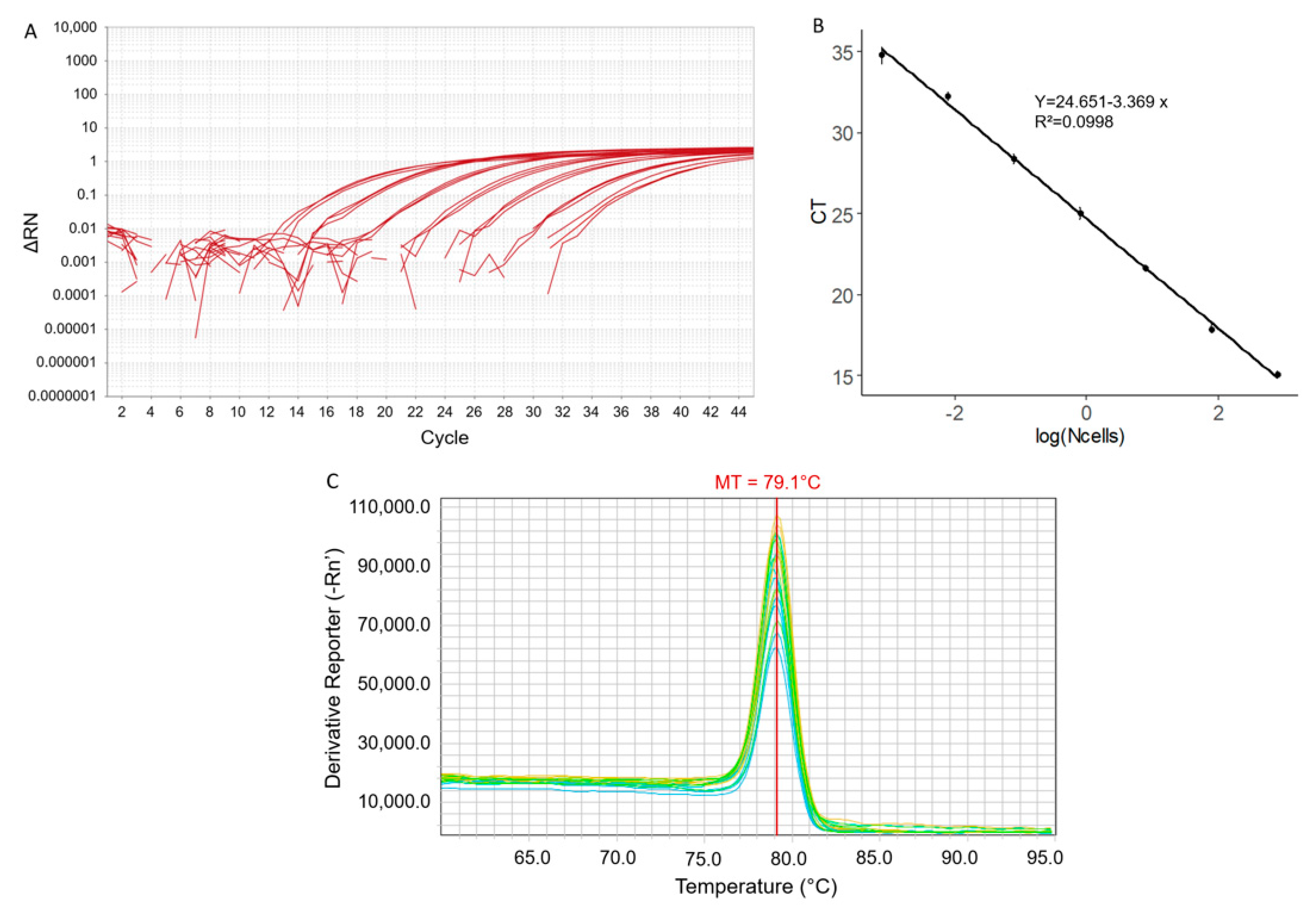
2.1. qPCR Parameters and Sensitivity

A qPCR assay was developed for the dinoflagellate V. rugosum using a selected designed primers pair. qPCR standard curves were established from a serial dilution of a DNA stock solution of V. rugosum (Figure 1A) for an optimal qPCR annealing temperature of 66 °C. A linear relationship between the Ct and the logarithm to base 10 of Equiv. cells of V. rugosum in 1 µL of extract was established (Figure 1B). The regression equation was y = 24.651 − 3.369x (R2 = 0.998), where y represents the Ct and x the log to base 10 of the numbers of equiv. cells in 1 µL of the DNA extract (Figure 1B). The efficiency of the reaction (E) was calculated to be 97.7% by using the formula E = 10(−1/m)−1, where m is the slope of the standard curve. From 35 to 40 cycles, the amplification results were considered positive but not quantifiable. Over 40 cycles, the results were considered negative. The limit of quantification was 0.00078 Equiv. cells corresponding to the 10−6 dilution. The melting temperature (MT) remained constant through the dilutions of the V. rugosum DNA and was calculated to be 79.1 ± 0.1 °C (Figure 1C).

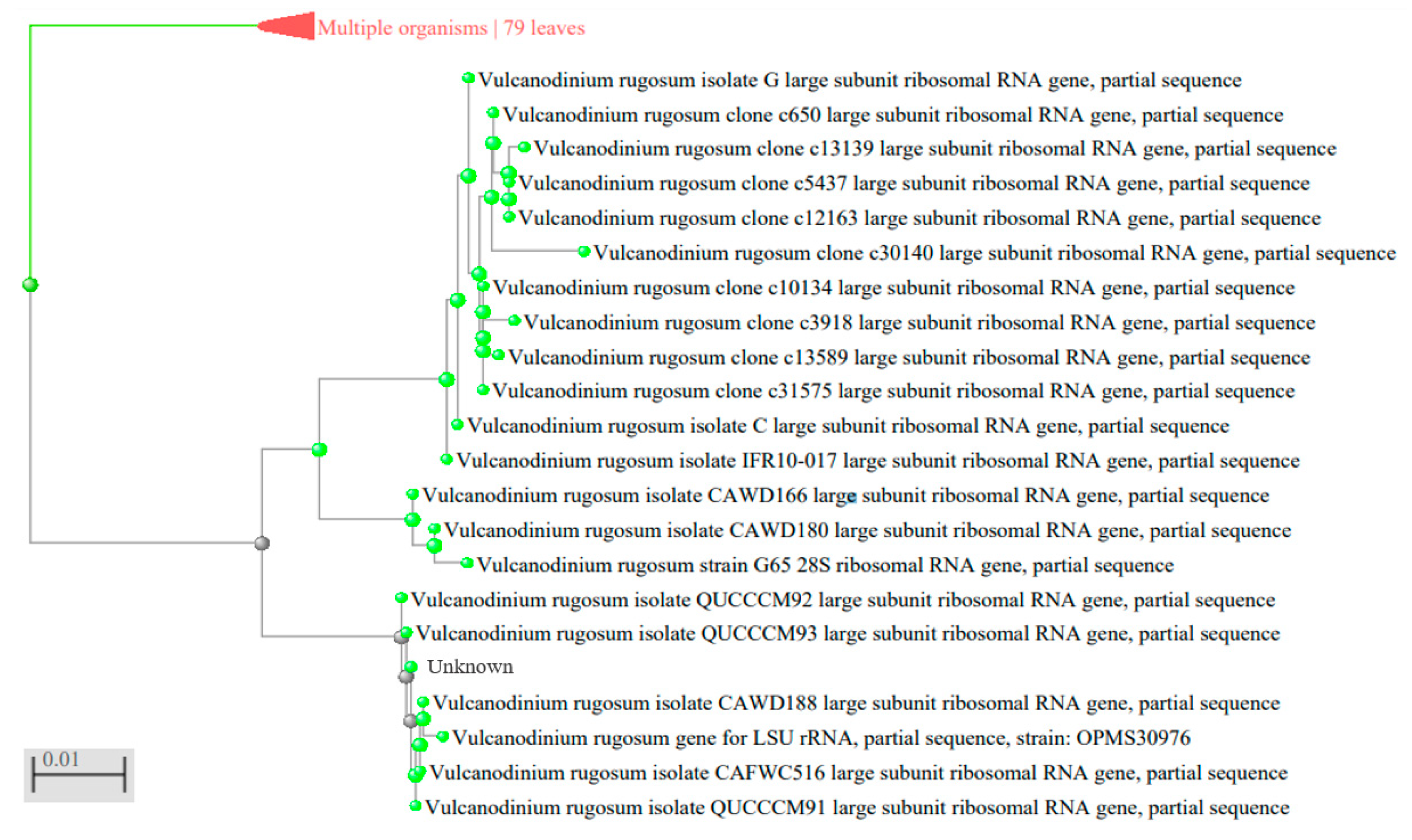
2.2. Specificity of the qPCR

In order to determine the specificity of the PCR product, qPCR was performed on DNAs from 20 dinoflagellates species (Table 1). The use of the primers developed here showed a specific amplification with the DNAs of three V. rugosum strains. The amplification products size and their sequences corresponded to those expected from the large sub unit (LSU) ribosomal RNA gene of V. rugosum (results of sequencing not shown). No PCR amplification product was evidenced with the DNA of the other dinoflagellate species (Table 1). This result showed that the PCR developed here is specific to V. rugosum strains.

2.3. Application of AS-qPCR for Field Survey

Artificial Substrates were set up and collected every two weeks in four Mediterranean lagoons over a year period. The numbers of cells in artificial substrates samples were determined using the real-time PCR assay and LM counting method. By using LM, we observed V. rugosum cells in a few samples in summer 2021 in numbers ranging from 0.036 (corresponding to 1 cell observed in the counting chamber) to 0.36 cells/cm2 substrate in Ingril, Prévost and Vic lagoons (Table 2). No cells were observed in the Thau lagoon. By using the qPCR assay, the presence of V. rugosum cells was detected in the same samples as with LM, along with many other samples in summer 2021 in the four lagoons. Most of the time, V. rugosum was detected at numbers below the relative quantification limit (0.057 Equiv. cells/cm2 substrate) (Table 2). The highest relative numbers were detected during the summer of 2021 in June at Ingril and in July at Vic, (0.18 and 0.26 Equiv. cells/cm2 of substrate, respectively) (Table 2). The qPCR method also allowed for the detection of cells in other seasons in lagoons: in winter (February and March) in Thau and from autumn to spring (November, February and April) in Vic (Table 2).

3. Discussion

We developed an original methodology for the detection and quantification of the toxic dinoflagellate Vulcanodinium rugosum in a marine environment. This method, based on artificial substrates coupled with qPCR identification (AS-qPCR), showed great sensitivity and specificity and was shown to be relevant for the detection of this harmful species even in very low densities.
The artificial substrate characteristics and deployment modalities were adequate for the specific monitoring of V. rugosum. Many types of material have been tested for harmful algae monitoring, including nylon ropes, test tube brushes, plastic plates and fiberglass screens [14,15,16,17]. This study showed that large pieces of nylon plankton net allowed for the collection of V. rugosum. Their wide dimensions are interesting for low-density species [16], and the large 1 mm mesh reduces clogging in highly turbid environment, such as lagoons [25]. Previous studies generally described 24 to 72 h deployments of artificial substrates [14,15,16,17]. This short-term period is adequate for species that shows a diel pattern in the frequency at which they enter the water column and then resettle, and for species that easily detach from their substrate, such as Ostreopsis spp. or Gambierdiscus spp. However, it requires returning to the sampling site after a short period to retrieve samples. V. rugosum is known to form clusters of temporary cysts embedded in highly adherent mucous on their substrate [18], which could prolong their settlement for several days. This constitutes a main advantage for monitoring programs, as it reduces the time and expense to obtain samples. Further studies focusing on the influence of incubation time could, however, allow to optimize the cells’ accumulation on substrates.
The qPCR developed here was shown to be very specific and sensitive to V. rugosum strains. It allowed to detect a very low number of cells and allowed for the quantification of a broad range of cell densities. The limit of quantification (0.00078 Equiv. cells per reaction) and its efficiency (97.7%) are in the range of other studies describing the detection of dinoflagellates through qPCR [21,22]. The ability to detect less than one cell was previously attributed to the multiple copies of the ribosomal genes and was observed with many other dinoflagellates [19,24]. The sensitivity level of this molecular method is a significant benefit in the monitoring of species developing in low densities. The qPCR assay was optimized with primers specific for V. rugosum. They were tested on a great number of microalgae, most of them isolated from the marine environment, where V. rugosum blooms occur, and no cross-reaction occurred with these microalgae. In addition, many field samples showed no amplification, whereas several dinoflagellate species could be identified through LM (Figure S1). Taken together, these results show that the qPCR method allows for specific and sensitive detection of V. rugosum cells, even in environmental samples containing various microalgae species. The quantification results of this method depend on the number of LSRU gene copies per genome and may vary depending on the strains. Thus, the assay cannot yet be used in an absolutely quantitative manner in field samples, but it can be used to determine the relative abundance of V. rugosum and allows for the statistical assessment of environmental parameters associated with blooms.
The artificial substrates combined with qPCR (As-qPCR) were shown to be a powerful tool for the detection of V. rugosum cells in the environment. Artificial substrates are mainly described for the count of benthic species [14,15,16,17] and are thus adequate for V. rugosum, given its low density in water [8,9]. The use of these devices requires, however, to discriminate V. rugosum benthic temporary cysts, which are particularly complex to distinguish from other species with LM. This is partly due to changes in cell size and shape in response to the use of fixatives [26,27]. The high specificity of qPCR eliminated the misidentification of V. rugosum cells. Even if the relative numbers of cells detected were quite low, the high sensitivity allowed for detection in many more samples than LM and for the relative quantification of a minimum of 0.057 Equiv. cells/cm2 of substrate. Cells detected through qPCR could either be resuspended benthic cysts, vegetative cells or cells that have encysted on the substrates. This method provides thus a global idea of V. rugosum state in the environment. In comparison with the search of benthic species on macrophytes, it also reduces the complexity associated with the patchy distribution of cells on macroalgae, removes issues due to macroalgae distribution, and allows for an easy standardization of counting cell abundance per unit of surface area. In regard to all these considerations, AS-qPCR seems to be an efficient tool for monitoring V. rugosum in the environment.
The field survey by AS-qPCR highlighted the presence of V. rugosum mainly during summer, from May to September in the Ingril, Thau, Vic and Prévost lagoons. This result is in agreement with the temporal distribution of this species observed in the Ingril lagoon [8,9], and with the contamination of bivalves by Pinnatoxins observed during summer seasons in Ingril, Vic and Thau lagoons (EMERGTOX monitoring program) [12]. However, no studies have yet shown the presence of this species in the Prévost lagoon. The extension of the microalgae distribution area has long been studied [28,29,30,31]. Recent studies showed that V. rugosum cells could be translocated through ballast water, and that their temporary cysts could survive the passage through the guts of migratory grey mullets Liza ramada or oysters Crassostrea gigas [7,32,33]. These species are massively present in Mediterranean lagoons and could thus participate in V. rugosum cells dispersion [7,32,33,34]. Taken together, the results show the potential of V. rugosum to colonize Mediterranean lagoons and coastal marine ecosystems.
The colonization of lagoons could lead to new sanitary issues. Shellfish harvesting has been banned in the Ingril lagoon since 2020 because of Pinnatoxins contamination; Vic and Prévost lagoons are significant shellfish harvesting areas, and the Thau lagoon is the most important shellfish farming area of the French Mediterranean coast. V. rugosum was moreover detected in low quantities also in autumn and winter in the Thau and Vic lagoons. Even in low abundance, V. rugosum can lead to shellfish contamination that could pose human risks [10]. These results thus remind us that hazards should not be underestimated outside periods and areas currently considered to be at risk. Given the economical and potential sanitary consequences of shellfish contamination, the AS-qPCR method developed here could be a very useful tool for monitoring V. rugosum in the environment.

4. Conclusions

In this work, we developed an AS-qPCR assay to detect the benthopelagic dinoflagellate V. rugosum in the environment. This species shows characteristics that have made it so far very challenging to monitor in its natural habitats, as it is sparsely present in the water column and complex to be identified. The combination of artificial substrates, which could integrate both vegetative and benthic densities, and of the very sensitive and specific qPCR allowed for the identification and quantification of cells in the environment. The results highlighted observations of V. rugosum in several Mediterranean lagoons and showed critical and novel information regarding its distribution. Further studies should focus on the calibration of the artificial substrate incubation time. The need to standardize collection methods for benthic harmful algae is commonly recognized, but the approaches to do so are still diverse and complex. After species-specific optimizations, AS-qPCR could represent a sensitive, standardized and easy-to-implement tool applicable for many benthic or benthopelagic microalgae species monitoring.

5. Materials and Methods

5.1. Dinoflagellate Strains Culture

Phytoplankton strains (Table 1) were cultivated in Enriched Natural Sea Water (ENSW) media, composed of Thau lagoon water (kept at obscurity for several months, filtered at 0.2 μm, and autoclaved) enriched with sodium nitrate, Ferric EDTA, monosodium phosphate, vitamins and other oligo-elements [35]. Monoclonal algal strains were grown in batch mode in Greiner bio-one GmbH culture flasks at 100 μmole photon m-2.S-1 (12:12 h light:dark) at a salinity of 35 and a temperature of 22 °C to 25 °C until DNA extraction.

5.2. Artificial Substrates Environmental Sampling

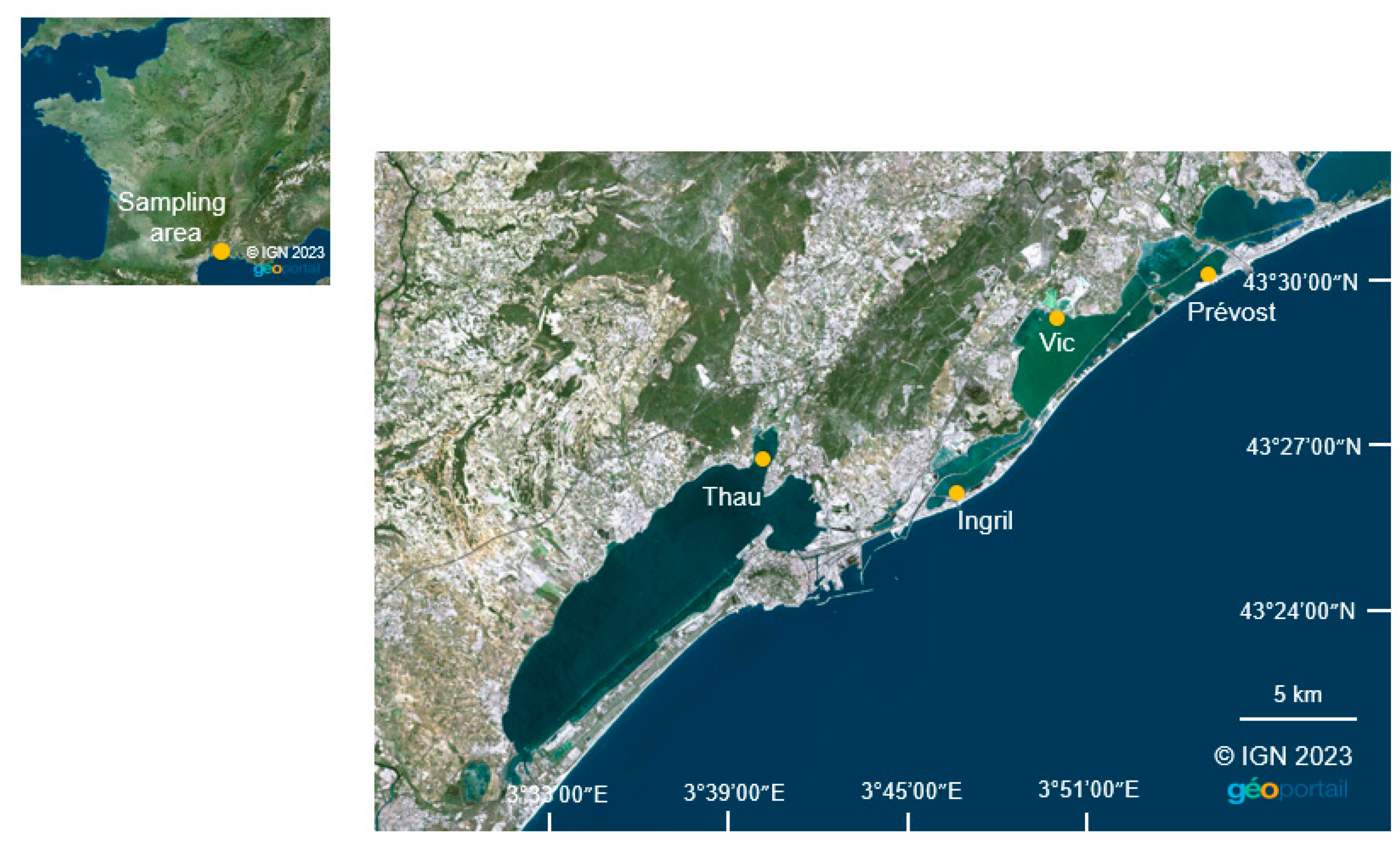
5.2.1. Sampling Area

Collecting stations were set up in the shallow French Mediterranean lagoons of Thau (7000 ha, mean depth 4 m, station localization: 43°27′02.8″ N 3°40′01.4″ E), Ingril (685 ha, mean depth 0.6, station localization: 43°26′09.6″ N 3°46′39.8″ E), Vic (1339 ha, mean depth 1 m, station localization: 43°30′32.8″ N 3°49′20.7″ E) and Prévost (245 ha, mean depth 0.8 m, station localization: 43°30′58.7″ N 3°54′06.7″ E) [36] (Figure 2). All of these lagoons are connected through the Canal du Rhône à Sète and, apart from the Vic lagoon, they connect to the sea through inlets. The stations were all located above muddy sediments at shallow depth and near coasts, as the highest abundance of V. rugosum in Mediterranean lagoons was found in such environments [8].

5.2.2. Deployment and Collection

One artificial substrate, made of rectangular pieces (10 cm × 14 cm) of nylon plankton net (1 mm mesh) attached to a rope with cable ties, was deposited in every station. They were collected and replaced from May 2021 to April 2022 every two weeks. The substrates were kept in plastic bottles filled with seawater. Back in the laboratory, the cells were extracted from the substrates using a modified version of the protocol developed by Jauzein et al. [15]. Briefly, the bottles were shaken vigorously for 20 s in order to detach phytoplankton cells, then the substrates were rinsed twice with 100 mL of FSW (filtered sea water). The water collected was mixed and filtered through a 125 µm filter to remove large particles, then the phytoplankton was concentrated through a 20 µm filter. Phytoplankton cells fixed on the filter were recovered using 50 mL of FSW. Then, 25 mL of water was centrifuged at 3600 rpm 30 min, and the cell pellet fixed in 1 mL of absolute ethanol and stored at −20 °C until DNA extraction. The remaining 25 mL of water was used for visual counting using inverted optical microscopy (LM) in 10 mL counting chambers after Lugol fixation, and for future V. rugosum cell isolation.

5.3. DNA Extraction

5.3.1. From Phytoplankton Culture Cells

DNA was extracted from phytoplankton cultures in their exponential growth phase. The V. rugosum cell concentration was determined in three 1 mL subsamples fixed with Lugol′s iodine solution in which cells were counted on a Sedgwick-Rafter counting chamber under an inverted light microscope. Three 10 mL subsamples were filtered on a 10 µm polycarbonate membrane. Membranes were incubated for 12 h at 57 °C with 500 µL of buffer (Tris-Base 10 mM, NACL 100 mM, EDTA dihydrate 25 mM, SDS 0.5%, Proteinase K 0.1 mg/L). Total nucleic acids were then sequentially purified with phenol/chloroform/isoamyl alcohol (25:24:1, v/v/v) and absolute chloroform. DNA samples were concentrated with 5 volumes of absolute ethanol for 24 h at −20 °C. The precipitates were rinsed with 70% ethanol and resuspended in 100 µL of DNase/RNase free water. DNA quantification was performed using a spectrophotometer (NanoDrop ND-1000) to check for DNA quantity and quality (A260/A280 ratio).

5.3.2. From Environmental Samples

To avoid the effect of possible interferents, DNA from environmental samples was extracted by the above phenol–chloroform method and purified on an affinity column. Thus, half the volume of each DNA extract was purified using the QIAamp® DNA Mini Kit then diluted 10 times before qPCR analysis. The relative number of equivalent cells (Equiv. cells) per cm2 of artificial substrates was calculated as follows:
Ncells / cm 2 AS = Ncells / µ L   ×   V   ×   F 1   ×   F 2   ×   F 3 AS   surface = Ncells / µ L   ×   200   ×   10   ×   2   ×   2 140
where
  • Ncells/cm2AS: Equiv. cells per cm2 of artificial substrate (in cells/cm2 substrate)
  • Ncells/µL: Equiv. cells per µL in diluted purified DNA extracts (in cells/µL)
  • V: Total Volume of diluted purified DNA extracts (in µL)
  • F1: Factor associated with the dilution of purified DNA extracts
  • F2: Factor associated with the volume of DNA extract used for purification
  • F3: Factor associated with the volume of water used for DNA extraction
  • AS surface: Artificial Substrates surface (in cm2)
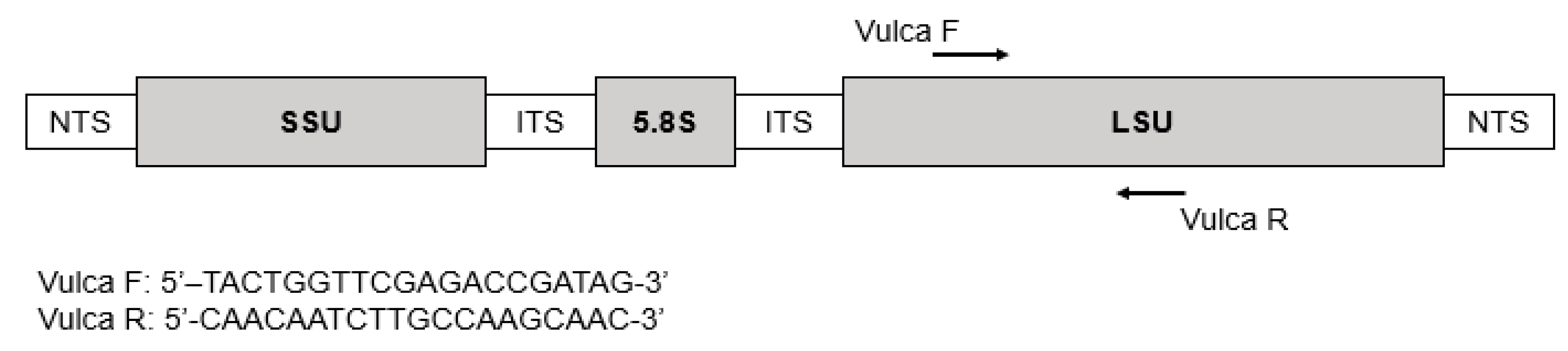
The limit of quantification in artificial substrates was calculated by applying this formula on the limit of quantification in 1 µL of V. rugosum DNA extract estimated with the standard curve.

5.4. Primers Design

V. rugosum Large Sub Unit (LSU) ribosomal RNA genes sequences were obtained from GenBank using the Basic Local Alignment Search Tool from NCBI (National Centre for Biotechnology Information) (Figure 3, Table 3) and aligned using Multalin [37]. Specific primers were designed in a conserved region to amplify a 132 bp fragment (Figure 4). The two primers were as follows: (VulcaF; 5′ TACTGGTTCGAGACCGATAG 3′) sens and (VulcaR: 5′ CAACAATCTTGCCAAGCAAC 3′) antisens.

5.5. qPCR Assay

Quantitative PCR was performed in 96-well plates using a QuantStudio3 thermocycler (Applied Biosystems TM). Each well contained 5 µL CYBERgreen mix (SYBERgreen, dNTP, Polymerase, buffer), 1 µL each of the sens and antisens primers (3.33 µM), 2 µL of DNase/RNase free water and 1 µL of the DNA template. The quantification cycling protocol was as follows: an initial denaturation step at 95 °C for 5 min, followed by 45 cycles of 10 s at 95 °C, 50 to 70 °C for 10 s and 72 °C for 10 s. The melting curve profile was generated by increasing temperature from 55 °C to 95 °C at 0.5 °C /s. Amplification products were analyzed using QuantStudio3 software V1.5.2 (Applied biosystemsTM).

5.6. Standard Curve and Specificity

To prepare DNA templates for the construction of the qPCR standard curve, DNA extracted from a culture of V. rugosum isolated in Ingril lagoon in 2010 by Nézan [12] was serially diluted from 10-1 to 10-6 in miliQ water. The qPCR was performed on the dilutions in triplicate. Cells were considered non-quantifiable when CT was ranged between 35 and 40 and non-detectable when CT was over 40.
In order to determine the specificity of the PCR product, DNA samples from 20 dinoflagellates species were used as templates for qPCR reaction (Table 1).

5.7. Data Analysis

Graphics and statistical analysis were performed using R software [38].

Supplementary Materials

The following supporting information can be downloaded at: https://www.mdpi.com/article/10.3390/toxins15030217/s1, Figure S1: Derivatives melting curve plot for A. Vulcanodinium rugosum DNA extract 2-fold diluted diluted, B. Field sample, Vic lagoon, 7 July 2021, C. Field sample, Vic lagoon, 2nd December 2021.

Author Contributions

Conceptualization, A.B., J.L.R. and E.A.; Methodology, A.B. and J.L.R., Investigation, A.B., J.L.R., C.R. and C.F.; Formal analysis, A.B.; Project administration, J.L.R., A.B., E.A. and M.L.; Data curation, A.B. and J.L.R.; Funding acquisition, J.L.R., E.A. and M.L.; Resources, J.L.R., E.A. and E.M.; Supervision, J.L.R. and A.B.; Validation, J.L.R., E.A., M.L. and E.M.; Visualization, A.B. and J.L.R.; Writing—original draft, A.B.; Writing—review and editing, J.L.R., M.L., E.M. and E.A. All authors have read and agreed to the published version of the manuscript.

Funding

This research was supported by the French Ministry of Agriculture and Food, the French research institute for the exploitation of the sea (IFREMER) and MARBEC (MARine Biodiversity, Exploitation and Conservation).

Institutional Review Board Statement

Not applicable.

Informed Consent Statement

Not applicable.

Acknowledgments

The authors would like to thank all members of the IFREMER Laboratoire Environnement Ressource Languedoc Roussillon for their help and support during this study.

Conflicts of Interest

The authors declare no conflict of interest.

References

  1. Moreira, A.R.; Comas, A.; Valle, A.; Seisdedo, M. Bloom of Vulcanodinium rugosum Linked to Skin Lesions in Cienfuegos Bay, Cuba. IOC Newsl. Toxic Algae Algal Bloom. 2016, 55, 10–11. [Google Scholar] [CrossRef]
  2. Rhodes, L.; Smith, K.; Selwood, A.; McNabb, P.; Munday, R.; Suda, S.; Molenaar, S.; Hallegraeff, G. Dinoflagellate Vulcanodinium rugosum Identified as the Causative Organism of Pinnatoxins in Australia, New Zealand and Japan. Phycologia 2011, 50, 624–628. [Google Scholar] [CrossRef] [PubMed]
  3. Smith, K.F.; Rhodes, L.L.; Suda, S.; Selwood, A.I. A Dinoflagellate Producer of Pinnatoxin G, Isolated from Sub-Tropical Japanese Waters. Harmful Algae 2011, 10, 702–705. [Google Scholar] [CrossRef]
  4. Zeng, N.; Gu, H.; Smith, K.F.; Rhodes, L.L.; Selwood, A.I.; Yang, W. The First Report of Vulcanodinium rugosum (Dinophyceae) from the South China Sea with a Focus on the Life Cycle. N. Z. J. Mar. Freshw. Res. 2012, 46, 511–521. [Google Scholar] [CrossRef] [Green Version]
  5. Hess, P.; Abadie, E.; Herve, F.; Berteaux, T.; Sechet, V.; Araoz, R.; Molgo, J.; Zakarian, A.; Sibat, M.; Rundberget, T.; et al. Is Responsible for Atypical Toxicity in Mussels (Mytilus galloprovincialis) and Clams (Venerupis decussata) from Ingril, a French Mediterranean Lagoon. Toxicon Off. J. Int. Soc. Toxinology 2013, 75, 16–26. [Google Scholar] [CrossRef] [Green Version]
  6. Munday, R.; Selwood, A.I.; Rhodes, L. Acute Toxicity of Pinnatoxins, E, F and G to Mice. Toxicon 2012, 60, 995–999. [Google Scholar] [CrossRef]
  7. Bouquet, A.; Perdrau, M.A.; Laabir, M.; Foucault, E.; Chomérat, N.; Rolland, J.L.; Abadie, E. Liza Ramada Juveniles after Exposure to the Toxic Dinoflagellate Vulcanodinium rugosum: Effects on Fish Viability, Tissue Contamination and Microalgae Survival after Gut Passage. Toxins 2022, 14, 401. [Google Scholar] [CrossRef]
  8. Abadie, E.; Chiantella, C.; Crottier, A.; Rhodes, L.; Masseret, E.; Berteau, T.; Laabir, M. What are the main environmental factors driving the development of the neurotoxic dinoflagellate Vulcanodinium rugosum in a Mediterranean ecosystem (Ingril lagoon, France)? Harmful Algae 2018, 75, 75–86. [Google Scholar] [CrossRef] [Green Version]
  9. REPHY—French Observation and Monitoring program for Phytoplankton and Hydrology in coastal waters. In REPHY Dataset—French Observation and Monitoring Program for Phytoplankton and Hydrology in Coastal Waters. Metropolitan Data; SEANOE, Brest, France: 2022. [CrossRef]
  10. Arnich, N.; Abadie, E.; Delcourt, N.; Fessard, V.; Fremy, J.-M.; Hort, V.; Lagrange, E.; Maignien, T.; Molgó, J.; Peyrat, M.-B.; et al. Health Risk Assessment Related to Pinnatoxins in French Shellfish. Toxicon 2020, 180, 1–10. [Google Scholar] [CrossRef]
  11. Nézan, E.; Chomérat, N. Vulcanodinium rugosum Gen. Nov., Sp. Nov. (Dinophyceae): A New Marine Dinoflagellate from the French Mediterranean Coast. Cryptogam. Algol. 2011, 32, 3–18. [Google Scholar] [CrossRef]
  12. Amzil, Z.; Derrien, A.; Terre Terrillon, A.; Duval, A.; Connes, C.; Marco-Miralles, F.; Nézan, E.; Mertens, K.N. Monitoring the Emergence of Algal Toxins in Shellfish: First Report on Detection of Brevetoxins in French Mediterranean Mussels. Mar. Drugs 2021, 19, 393. [Google Scholar] [CrossRef]
  13. Ben Gharbia, H.; Yahia, O.K.D.; Cecchi, P.; Masseret, E.; Amzil, Z.; Herve, F.; Rovillon, G.; Nouri, H.; M’Rabet, C.; Couet, D.; et al. New Insights on the Species-Specific Allelopathic Interactions between Macrophytes and Marine HAB Dinoflagellates. PLoS ONE 2017, 12, 28. [Google Scholar] [CrossRef] [Green Version]
  14. Tester, P.A.; Kibler, S.R.; Holland, W.C.; Usup, G.; Vandersea, M.W.; Leaw, C.P.; Teen, L.P.; Larsen, J.; Mohammad-Noor, N.; Faust, M.A.; et al. Sampling Harmful Benthic Dinoflagellates: Comparison of Artificial and Natural Substrate Methods. Harmful Algae 2014, 39, 8–25. [Google Scholar] [CrossRef]
  15. Jauzein, C.; Fricke, A.; Mangialajo, L.; Lemée, R. Sampling of Ostreopsis Cf. ovata Using Artificial Substrates: Optimization of Methods for the Monitoring of Benthic Harmful Algal Blooms. Mar. Pollut. Bull. 2016, 107, 300–304. [Google Scholar] [CrossRef] [PubMed]
  16. Tester, P.A.; Litaker, R.W.; Soler-Onís, E.; Fernández-Zabala, J.; Berdalet, E. Using Artificial Substrates to Quantify Gambierdiscus and Other Toxic Benthic Dinoflagellates for Monitoring Purposes. Harmful Algae 2022, 120, 102351. [Google Scholar] [CrossRef] [PubMed]
  17. Fernández-Zabala, J.; Tuya, F.; Amorim, A.; Soler-Onís, E. Benthic Dinoflagellates: Testing the Reliability of the Artificial Substrate Method in the Macaronesian Region. Harmful Algae 2019, 87, 101634. [Google Scholar] [CrossRef]
  18. Hernandez-Becerril, D.U.; Rodriguez-Palacio, M.C.; Lozano-Ramirez, C. Morphology and Life Stages of the Potentially Pinnatoxin-Producing Thecate Dinoflagellate Vulcanodinium rugosum from the Tropical Mexican Pacific. Bot. Mar. 2013, 56, 535–540. [Google Scholar] [CrossRef]
  19. Smith, K.F.; de Salas, M.; Adamson, J.; Rhodes, L.L. Rapid and accurate identification by real-time PCR of biotoxin-producing dinoflagellates from the family gymnodiniaceae. Mar. Drugs 2014, 12, 1361–1376. [Google Scholar] [CrossRef] [Green Version]
  20. Galluzzi, L.; Penna, A.; Bertozzini, E.; Vila, M.; Garcés, E.; Magnani, M. Development of a Real-Time PCR Assay for Rapid Detection and Quantification of Alexandrium minutum (a Dinoflagellate). Appl. Environ. Microbiol. 2004, 70, 1199–1206. [Google Scholar] [CrossRef] [Green Version]
  21. Perini, F.; Casabianca, A.; Battocchi, C.; Accoroni, S.; Totti, C.; Penna, A. New Approach Using the Real-Time PCR Method for Estimation of the Toxic Marine Dinoflagellate Ostreopsis Cf. ovata in Marine Environment. PLoS ONE 2011, 6, e17699. [Google Scholar] [CrossRef] [Green Version]
  22. Yuan, J.; Mi, T.; Zhen, Y.; Yu, Z. Development of a Rapid Detection and Quantification Method of Karenia mikimotoi by Real-Time Quantitative PCR. Harmful Algae 2012, 17, 83–91. [Google Scholar] [CrossRef]
  23. Bowers, H.A.; Tengs, T.; Glasgow, H.B.; Burkholder, J.M.; Rublee, P.A.; Oldach, D.W. Development of Real-Time PCR Assays for Rapid Detection of Pfiesteria Piscicida and Related Dinoflagellates. Appl. Environ. Microbiol. 2000, 66, 4641–4648. [Google Scholar] [CrossRef] [PubMed] [Green Version]
  24. Erdner, D.L.; Percy, L.; Keafer, B.; Lewis, J.; Anderson, D.M. A Quantitative Real-Time PCR Assay for the Identification and Enumeration of Alexandrium Cysts in Marine Sediments. Deep Sea Res. Part II Top. Stud. Oceanogr. 2010, 57, 279–287. [Google Scholar] [CrossRef] [PubMed] [Green Version]
  25. Smith, P.E.; Counts, R.C.; Clutter, R.I. Changes in Filtering Efficiency of Plankton Nets Due to Clogging Under Tow. ICES J. Mar. Sci. 1968, 32, 232–248. [Google Scholar] [CrossRef]
  26. Leakey, R.J.G.; Burkill, P.H.; Sleigh, M.A. A Comparison of Fixatives for the Estimation of Abundance and Biovolume of Marine Planktonic Ciliate Populations. J. Plankton Res. 1994, 16, 375–389. [Google Scholar] [CrossRef]
  27. Menden-Deuer, S.; Lessard, E.J.; Satterberg, J. Effect of Preservation on Dinoflagellate and Diatom Cell Volume and Consequences for Carbon Biomass Predictions. Mar. Ecol. Prog. Ser. 2001, 222, 41–50. [Google Scholar] [CrossRef]
  28. Faust, M.A. Ciguatera-Causing Dinoflagellates in a Coral-Reef Mangrove Ecosystem, Belize. Atoll Res. Bull. 2009, 569, 1–32. [Google Scholar] [CrossRef]
  29. Montagnes, D.J.S.; Berges, J.A.; Harrison, P.J.; Taylor, F.J.R. Estimating Carbon, Nitrogen, Protein, and Chlorophyll a from Volume in Marine Phytoplankton. Limnol. Oceanogr. 1994, 39, 1044–1060. [Google Scholar] [CrossRef] [Green Version]
  30. Bolch, C.J.S.; de Salas, M.F. A Review of the Molecular Evidence for Ballast Water Introduction of the Toxic Dinoflagellates Gymnodinium catenatum and the Alexandrium tamarensis Complex to Australasia. Harmful Algae 2007, 6, 465–485. [Google Scholar] [CrossRef]
  31. Glibert, P.M. Harmful Algae at the Complex Nexus of Eutrophication and Climate Change. Harmful Algae 2020, 91, 101583. [Google Scholar] [CrossRef]
  32. Garret, M.J.; Puchulutegui, C.; Selwood, A.I.; Wolny, J.L. Identification of the harmful dinoflagellate Vulcanodinium rugosum recovered from a ballast tank of a globally travelled ship in Port Tampa Bay, USA. Harmful Algae 2014, 39, 202–209. [Google Scholar] [CrossRef]
  33. Abadie, E. Etude de Vulcanodinium rugosum (Dinoflagellé Producteur de Pinnatoxines) se Développant dans la Lagune Méditerranéenne de l’Ingril. Ph.D. Thesis, Université de Montpellier, Montpellier, France, 2015. Available online: https://archimer.ifremer.fr/doc/00473/58425 (accessed on 10 March 2023).
  34. Newton, A.; Icely, J.; Cristina, S.; Brito, A.; Cardoso, A.C.; Colijn, F.; Riva, S.D.; Gertz, F.; Hansen, J.W.; Holmer, M.; et al. An overview of ecological status, vulnerability and future perspectives of European large shallow, semi enclosed coastal systems, lagoons and transitional waters. Estuar. Coast. Shelf Sci. 2014, 140, 95–122. [Google Scholar] [CrossRef]
  35. Harrison, P.J.; Waters, R.E.; Taylor, F.J.R. A Broad-Spectrum Artificial Seawater Medium for Coastal and Open Ocean Phytoplankton. J. Phycol. 1980, 16, 28–35. [Google Scholar] [CrossRef]
  36. Le Fur, I.; De Wit, R.; Plus, M.; Oheix, J.; Simier, M.; Ouisse, V. Submerged Benthic Macrophytes in Mediterranean Lagoons: Distribution Patterns in Relation to Water Chemistry and Depth. Hydrobiologia 2018, 808, 175–200. [Google Scholar] [CrossRef]
  37. Corpet, F. Multiple Sequence Alignment with Hierarchical Clustering. Nucleic Acids Res. 1988, 16, 10881–10890. [Google Scholar] [CrossRef]
  38. R Core Team. R: A Language and Environment for Statistical Computing; R Core Team: Vienna, Austria, 2020; Available online: https://www.r-project.org/ (accessed on 10 March 2023).
Figure 1. Amplification results of qPCR products from 6-fold diluted Vulcanodinium rugosum DNA extract: (A) Amplification curves plot, (B) standard curve, and (C) derivative melting curves plot. The error bars represent ± standard deviation from triplicate mean values. MT: melting temperature.
Toxins 15 00217 g001
Figure 2. Collecting stations in Thau, Ingril, Vic and Prévost.
Toxins 15 00217 g002
Figure 3. Phylogenetic distance tree of Vulcanodinium rugosum LSRU genes used to design PCR primers.
Toxins 15 00217 g003
Figure 4. Structure of Vulcanodinium rugosum species rDNA and location of primers for the V. rugosum genus-specific PCR. NTS: non-transcribed spacer; SSU: small subunit; LSU: large subunit.
Toxins 15 00217 g004
Table 1. PCR amplification results on different dinoflagellate species. Nd: not detectable.
SpeciesStrains and/or OriginsCollectionsTM (°C)
Fukyuoa paulensisRCC-6550Roscoff Culture Collection (RCC)Nd
Fukyuoa sp.RCC-6548RCC Nd
Gambierdiscus belizeanusRCC-6344RCCNd
Gambierdiscus carolinianusRCC-6338RCC Nd
Gambierdiscus spp.RCC-6328RCCNd
Alexandrium minutumBritany, FranceMARBEC, University of Montpellier Collection (UMC)Nd
Alexandrium pseudogonyaulaxBizerte lagoon, TunisiaMarbec, UMCNd
Alexandrium pacificumACT03, Thau, FranceMarbec, UMCNd
Alexandrium pacificumBizerte lagoon, TunisiaMarbec, UMCNd
Amphidinum carteraeSAMS, Oban, UKMarbec, UMCNd
Coolia monotisCMBZT14, Bizerte, TunisiaMarbec, UMC Nd
Gymnodinium catenatumM'diq Bay, MoroccoMarbec, UMCNd
Gyrodinium impudicumGulf of Tunis, TunisiaMarbec, UMCNd
Karenia selliformisGulf of Gabes, TunisiaMarbec, UMCNd
Prorocentrum limaPLBZT14, Bizerte lagoon, TunisiaMarbec, UMCNd
Prorocentrum micansH4-3, Thau lagoon, FranceMarbec, UMCNd
Ostreopsis siamensis.P550, Gbraltar straitMarbec, UMCNd
Scrippsiella trochoideaST17, Mellah, AlgeriaMarbec, UMCNd
Scrippsiella accuminataChina SeaMarbec, UMCNd
Vulcanodinium rugosumIFR-VRU-01, Ingril, FranceMarbec, UMC79.1
Vulcanodinium rugosum15-Ing-5.48, Ingril, FranceMarbec, UMC79.1
Vulcanodinium rugosum21-Ing-1.96, Ingril, FranceMarbec, UMC79.1
Vulcanodinium rugosum21-Vic-2.96, Vic, FranceMarbec, UMC79.1
Table 2. Detection and quantification of Vulcanodinium rugosum cells during one year of monitoring in four French Mediterranean lagoons: by light microscopy (LM, in cells/cm2 substrate) and by qPCR (relative quantification in Equiv. cells/cm2 substrate). Nd: not detectable.
Collection DateLagoons
ThauIngrilVicPrévost
YearMonthDayLM qPCRLMqPCRLMqPCRLMqPCR
2021May4thNdNdNdNdNdNdNdNd
20thNdNd0.11<0.057NdNdNdNd
Jun.3rdNdNdNdNdNd<0.057Nd<0.057
16thNdNd0.360.26Nd<0.057NdNd
29thNdNdNd<0.057Nd<0.0570.11<0.057
Jul.7thNdNdNd<0.0570.250.18NdNd
27thNdNdNd<0.0570.071<0.057NdNd
Aug.10thNd<0.0570.036<0.0570.036<0.057Nd<0.057
25thNdNdNdNd0.036<0.057NdNd
Sep.8thNdNdNdNdNdNdNd<0.057
21thNd<0.057NdNdNdNdNdND
Oct.4thNdNdNdNdNdNdNdNd
19thNdNdNdNdNdNdNdNd
Nov.3rdNdNdNdNdNd<0.057NdNd
19thNdNdNdNdNdNdNdNd
Dec.2ndNdNdNdNdNdNdNdNd
16thNdNdNdNdNdNdNdNd
2022Jan.4thNdNdNdNdNdNdNdNd
20thNdNdNdNdNdNdNdNd
Feb.3rdNdNdNdNdNd<0.057NdNd
15thNd<0.057NdNdNdNdNdNd
Mar.1stNd<0.057NdNdNdNdNdNd
17thNdNdNdNdNdNdNdNd
29thNdNdNdNdNdNdNdNd
Apr.12thNdNdNdNdNd<0.057NdNd
Table 3. Sequences and lengths of Vulcanodinium rugosum LSRU genes obtained from GenBank with Basic Local Alignment Search Tool from NCBI (National Centre for Biotechnology Information).
LSRU GenesSequencesSequences Lengths
Isolate GMG826107.11351
Clone c650MK236574.1620
Clone c13139MK236578.1620
Clone c5437MK236579.1619
Clone c12163MK236577.1622
Clone c30140MK236582.1619
Clone c10134MK236580.1617
Clone c3918MK236576.1619
Clone c13589MK236581.1620
Clone c31575MK236575.1618
Isolate CMG826106.1928
Isolate IFR10-017HQ622103.11384
Isolate CAWD166JF267773.1805
Isolate CAWD180JF683380.1788
Strain G65JX457352.1596
Isolate QQCCCM92KX853181.1856
Isolate QQCCCM93KX853179.1846
Isolate CAWD188JF683382.1788
Strain OPMS30976LC228963.1599
Isolate CAFWC516KM252944.1845
Isolate QUCCCM91KX853180.1847
Disclaimer/Publisher’s Note: The statements, opinions and data contained in all publications are solely those of the individual author(s) and contributor(s) and not of MDPI and/or the editor(s). MDPI and/or the editor(s) disclaim responsibility for any injury to people or property resulting from any ideas, methods, instructions or products referred to in the content.

