Review

Inflammation and Nutrition: Friend or Foe?

1
Medical University Department of Medicine, Division of General Internal and Emergency Medicine, Kantonsspital Aarau, 5001 Aarau, Switzerland
2
Department of Nutritional Medicine and Prevention, Institute of Clinical Nutrition, University of Hohenheim, 70599 Stuttgart, Germany
3
Medical Faculty, University of Basel, 4056 Basel, Switzerland
*
Author to whom correspondence should be addressed.
Nutrients 2023, 15(5), 1159; https://doi.org/10.3390/nu15051159
Submission received: 31 January 2023 / Revised: 21 February 2023 / Accepted: 22 February 2023 / Published: 25 February 2023
(This article belongs to the Special Issue Hot Topics in Clinical Nutrition)

Abstract

:
The importance of the interplay between inflammation and nutrition has generated much interest in recent times. Inflammation has been identified as a key driver for disease-related malnutrition, leading to anorexia, reduced food intake, muscle catabolism, and insulin resistance, which are stimulating a catabolic state. Interesting recent data suggest that inflammation also modulates the response to nutritional treatment. Studies have demonstrated that patients with high inflammation show no response to nutritional interventions, while patients with lower levels of inflammation do. This may explain the contradictory results of nutritional trials to date. Several studies of heterogeneous patient populations, or in the critically ill or advanced cancer patients, have not found significant benefits on clinical outcome. Vice versa, several dietary patterns and nutrients with pro- or anti-inflammatory properties have been identified, demonstrating that nutrition influences inflammation. Within this review, we summarize and discuss recent advances in both the role of inflammation in malnutrition and the effect of nutrition on inflammation.

1. Introduction

Disease-related malnutrition (DRM) is a common syndrome in patients with acute and chronic illnesses. Prevalence rates are approximately 30% among medical inpatients and rise higher among the elderly or critically ill [1,2]. Left untreated, DRM is associated with poor outcomes, such as higher mortality and prolonged intensive care unit (ICU) and hospital stays [3,4]. Inflammation, undernutrition-driven catabolism, and inadequate dietary intake are key drivers of DRM [1]. While medical inpatients with malnutrition have been shown to benefit from nutritional treatment, this may not be equally true for other patient populations, such as those in the ICU [1,5]. A Cochrane review from 2017 on a more heterogenic patient population, which included highly inflamed patients such as those in the ICU, only found limited positive effects of nutritional treatment on clinical outcomes [6].
The focus on inflammation as a key driver of DRM has grown due to the growing understanding of DRM and its pathophysiology. Recent studies have shown associations between inflammatory processes measured by inflammation biomarkers, such as C-reactive protein (CRP), and responses to nutritional therapy [7]. The many possible reasons will be discussed within the scope of this review, in conjunction with the pathophysiological connection of inflammatory responses in illness and DRM. As researchers strive to elucidate the role of inflammation in nutrition, the interest in potential anti-inflammatory properties of nutrition also grows. We aim to provide an overview of the influence of nutrition on inflammation through a discussion of dietary patterns such as the Mediterranean diet (MD), and various indices such as the Dietary Inflammatory Index (DII) and nutrients (ex. fatty acids [FA]).

2. Malnutrition and Inflammation

DRM is a complex syndrome resulting from the inadequate intake of nutrients required to support physiological function and disease-related systemic inflammatory responses. The European Society of Clinical Nutrition and Metabolism (ESPEN) defines malnutrition as “a state resulting from lack of intake or uptake of nutrition that leads to altered body composition (decreased fat free mass) and body cell mass leading to diminished physical and mental function and impaired clinical outcome from disease” [8]. There remains considerable scope for confusion and misunderstanding; however, this is only one of several definitions found in the literature.
In hospitalized patients, malnutrition is not only due to inadequate nutritional intake, but is often disease-related and associated with complex pathophysiological mechanisms. These mechanisms can differ depending on the underlying disease and its treatment. Inadequate dietary intake has also been shown to lead to immune system dysfunction and mucosal damage in the gut [9]. In the presence of DRM, hospital food, gastrointestinal symptoms, and dysfunction can contribute to reduced appetite, food intake, and nutrient assimilation. The lack of movement can further contribute to malnutrition [1].

2.1. Malnutrition—Risk Factors and Diagnosis

DRM is multifactorial: risk factors include polypharmacy, disease-related inflammatory mechanisms, compromised intake or assimilation of nutrients, immobility associated muscle wasting, older age, and social isolation [1,10]. In addition to an already high prevalence of malnutrition upon admission, nutritional states may be further aggravated during hospitalization due to illness-related loss of appetite, fasting for diagnostic tests, drug-induced side effects, diseases that affect gastrointestinal function, or other factors associated with hospitalization [1]. To diagnose malnutrition, the Global Leadership Initiative on Malnutrition (GLIM) proposes a two-step approach consisting of nutritional risk screening followed by a more thorough evaluation. There is no one universal screening method for malnutrition but rather a number of different tools which have been validated for different settings, including the NRS-2002 [11], SGA [12], MUST [13] or MNA-SF [14]. If nutritional risk is identified, a nutritional assessment to confirm a diagnosis should be performed, including etiological (reduced food intake or assimilation and disease burden/inflammation) and phenotypic (non-volitional weight loss, low BMI and reduced muscle mass) criterions. According to GLIM, a diagnosis of malnutrition is fulfilled if one etiological and one phenotypic criterion apply for the patient [15].

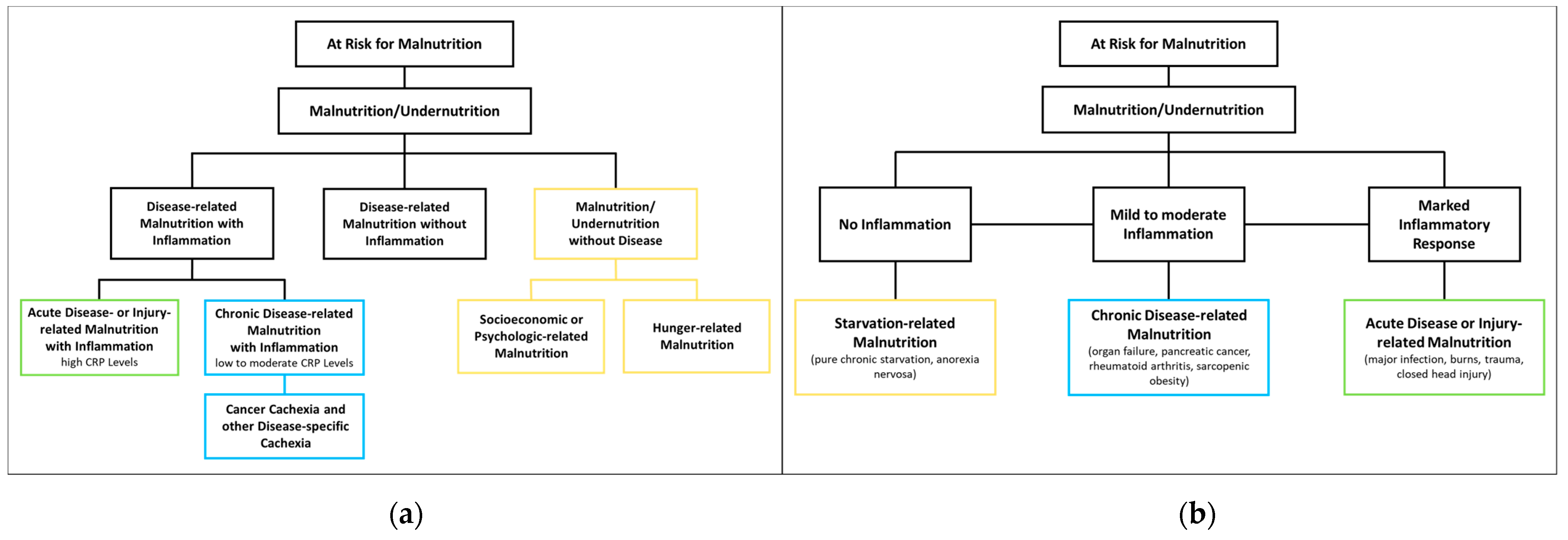
2.2. Malnutrition—Classification

The ESPEN proposes three etiological groups: DRM with inflammation, DRM without inflammation, and malnutrition/undernutrition without disease (Figure 1a). DRM with inflammation can be divided further into acute and chronic forms. Chronically malnourished patient groups typically affected by inflammation (and thus cachexia) include patients with cancer, chronic obstructive pulmonary disease (COPD), inflammatory bowel diseases, congestive heart failure, chronic kidney disease, and other end-stage organ diseases. Inflammation in these patients is often milder, with CRP levels of up to 40 mg/l. DRM with inflammation due to acute disease or injury typically affects critically ill patients or post major surgery, and is accompanied by higher levels of CRP [8]. The American Society for Parenteral and Enteral Nutrition (ASPEN) uses a similar approach, categorizing according to (I) social and environmental circumstances, (II) chronic illness and (III) acute illness or injury (Figure 1b) [16]. They also specify that malnutrition caused by social and environmental circumstances is not related to disease and corresponds to “malnutrition without disease”. As the latter is not relevant in the context of disease, it will not be discussed in this review.

2.3. Malnutrition—Therapy and Clinical Outcomes

Malnutrition has been shown to be a risk factor for adverse outcomes such as increased mortality, a higher risk of readmission within 30 days, prolonged hospital and ICU stays, loss of function, and infection rates [2,3,4]. While the possible benefits of nutritional therapy in malnourished patients have long been unclear [6,17], recent evidence in favor of applying nutritional therapy in medical inpatient settings has been growing, in part due to large-scale RCTs including the EFFORT [18] and the NOURISH trials [19]. In a recent meta-analysis, Gomes et al. found that nutritional therapy significantly improves functional outcome and reduces loss of body weight, risk of complications, and hospital readmission. Recent trials included in the meta-analysis have indicated a marked decrease in risk of mortality compared to older studies (OR 0.47, 95% CI 0.28–0.79) [20]. In addition, trials with higher protein intake and longer intervention times have been associated with a stronger impact of nutritional support on clinical outcomes [21]. All the above-mentioned reasons make early recognition and adequate treatment of malnutrition vital for each patient’s individual treatment plan.

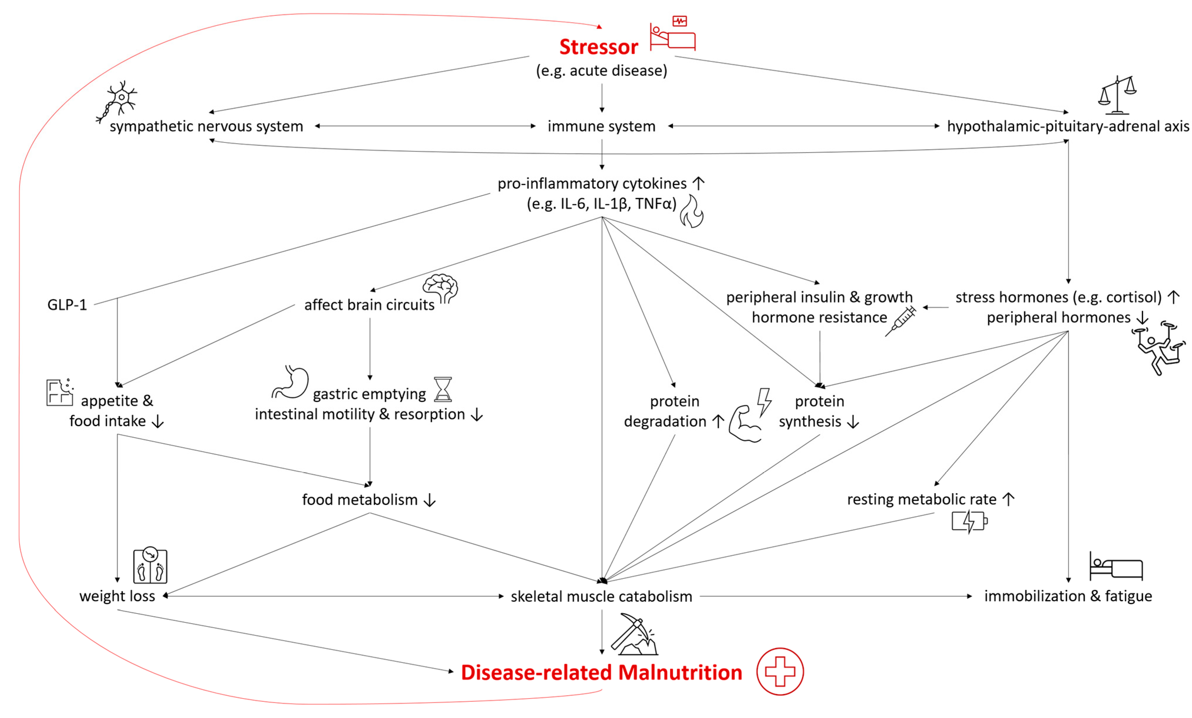
2.4. Inflammation in Malnutrition

In both DRM with acute and chronic inflammation, the sympathetic nervous system, the immune system, and the hypothalamic–pituitary–adrenal axis are activated as a systemic response to a stressor and disease [22,23]. As they are connected both anatomically and functionally, they interact in the response to the stressor [24]. The modulation of the hypothalamic–pituitary–adrenal axis stimulates the release of stress hormones, including cortisol, catecholamines, and suppresses other hormones regulating sex, thyroid, and other peripheral functions [22]. In malnutrition, the deiodination of thyroxine (T4) to triiodothyronine (T3) was shown to be down regulated—a process called “low T3 syndrome” which is an adaptive metabolic mechanism to reduce energy expenditure and prevent catabolism [25]. Catecholamines and cortisol increase glycogenolysis and gluconeogenesis in the liver while simultaneously inducing peripheral insulin resistance and inhibiting glucose from entering cells [22]. Furthermore, insulin-dependent glucose transporters in peripheral tissues are downregulated, causing stress hyperglycemia [26]. Pro-inflammatory cytokines including interleukin 6 (IL-6), interleukin 1β (IL-1β) and tumor necrosis factor α (TNF-α) are released, triggering several mechanisms which contribute to the pathogenesis of malnutrition (Figure 2). Pro-inflammatory cytokines also affect brain circuits which control food intake, cause delayed gastric emptying and increase skeletal muscle catabolism [1,23,27,28]. Furthermore, researchers have identified an interaction of pro-inflammatory cytokines (mainly IL-6 and IL-1β) and gut tissue-released glucagon-like peptide-1 (GLP-1), resulting in reduced food intake and unintentional weight loss [29]. Muscle degradation is triggered by decreased synthesis of muscle protein and the increased degradation of proteins such as myosin heavy chains [9]. These endocrine changes further advance catabolism and lead to fatigue and immobilization [1,23]. The combination of these mechanisms leads to compromised food metabolism, a hypercatabolic state, and eventually to DRM.

3. Is Nutrition a Friend? How Nutrition influences Inflammation

Primarily, nutrition serves as the source of essential nutrients, providing energy and substrates for the numerous metabolic functions. However, as nutrition has long been a topic of scientific interest, knowledge about specific properties of nutrition or nutrients has grown with its anti-inflammatory properties gaining much attention [30]. While inflammation is an acute reaction to stressors such as trauma or infection, an increasing number of chronic diseases such as cardiovascular diseases, vascular cognitive impairment, and dementia have also been associated with chronic (often subclinical) inflammation [31].

3.1. Anti-Inflammatory Potential of Nutrients and Other Food Components

Numerous food components have been investigated for their anti-inflammatory properties and potential use in nutritional therapy. Research has focused not only on macronutrients such as fatty acids or amino acids and micronutrients such as vitamin D, but also included other food components such as fiber and phytochemicals [32,33,34,35]. A selection of these is described in more detail below.

3.1.1. Omega-3 and Omega-6 Fatty Acids

Polyunsaturated fatty acids (PUFA), especially omega-3 (n-3 FA) and omega-6 fatty acids (n-6 FA), are amongst the most studied macronutrients in this context. As the human body is not able to synthesize these essential elements, they must be ingested from an outside source. Long-chain n-3 FA (LC n-3 FA) such as eicosapentaenoic acid (EPA), docosapentaenoic acid (DPA), or docosahexaenoic acid (DHA) are found in aquatic organisms or can be metabolized from plant-derived α-linolenic acid (ALA) [36]. While n-6 FA linolenic acid (LA) is commonly found in vegetable oils such as sunflower oil, long-chain n-6 FA (LC n-6 FA), arachidonic acid (AA) is found in animal products such as meat or egg or is biosynthesized from LA. LC n-3 FA and LC n-6 FA are then used as substrates for mediators such as prostaglandins, thromboxanes, and leukotrienes. While these n-6 FA products are considered pro-inflammatory, products within the n-3 FA pathway are considered anti-inflammatory. Due to the role of the same enzymes in both pathways, n-3 FA possesses the potential to competitively reduce the metabolism of n-6 FA to pro-inflammatory mediators. However, results on the pro-inflammatory effect of n-6 FA are conflicting, as some studies did not find a significant association with inflammation biomarkers or even reported anti-inflammatory effects [37,38]. This is also reflected in the literature-based DII (described in detail below) which calculated an anti-inflammatory potential for n-6 FA [39]. The effects of n-3 and n-6 FA have been extensively studied in relation to cardiovascular diseases [40], as well as other chronic illnesses associated with inflammation such as rheumatoid arthritis [41] or cancer cachexia [42,43].
A growing number of studies on various cancer entities show n-3 FA supplementation to have a positive effect, e.g., reduced inflammatory markers and increased fat, skeletal muscle, and lean body mass [42,43]. Despite the physiological rationale and considerable number of high-quality trials on the beneficial effects of n-3 FA in cancer patients, the amount of evidence remains low to moderate overall, with the ESPEN guideline on clinical nutrition in cancer only publishing a weak recommendation for n-3 FA usage [42]. Similarly, two recent Cochrane reviews on the preventive effects of n-3-FA and n-6-FA on cardiovascular disease [32,33] found only low to moderate certainty evidence for their respective effects. They did however conclude that n-3 FA supplementation may reduce the risk of coronary heart disease, mortality, and cardiovascular events, and lead to a reduction in serum triglycerides. Additionally, substitution of saturated FA with n-6 FA may decrease the rate of myocardial infarctions and lead to a lower serum cholesterol [32,33].

3.1.2. Saturated and Trans-Fatty Acids

In contrast to the n-3 FA, other FA such as trans-FA seem to have predominantly pro-inflammatory properties. In “Western” diets, the main source of trans-FA are partially hydrogenated oils, usually the result of industrial processing, and partly derived by microbial processes in ruminant animal products [44]. They lead to cell toxicity through increased oxidative stress, increased radical oxygen species (ROS) production, or damage of the endoplasmic reticulum. Furthermore, incorporation of trans-FA into components of the cell membrane may lead to modulation of cellular signaling pathways related to inflammation. In contrast, the effects of saturated FA on inflammation are not yet clear [30]. Most studies exploring their effect on inflammation focus on entire meals high in saturated FA rather than on the individual FAs. Current evidence suggests that LC-saturated FA exert a pro-inflammatory effect due to an increased production of ROS and an activation of pro-inflammatory pathways. Short- and medium-chain saturated FA on the other hand seem to have no such effect, and may potentially possess anti-inflammatory properties [30].

3.1.3. Carbohydrates and Fiber

Fiber is another nutritional component, known to have anti-inflammatory properties [45]. Fiber-rich diets are often associated with a high intake of polyphenols and complex carbohydrates, both of which may affect inflammation positively. One anti-inflammatory mechanism of fiber is due to the conversion of non-digestible carbohydrates into immune-regulating substances (ex. short-chain FA [SCFA]) by the gut microbiota. These SCFA are converted into acetyl-CoA, which can activate signaling pathways via G protein-coupled receptors. Activation can promote gene transcription in the nucleus by inhibiting histone deacetylases and is followed by activation of the peroxisomal proliferator-activated receptor gamma (PPAR-γ), and inhibition of nuclear factor-kappa B (NF-κB) activation. This decreases the inflammatory response by reducing cytokine, TNF-α, MCP-1 or IL-6 production [30]. They may also increase the intrinsic availability of antioxidant substances such as vitamins or carotenoids by carrying them into the gastrointestinal tract where they help to maintain a normal intestinal flora. Furthermore, foods containing complex carbohydrates and fiber have been reported to reduce low-density lipoprotein (VDL) as well as inflammatory markers such as CRP, plasminogen activator inhibitor, Il-6 or TNF-α [46]. In diabetic patients, for instance, an increase in fiber consumption resulted in decreased CRP levels [30,47].
Products with high levels of free added sugar, on the other hand, seem to have enhanced pro-inflammatory effects. An increase in blood glucose caused by sugar-rich food can lead to the formation of advanced glycation end products (AGE), caused by non-enzymatic interaction between glucose and amino acids, proteins, or peptides. Some AGE (termed toxic AGE) may cause oxidative stress, trigger inflammatory processes, or induce cell death. Several possible mechanisms of inflammation have been identified, including the binding of AGE to the receptor for AGE (RAGE). This in turn leads to intracellular signal transduction, the activation of NF-κB, which then translocates to the nucleus and modulates gene expression as a transcription factor. RAGE can also regulate TNF-α expression. Other receptors such as lipoprotein receptor 1 (LOX-1) can also activate NF-κB by increasing production of intracellular reactive oxygen or reducing nitric oxide within the cell [48]. AGEs may also be linked to the development of chronic diseases associated with inflammatory processes such as atherosclerosis, cancer, Alzheimer’s disease, or diabetic retinopathy [49]. Moreover, an increase in insulin production caused by hyperglycemia may increase the endogen production of AA, as insulin influences the rate limiting enzymes responsible for the conversion of LA to AA, thereby promoting an inflammatory effect [50].

3.1.4. Polyphenols

Polyphenols are a heterogeneous group of bioactive substances that are found in plant-based foods. Termed secondary phytochemicals, they can be subdivided into flavonoids, lignans, stilbenes, and phenolic acids, and are known to have a wide spectrum of benefits on health including antioxidant and anti-inflammatory effects [30,51,52]. Antioxidant properties are attributed to their ability to scavenge a wide range of ROS and chelate metal ions. Furthermore, polyphenols interact with a range of pathways (ex. NF-κB or MAPK) and have modulatory effects on cyclooxygenases (COXs), which decreases inflammation [51]. This anti-inflammatory potential was demonstrated in recent clinical trials where polyphenols reduced inflammatory markers such as TNF-α or IL-6 in elderly adults, with and without metabolic syndrome [53,54]. Additionally, polyphenols have a regulatory effect on the gut microbiota. Around 90% are not absorbed in the small intestine but rather transported to the large intestine where they are metabolized by microbes into metabolites such as SCFA. In addition, polyphenols may also have a beneficial effect on the composition of the microbiota, promoting growth of beneficial bacteria such as lactobacillus [55].

3.2. Anti-Inflammatory Potential of Dietary Patterns

Due to the complex interactions of different nutrients within a particular diet, the focus has shifted towards research on the effects of dietary patterns instead of single nutrients [56]. Several inflammatory scores have been developed to quantify the inflammatory potential of a diet, such as the DII and the Empirical Dietary Inflammatory Index (EDII). While the DII calculates the inflammatory potential of diet using single components such as spices or fatty acids [39], the EDII targets food groups such as processed meats or leafy green vegetables (Table 1) [57]. Both use a scoring system attributing a specific value to different food groups or components, depending on their inflammatory potential. These values are summarized according to the respective diet, generating a score representing inflammatory potential. Higher scores indicate a higher pro-inflammatory potential and are associated with higher inflammatory markers. The most extensively examined dietary pattern in terms of its association with inflammation is the Mediterranean diet (MD), characterized by a high intake of vegetables, legumes, fruits, olive oil, fish, and grains [58]. Plant-based dietary patterns such as the MD or the DASH (Dietary Approaches to Stop Hypertension) have been shown to be inversely correlated to inflammatory and oxidative markers. A high adherence to MD is associated with a decrease in CRP, IL-6, TNF-α, as well as biomarkers indicating oxidative stress such as ox-LDL, 8-OH-dG, and others. Simultaneously, there was a positive correlation for an increase in markers associated with radical oxygen species (ROS) detoxification [56,59]. Another dietary pattern studied for its anti-inflammatory potential is the ketogenic diet. Its main characteristic is the limitation of carbohydrates to 20–50 g per day, forcing the body into a ketogenic state where ketone bodies are produced by oxidizing fatty acids to form a source of energy [60]. Growing evidence of this diet’s anti-inflammatory properties highlight various mechanisms including a shift in the gut microbiota causing increases in folate production, inhibited assembly of certain inflammasomes, and/or activation of a specific G-Protein coupled receptor expressed on several immune cells [60,61]. Similarly, adherence to a Paleolithic diet, rich in plant-based and non-processed animal products but avoiding grain-based and processed foods, added sugar, salt, and dairy, has also been linked to a decrease in inflammation markers, especially CRP and oxidative biomarkers [62,63]. The consistent anti-inflammatory effects of plant-rich diets may be explained by the high content of anti-inflammatory nutrients mentioned above: ex. certain PUFAs, fibers, complex carbohydrates, and polyphenols. In contrast, there are multiple trials linking the “Western dietary pattern”, rich in processed meats, refined grains, or sugary beverages to an increase in inflammatory markers [59,64]. These results largely correspond with patterns in the EDII and the DII [65]. However, some EDII findings are highly controversial and counterintuitive, including a suggested pro-inflammatory effect of fish despite its high (anti-inflammatory) n-3 FA content. Findings of the epidemiological-based EDII might have been triggered by the food preparation methods (e.g., deep-frying). Similarly, a marginal pro-inflammatory effect of tomato-based products has been reported in a recent meta-analysis, but is not yet confirmed [66].

3.3. Immunonutrition

Anti-inflammatory or immune-modulating nutrients have already been applied in immunonutrition, which has the potential to influence immune system activity. There is no standard immunonutrition in terms of nutrients included and their concentrations. However, the formulae all combine several nutrients rather than single ones, including n-3 FA, vitamin D, selenium, nucleotides, and sulfur-containing amino acids or glutamine and arginine, which are given in supranormal dosages to induce a pharmacological effect [67,68,69,70]. Immunonutrition or immune-enhanced nutrition has become a topic of interest particularly in oncology, and in surgical or critically ill patients. Nevertheless, despite the identified anti-inflammatory properties, the use of immune-enhancing nutrition products in research and clinical practice produces conflicting results depending on the patient population. Moreover, the composition, amount, and timing are still under discussion, which also possibly contribute to the varying findings [71]. A recent meta-analysis of usage in esophageal cancer patients undergoing esophagectomy was unable to show a benefit on post-op infection rates compared to standard nutrition [72]. For head and neck cancer patients undergoing surgery, a Cochrane review found no improvement in length of stay or post-operative infection, but a possible benefit regarding fistula formation. Overall, the levels of evidence, however, were low [73]. Another systematic review by Yang et al. [74] showed that immunonutrition might decrease post-operative wound infections and shorten the length of hospital stay in patients undergoing surgery for pancreatic cancer. These results are in line with a comprehensive meta-analysis from 2020, which included 5983 cancer patients with surgery in 61 RCTs and reported positive effects on several clinical outcomes. Significant benefits on sepsis or all-cause mortality were not detected [70]. Correspondingly, ESPEN currently recommends the use of immunonutrition especially for upper GI cancer patients undergoing surgical treatment [75], or for malnourished cancer patients undergoing major surgery [76]. ESPEN guidelines for critical care [77] only recommend glutamine with a possible supplement of n-3 FA in burn and trauma cases, while the German Association for Clinical Nutrition (DGEM) [78] recommends excluding immune-modulating enteral nutrition and only advises restricted use in parenteral nutrition. In conclusion, the current evidence for beneficial effect of immunonutrition is still inconsistent and largely depends on the patient population.

4. Is Nutrition a Foe? How Inflammation Influences Response to Therapy

Although growing numbers of trials increase and strengthen knowledge on the anti-inflammatory effects of nutrients and dietary patterns, nutrition seems to have an opposite influence in inflammatory malnutrition. If DRM occurs with systemic inflammation, inflammatory mechanisms further aggravate malnutrition. When planning a nutritional intervention, it is crucial to classify a patient’s malnutrition based on etiology, even though it can be challenging in clinical practice [1]. Results of clinical trials support ESPEN and ASPEN classifications of malnutrition, showing differences in biomarker levels in acute versus chronic malnutrition and thus suggesting differences in pathophysiological pathways activated in each of these categories. The different pathophysiological pathways might also explain why varying types of malnutrition respond differently to nutritional support [79]. For nutritional therapy to be an effective integral part of a treatment plan, a “one fits all” approach has proven insufficient. Rather, influencing factors, underlying mechanisms, and biological parameters for (non)response should be identified and further explored in order to improve and individualize nutritional management [18,80].

4.1. Research on Predictors for Treatment Response

Evidence of improved clinical outcomes following nutritional therapy in malnourished patients has been strengthened by recent high-quality RCTs [20]. However, though overall medical inpatient populations with malnutrition have been shown to benefit from nutritional treatment, the heterogeneity of study populations and interventions produced contradictory findings in the past [6,17,20,81,82]. Not every patient population reacts to nutritional therapy in the same way, as nutritional and metabolic needs seem to differ. Even within a relatively homogenous group, response to nutritional therapy can vary depending on factors such as malnutrition severity [83,84] or kind of disease [25,85,86,87,88], as seen in secondary analyses of trials including EFFORT. One particular meta-analysis showed stronger beneficial effects of nutritional therapy in patients with established malnutrition compared to those only at nutritional risk [20]. In the search for predictors, inflammation is another identified factor that influences responses to nutritional therapy.

4.2. Inflammation as a Predictor for Treatment Response

The persistent catabolism during inflammation leads to loss of muscle mass if muscle proteolysis exceeds muscle protein synthesis. While nutritional support can potentially reverse this imbalance [20], in highly inflamed patients, the catabolic process seems to be irreversible, even under nutritional support [89]. This can result in nonresponse [90,91] or even harmful effects due to a possible overfeeding [5,91,92,93,94,95,96,97]. Constant and extensive systemic inflammation in acutely malnourished ICU and surgical patients [6] is the considered main reason for the nonresponse to nutritional therapy mentioned above [5]. Similarly, acute malnutrition in medical inpatients was associated with changes in biomarkers, which reflect inflammatory or infectious processes [79]. When seeking an explanation for the conflicting results concerning the effect of nutritional therapy, it is thus important to consider ESPEN and ASPEN’s proposed etiologic classification of malnutrition, which distinguishes between DRM with or without inflammation and malnutrition due to acute or chronic disease, respectively.
Indeed, the response to nutritional therapy depends on the inflammatory status. As an acute-phase protein, CRP is one of the most popular biomarkers for inflammation. The production of acute-phase proteins in the liver is induced by pro-inflammatory cytokines [98]. CRP levels of >100 mg/L (an indication of high inflammation) have been shown to be associated with a lower treatment response in malnourished medical inpatients who did not benefit significantly from nutritional therapy in EFFORT. However, 30-day mortality in patients with low and moderate CRP levels was significantly reduced (OR 0.34 and 0.41, respectively) [7]. Similarly, comparing CRP levels in the cancer patients subgroup, those with CRP > 100 mg/L showed no response to treatment, suggesting that inflammation is an important driver in addition to the main diagnosis [99]. These results are in line with former trials, e.g., by Gariballa and Forster, in which acute-phase response defined by CRP >10 mg/L had significant negative effects on nutritional status and clinical outcome [100].
Albumin is another acute-phase protein and established biomarker for inflammation. It is also associated with inflammatory DRM, as low levels in acute illness are mainly caused by inflammation and it is a strong prognostic marker for mortality. In contrast to CRP, albumin, however, did not predict the response to nutritional therapy within the EFFORT cohort. Stratifying by CRP, only low and moderately inflamed patients responded while highly inflamed did not. Additional stratification by albumin concentration added no further informational value. Albumin can therefore not be considered a suitable inflammatory marker for response to nutritional therapy [98,101].

4.3. Explanatory Approaches for Non-Response in Highly Inflamed Patients

Inflammation, reflected by elevated CRP and decreased albumin levels, may at least partly explain nonresponse to nutritional therapy within highly inflamed patients such as the critically ill [8]. The influence of unbalanced autophagy has been discussed as a contributing factor, as its balance is reported to be crucial in the inflammatory response [102]. This cellular self-degradative process is induced by stressors (including underfeeding) and is an essential adaptation mechanism for cell detoxification during acute disease and inflammation [95,103]. Meanwhile, the products of this breakdown are reused in cellular metabolism and serve as an energy source during starvation. Food intake is a well-known suppressor of autophagy [95], leading to an “inadequate clearance of cell damage and microorganisms” [104]. In critically ill patients, overfeeding by excessive nutrition during acute phase [105] has been shown to impair autophagy [106]. On the other hand, He et al. suggested that overfeeding could also induce autophagy, leading to unbalanced “over-autophagy”, with subsequent excessive cellular breakdown and cell death—again highlighting the importance of a balanced autophagy [107]. As a consequence, disease-related anorexia and subsequent downregulation of nutritional intake by cytokines may have physiological benefits on this process and clinical outcome [95,104]. However, when severity of illness increases, autophagy can become excessive, resulting in a pathological mechanism and increased cell death. This implies that autophagy in critical illness is not beneficial or harmful per se, but implies that adequate nutritional approaches should be taken into consideration to achieve a balanced autophagy [108].
Another possible explanation for nonresponse is the higher prevalence of refeeding syndrome and the use of PN in highly inflamed, severely malnourished patients, which can result in higher complication rates and potentially lead to contrary outcomes [92,95,109]. Lastly, feeding via continuous PN or EN may blunt protein synthesis and thus contribute to the imbalance in muscle protein degradation and synthesis [5,91].

5. Clinical Practice—Nutritional Therapy for Severely Inflamed Patients

ICU patients are at risk for malnutrition and still require nutritional therapy to prevent impaired recovery from critical illness [77]. Clearly, the phase and severity of the acute illness and inflammatory response play important roles in terms of nutritional needs and response to nutritional therapy. Although it can be challenging in clinical practice, a consistent adaptation of nutritional management during the course of critical illness is crucial [92,108,110,111].
Despite growing knowledge about connections between inflammation, malnutrition, and low response to nutritional therapy in highly inflamed patients, a satisfactory approach is still unclear. Nutritional strategies remain controversial, including questions about timing, amount, and routes of administration. Several trials have been conducted to resolve conflicting recommendations by medical guidelines on nutritional therapy in highly inflamed patients [89,96,97,100,101,105,112]. Some suggested withholding nutritional therapy until systemic inflammation has subsided. For instance, the EPaNIC trial, including approximately 5000 patients, compared late initiation of additional PN (recommended by American and Canadian guidelines) in patients not reaching caloric targets with EN to early substitution (ESPEN guideline) [92]. Despite higher acute inflammation levels and incidence of hypoglycemia, overall late initiation was superior, resulting in shorter length of hospital and ICU stay, and reduced mechanical ventilation and renal replacement therapy compared to early initiation. Rates of ICU infections and cholestasis, as well as health care costs were also lower. In contrast, Patel et al. pointed out that the effect of autophagy depends on the stages of disease, as mentioned above. Withholding nutritional therapy to preserve autophagy might only be beneficial in an early phase in mild critical illness, while not delaying nutritional therapy for as long as multiple days [108]. Meanwhile, the PermiT trial demonstrated that underfeeding while upholding protein targets in the critically ill did not improve survival [113]. However, nutritional support has also been reported to decrease complication rates in critically ill, undernourished patients [94,114].
As ICU patients are typically highly inflamed, research on the interaction between inflammation, nutritional status, and response to therapy is often focused on critical care. However, pathophysiological mechanisms in other highly inflamed patients are similar and it is plausible that proposed explanations for nonresponse and approaches for nutritional therapy also apply to them. This might also be the case in the medical inpatient cohort in EFFORT [7]. The high heterogeneity of this patient group (comorbidities and state of disease) still requires incorporation of their subsequent individual nutritional needs into future nutritional strategies [69].

6. Personalized Nutrition

Despite recent promising results and the possibility of stratifying patients, the implications for clinical practice must still be determined [1,5]. Current evidence suggests that specific nutritional needs are not only based on traditional parameters such as body weight, sex, and age for calculating caloric targets, but also on illness-specific (e.g., comorbidities and acute vs. chronic course) or other patient-specific factors (e.g., genetic traits or metabolomic markers), including inflammatory status [115,116]. Identifying underlying factors, mechanisms, and biological parameters and using them to improve the quality of nutritional therapy will help adapt ideally to individual patients’ needs.
However, identification and stratification do not mean withholding nutritional therapy from non-responders, such as patients with CRP > 100 mg/L. Rather, they further emphasize the need for thorough evaluation of the underlying causes, followed by an appropriate, individualized treatment. Even though it seems counter-intuitive, this corresponding treatment may, for instance, involve withholding nutritional treatment during acute-phase response in ICU patients. However, more research is needed on how to feed highly inflamed malnourished patients [1,20].
The concept of precision or personalized nutrition takes into account that not all patients show the same response to an intervention, and that it is necessary to provide them with “personalized” nutritional therapy based on their individual condition and requirements. Following the identification of relevant factors and associated biomarkers, patients may be stratified into subgroups according to their presumed response to nutritional therapy [1,117]. In addition to inflammation represented by CRP, other biomarkers have been found to be associated with DRM, including procalcitonin, proadrenomedullin, and copeptin [79]. Other biomarkers also predict response to nutritional support such as handgrip strength [118], sarcopenia [1,119], kidney function by estimated glomerular filtration rates (eGFR) [88], or triiodothyronine (T3) serum concentration [25]. Nutritional interventions may be adapted based on these findings. If patients, e.g., with low handgrip strength, do not respond to the nutritional intervention, they may require a specialized protocol or nutrient composition to fulfill their individual needs.
Once established, recommendations for evidence-based personalized nutrition may enable clinicians to treat all patients effectively, including those who do not benefit or are even harmed by “traditional” interventions.

7. Conclusions

There is increasing evidence highlighting that inflammation and nutrition are strongly linked: nutrition influences the body’s inflammatory reaction, and inflammation influences the effects of nutrition on many different levels. Research suggests that patients with high inflammation—including cancer patients—may not benefit from current nutritional treatment plans, and optimal approaches for the use of nutritional therapy in highly inflamed patients are still inconsistently understood. Based on the existing literature, it is reasonable, however, to stratify malnourished patients according to inflammatory status and anticipated response to the therapy. In the future, more personalized nutritional approaches must be developed to specify type, amount, composition, and timing. These adaptations represent important steps toward individualized nutritional management, and the provision of effective nutritional therapy to highly inflamed patients who do not respond to existing nutritional strategies. Future research must focus on this highly vulnerable group of patients at high risk for malnutrition-related adverse clinical outcomes, and then interventions should be applied to determine the best clinical approach.

Author Contributions

All authors have significantly contributed to this review. All authors have read and agreed to the published version of the manuscript.

Funding

This research received no external funding.

Institutional Review Board Statement

Not applicable.

Informed Consent Statement

Not applicable.

Conflicts of Interest

The authors declare no conflict of interest.

Disclosures

We confirm that within the past 3 years, none of the authors has been in possession of stocks, equity, a contract of employment, or a position on the board of any relevant company or competitor. Furthermore, none of the authors have been asked by any organization other than Nutrition to write, be named in, or to submit the paper. PS is supported by grants of the Swiss National Science Foundation (SNSF Professorship, PP00P3_150531) and the Research Committee of the Kantonsspital Aarau (1410.000.058 and 1410.000.044). The institution of PS has previously received unrestricted grant money from Neste Health Science and Abbott Nutrition. All other authors report no conflicts of interest within the last 36 months.

References

  1. Schuetz, P.; Seres, D.; Lobo, D.N.; Gomes, F.; Kaegi-Braun, N.; Stanga, Z. Management of disease-related malnutrition for patients being treated in hospital. Lancet 2021, 398, 1927–1938. [Google Scholar] [CrossRef]
  2. Lew, C.C.H.; Yandell, R.; Fraser, R.J.L.; Chua, A.P.; Chong, M.F.F.; Miller, M. Association Between Malnutrition and Clinical Outcomes in the Intensive Care Unit: A Systematic Review. JPEN J. Parenter. Enteral. Nutr. 2017, 41, 744–758. [Google Scholar] [CrossRef] [PubMed]
  3. Felder, S.; Lechtenboehmer, C.; Bally, M.; Fehr, R.; Deiss, M.; Faessler, L.; Kutz, A.; Steiner, D.; Rast, A.C.; Laukemann, S.; et al. Association of nutritional risk and adverse medical outcomes across different medical inpatient populations. Nutrition 2015, 31, 1385–1393. [Google Scholar] [CrossRef] [PubMed]
  4. Hiura, G.; Lebwohl, B.; Seres, D.S. Malnutrition Diagnosis in Critically Ill Patients Using 2012 Academy of Nutrition and Dietetics/American Society for Parenteral and Enteral Nutrition Standardized Diagnostic Characteristics Is Associated With Longer Hospital and Intensive Care Unit Length of Stay and Increased In-Hospital Mortality. JPEN J. Parenter. Enteral. Nutr. 2020, 44, 256–264. [Google Scholar] [CrossRef] [PubMed]
  5. Marik, P.E. Nutritional Support Among Medical Inpatients-Feed the Cold (and Malnourished) and Starve the Febrile. JAMA Netw. Open 2019, 2, e1915707. [Google Scholar] [CrossRef] [PubMed] [Green Version]
  6. Feinberg, J.; Nielsen, E.E.; Korang, S.K.; Halberg Engell, K.; Nielsen, M.S.; Zhang, K.; Didriksen, M.; Lund, L.; Lindahl, N.; Hallum, S.; et al. Nutrition support in hospitalised adults at nutritional risk. Cochrane Database Syst. Rev. 2017, 5, Cd011598. [Google Scholar] [CrossRef] [PubMed]
  7. Merker, M.; Felder, M.; Gueissaz, L.; Bolliger, R.; Tribolet, P.; Kagi-Braun, N.; Gomes, F.; Hoess, C.; Pavlicek, V.; Bilz, S.; et al. Association of Baseline Inflammation With Effectiveness of Nutritional Support Among Patients With Disease-Related Malnutrition: A Secondary Analysis of a Randomized Clinical Trial. JAMA Netw. Open 2020, 3, e200663. [Google Scholar] [CrossRef] [PubMed]
  8. Cederholm, T.; Barazzoni, R.; Austin, P.; Ballmer, P.; Biolo, G.; Bischoff, S.C.; Compher, C.; Correia, I.; Higashiguchi, T.; Holst, M.; et al. ESPEN guidelines on definitions and terminology of clinical nutrition. Clin. Nutr. 2017, 36, 49–64. [Google Scholar] [CrossRef]
  9. Schuetz, P.; Bally, M.; Stanga, Z.; Keller, U. Loss of appetite in acutely ill medical inpatients: Physiological response or therapeutic target? Swiss Med Wkly. 2014, 144, w13957. [Google Scholar] [CrossRef]
  10. Corkins, M.R.; Guenter, P.; DiMaria-Ghalili, R.A.; Jensen, G.L.; Malone, A.; Miller, S.; Patel, V.; Plogsted, S.; Resnick, H.E. Malnutrition diagnoses in hospitalized patients: United States, 2010. JPEN J. Parenter. Enteral. Nutr. 2014, 38, 186–195. [Google Scholar] [CrossRef] [Green Version]
  11. Kondrup, J.; Rasmussen, H.H.; Hamberg, O.; Stanga, Z.; Ad Hoc, E.W.G. Nutritional risk screening (NRS 2002): A new method based on an analysis of controlled clinical trials. Clin. Nutr. 2003, 22, 321–336. [Google Scholar] [CrossRef]
  12. Detsky, A.S.; McLaughlin, J.R.; Baker, J.P.; Johnston, N.; Whittaker, S.; Mendelson, R.A.; Jeejeebhoy, K.N. What is subjective global assessment of nutritional status? JPEN J. Parenter. Enteral. Nutr. 1987, 11, 8–13. [Google Scholar] [CrossRef] [PubMed] [Green Version]
  13. Weekes, C.E.; Elia, M.; Emery, P.W. The development, validation and reliability of a nutrition screening tool based on the recommendations of the British Association for Parenteral and Enteral Nutrition (BAPEN). Clin. Nutr. 2004, 23, 1104–1112. [Google Scholar] [CrossRef] [PubMed]
  14. Rubenstein, L.Z.; Harker, J.O.; Salva, A.; Guigoz, Y.; Vellas, B. Screening for undernutrition in geriatric practice: Developing the short-form mini-nutritional assessment (MNA-SF). J. Gerontol. A Biol. Sci. Med. Sci. 2001, 56, M366–M372. [Google Scholar] [CrossRef] [Green Version]
  15. Cederholm, T.; Jensen, G.L.; Correia, M.; Gonzalez, M.C.; Fukushima, R.; Higashiguchi, T.; Baptista, G.; Barazzoni, R.; Blaauw, R.; Coats, A.J.S.; et al. GLIM criteria for the diagnosis of malnutrition—A consensus report from the global clinical nutrition community. J. Cachexia Sarcopenia Muscle 2019, 10, 207–217. [Google Scholar] [CrossRef] [Green Version]
  16. White, J.V.; Guenter, P.; Jensen, G.; Malone, A.; Schofield, M.; Academy Malnutrition Work Group; A.S.P.E.N. Malnutrition Task Force; A.S.P.E.N. Board of Directors. Consensus statement of the Academy of Nutrition and Dietetics/American Society for Parenteral and Enteral Nutrition: Characteristics recommended for the identification and documentation of adult malnutrition (undernutrition). J. Acad. Nutr. Diet 2012, 112, 730–738. [Google Scholar] [CrossRef] [PubMed]
  17. Bally, M.R.; Blaser Yildirim, P.Z.; Bounoure, L.; Gloy, V.L.; Mueller, B.; Briel, M.; Schuetz, P. Nutritional Support and Outcomes in Malnourished Medical Inpatients: A Systematic Review and Meta-analysis. JAMA Intern. Med. 2016, 176, 43–53. [Google Scholar] [CrossRef] [PubMed] [Green Version]
  18. Schuetz, P.; Fehr, R.; Baechli, V.; Geiser, M.; Deiss, M.; Gomes, F.; Kutz, A.; Tribolet, P.; Bregenzer, T.; Braun, N.; et al. Individualised nutritional support in medical inpatients at nutritional risk: A randomised clinical trial. Lancet 2019, 393, 2312–2321. [Google Scholar] [CrossRef]
  19. Deutz, N.E.; Matheson, E.M.; Matarese, L.E.; Luo, M.; Baggs, G.E.; Nelson, J.L.; Hegazi, R.A.; Tappenden, K.A.; Ziegler, T.R. Readmission and mortality in malnourished, older, hospitalized adults treated with a specialized oral nutritional supplement: A randomized clinical trial. Clin. Nutr. 2016, 35, 18–26. [Google Scholar] [CrossRef] [Green Version]
  20. Gomes, F.; Baumgartner, A.; Bounoure, L.; Bally, M.; Deutz, N.E.; Greenwald, J.L.; Stanga, Z.; Mueller, B.; Schuetz, P. Association of Nutritional Support With Clinical Outcomes Among Medical Inpatients Who Are Malnourished or at Nutritional Risk: An Updated Systematic Review and Meta-analysis. JAMA Netw. Open 2019, 2, e1915138. [Google Scholar] [CrossRef] [Green Version]
  21. Kaegi-Braun, N.; Faessli, M.; Kilchoer, F.; Dragusha, S.; Tribolet, P.; Gomes, F.; Bretscher, C.; Germann, S.; Deutz, N.E.; Stanga, Z.; et al. Nutritional trials using high protein strategies and long duration of support show strongest clinical effects on mortality.: Results of an updated systematic review and meta-analysis. Clin. Nutr. ESPEN 2021, 45, 45–54. [Google Scholar] [CrossRef] [PubMed]
  22. Morley, J.E.; Thomas, D.R.; Wilson, M.M. Cachexia: Pathophysiology and clinical relevance. Am. J. Clin. Nutr. 2006, 83, 735–743. [Google Scholar] [CrossRef] [PubMed] [Green Version]
  23. Preiser, J.C.; Ichai, C.; Orban, J.C.; Groeneveld, A.B. Metabolic response to the stress of critical illness. Br. J. Anaesth. 2014, 113, 945–954. [Google Scholar] [CrossRef] [Green Version]
  24. Mueller, B.; Figueroa, A.; Robinson-Papp, J. Structural and functional connections between the autonomic nervous system, hypothalamic-pituitary-adrenal axis, and the immune system: A context and time dependent stress response network. Neurol. Sci. 2022, 43, 951–960. [Google Scholar] [CrossRef] [PubMed]
  25. Muller, N.A.; Kaegi-Braun, N.; Durmisi, M.; Gressies, C.; Tribolet, P.; Stanga, Z.; Mueller, B.; Schuetz, P. Low T3 syndrome upon admission and response to nutritional support in malnourished medical inpatients. J. Clin. Endocrinol. Metab. 2022. [Google Scholar] [CrossRef] [PubMed]
  26. Lheureux, O.; Preiser, J.C. Role of Nutrition Support in Inflammatory Conditions. Nutr. Clin. Pract. 2017, 32, 310–317. [Google Scholar] [CrossRef]
  27. Kuhlmann, M.K.; Levin, N.W. Potential interplay between nutrition and inflammation in dialysis patients. Contrib. Nephrol. 2008, 161, 76–82. [Google Scholar] [CrossRef] [PubMed]
  28. Oner-Iyidogan, Y.; Gurdol, F.; Kocak, H.; Oner, P.; Cetinalp-Demircan, P.; Caliskan, Y.; Kocak, T.; Turkmen, A. Appetite-regulating hormones in chronic kidney disease patients. J. Ren. Nutr. 2011, 21, 316–321. [Google Scholar] [CrossRef] [PubMed]
  29. Ellingsgaard, H.; Hauselmann, I.; Schuler, B.; Habib, A.M.; Baggio, L.L.; Meier, D.T.; Eppler, E.; Bouzakri, K.; Wueest, S.; Muller, Y.D.; et al. Interleukin-6 enhances insulin secretion by increasing glucagon-like peptide-1 secretion from L cells and alpha cells. Nat. Med. 2011, 17, 1481–1489. [Google Scholar] [CrossRef] [Green Version]
  30. Grosso, G.; Laudisio, D.; Frias-Toral, E.; Barrea, L.; Muscogiuri, G.; Savastano, S.; Colao, A. Anti-Inflammatory Nutrients and Obesity-Associated Metabolic-Inflammation: State of the Art and Future Direction. Nutrients 2022, 14, 1137. [Google Scholar] [CrossRef]
  31. Simonetto, M.; Infante, M.; Sacco, R.L.; Rundek, T.; Della-Morte, D. A Novel Anti-Inflammatory Role of Omega-3 PUFAs in Prevention and Treatment of Atherosclerosis and Vascular Cognitive Impairment and Dementia. Nutrients 2019, 11, 2279. [Google Scholar] [CrossRef] [PubMed] [Green Version]
  32. Abdelhamid, A.S.; Brown, T.J.; Brainard, J.S.; Biswas, P.; Thorpe, G.C.; Moore, H.J.; Deane, K.H.; Summerbell, C.D.; Worthington, H.V.; Song, F.; et al. Omega-3 fatty acids for the primary and secondary prevention of cardiovascular disease. Cochrane Database Syst. Rev. 2020, 3, Cd003177. [Google Scholar] [CrossRef]
  33. Hooper, L.; Al-Khudairy, L.; Abdelhamid, A.S.; Rees, K.; Brainard, J.S.; Brown, T.J.; Ajabnoor, S.M.; O’Brien, A.T.; Winstanley, L.E.; Donaldson, D.H.; et al. Omega-6 fats for the primary and secondary prevention of cardiovascular disease. Cochrane Database Syst. Rev. 2018, 7, Cd011094. [Google Scholar] [CrossRef] [Green Version]
  34. Noor, S.; Piscopo, S.; Gasmi, A. Nutrients Interaction with the Immune System. Arch. Razi Inst. 2021, 76, 1579–1588. [Google Scholar] [CrossRef] [PubMed]
  35. Poles, J.; Karhu, E.; McGill, M.; McDaniel, H.R.; Lewis, J.E. The effects of twenty-four nutrients and phytonutrients on immune system function and inflammation: A narrative review. J. Clin. Transl. Res. 2021, 7, 333–376. [Google Scholar] [PubMed]
  36. Shahidi, F.; Ambigaipalan, P. Omega-3 Polyunsaturated Fatty Acids and Their Health Benefits. Annu. Rev. Food Sci. Technol. 2018, 9, 345–381. [Google Scholar] [CrossRef]
  37. Innes, J.K.; Calder, P.C. Omega-6 fatty acids and inflammation. Prostaglandins Leukot Essent Fat. Acids 2018, 132, 41–48. [Google Scholar] [CrossRef] [Green Version]
  38. Marventano, S.; Kolacz, P.; Castellano, S.; Galvano, F.; Buscemi, S.; Mistretta, A.; Grosso, G. A review of recent evidence in human studies of n-3 and n-6 PUFA intake on cardiovascular disease, cancer, and depressive disorders: Does the ratio really matter? Int. J. Food Sci. Nutr. 2015, 66, 611–622. [Google Scholar] [CrossRef]
  39. Shivappa, N.; Steck, S.E.; Hurley, T.G.; Hussey, J.R.; Hébert, J.R. Designing and developing a literature-derived, population-based dietary inflammatory index. Public Health Nutr. 2014, 17, 1689–1696. [Google Scholar] [CrossRef] [Green Version]
  40. Bäck, M.; Hansson, G.K. Omega-3 fatty acids, cardiovascular risk, and the resolution of inflammation. Faseb. J. 2019, 33, 1536–1539. [Google Scholar] [CrossRef] [PubMed]
  41. Raad, T.; Griffin, A.; George, E.S.; Larkin, L.; Fraser, A.; Kennedy, N.; Tierney, A.C. Dietary Interventions with or without Omega-3 Supplementation for the Management of Rheumatoid Arthritis: A Systematic Review. Nutrients 2021, 13, 3506. [Google Scholar] [CrossRef] [PubMed]
  42. Shirai, Y.; Okugawa, Y.; Hishida, A.; Ogawa, A.; Okamoto, K.; Shintani, M.; Morimoto, Y.; Nishikawa, R.; Yokoe, T.; Tanaka, K.; et al. Fish oil-enriched nutrition combined with systemic chemotherapy for gastrointestinal cancer patients with cancer cachexia. Sci. Rep. 2017, 7, 4826. [Google Scholar] [CrossRef] [PubMed] [Green Version]
  43. Solís-Martínez, O.; Plasa-Carvalho, V.; Phillips-Sixtos, G.; Trujillo-Cabrera, Y.; Hernández-Cuellar, A.; Queipo-García, G.E.; Meaney-Mendiolea, E.; Ceballos-Reyes, G.M.; Fuchs-Tarlovsky, V. Effect of Eicosapentaenoic Acid on Body Composition and Inflammation Markers in Patients with Head and Neck Squamous Cell Cancer from a Public Hospital in Mexico. Nutr. Cancer 2018, 70, 663–670. [Google Scholar] [CrossRef] [PubMed]
  44. Micha, R.; Mozaffarian, D. Trans fatty acids: Effects on metabolic syndrome, heart disease and diabetes. Nat. Rev. Endocrinol. 2009, 5, 335–344. [Google Scholar] [CrossRef] [PubMed]
  45. Venter, C.; Eyerich, S.; Sarin, T.; Klatt, K.C. Nutrition and the Immune System: A Complicated Tango. Nutrients 2020, 12, 818. [Google Scholar] [CrossRef] [Green Version]
  46. Di Giosia, P.; Stamerra, C.A.; Giorgini, P.; Jamialahamdi, T.; Butler, A.E.; Sahebkar, A. The role of nutrition in inflammaging. Ageing Res. Rev. 2022, 77, 101596. [Google Scholar] [CrossRef]
  47. Ojo, O.; Ojo, O.O.; Wang, X.H.; Adegboye, A.R.A. The Effects of a Low GI Diet on Cardiometabolic and Inflammatory Parameters in Patients with Type 2 and Gestational Diabetes: A Systematic Review and Meta-Analysis of Randomised Controlled Trials. Nutrients 2019, 11, 1584. [Google Scholar] [CrossRef] [Green Version]
  48. Twarda-Clapa, A.; Olczak, A.; Białkowska, A.M.; Koziołkiewicz, M. Advanced Glycation End-Products (AGEs): Formation, Chemistry, Classification, Receptors, and Diseases Related to AGEs. Cells 2022, 11, 1312. [Google Scholar] [CrossRef]
  49. Kuzan, A. Toxicity of advanced glycation end products (Review). Biomed. Rep. 2021, 14, 46. [Google Scholar] [CrossRef]
  50. Ricker, M.A.; Haas, W.C. Anti-Inflammatory Diet in Clinical Practice: A Review. Nutr. Clin. Pract. 2017, 32, 318–325. [Google Scholar] [CrossRef]
  51. Yahfoufi, N.; Alsadi, N.; Jambi, M.; Matar, C. The Immunomodulatory and Anti-Inflammatory Role of Polyphenols. Nutrients 2018, 10, 1618. [Google Scholar] [CrossRef] [PubMed] [Green Version]
  52. Rana, A.; Samtiya, M.; Dhewa, T.; Mishra, V.; Aluko, R.E. Health benefits of polyphenols: A concise review. J. Food Biochem. 2022, 46, e14264. [Google Scholar] [CrossRef]
  53. Medina-Remón, A.; Casas, R.; Tressserra-Rimbau, A.; Ros, E.; Martínez-González, M.A.; Fitó, M.; Corella, D.; Salas-Salvadó, J.; Lamuela-Raventos, R.M.; Estruch, R. Polyphenol intake from a Mediterranean diet decreases inflammatory biomarkers related to atherosclerosis: A substudy of the PREDIMED trial. Br. J. Clin. Pharmacol. 2017, 83, 114–128. [Google Scholar] [CrossRef] [PubMed] [Green Version]
  54. Pastor, R.F.; Repetto, M.G.; Lairion, F.; Lazarowski, A.; Merelli, A.; Manfredi Carabetti, Z.; Pastor, I.; Pastor, E.; Iermoli, L.V.; Bavasso, C.A.; et al. Supplementation with Resveratrol, Piperine and Alpha-Tocopherol Decreases Chronic Inflammation in a Cluster of Older Adults with Metabolic Syndrome. Nutrients 2020, 12, 3149. [Google Scholar] [CrossRef] [PubMed]
  55. Wang, X.; Qi, Y.; Zheng, H. Dietary Polyphenol, Gut Microbiota, and Health Benefits. Antioxidants 2022, 11, 1212. [Google Scholar] [CrossRef] [PubMed]
  56. Norde, M.M.; Collese, T.S.; Giovannucci, E.; Rogero, M.M. A posteriori dietary patterns and their association with systemic low-grade inflammation in adults: A systematic review and meta-analysis. Nutr. Rev. 2021, 79, 331–350. [Google Scholar] [CrossRef] [PubMed]
  57. Tabung, F.K.; Smith-Warner, S.A.; Chavarro, J.E.; Wu, K.; Fuchs, C.S.; Hu, F.B.; Chan, A.T.; Willett, W.C.; Giovannucci, E.L. Development and Validation of an Empirical Dietary Inflammatory Index. J. Nutr. 2016, 146, 1560–1570. [Google Scholar] [CrossRef] [PubMed] [Green Version]
  58. Sureda, A.; Bibiloni, M.D.M.; Julibert, A.; Bouzas, C.; Argelich, E.; Llompart, I.; Pons, A.; Tur, J.A. Adherence to the Mediterranean Diet and Inflammatory Markers. Nutrients 2018, 10, 62. [Google Scholar] [CrossRef] [Green Version]
  59. Aleksandrova, K.; Koelman, L.; Rodrigues, C.E. Dietary patterns and biomarkers of oxidative stress and inflammation: A systematic review of observational and intervention studies. Redox. Biol. 2021, 42, 101869. [Google Scholar] [CrossRef]
  60. Watanabe, M.; Tozzi, R.; Risi, R.; Tuccinardi, D.; Mariani, S.; Basciani, S.; Spera, G.; Lubrano, C.; Gnessi, L. Beneficial effects of the ketogenic diet on nonalcoholic fatty liver disease: A comprehensive review of the literature. Obes. Rev. 2020, 21, e13024. [Google Scholar] [CrossRef] [Green Version]
  61. Mundi, M.S.; Mohamed Elfadil, O.; Patel, I.; Patel, J.; Hurt, R.T. Ketogenic diet and cancer: Fad or fabulous? JPEN J. Parenter. Enter. Nutr. 2021, 45, 26–32. [Google Scholar] [CrossRef]
  62. Ghaedi, E.; Mohammadi, M.; Mohammadi, H.; Ramezani-Jolfaie, N.; Malekzadeh, J.; Hosseinzadeh, M.; Salehi-Abargouei, A. Effects of a Paleolithic Diet on Cardiovascular Disease Risk Factors: A Systematic Review and Meta-Analysis of Randomized Controlled Trials. Adv. Nutr. 2019, 10, 634–646. [Google Scholar] [CrossRef]
  63. Whalen, K.A.; McCullough, M.L.; Flanders, W.D.; Hartman, T.J.; Judd, S.; Bostick, R.M. Paleolithic and Mediterranean Diet Pattern Scores Are Inversely Associated with Biomarkers of Inflammation and Oxidative Balance in Adults. J. Nutr. 2016, 146, 1217–1226. [Google Scholar] [CrossRef] [PubMed] [Green Version]
  64. Barbaresko, J.; Rienks, J.; Oluwagbemigun, K.; Jacobs, G.; Lieb, W.; Laudes, M.; Nöthlings, U. Dietary patterns associated with inflammatory biomarkers in a Northern German population. Eur. J. Nutr. 2020, 59, 1433–1441. [Google Scholar] [CrossRef] [PubMed]
  65. Tabung, F.K.; Smith-Warner, S.A.; Chavarro, J.E.; Fung, T.T.; Hu, F.B.; Willett, W.C.; Giovannucci, E.L. An Empirical Dietary Inflammatory Pattern Score Enhances Prediction of Circulating Inflammatory Biomarkers in Adults. J. Nutr. 2017, 147, 1567–1577. [Google Scholar] [CrossRef] [PubMed] [Green Version]
  66. Widjaja, G.; Doewes, R.I.; Rudiansyah, M.; Sultan, M.Q.; Ansari, M.J.; Izzat, S.E.; Al Jaber, M.S.; Kzar, H.H.; Mustafa, Y.F.; Hammid, A.T.; et al. Effect of tomato consumption on inflammatory markers in health and disease status: A systematic review and meta-analysis of clinical trials. Clin. Nutr. ESPEN 2022, 50, 93–100. [Google Scholar] [CrossRef]
  67. Calder, P.C. Immunonutrition. Bmj 2003, 327, 117–118. [Google Scholar] [CrossRef] [PubMed]
  68. Grimble, R.F. Immunonutrition. Curr. Opin. Gastroenterol. 2005, 21, 216–222. [Google Scholar] [CrossRef]
  69. Hill, A.; Elke, G.; Weimann, A. Nutrition in the Intensive Care Unit-A Narrative Review. Nutrients 2021, 13, 2851. [Google Scholar] [CrossRef]
  70. Yu, K.; Zheng, X.; Wang, G.; Liu, M.; Li, Y.; Yu, P.; Yang, M.; Guo, N.; Ma, X.; Bu, Y.; et al. Immunonutrition vs Standard Nutrition for Cancer Patients: A Systematic Review and Meta-Analysis (Part 1). JPEN J. Parenter. Enter. Nutr. 2020, 44, 742–767. [Google Scholar] [CrossRef]
  71. Stuever, M.F.; Kidner, R.F.; Chae, F.E.; Evans, D.C. Full Nutrition or Not? Nutr. Clin. Pract. 2018, 33, 333–338. [Google Scholar] [CrossRef]
  72. Mingliang, W.; Zhangyan, K.; Fangfang, F.; Huizhen, W.; Yongxiang, L. Perioperative immunonutrition in esophageal cancer patients undergoing esophagectomy: The first meta-analysis of randomized clinical trials. Dis. Esophagus 2020, 33, doz111. [Google Scholar] [CrossRef]
  73. Howes, N.; Atkinson, C.; Thomas, S.; Lewis, S.J. Immunonutrition for patients undergoing surgery for head and neck cancer. Cochrane Database Syst. Rev. 2018, 8, Cd010954. [Google Scholar] [CrossRef] [PubMed] [Green Version]
  74. Yang, F.A.; Chen, Y.C.; Tiong, C. Immunonutrition in Patients with Pancreatic Cancer Undergoing Surgical Intervention: A Systematic Review and Meta-Analysis of Randomized Controlled Trials. Nutrients 2020, 12, 2798. [Google Scholar] [CrossRef]
  75. Muscaritoli, M.; Arends, J.; Bachmann, P.; Baracos, V.; Barthelemy, N.; Bertz, H.; Bozzetti, F.; Hütterer, E.; Isenring, E.; Kaasa, S.; et al. ESPEN practical guideline: Clinical Nutrition in cancer. Clin. Nutr. 2021, 40, 2898–2913. [Google Scholar] [CrossRef] [PubMed]
  76. Weimann, A.; Braga, M.; Carli, F.; Higashiguchi, T.; Hübner, M.; Klek, S.; Laviano, A.; Ljungqvist, O.; Lobo, D.N.; Martindale, R.; et al. ESPEN guideline: Clinical nutrition in surgery. Clin. Nutr. 2017, 36, 623–650. [Google Scholar] [CrossRef] [Green Version]
  77. Singer, P.; Blaser, A.R.; Berger, M.M.; Alhazzani, W.; Calder, P.C.; Casaer, M.P.; Hiesmayr, M.; Mayer, K.; Montejo, J.C.; Pichard, C.; et al. ESPEN guideline on clinical nutrition in the intensive care unit. Clin. Nutr. 2019, 38, 48–79. [Google Scholar] [CrossRef] [Green Version]
  78. Elke, G.; Hartl, W.H.; Kreymann, K.G.; Adolph, M.; Felbinger, T.W.; Graf, T.; de Heer, G.; Heller, A.R.; Kampa, U.; Mayer, K.; et al. Clinical Nutrition in Critical Care Medicine—Guideline of the German Society for Nutritional Medicine (DGEM). Clin. Nutr. ESPEN 2019, 33, 220–275. [Google Scholar] [CrossRef] [PubMed]
  79. Felder, S.; Braun, N.; Stanga, Z.; Kulkarni, P.; Faessler, L.; Kutz, A.; Steiner, D.; Laukemann, S.; Haubitz, S.; Huber, A.; et al. Unraveling the Link between Malnutrition and Adverse Clinical Outcomes: Association of Acute and Chronic Malnutrition Measures with Blood Biomarkers from Different Pathophysiological States. Ann. Nutr. Metab. 2016, 68, 164–172. [Google Scholar] [CrossRef] [Green Version]
  80. Keller, U. Nutritional Laboratory Markers in Malnutrition. J. Clin. Med. 2019, 8, 775. [Google Scholar] [CrossRef] [Green Version]
  81. Koretz, R.L.; Avenell, A.; Lipman, T.O.; Braunschweig, C.L.; Milne, A.C. Does enteral nutrition affect clinical outcome? A systematic review of the randomized trials. Am. J. Gastroenterol. 2007, 102, 412–429, quiz 468. [Google Scholar] [CrossRef] [PubMed]
  82. Milne, A.C.; Potter, J.; Vivanti, A.; Avenell, A. Protein and energy supplementation in elderly people at risk from malnutrition. Cochrane Database Syst. Rev. 2009, 2009, CD003288. [Google Scholar] [CrossRef]
  83. Stalder, L.; Kaegi-Braun, N.; Gressies, C.; Gregoriano, C.; Tribolet, P.; Lobo, D.N.; Gomes, F.; Hoess, C.; Pavlicek, V.; Bilz, S.; et al. Prospective validation of five malnutrition screening and assessment instruments among medical inpatients: Secondary analysis of a randomized clinical trial. Clin. Nutr. 2022, 41, 1307–1315. [Google Scholar] [CrossRef] [PubMed]
  84. Kaegi-Braun, N.; Boesiger, F.; Tribolet, P.; Gomes, F.; Kutz, A.; Hoess, C.; Pavlicek, V.; Bilz, S.; Sigrist, S.; Brandle, M.; et al. Validation of modified GLIM criteria to predict adverse clinical outcome and response to nutritional treatment: A secondary analysis of a randomized clinical trial. Clin. Nutr. 2022, 41, 795–804. [Google Scholar] [CrossRef]
  85. Hersberger, L.; Dietz, A.; Burgler, H.; Bargetzi, A.; Bargetzi, L.; Kagi-Braun, N.; Tribolet, P.; Gomes, F.; Hoess, C.; Pavlicek, V.; et al. Individualized Nutritional Support for Hospitalized Patients With Chronic Heart Failure. J. Am. Coll Cardiol. 2021, 77, 2307–2319. [Google Scholar] [CrossRef]
  86. Baumgartner, A.; Hasenboehler, F.; Cantone, J.; Hersberger, L.; Bargetzi, A.; Bargetzi, L.; Kaegi-Braun, N.; Tribolet, P.; Gomes, F.; Hoess, C.; et al. Effect of nutritional support in patients with lower respiratory tract infection: Secondary analysis of a randomized clinical trial. Clin. Nutr. 2021, 40, 1843–1850. [Google Scholar] [CrossRef] [PubMed]
  87. Bargetzi, L.; Brack, C.; Herrmann, J.; Bargetzi, A.; Hersberger, L.; Bargetzi, M.; Kaegi-Braun, N.; Tribolet, P.; Gomes, F.; Hoess, C.; et al. Nutritional support during the hospital stay reduces mortality in patients with different types of cancers: Secondary analysis of a prospective randomized trial. Ann. Oncol. 2021, 32, 1025–1033. [Google Scholar] [CrossRef]
  88. Bargetzi, A.; Emmenegger, N.; Wildisen, S.; Nickler, M.; Bargetzi, L.; Hersberger, L.; Segerer, S.; Kaegi-Braun, N.; Tribolet, P.; Gomes, F.; et al. Admission kidney function is a strong predictor for the response to nutritional support in patients at nutritional risk. Clin. Nutr. 2021, 40, 2762–2771. [Google Scholar] [CrossRef]
  89. Puthucheary, Z.A.; Rawal, J.; McPhail, M.; Connolly, B.; Ratnayake, G.; Chan, P.; Hopkinson, N.S.; Phadke, R.; Dew, T.; Sidhu, P.S.; et al. Acute skeletal muscle wasting in critical illness. JAMA 2013, 310, 1591–1600. [Google Scholar] [CrossRef] [Green Version]
  90. Chapman, M.; Peake, S.L.; Bellomo, R.; Davies, A.; Deane, A.; Horowitz, M.; Hurford, S.; Lange, K.; Little, L.; Mackle, D.; et al. Energy-Dense versus Routine Enteral Nutrition in the Critically Ill. N. Engl. J. Med. 2018, 379, 1823–1834. [Google Scholar] [CrossRef]
  91. Marik, P.E.; Hooper, M.H. Normocaloric versus hypocaloric feeding on the outcomes of ICU patients: A systematic review and meta-analysis. Intensive Care Med. 2016, 42, 316–323. [Google Scholar] [CrossRef]
  92. Casaer, M.P.; Mesotten, D.; Hermans, G.; Wouters, P.J.; Schetz, M.; Meyfroidt, G.; Van Cromphaut, S.; Ingels, C.; Meersseman, P.; Muller, J.; et al. Early versus late parenteral nutrition in critically ill adults. N. Engl. J. Med. 2011, 365, 506–517. [Google Scholar] [CrossRef] [Green Version]
  93. Hooper, M.H.; Marik, P.E. Controversies and Misconceptions in Intensive Care Unit Nutrition. Clin. Chest Med. 2015, 36, 409–418. [Google Scholar] [CrossRef]
  94. Heyland, D.; Muscedere, J.; Wischmeyer, P.E.; Cook, D.; Jones, G.; Albert, M.; Elke, G.; Berger, M.M.; Day, A.G.; Canadian Critical Care Trials Group. A randomized trial of glutamine and antioxidants in critically ill patients. N. Engl. J. Med. 2013, 368, 1489–1497. [Google Scholar] [CrossRef] [PubMed] [Green Version]
  95. Schetz, M.; Casaer, M.P.; Van den Berghe, G. Does artificial nutrition improve outcome of critical illness? Crit. Care 2013, 17, 302. [Google Scholar] [CrossRef] [Green Version]
  96. Casaer, M.P.; Van den Berghe, G. Nutrition in the acute phase of critical illness. N. Engl. J. Med. 2014, 370, 1227–1236. [Google Scholar] [CrossRef]
  97. Rice, T.W.; Wheeler, A.P.; Thompson, B.T.; Steingrub, J.; Hite, R.D.; Moss, M.; Morris, A.; Dong, N.; Rock, P.; National Heart, L.; et al. Initial trophic vs full enteral feeding in patients with acute lung injury: The EDEN randomized trial. JAMA 2012, 307, 795–803. [Google Scholar] [CrossRef] [PubMed] [Green Version]
  98. Mitch, W.E.; Goldberg, A.L. Mechanisms of muscle wasting. The role of the ubiquitin-proteasome pathway. N. Engl. J. Med. 1996, 335, 1897–1905. [Google Scholar] [CrossRef] [PubMed]
  99. Bargetzi, L.; Bargetzi, M.; Laviano, A.; Stanga, Z.; Schuetz, P. Inflammation reduces the effect of nutritional therapy on clinical outcomes in cancer patients. Ann. Oncol. 2021, 32, 1451–1452. [Google Scholar] [CrossRef]
  100. Gariballa, S.; Forster, S. Effects of acute-phase response on nutritional status and clinical outcome of hospitalized patients. Nutrition 2006, 22, 750–757. [Google Scholar] [CrossRef]
  101. Bretschera, C.; Boesiger, F.; Kaegi-Braun, N.; Hersberger, L.; Lobo, D.N.; Evans, D.C.; Tribolet, P.; Gomes, F.; Hoess, C.; Pavlicek, V.; et al. Admission serum albumin concentrations and response to nutritional therapy in hospitalised patients at malnutrition risk: Secondary analysis of a randomised clinical trial. EClinicalMedicine 2022, 45, 101301. [Google Scholar] [CrossRef] [PubMed]
  102. Levine, B.; Mizushima, N.; Virgin, H.W. Autophagy in immunity and inflammation. Nature 2011, 469, 323–335. [Google Scholar] [CrossRef]
  103. He, C. Balancing nutrient and energy demand and supply via autophagy. Curr. Biol. 2022, 32, R684–R696. [Google Scholar] [CrossRef] [PubMed]
  104. Rabinowitz, J.D.; White, E. Autophagy and metabolism. Science 2010, 330, 1344–1348. [Google Scholar] [CrossRef] [PubMed] [Green Version]
  105. Vincent, J.L.; Preiser, J.C. When should we add parenteral to enteral nutrition? Lancet 2013, 381, 354–355. [Google Scholar] [CrossRef]
  106. Vanhorebeek, I.; Gunst, J.; Derde, S.; Derese, I.; Boussemaere, M.; Guiza, F.; Martinet, W.; Timmermans, J.P.; D’Hoore, A.; Wouters, P.J.; et al. Insufficient activation of autophagy allows cellular damage to accumulate in critically ill patients. J. Clin. Endocrinol. Metab. 2011, 96, E633–E645. [Google Scholar] [CrossRef] [PubMed] [Green Version]
  107. He, L.; Zhang, J.; Zhao, J.; Ma, N.; Kim, S.W.; Qiao, S.; Ma, X. Autophagy: The Last Defense against Cellular Nutritional Stress. Adv. Nutr. 2018, 9, 493–504. [Google Scholar] [CrossRef] [Green Version]
  108. Patel, J.J.; Martindale, R.G.; McClave, S.A. Controversies Surrounding Critical Care Nutrition: An Appraisal of Permissive Underfeeding, Protein, and Outcomes. JPEN J. Parenter. Enteral. Nutr. 2018, 42, 508–515. [Google Scholar] [CrossRef] [Green Version]
  109. Berlana, D. Parenteral Nutrition Overview. Nutrients 2022, 14, 4480. [Google Scholar] [CrossRef]
  110. Ridley, E.J.; Lambell, K. Nutrition before, during and after critical illness. Curr. Opin. Crit. Care 2022, 28, 395–400. [Google Scholar] [CrossRef]
  111. Gressies, C.; Schuetz, P. Nutritional issues concerning general medical ward patients: Feeding patients recovering from critical illness. Curr. Opin. Clin. Nutr. Metab. Care 2023. [Google Scholar] [CrossRef]
  112. Bistrian, B.R.; Grimble, R.F. Nutrition and Immune and Inflammatory Systems. In Clinical Nutrition; Elia, M., Ljungqvist, O., Stratton, R.J., Lanham-New, S.A., Davies, S.C., Eds.; Elsevier: Southampton, UK; Örebro, Sweden; Surrey, UK, 2015; pp. 276–299. [Google Scholar]
  113. Arabi, Y.M.; Aldawood, A.S.; Al-Dorzi, H.M.; Tamim, H.M.; Haddad, S.H.; Jones, G.; McIntyre, L.; Solaiman, O.; Sakkijha, M.H.; Sadat, M.; et al. Permissive Underfeeding or Standard Enteral Feeding in High- and Low-Nutritional-Risk Critically Ill Adults. Post Hoc Analysis of the PermiT Trial. Am. J. Respir. Crit. Care Med. 2017, 195, 652–662. [Google Scholar] [CrossRef] [PubMed]
  114. Gramlich, L.; Kichian, K.; Pinilla, J.; Rodych, N.J.; Dhaliwal, R.; Heyland, D.K. Does enteral nutrition compared to parenteral nutrition result in better outcomes in critically ill adult patients? A systematic review of the literature. Nutrition 2004, 20, 843–848. [Google Scholar] [CrossRef]
  115. Gressies, C.; Tribolet, P.; Schuetz, P. Nutrition issues in the general medical ward patient: From general screening to specific diagnosis and individualized treatment. JPEN J. Parenter. Enteral. Nutr. 2022, 47, S16–S23. [Google Scholar] [CrossRef] [PubMed]
  116. Merker, M.; Gomes, F.; Stanga, Z.; Schuetz, P. Evidence-based nutrition for the malnourished, hospitalised patient: One bite at a time. Swiss Med. Wkly. 2019, 149, w20112. [Google Scholar] [CrossRef] [PubMed] [Green Version]
  117. Zeisel, S.H. Precision (Personalized) Nutrition: Understanding Metabolic Heterogeneity. Annu. Rev. Food Sci. Technol. 2020, 11, 71–92. [Google Scholar] [CrossRef] [Green Version]
  118. Kaegi-Braun, N.; Tribolet, P.; Baumgartner, A.; Fehr, R.; Baechli, V.; Geiser, M.; Deiss, M.; Gomes, F.; Kutz, A.; Hoess, C.; et al. Value of handgrip strength to predict clinical outcomes and therapeutic response in malnourished medical inpatients: Secondary analysis of a randomized controlled trial. Am. J. Clin. Nutr. 2021, 114, 731–740. [Google Scholar] [CrossRef]
  119. Arends, J.; Strasser, F.; Gonella, S.; Solheim, T.S.; Madeddu, C.; Ravasco, P.; Buonaccorso, L.; de van der Schueren, M.A.E.; Baldwin, C.; Chasen, M.; et al. Cancer cachexia in adult patients: ESMO Clinical Practice Guidelines. ESMO Open 2021, 6, 100092. [Google Scholar] [CrossRef]
Figure 1. Classification of Malnutrition by (a) European Society of Clinical Nutrition and Metabolism (ESPEN) [8] and (b) American Society for Parenteral and Enteral Nutrition (ASPEN) [16].
Nutrients 15 01159 g001
Figure 2. Selected Pathways in the Interplay of Inflammation and the Pathophysiology of Disease-related Malnutrition. IL-6, interleukin 6; IL-1β, interleukin 1β; TNF-α, tumor necrosis factor α; GLP-1, glucagon-like Peptide-1.
Nutrients 15 01159 g002
Table 1. Overview of selected pro-inflammatory and anti-inflammatory food components according to the Dietary Inflammatory Index (DII) [39] and food groups according to the Empirical Dietary Inflammatory Index (EDII) [57].
Pro-InflammatoryAnti-Inflammatory
Food ComponentsFood GroupsFood ComponentsFood Groups
Saturated fatty acidsRefined grainsFlavonoidsCoffee
Trans fatty acidsCarbonated BeveragesFibreWine
CholesterolMeat, especially red or processedPoly-unsaturated fatty acidsBeer
Vitamin B12Organ meatOmega-3 fatty acidsFruit juice
Iron Omega-6 fatty acidsTea
Turmeric, garlic, gingerLeafy green vegetables
Vitamin A, D and E, β-CaroteneDark yellow vegetables
Vitamin C, B6, niacin
Magnesium, zinc
Disclaimer/Publisher’s Note: The statements, opinions and data contained in all publications are solely those of the individual author(s) and contributor(s) and not of MDPI and/or the editor(s). MDPI and/or the editor(s) disclaim responsibility for any injury to people or property resulting from any ideas, methods, instructions or products referred to in the content.

