Article

The Prevalence of Small for Gestational Age and Extrauterine Growth Restriction among Extremely and Very Preterm Neonates, Using Different Growth Curves, and Its Association with Clinical and Nutritional Factors

by
Ioanna Kakatsaki
1,
Styliani Papanikolaou
1,
Theano Roumeliotaki
2,
Nicolina Hilda Anagnostatou
1,3,
Ioanna Lygerou
1 and
Eleftheria Hatzidaki
1,3,*
1
Neonatal Intensive Care Unit, Department of Neonatology, University General Hospital of Heraklion, 71500 Crete, Greece
2
Clinic of Preventive Medicine and Nutrition, Division of Social Medicine, School of Medicine, University of Crete, 70013 Crete, Greece
3
Neonatology, School of Medicine, University of Crete, 70013 Crete, Greece
*
Author to whom correspondence should be addressed.
Nutrients 2023, 15(15), 3290; https://doi.org/10.3390/nu15153290
Submission received: 30 June 2023 / Revised: 19 July 2023 / Accepted: 20 July 2023 / Published: 25 July 2023

Abstract

:
Monitoring the growth of neonates in the Neonatal Intensive Care Unit (NICU) using growth charts constitutes an essential part of preterm infant care. Preterm infants are at increased risk for extrauterine growth restriction (EUGR) due to increased energy needs and clinical complications. This retrospective study compares the prevalence of small for gestational age (SGA) at birth and EUGR at discharge in extremely and very preterm neonates hospitalized in the NICU of a tertiary hospital in Greece, using different growth curves, and it examines the associated nutritional and clinical factors. Fenton2013 and INTERGROWTH-21st growth curves were used to calculate z-scores of birth weight (BW) and weight, length, and head circumference at discharge. The study includes 462 newborns with a mean BW of 1341.5 g and mean GA of 29.6 weeks. At birth, 6.3% of neonates were classified as SGA based on Fenton2013 curves compared to 9.3% with INTERGROWTH-21st growth curves. At discharge, 45.9% of neonates were characterized as having EUGR based on the Fenton2013 weight curves and 29.2% were characterized based οn INTERGROWTH-21st curves. Nutritional factors such as the day of initiation, attainment of full enteral feeding, and the duration of parenteral nutrition were associated with EUGR by both curves. The prevalence of SGA and EUGR neonates differs between the two growth references. This shows that further evaluation of these charts is needed to determine the most appropriate way to monitor infant growth.

1. Introduction

Assessing and monitoring the postnatal growth of prematurely born infants is fundamental to Neonatal Intensive Care Units (NICUs). However, there is no standardized approach among neonatologists regarding how the postnatal growth of preterm infants should be monitored, and the optimum pattern of growth has yet to be determined [1,2].
Growth curves serve as monitoring tools for assessing the growth of preterm infants at birth and in the postnatal period. In clinical practice, two widely utilized growth charts are the Fenton 2013 and the INTERGROWTH-21st growth charts [3]. The 2013 Fenton growth charts are one of the most commonly used reference charts and were created based on the theory that the growth of a preterm infant should follow that of a normal human fetus [4]. The use of these growth charts has been questioned in recent years since the growth of a fetus in utero and the growth of an infant ex utero are different biological processes due to environmental and nutritional factors [5]. Later developed growth monitoring tools, such as the INTERGROWTH-21st preterm postnatal growth standards, are prospectively constructed standard curves based on a subpopulation of preterm infants born from healthy mothers with no related complications in pregnancy and no evidence of fetal growth restriction [6]
Feeding practices in NICUs have exhibited significant variation, particularly in previous times [7]. Traditionally, a conservative feeding approach involved fasting shortly after birth, introducing feeds after four days, and gradually increasing feeding volume at a maximum rate of 24 mL/kg/day. This approach aimed to minimize the risks of necrotizing enterocolitis (NEC) and early onset sepsis [8,9]. Over time, a more liberal approach was followed, which involves early trophic feeding within 24–48 h after birth and gradual advancement in an effort to achieve full enteral feed in a shorter period [10].
Preterm infants are at increased risk for extrauterine growth restriction (EUGR) due to often being born small for gestational age (SGA), increased energy needs and prematurity-associated morbidities [11]. The identification of EUGR and SGA neonates is crucial as it carries significant implications to their health in the neonatal period and later in life [12,13,14]. Nonetheless, the prevalence of SGA and EUGR exhibits considerable variation across studies, which is closely related to the choice of a specific growth chart for assessment [15].
The aim of this study was to compare the prevalence of SGA and EUGR in infants born before the 32nd week of gestation, using the Fenton2013 and INTERGROWTH-21st growth reference curves, and determine the nutritional and clinical factors associated with restricted postnatal growth by both charts.

2. Materials and Methods

2.1. Study Setting and Population

This is a retrospective study conducted at the Department of Neonatology/Neonatal Intensive Care Unit of the University General Hospital of Heraklion (PAGNI), a tertiary referral hospital in Crete, Greece, with nearly 600 admissions annually. Preterm infants born before 32 weeks admitted to the NICU from January 2008 to December 2022, who survived until discharge to home, were considered eligible participants for the study. Exclusion criteria were major congenital malformations and genetic syndromes. The final study population consists of 462 extremely and very preterm infants. Ethical approval was obtained from the Scientific Council of the Hospital (protocol number 23000).

2.2. Indicators of Growth and Development

Birth weight, gestational age, and child’s biological sex were recorded by hospital staff within 1 h after delivery. Weight, length, and head circumference (HC) were measured at discharge following standard clinical protocols. Birth and discharge weight were measured using adapted patient incubator electronic and digital infant weighing scales.
Gestational age (GA) was determined according to the last menstrual period, an early prenatal ultrasound or calculated directly in case of in vitro fertilization. Small for gestational age (SGA) was defined when birth weight was below the 10th sex-specific percentile for GA. Postmenstrual age (PMA) at discharge was calculated as gestational age plus chronological age in weeks [16]. Extrauterine growth restriction (EUGR) was defined as weight, length, or HC at discharge below the 10th sex- and postmenstrual age-specific percentile. Percentile and z-scores of weight at birth were calculated using both the Fenton growth charts and the INTERGROWTH-21st very preterm size charts [4,17]. Size at discharge was also calculated using the Fenton charts and INTERGROWTH-21st postnatal growth standards for preterm infants [4,18].

2.3. Nutritional Data

Assessed nutritional factors included in our study were the day of life that enteral and parenteral nutrition were initiated, the time to reach full feeds and the duration of parenteral nutrition. It should be noted that in the studied period of 15 years, nutrition protocols have changed, adapting to current international recommendations regarding enteral and parenteral feeding [19,20,21,22,23,24,25,26]. During the recent years, total parenteral nutrition (TPN) was initiated on the first day of life, and enteral trophic feeding started as soon as possible, either with expressed own mother’s milk, when available, or with formula designed for premature infants [27]. Enteral feed volumes were gradually increased on a daily basis for each infant, provided that they tolerated the feeding, while simultaneously reducing the amount of parenteral nutrition [28].

2.4. Clinical Data

Additional information recorded for each infant included multiple gestation, duration of hospitalization (days), and morbidity data such as pneumothorax [29], bronchopulmonary dysplasia (BPD), early (<72 h) and late (≥72 h) suspected and culture-proven sepsis, anemia requiring at least one blood transfusion [30], necrotizing enterocolitis (NEC) defined according to modified Bell’s criteria (stage IΙIB) [31], hemodynamically significant patent ductus arteriosus (hsPDA) determined by a combination of echocardiographic and clinical criteria and requiring pharmacological treatment [32], retinopathy of prematurity (ROP) (≥Stage II) [33,34], cystic periventricular leukomalacia (PVL) and intraventricular hemorrhage (IVH) grade I–IV based on ultrasound diagnosis [35,36]. Finally, the duration in days of respiratory support, mechanical ventilation, non-invasive ventilation, and oxygen administration was recorded.
Different criteria were used to define some of the above morbidities over the years of the study. BPD until 2018 was defined as the need for supplemental oxygen for 28 consecutive days or supplemental oxygen at 36 weeks PMA; after 2019, according to Jensen et al., the definition of BPD was modified and included respiratory support plus oxygen versus oxygen only in order to classify BPD at 36 weeks PMA [37]. Sepsis is a clinical syndrome in a neonate characterized by a combination of clinical, biochemical, and microbiological data. In our study, both suspected with negative blood culture and confirmed by blood culture sepsis were assessed [38,39].

2.5. Statistical Analysis

Descriptive statistics are expressed as mean and standard deviation for continuous variables and as frequencies and percentages for binary or categorical data. The paired t-test was used to compare sex-specific z-scores at birth and discharge between the two growth references (Fenton2013 and INTERGROWTH-21st). Cochran’s Q test was used to compare the prevalence of SGA and EUGR in the study population based on the different growth references. Bivariate comparisons of nutritional and clinical factors between the EUGR and non-EUGR groups previously defined were conducted using the parametric Student’s t-test or non-parametric Mann–Whitney for continuous variables, and Fisher’s exact test was used for categorical variables.
We used multivariate linear regression models to explore the association of sex-specific z-scores of weight, length, and HC at discharge with nutritional and clinical factors. Estimations are presented regarding β coefficients and their 95% confidence intervals (CI). For the binary outcomes of EUGR, logistic regression models were applied, and estimations are presented in terms of odds ratio (OR) and their 95% confidence interval (CI). Possible factors identified from the univariate comparisons were entered into the regression models for the multivariate models. Due to highly correlated factors, some variables were excluded according to the correlation matrix and the variance inflation factor (VIF), and only one of the highly correlated variables was included in the model. Additional sensitivity analysis was performed by stratifying admissions before and after 2018 in order to examine possible modification due to the change in nutritional feeding practices during the course of the study.
All hypothesis testing was conducted, assuming a 0.05 significance level and a 2-sided alternative hypothesis. All statistical analyses were performed using Stata Software, version 13 (Stata Corp LP, College Station, TX, USA).

3. Results

3.1. Infant Characteristics

Perinatal, clinical, and nutritional characteristics of the study population are presented in Table 1. The mean (±SD) gestational age was 29.6 (±1.7) weeks, and 17.5% of infants were born extremely preterm (<28 weeks). Newborns had a mean BW of 1341.5 (±363.3) grams, and 55.8% were male. The mean duration of hospitalization was 54 (±25.8) days. At discharge, the mean PMA was 37.3 (±2.9) weeks, and the mean weight was 2415.9 (±406.7) grams. Nutritional data indicate that parenteral feeding was introduced the first day, with a median (IQR) duration of 9 (5–18) days. Enteral feeding starting age was 4 (2–6) days, and full enteral feeding was achieved at 13 (9–22) days. The most common morbidities among preterm infants were anemia (45.7%) and late-onset sepsis (26.4%). BPD and ROP were present at 14.3% and 6.7% of cases, respectively. Mean (±SD) duration of invasive mechanical ventilation was 3.3 (±7.6) days, and non-invasive ventilation was 9.7 (±13.2) days, whereas oxygen administration was used 11.6 (±18.3) days.
In the course of years of the study, nutrition protocols strain to adapt to international recommendations regarding enteral and parenteral feeding, and this change is depicted in Figure 1. A significant decline is observed after year 2018 (p < 0.001) for the median day of enteral feeding initiation (median (IQR) before 2018: 5 (3–7) days; after 2018: 2 (2–4) days) and full enteral feeding achieved (before 2018: 14 (9–24) days; after 2018: 11 (8–17) days).

3.2. The Fenton2013 and INTERGROWTH-21st Growth References

Infant size at birth and at discharge was examined by applying the Fenton2013 and INTERGROWTH-21st growth references, as previously described, and the results are presented in Table 2 and Figure 2. The prevalence of SGA infants at birth is 6.3% based on Fenton2013 curves and 9.3% based on INTERGROWTH-21st very preterm size at birth reference charts. Extrauterine growth, assessed by means of weight, length, and head circumference measured at discharge, was found to be significantly different between the two growth standards. The Fenton2013 growth curves produced a higher prevalence of EUGR compared to INTERGROWTH-21st for the weight (45.9% and 29.2%, respectively) and length (36% and 34.4%, respectively). Regarding HC, our study revealed a higher prevalence of EUGR when the INTERGROWTH-21st postnatal growth charts were used.

3.3. Nutritional and Clinical Factors Associated with EUGR

Bivariate comparisons between perinatal, nutritional, and clinical information with EUGR (Table 3) showed that postnatal growth failure was more prevalent in extremely preterm infants (by Fenton, p = 0.003 and by INTERGROWTH-21st, p < 0.001) and those who had stayed longer in the NICU (p < 0.001, by both reference curves). Clinical conditions associated with restricted postnatal growth by both growth references were BPD, late-onset sepsis, anemia, hsPDA and ROP. In addition, EUGR infants needed more days of respiratory support, mechanical or non-invasive ventilation and oxygen supply.
All three nutritional factors accounted for were significantly related to postnatal growth failure with parenteral supported feeding lasting longer and full enteral feeding being achieved later by at least one week (p < 0.001). Further analysis revealed a significant decrease in the prevalence of EUGR after the year 2018 when nutritional practices altered toward international standards compared to previous years (36.6% compared to 51.2% by Fenton2013 (p = 0.003); 21.3% compared to 33.7% by INTERGROWTH-21st (p = 0.005)).

3.4. Multivariate Analysis

In the multivariate analysis presented in Table 4, 1 SD change of weight at discharge was increased for extreme prematurity by 0.53 (95% CI: 0.23 to 0.84) and 0.48 (95% CI: 0.17 to 0.80) based on the Fenton2013 and INTERGROWTH-21st growth references, respectively, but no effect was observed on the prevalence of EUGR. Birth weight was positively associated with weight at discharge and had a protective effect on EUGR (OR: 0.21 (95% CI: 0.14 to 0.31) by Fenton2013; OR: 0.27 (95% CI: 0.19 to 0.39) by INTERGROWTH-21st). On the contrary, long-term hospitalization was a burdensome factor for decreased weight at discharge, which was expressed in SD change by −0.04 (−95% CI: 0.04 to −0.03) and risk of EUGR (OR: 1.05 (95% CI: 1.03 to 1.07).
Weight at discharge SD score was inversely associated with the number of days needed to achieve full enteral feeding (EN), and the duration of parenteral feeding (PN) increased infant size. The day of EN initiation was a risk factor for EUGR by INTERGROWTH-21st reference (OR: 1.16 (95% CI: 1.05 to 1.25), whereas the risk of EUGR by Fenton2013 reference was higher by 7% for each day that EN was delayed (OR: 1.07 (95% CI: 1.03 to 1.12).
Lastly, the clinical conditions that retained significance in the multivariate models were anemia (β: 0.33 (95% CI: 0.10 to 0.56) and β: 0.31 (95% CI: 0.06 to 0.55)) and hsPDA (β: −0.41 (95% CI: −0.75 to −0.07) and β: −0.39 (95% CI: −0.75 to −0.04)) for Fenton2013 and INTERGROWTH-21st weight SD change, respectively. BPD was associated only with weight SD change based on INTERGROWTH-21st curves (β: 0.37 (95% CI: 0.00 to 0.73)). The risk of EUGR based on Fenton2013 curves was inversely related to anemia (OR: 0.49 (95% CI: 0.25 to 0.95)), whereas based on INTERGROWTH-21st curves, it was tripled for infants developing late-onset sepsis (OR: 3.08 (95% CI: 1.56 to 6.08)).
Sensitivity analysis by data stratification according to the year of admission (before or after 2018) showed that the perinatal factors accounted for remained highly significant for both growth references for weight z-scores (Supplementary Table S1). Similar effects were observed for the nutritional and clinical factors, even if significance was not attained for all predictors, which was possibly due to sample size reduction. The prevalence of EUGR was lower for admissions after 2018 by both Fenton2013 (51.2% vs. 36.6%) and INTERGROWTH-21st (33.7% vs. 21.3%) weight growth references (p = 0.003 and p = 0.005, respectively). Estimates from the stratified analysis of predictors were not meaningfully changed by the year of admission (Supplementary Table S2).

4. Discussion

The present study demonstrated a difference in the proportion of infants identified as SGA at birth on the usage of INTERGROWTH-21st growth charts compared to Fenton2013, with SGA being higher on INTERGROWTH-21st. This finding is in line with the study of Tuzun et al. which revealed that one in four cases evaluated as SGA based on the INTERGROWTH-21st curves fell within the normal range according to Fenton’s standards. Notably, these SGA infants did not have an increased risk of early morbidities [40]. On the contrary, in another study, Reddy et al. pointed out that neonates classified as SGA at birth by INTERGROWTH-21st and not by Fenton2013 growth charts had a higher incidence of morbidities such as sepsis and ROP [41]. In other studies, no significant difference was observed for the classification of birth weight for GA in preterm neonates born before 33 weeks between the two assessment growth charts [42,43]. It is very essential to accurately define SGA with appropriate charts, since being born SGA is associated with an increased risk of higher mortality, postnatal growth failure, and neurodevelopmental impairment at 18–22 months of corrected age [44]. Also, it has been shown that increasing SGA severity had a significant impact on neonatal outcomes among very preterm infants [45].
Our study revealed that extremely and very preterm neonates had a higher prevalence of EUGR at discharge concerning weight, according to Fenton2013 charts compared to INTERGROWTH-21st charts. This is in accordance with other studies [46,47,48], and particularly, a recent study by Starc et al. reported a decreased prevalence of EUGR of weight when INTERGROWTH-21st charts were used in comparison to Fenton. The differences in outcomes between these two growth charts were expected and can be attributed to the fundamental disparities in the way they were created. The INTERGROWTH-21st charts were developed based on the growth of preterm infants developing in an extrauterine environment, while the Fenton charts were based on the growth of fetuses developing within the confines of the intrauterine setting. The extrauterine environment presents a completely different context for preterm babies, exposing them to unique metabolic responses and morbidities, which contrast significantly with the conditions experienced by fetuses in utero. A retrospective study published in 2021 presented a lower percentage of EUGR with respect to weight and length at discharge when INTERGROWTH-21st curves were used, compared to Fenton2013 curves, and this was associated with poorer language development assessed either at 12 or 24 months [43]. It is important to note that the main limitation of the INTERGROWTH-21st preterm postnatal standards is the limited number of infants born before 33 weeks’ gestation who contributed to the development of the growth curves, which potentially affects the validity of the standards. Therefore, further research with large multicenter population-based data is needed to investigate the implications and effectiveness of utilizing these preterm postnatal growth curves, especially for monitoring growth in preterm infants at the earliest gestational ages [49]. In terms of HC, our study revealed a higher prevalence of EUGR when the INTERGROWTH-21st postnatal growth charts were used as the reference. However, other studies found no significant difference in the prevalence of EUGR concerning HC at discharge between the Fenton2013 and INTERGROWTH-21st growth charts [40,50].
Enteral feeding of preterm neonates has undergone significant advancements over time, and these progressions are evident in our study, which spanned fifteen years. Our study findings indicate a substantial decrease, after the year 2018, in the time it took to initiate enteral feeding and a notable reduction in the duration required to achieve full enteral feeding. According to recent meta-analyses, the prompt initiation of enteral feeding, preferably with mother’s own milk, combined with a faster increment in enteral feed volumes, has been associated with a shorter duration to attain full enteral feeding, decreased length of hospitalization, and a potential decrease in the occurrence of invasive infections [51]. The previous assumption that gradually advancing milk feeds at a slow rate effectively decreases the risk of necrotizing enterocolitis in very low birth weight infants has been found to be ineffective [52].
In our study, statistically significant associations were observed between a higher decrease in weight at discharge and specific factors, including delayed initiation of enteral feeding, delayed attainment of full feedings by at least one week, and a longer duration of parenteral feeding with both Fenton and INTERGROWTH-21st charts. Furthermore, based on the Fenton 2013 reference, there was a notable 7% increase in the risk of EUGR for each day of delayed enteral nutrition initiation. Since enteral feeding is insufficient, the nutrients are delivered parenterally, and prolonged parenteral nutrition in combination with long periods of interrupted enteral feeding can lead to decreased feeding tolerance and increased risk of infection, which can negatively impact growth and development in preterm infants, as demonstrated in our research findings and other studies as well [53,54]. Indeed, an early initiation of enteral feeding is important in reducing the risk of infections, improving intestinal development and maturation, stimulating microbiome development, and simultaneously promoting growth in preterm infants [55].
Our study investigated various clinical factors that are linked to EUGR using both growth references. These factors included BPD, late-onset sepsis, anemia, hsPDA and ROP. Notably, anemia and hsPDA were identified as risk factors, as they were associated with decreased weight at discharge, according to both growth charts. Premature neonates, specifically those with hsPDA, experience reduced blood flow in the superior mesenteric artery, which raises the likelihood of developing feeding problems and consequently EUGR. Additionally, the administration of nonsteroidal drugs for PDA treatment can potentially lead to gastrointestinal bleeding, causing these newborns to restrict their milk intake and delay the progression of enteral feeding [56]. Moreover, it is noteworthy that infants who experienced late-onset sepsis exhibited a threefold increased risk of developing EUGR when evaluated using the INTERGROWTH-21st growth curves. According to other studies, it was indicated that despite the lower prevalence of EUGR neonates based on weight according to INTERGROWTH-21st growth charts compared to Fenton, these neonates presented with a higher incidence of morbidities, and thus, these charts may be associated with adverse clinical outcomes during hospitalization [50,57].
Our research concluded that the prevalence of SGA and EUGR neonates differs between the Fenton2013 and INTERGROWTH-21st growth charts, reinforcing the importance of optimizing in-hospital and post-discharge nutritional support. Choosing an appropriate growth assessment tool for monitoring the postnatal growth of preterm infants is crucial to promote optimal neurodevelopmental outcomes and avoid excessive caloric intake that is linked to increased cardiometabolic risk later in life [58,59]. Prematurity constitutes an independent risk factor for the development of cardiovascular disease and metabolic syndrome regardless of birth weight [60]. Indeed, children born SGA, especially those who undergo rapid weight catch-up during early life, are prone to developing insulin resistance and central adiposity since early childhood. Furthermore, they may also experience cardiovascular dysfunctions in their adulthood [61]. Therefore, the best approach to address this barrier is implementing an evidenced-based enteral feeding protocol within each NICU and individualized care meeting each patient’s medical needs.
Our study has several limitations. As only body sizes at birth and discharge were registered in the database, information on growth patterns such as weight loss during the first few days after delivery, body sizes at specified timing (i.e., PMA 36 weeks), or other growth indicators such as growth velocity were not provided. This restraint of available data pinpoints the importance of maintaining more comprehensive and detailed records, which could be useful for future reference to the health professionals and researchers, who can utilize this data to enhance the care and treatment offered to patients. Additionally, the present study did not investigate long-term outcomes of growth and neurodevelopment. Finally, regarding dietary data, our study did not include the advancement of enteral feed volume, the type of enteral feeding (breast milk, formula feeding) and the content of parenteral nutrition in protein, lipids, and energy.

5. Conclusions

In our study, despite the observed differences between the Fenton 2013 and INTERGROWTH-21st growth charts, it is not possible to definitively conclude that one graph is superior to the other. To evaluate the practical implications of the INTERGROWTH-21st growth charts specifically for very and extremely preterm neonates, additional validation using a large multicenter population-based dataset is necessary. It is crucial to closely monitor the growth of preterm infants using an appropriate growth curve to ensure optimal developmental and growth potential. Further research is warranted to deepen our understanding of the intricate relationship between nutrition and growth outcomes in preterm infants. By continuously refining dietary protocols and closely monitoring growth parameters, healthcare providers can enhance their ability to guide the nutritional management of them effectively. This ongoing effort is vital to improving the long-term health and overall well-being of these vulnerable individuals.

Supplementary Materials

The following supporting information can be downloaded at: https://www.mdpi.com/article/10.3390/nu15153290/s1, Supplementary Table S1: Associations of perinatal, clinical and nutritional factors with weight at discharge z scores based on the Fenton2013 and INTERGROWTH-21st weight growth references, stratified by year of hospitalization (before or after 2018); Supplementary Table S2: Associations of perinatal, clinical and nutritional factors with extrauterine growth restriction, based on the Fenton2013 and INTERGROWTH-21st weight growth references, stratified by year of hospitalization (before or after 2018).

Author Contributions

Conceptualization, I.K. and E.H.; Data curation, I.K., S.P., T.R., I.L. and E.H.; Formal analysis, T.R.; Supervision, E.H.; Writing—original draft, I.K. and T.R.; Writing—review and editing, I.K., T.R., N.H.A. and E.H. All authors have read and agreed to the published version of the manuscript.

Funding

This research received funding from the Special Account for Research Funds of University of Crete (SARF UoC), University Campus, Voutes, Heraklion P.C. 70013. (KA10773).

Institutional Review Board Statement

Ethical approval was obtained from the Scientific Council of the University General Hospital of Heraklion (PAGNI). Obtained protocol number: 23000.

Informed Consent Statement

Patient consent was waived due to the nature of the research being retrospective and anonymous. The study analyzed pre-existing data from hospital archives, and no new data was collected directly from individuals for the study. The data was already anonymized and devoid of any identifiable personal information, ensuring that the privacy and confidentiality of the individuals involved were preserved.

Data Availability Statement

The data presented in this study are available on request from the corresponding author. The data are not publicly available due to privacy or ethical restrictions.

Conflicts of Interest

All authors declare no conflict of interest.

References

  1. Clark, R.H.; Olsen, I.E.; Spitzer, A.R. Assessment of neonatal growth in prematurely born infants. Clin. Perinatol. 2014, 41, 295–307. [Google Scholar] [CrossRef] [PubMed]
  2. Greer, F.R.; Olsen, I.E. How Fast Should the Preterm Infant Grow? Curr. Pediatr. Rep. 2013, 1, 240–246. [Google Scholar] [CrossRef] [Green Version]
  3. Silveira, R.C.; Procianoy, R.S. Preterm newborn’s postnatal growth patterns: How to evaluate them. J. Pediatr. 2019, 95, 42–48. [Google Scholar] [CrossRef]
  4. Fenton, T.R.; Kim, J.H. A systematic review and meta-analysis to revise the Fenton growth chart for preterm infants. BMC Pediatr. 2013, 13, 59. [Google Scholar] [CrossRef] [PubMed] [Green Version]
  5. Villar, J.; Giuliani, F.; Barros, F.; Roggero, P.; Coronado Zarco, I.A.; Rego, M.A.S.; Ochieng, R.; Gianni, M.L.; Rao, S.; Lambert, A.; et al. Monitoring the Postnatal Growth of Preterm Infants: A Paradigm Change. Pediatrics 2018, 141, e20172467. [Google Scholar] [CrossRef] [Green Version]
  6. Papageorghiou, A.T.; Kennedy, S.H.; Salomon, L.J.; Altman, D.G.; Ohuma, E.O.; Stones, W.; Gravett, M.G.; Barros, F.C.; Victora, C.; Purwar, M.; et al. The INTERGROWTH-21st fetal growth standards: Toward the global integration of pregnancy and pediatric care. Am. J. Obstet. Gynecol. 2018, 218, S630–S640. [Google Scholar] [CrossRef] [Green Version]
  7. Klingenberg, C.; Embleton, N.D.; Jacobs, S.E.; O’Connell, L.A.F.; Kuschel, C.A. Enteral feeding practices in very preterm infants: An international survey. Arch. Dis. Child. Fetal Neonatal Ed. 2012, 97, F56–F61. [Google Scholar] [CrossRef] [Green Version]
  8. Berman, L.; Moss, R.L. Necrotizing enterocolitis: An update. Semin. Fetal. Neonatal Med. 2011, 16, 145–150. [Google Scholar] [CrossRef]
  9. Kwok, T.C.; Dorling, J.; Gale, C. Early enteral feeding in preterm infants. Semin. Perinatol. 2019, 43, 151159. [Google Scholar] [CrossRef]
  10. Brune, K.D.; Donn, S.M. Enteral Feeding of the Preterm Infant. NeoReviews 2018, 19, e645–e653. [Google Scholar] [CrossRef]
  11. Ruth, V.A. Extrauterine growth restriction: A review of the literature. Neonatal Netw. NN 2008, 27, 177–184. [Google Scholar] [CrossRef] [PubMed]
  12. Hokken-Koelega, A.C.S.; van der Steen, M.; Boguszewski, M.C.S.; Cianfarani, S.; Dahlgren, J.; Horikawa, R.; Mericq, V.; Rapaport, R.; Alherbish, A.; Braslavsky, D.; et al. International Consensus Guideline on Small for Gestational Age: Etiology and Management From Infancy to Early Adulthood. Endocr. Rev. 2023, 44, 539–565. [Google Scholar] [CrossRef] [PubMed]
  13. Lucaccioni, L.; Iughetti, L.; Berardi, A.; Predieri, B. Challenges in the growth and development of newborns with extra-uterine growth restriction. Expert Rev. Endocrinol. Metab. 2022, 17, 415–423. [Google Scholar] [CrossRef]
  14. Ordóñez-Díaz, M.D.; Pérez-Navero, J.L.; Flores-Rojas, K.; Olza-Meneses, J.; Muñoz-Villanueva, M.C.; Aguilera-García, C.M.; Gil-Campos, M. Prematurity with Extrauterine Growth Restriction Increases the Risk of Higher Levels of Glucose, Low-Grade of Inflammation and Hypertension in Prepubertal Children. Front. Pediatr. 2020, 8, 180. [Google Scholar] [CrossRef] [Green Version]
  15. Murray, Y.L.; Paul, I.M.; Miller, J.R.; Thrash, S.Z.; Kaiser, J.R. Variability in the use of growth curves between preterm and term infants in NICUs and newborn nurseries. Pediatr. Res. 2021, 89, 711–713. [Google Scholar] [CrossRef] [PubMed]
  16. Engle, W.A. American Academy of Pediatrics Committee on Fetus and Newborn Age terminology during the perinatal period. Pediatrics 2004, 114, 1362–1364. [Google Scholar] [CrossRef] [Green Version]
  17. Villar, J.; Giuliani, F.; Fenton, T.R.; Ohuma, E.O.; Ismail, L.C.; Kennedy, S.H. INTERGROWTH-21st very preterm size at birth reference charts. Lancet 2016, 387, 844–845. [Google Scholar] [CrossRef] [Green Version]
  18. Villar, J.; Giuliani, F.; Bhutta, Z.A.; Bertino, E.; Ohuma, E.O.; Ismail, L.C.; Barros, F.C.; Altman, D.G.; Victora, C.; Noble, J.A.; et al. Postnatal growth standards for preterm infants: The Preterm Postnatal Follow-up Study of the INTERGROWTH-21(st) Project. Lancet Glob. Health 2015, 3, e681–e691. [Google Scholar] [CrossRef] [Green Version]
  19. Agostoni, C.; Buonocore, G.; Carnielli, V.P.; De Curtis, M.; Darmaun, D.; Decsi, T.; Domellöf, M.; Embleton, N.D.; Fusch, C.; Genzel-Boroviczeny, O.; et al. Enteral nutrient supply for preterm infants: Commentary from the European Society of Paediatric Gastroenterology, Hepatology and Nutrition Committee on Nutrition. J. Pediatr. Gastroenterol. Nutr. 2010, 50, 85–91. [Google Scholar] [CrossRef]
  20. Embleton, N.D.; Moltu, S.J.; Lapillonne, A.; van den Akker, C.H.P.; Carnielli, V.; Fusch, C.; Gerasimidis, K.; van Goudoever, J.B.; Haiden, N.; Iacobelli, S.; et al. Enteral Nutrition in Preterm Infants (2022): A Position Paper from the ESPGHAN Committee on Nutrition and invited experts. J. Pediatr. Gastroenterol. Nutr. 2022; Ahead of Print. [Google Scholar] [CrossRef]
  21. van Goudoever, J.B.; Carnielli, V.; Darmaun, D.; de Pipaon, M.S.; Braegger, C.; Bronsky, J.; Cai, W.; Campoy, C.; Carnielli, V.; Darmaun, D.; et al. ESPGHAN/ESPEN/ESPR/CSPEN guidelines on pediatric parenteral nutrition: Amino acids. Clin. Nutr. 2018, 37, 2315–2323. [Google Scholar] [CrossRef] [Green Version]
  22. Iacobelli, S.; Bonsante, F.; Gouyon, J.B. Fluid and electrolyte intake during the first week of life in preterm infants receiving parenteral nutrition according current guidelines. Minerva Pediatr. 2010, 62, 203–204. [Google Scholar] [PubMed]
  23. Lapillonne, A.; Mis, N.F.; Goulet, O.; van den Akker, C.H.P.; Wu, J.; Koletzko, B.; Braegger, C.; Bronsky, J.; Cai, W.; Campoy, C.; et al. ESPGHAN/ESPEN/ESPR/CSPEN guidelines on pediatric parenteral nutrition: Lipids. Clin. Nutr. 2018, 37, 2324–2336. [Google Scholar] [CrossRef]
  24. Mesotten, D.; Joosten, K.; van Kempen, A.; Verbruggen, S.; Braegger, C.; Bronsky, J.; Cai, W.; Campoy, C.; Carnielli, V.; Darmaun, D.; et al. ESPGHAN/ESPEN/ESPR/CSPEN guidelines on pediatric parenteral nutrition: Carbohydrates. Clin. Nutr. 2018, 37, 2337–2343. [Google Scholar] [CrossRef] [PubMed] [Green Version]
  25. Patel, P.; Bhatia, J. Total parenteral nutrition for the very low birth weight infant. Semin. Fetal. Neonatal Med. 2017, 22, 2–7. [Google Scholar] [CrossRef]
  26. Uthaya, S.; Modi, N. Practical preterm parenteral nutrition: Systematic literature review and recommendations for practice. Early Hum. Dev. 2014, 90, 747–753. [Google Scholar] [CrossRef] [PubMed]
  27. De Curtis, M.; Rigo, J. The nutrition of preterm infants. Early Hum. Dev. 2012, 88 (Suppl. S1), S5–S7. [Google Scholar] [CrossRef]
  28. Parker, L.A.; Desorcy-Scherer, K.; Magalhães, M. Feeding Strategies in Preterm Very Low Birth-Weight Infants: State-of-the-Science Review. Adv. Neonatal Care 2021, 21, 493–502. [Google Scholar] [CrossRef]
  29. Carey, B. Neonatal Air Leaks: Pneumothorax, Pneumomediastinum, Pulmonary Interstitial Emphysema, Pneumopericardium. Neonatal Netw. 1999, 18, 81–84. [Google Scholar] [CrossRef]
  30. Howarth, C.; Banerjee, J.; Aladangady, N. Red Blood Cell Transfusion in Preterm Infants: Current Evidence and Controversies. Neonatology 2018, 114, 7–16. [Google Scholar] [CrossRef]
  31. Müller, M.J.; Paul, T.; Seeliger, S. Necrotizing enterocolitis in premature infants and newborns. J. Neonatal Perinat. Med. 2016, 9, 233–242. [Google Scholar] [CrossRef]
  32. Hamrick, S.E.G.; Sallmon, H.; Rose, A.T.; Porras, D.; Shelton, E.L.; Reese, J.; Hansmann, G. Patent Ductus Arteriosus of the Preterm Infant. Pediatrics 2020, 146, e20201209. [Google Scholar] [CrossRef] [PubMed]
  33. Chiang, M.F.; Quinn, G.E.; Fielder, A.R.; Ostmo, S.R.; Paul Chan, R.V.; Berrocal, A.; Binenbaum, G.; Blair, M.; Peter Campbell, J.; Capone, A.; et al. International Classification of Retinopathy of Prematurity, Third Edition. Ophthalmology 2021, 128, e51–e68. [Google Scholar] [CrossRef]
  34. Gole, G.A.; Ells, A.L.; Katz, X.; Holmstrom, G.; Fielder, A.R.; Capone Jr, A.; Flynn, J.T.; Good, W.G.; Holmes, J.M.; McNamara, J.A.; et al. The International Classification of Retinopathy of Prematurity revisited. Arch. Ophthalmol. 2005, 123, 991–999. [Google Scholar] [CrossRef]
  35. Agut, T.; Alarcon, A.; Cabañas, F.; Bartocci, M.; Martinez-Biarge, M.; Horsch, S. Preterm white matter injury: Ultrasound diagnosis and classification. Pediatr. Res. 2020, 87, 37–49. [Google Scholar] [CrossRef] [PubMed] [Green Version]
  36. Papile, L.A.; Burstein, J.; Burstein, R.; Koffler, H. Incidence and evolution of subependymal and intraventricular hemorrhage: A study of infants with birth weights less than 1500 gm. J. Pediatr. 1978, 92, 529–534. [Google Scholar] [CrossRef]
  37. Thébaud, B.; Goss, K.N.; Laughon, M.; Whitsett, J.A.; Abman, S.H.; Steinhorn, R.H.; Aschner, J.L.; Davis, P.G.; McGrath-Morrow, S.A.; Soll, R.F.; et al. Bronchopulmonary dysplasia. Nat. Rev. Dis. Primer 2019, 5, 78. [Google Scholar] [CrossRef]
  38. Bethou, A.; Bhat, B.V. Neonatal Sepsis—Newer Insights. Indian J. Pediatr. 2022, 89, 267–273. [Google Scholar] [CrossRef]
  39. McGovern, M.; Giannoni, E.; Kuester, H.; Turner, M.A.; van den Hoogen, A.; Bliss, J.M.; Koenig, J.M.; Keij, F.M.; Mazela, J.; Finnegan, R.; et al. Challenges in developing a consensus definition of neonatal sepsis. Pediatr. Res. 2020, 88, 14–26. [Google Scholar] [CrossRef]
  40. Tuzun, F.; Yucesoy, E.; Baysal, B.; Kumral, A.; Duman, N.; Ozkan, H. Comparison of INTERGROWTH-21 and Fenton growth standards to assess size at birth and extrauterine growth in very preterm infants. J. Matern. Fetal Neonatal Med. 2018, 31, 2252–2257. [Google Scholar] [CrossRef]
  41. Reddy, K.V.; Sharma, D.; Vardhelli, V.; Bashir, T.; Deshbotla, S.K.; Murki, S. Comparison of Fenton 2013 growth curves and Intergrowth-21 growth standards to assess the incidence of intrauterine growth restriction and extrauterine growth restriction in preterm neonates ≤32 weeks. J. Matern. Fetal Neonatal Med. 2021, 34, 2634–2641. [Google Scholar] [CrossRef]
  42. Lebrão, C.W.; Suano-Souza, F.I.; Sarni, R.O.S. Is the Intrauterine INTERGROWTH-21 Growth Curve Better Than Fenton’s for the Classification at Birth and Prediction of Postnatal Growth in Preterm Infants? Matern. Child Health J. 2020, 24, 1446–1453. [Google Scholar] [CrossRef]
  43. Yitayew, M.; Chahin, N.; Rustom, S.; Thacker, L.R.; Hendricks-Muñoz, K.D. Fenton vs. Intergrowth-21st: Postnatal Growth Assessment and Prediction of Neurodevelopment in Preterm Infants. Nutrients 2021, 13, 2841. [Google Scholar] [CrossRef]
  44. De Jesus, L.C.; Pappas, A.; Shankaran, S.; Li, L.; Das, A.; Bell, E.F.; Stoll, B.J.; Laptook, A.R.; Walsh, M.C.; Hale, E.C.; et al. Outcomes of small for gestational age infants born at <27 weeks’ gestation. J. Pediatr. 2013, 163, 55–60.e3. [Google Scholar] [CrossRef] [Green Version]
  45. Minor, K.C.; Bianco, K.; Sie, L.; Druzin, M.L.; Lee, H.C.; Leonard, S.A. Severity of small-for-gestational-age and morbidity and mortality among very preterm neonates. J. Perinatol. 2023, 43, 437–444. [Google Scholar] [CrossRef]
  46. Ribas, S.A.; Paravidino, V.B.; Soares, F.V.M. Comparison of growth curves in very low birth weight preterm infants after hospital discharge. Eur. J. Pediatr. 2022, 181, 149–157. [Google Scholar] [CrossRef]
  47. Starc, M.; Giangreco, M.; Centomo, G.; Travan, L.; Bua, J. Extrauterine growth restriction in very low birth weight infants according to different growth charts: A retrospective 10 years observational study. PLoS ONE 2023, 18, e0283367. [Google Scholar] [CrossRef]
  48. Yazici, A.; Buyuktiryaki, M.; Sari, F.N.; Akin, M.S.; Ertekin, O.; Alyamac Dizdar, E. Comparison of different growth curves in the assessment of extrauterine growth restriction in very low birth weight preterm infants. Arch. Pediatr. 2023, 30, 31–35. [Google Scholar] [CrossRef]
  49. Cordova, E.G.; Belfort, M.B. Updates on Assessment and Monitoring of the Postnatal Growth of Preterm Infants. NeoReviews 2020, 21, e98–e108. [Google Scholar] [CrossRef]
  50. Kim, Y.-J.; Shin, S.H.; Cho, H.; Shin, S.H.; Kim, S.H.; Song, I.G.; Kim, E.-K.; Kim, H.-S. Extrauterine growth restriction in extremely preterm infants based on the Intergrowth-21st Project Preterm Postnatal Follow-up Study growth charts and the Fenton growth charts. Eur. J. Pediatr. 2021, 180, 817–824. [Google Scholar] [CrossRef]
  51. Hay, W.W. Nutritional Support Strategies for the Preterm Infant in the Neonatal Intensive Care Unit. Pediatr. Gastroenterol. Hepatol. Nutr. 2018, 21, 234–247. [Google Scholar] [CrossRef]
  52. Oddie, S.J.; Young, L.; McGuire, W. Slow advancement of enteral feed volumes to prevent necrotising enterocolitis in very low birth weight infants. Cochrane Database Syst. Rev. 2017, 2017, CD001241. [Google Scholar] [CrossRef]
  53. Xiang, Y.; Tang, Q.; Wang, Y.; Cai, W. Nutrition profile of very low birth weight infants with extrauterine growth restriction in NICU. Clin. Nutr. ESPEN 2021, 42, 252–257. [Google Scholar] [CrossRef] [PubMed]
  54. Uthaya, S. Better preterm parenteral nutrition practice. Early Hum. Dev. 2021, 162, 105468. [Google Scholar] [CrossRef]
  55. Thoene, M.; Anderson-Berry, A. Early Enteral Feeding in Preterm Infants: A Narrative Review of the Nutritional, Metabolic, and Developmental Benefits. Nutrients 2021, 13, 2289. [Google Scholar] [CrossRef] [PubMed]
  56. McCurnin, D.; Clyman, R.I. Effects of a patent ductus arteriosus on postprandial mesenteric perfusion in premature baboons. Pediatrics 2008, 122, e1262–e1267. [Google Scholar] [CrossRef] [Green Version]
  57. Anne, R.P.; Vardhelli, V.; Murki, S.; Deshabhotla, S.K.; Oleti, T.P. Comparison of Fenton, INTERGROWTH-21st, and Population-Based Growth Charts in Predicting Outcomes of Very Preterm Small-for-Gestational-Age Neonates. Indian J. Pediatr. 2022, 89, 1034–1036. [Google Scholar] [CrossRef] [PubMed]
  58. Christmann, V.; Roeleveld, N.; Visser, R.; Janssen, A.J.W.M.; Reuser, J.J.C.M.; van Goudoever, J.B.; van Heijst, A.F.J. The early postnatal nutritional intake of preterm infants affected neurodevelopmental outcomes differently in boys and girls at 24 months. Acta Paediatr. 2017, 106, 242–249. [Google Scholar] [CrossRef] [Green Version]
  59. Martínez-Jiménez, M.; Gómez-García, F.; Gil-Campos, M.; Pérez-Navero, J. Comorbidities in childhood associated with extrauterine growth restriction in preterm infants: A scoping review. Eur. J. Pediatr. 2020, 179, 1255–1265. [Google Scholar] [CrossRef]
  60. García, H.; Loureiro, C.; Poggi, H.; D’Apremont, I.; Moore, R.; Ossa, J.T.; Bruera, M.J.; Peredo, S.; Carvajal, J.; Trincado, C.; et al. Insulin resistance parameters in children born very preterm and adequate for gestational age. Endocrinol. Diabetes Metab. 2022, 5, e00329. [Google Scholar] [CrossRef]
  61. Cauzzo, C.; Chiavaroli, V.; Di Valerio, S.; Chiarelli, F. Birth size, growth trajectory and later cardio-metabolic risk. Front. Endocrinol. 2023, 14, 1187261. [Google Scholar] [CrossRef]
Figure 1. Median number of days of parenteral feeding duration, introduction of enteral feeding and full enteral feeding achieved by year of study.
Nutrients 15 03290 g001
Figure 2. Prevalence of small for gestational age (SGA) and extrauterine growth restriction (EUGR) by Fenton2013 and INTERGROWTH-21st growth references.
Nutrients 15 03290 g002
Table 1. Perinatal, clinical, and nutritional characteristics of the study population (n = 462).
Mean ± SD or n (%)
Perinatal characteristics
Gestational age (weeks)29.6 ± 1.7
Extremely preterm (<28 weeks)81 (17.5)
Birthweight (g)1341.5 ± 363.3
Male sex258 (55.8)
Caesarean section406 (88.3)
Multiple birth194 (43.6)
At discharge
Postmenstrual age (weeks)37.3 ± 2.9
Weight (g)2415.9 ± 406.7
Length (cm)46 ± 2.8
Head circumference (cm)33.1 ± 1.8
Nutritional information
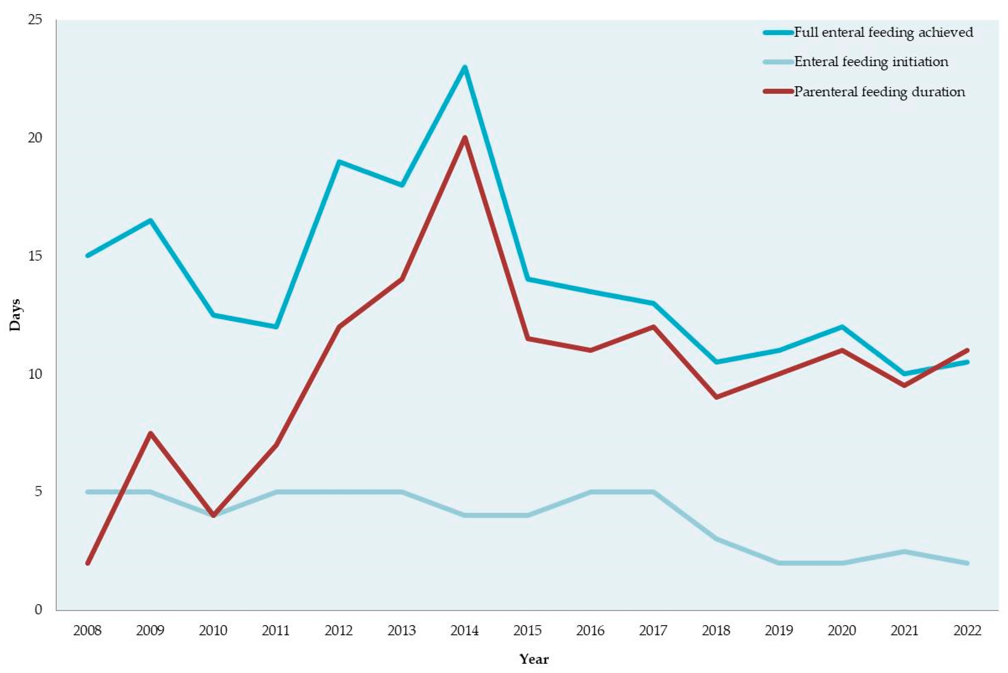
Parenteral feeding duration (days)15 ± 14.5
Enteral feeding starting time (days)4.8 ± 4.2
Full enteral feeding achieved (days)16.4 ± 11.3
Clinical information
Duration of hospitalization (days)54 ± 25.8
BPD66 (14.3)
hsPDA43 (9.3)
Early-onset sepsis (suspected or culture proved)49 (10.6)
Late-onset sepsis (suspected or culture proved)122 (26.4)
NEC8 (1.7)
IVH (≥Grade II)53 (11.5)
ROP (≥Stage II)31 (6.7)
Pneumothorax21 (4.5)
Duration of respiratory support (days)12.9 ± 17.0
Duration of mechanical ventilation (days)3.3 ± 7.6
Duration of non-invasive ventilation (days)9.7 ± 13.3
Duration of oxygen therapy (days)11.6 ± 18.3
Anemia211 (45.7)
Cystic PVL30 (6.5)
Abbreviations: BPD, bronchopulmonary dysplasia; hsPDA, hemodynamically significant patent ductus arteriosus; NEC, necrotizing enterocolitis; IVH, intraventricular hemorrhage; ROP, retinopathy of prematurity (≥Stage II); PVL, periventricular leukomalacia.
Table 2. Infant size at birth and discharge by the Fenton2013 and INTERGROWTH-21st growth references.
Fenton2013INTERGROWTH-21stp-Value *
At birth
Birth weight z, mean ± SD0.1 ± 0.90.1 ± 1.10.001
SGA weight, n (%)29 (6.3)43 (9.3)<0.001
At discharge
Weight z score, mean ± SD−1.2 ± 1.4−0.7 ± 1.5<0.001
Length z score, mean ± SD−0.9 ± 1.5−0.7 ± 2.0<0.001
HC z score, mean ± SD−0.1 ± 1.20.0 ± 1.6<0.001
EUGR weight, n (%)209 (45.9)133 (29.2)<0.001
EUGR length, n (%)165 (36.0)158 (34.4)0.039
EUGR HC, n (%)50 (10.9)70 (15.3)<0.001
Abbreviations: SGA, small for gestational age; HC, head circumference; EUGR, extrauterine growth restriction. * p-values from paired t-tests were used to compare sex-specific z-scores at birth and discharge, and Cochran’s Q test was used to compare the prevalence of SGA and EUGR between growth references (Fenton2013 and INTERGROWTH-21st).
Table 3. Perinatal, infant nutrition and clinical factors 1 by extrauterine restricted growth (EUGR) based on weight at discharge by the Fenton2013 and INTERGROWTH-21st growth references.
Fenton2013INTERGROWTH-21st
RestrictedNon Restrictedp-Value 2RestrictedNon Restrictedp-Value 2
Perinatal factors
Male sex111 (53.1)141 (57.3)0.39581 (60.9)171 (53.1)0.147
Gestational age (weeks)29.2 ± 1.830.0 ± 1.5<0.00128.9 ± 1.929.9 ± 1.5<0.001
Extremely preterm (<28 weeks)48 (23.0)30 (12.2)0.00338 (28.6)40 (12.4)<0.001
Birth weight (g)1144.3 ± 288.11510.9 ± 334.6<0.0011064.2 ± 276.51457.5 ± 331.3<0.001
BW z-score−0.3 ± 0.80.5 ± 0.9<0.001−0.7 ± 1.40.4 ± 0.8<0.001
SGA26 (12.4)3 (1.2)<0.00135 (26.3)8 (2.5)<0.001
Caesarean section186 (89.0)213 (87.3)0.663116 (87.2)283 (88.4)0.751
Multiple birth114 (56.4)131 (55.5)0.84878 (60.0)167 (54.2)0.293
Duration of hospitalization (days)66.3 ± 27.543.5 ± 19.1<0.00175.1 ± 29.445.2 ± 18.2<0.001
Nutritional factors
Parenteral feeding duration (days)11.5 (6–24)8 (4–13)<0.00114 (8–30)8 (4–13.5)<0.001
Enteral feeding initiation (days)4 (2–8)3 (2–5)<0.0015 (3–9)3 (2–5)<0.001
Full enteral feeding achieved (days)18 (11–27)11 (8–16)<0.00119 (12–30)11 (8–18)<0.001
Clinical factors
BPD38 (18.2)28 (11.4)0.04529 (21.8)37 (11.5)0.008
Early-onset sepsis29 (13.9)20 (8.1)0.06816 (12.0)33 (10.3)0.619
Late-onset sepsis74 (35.4)46 (18.7)<0.00164 (48.1)56 (17.4)<0.001
Anemia117 (56.0)92 (37.4)<0.00190 (67.7)119 (37.0)<0.001
NEC8 (3.8)0 (0.0)0.0027 (5.3)1 (0.3)0.001
hsPDA31 (14.8)11 (4.5)<0.00125 (18.8)17 (5.3)<0.001
ROP22 (10.5)9 (3.7)0.00517 (12.8)14 (4.4)0.002
Respiratory support (days)17.0 ± 20.59.5 ± 12.5<0.00120.7 ± 21.19.7 ± 13.9<0.001
Mechanical ventilation (days)5.5 ± 10.41.5 ± 2.9<0.0017.0 ± 11.61.8 ± 4.4<0.001
Non-invasive ventilation(days)11.7 ± 14.88.0 ± 11.50.00313.9 ± 15.38.0 ± 11.9<0.001
Oxygen administration (days)16.8 ± 23.07.2 ± 11.7<0.00121.0 ± 24.97.7 ± 13.2<0.001
Cystic PVL18 (8.6)12 (4.9)0.13013 (9.8)17 (5.3)0.096
IVH (Grade ≥ II)11 (5.3)12 (4.9)0.9996 (4.5)17 (5.3)0.818
Abbreviations: BPD, bronchopulmonary dysplasia; BW, birth weight; hsPDA, hemodynamically significant patent ductus arteriosus; IVH, intraventricular hemorrhage (grade ≥ II); NEC, necrotizing enterocolitis; ROP, retinopathy of prematurity (≥Stage II); cystic PVL, cystic periventricular leukomalacia. 1 Numbers represent mean ± SD for parametric continuous variables, median (IQR) for non-parametric continuous variables and n (%) for categorical variables. 2 p-values obtained from independent samples t-test or Mann–Whitney non-parametric test, used to compare continuous variables between two groups, and Fisher’s exact test, used to compare categorical variables.
Table 4. Associations * of clinical and nutritional factors with extrauterine growth restriction based on the Fenton2013 and INTERGROWTH-21st weight growth references.
Fenton2013INTERGROWTH-21st
Weight z-ScoreEUGRWeight z-ScoreEUGR
β (95% CI)OR (95% CI)β (95% CI)OR (95% CI)
Perinatal factors
Extremely preterm0.53 (0.23, 0.84)0.49 (0.19, 1.27)0.48 (0.17, 0.80)0.67 (0.26, 1.76)
BW z-score0.36 (0.26, 0.45)0.21 (0.14, 0.31)0.43 (0.33, 0.53)0.27 (0.19, 0.39)
Hospitalization (days)−0.04 (−0.04, −0.03)1.05 (1.03, 1.07)−0.04 (−0.04, −0.03)1.05 (1.03, 1.08)
Nutritional factors
PN duration (days)0.01 (0.00, 0.02)0.97 (0.94, 1.01)0.01 (−0.00, 0.02)0.99 (0.95, 1.03)
EN initiation (days)−0.02 (−0.05, 0.00)1.04 (0.94, 1.14)−0.03 (−0.06, 0.00)1.16 (1.05, 1.28)
Full EN achieved (days)−0.02 (−0.03, −0.01)1.07 (1.03, 1.12)−0.02 (−0.04, −0.01)1.03 (0.99, 1.07)
Clinical factors
BPD0.28 (−0.07, 0.63)0.81 (0.27, 2.40)0.37 (0.00, 0.73)0.98 (0.33, 2.97)
Late-onset sepsis−0.07 (−0.30, 0.15)1.33 (0.70, 2.52)−0.13 (−0.37, 0.10)3.08 (1.56, 6.08)
Anemia0.33 (0.10, 0.56)0.49 (0.25, 0.95)0.31 (0.06, 0.55)0.55 (0.25, 1.19)
hsPDA−0.41 (−0.75, −0.07)2.59 (0.88, 7.61)−0.39 (−0.75, −0.04)1.67 (0.56, 4.95)
ROP0.14 (−0.29, 0.57)2.04 (0.52, 8.07)0.08 (−0.36, 0.53)0.99 (0.25, 3.86)
Respiratory support (days)0.00 (−0.01, 0.01)0.99 (0.95, 1.02)0.00 (−0.01, 0.01)1.00 (0.97, 1.03)
Oxygen administration (days)0.00 (−0.00, 0.01)1.00 (0.97, 1.03)0.00 (−0.01, 0.01)0.99 (0.96, 1.01)
Abbreviations: SGA, small for gestational age; PN, parenteral nutrition; EN, enteral nutrition; BPD, bronchopulmonary dysplasia; hsPDA, hemodynamically significant patent ductus arteriosus; ROP, retinopathy of prematurity (≥Stage II); cystic PVL, cystic periventricular leukomalacia. * Beta coefficients (β) and odds ratios (OR) and corresponding confidence intervals (CI) obtained using linear and logistic regression models, respectively.
Disclaimer/Publisher’s Note: The statements, opinions and data contained in all publications are solely those of the individual author(s) and contributor(s) and not of MDPI and/or the editor(s). MDPI and/or the editor(s) disclaim responsibility for any injury to people or property resulting from any ideas, methods, instructions or products referred to in the content.

