Article

The Association between Gut Microbiome and Pregnancy-Induced Hypertension: A Nested Case–Control Study

1
Hunan Provincial Key Laboratory of Clinical Epidemiology, Xiangya School of Public Health, Central South University, Changsha 410000, China
2
Department of Epidemiology and Health Statistics, Xiangya School of Public Health, Central South University, Changsha 410000, China
3
Clinical Research Center for Reproduction and Genetics in Hunan Province, Reproductive and Genetic Hospital of CITIC-Xiangya, Changsha 410000, China
*
Authors to whom correspondence should be addressed.
These authors contributed equally to this work.
Nutrients 2022, 14(21), 4582; https://doi.org/10.3390/nu14214582
Submission received: 29 September 2022 / Revised: 25 October 2022 / Accepted: 26 October 2022 / Published: 1 November 2022
(This article belongs to the Section Nutrition in Women)

Abstract

:
(1) Background: Pregnancy-induced hypertension (PIH) is associated with obvious microbiota dysbiosis in the third trimester of pregnancy. However, the mechanisms behind these changes remain unknown. Therefore, this study aimed to explore the relationship between the gut microbiome in early pregnancy and PIH occurrence. (2) Methods: A nested case–control study design was used based on the follow-up cohort. Thirty-five PIH patients and thirty-five matched healthy pregnant women were selected as controls. The gut microbiome profiles were assessed in the first trimester using metagenomic sequencing. (3) Results: Diversity analyses showed that microbiota diversity was altered in early pregnancy. At the species level, eight bacterial species were enriched in healthy controls: Alistipes putredinis, Bacteroides vulgatus, Ruminococcus torques, Oscillibacter unclassified, Akkermansia muciniphila, Clostridium citroniae, Parasutterella excrementihominis and Burkholderiales bacterium_1_1_47. Conversely, Eubacterium rectale, and Ruminococcus bromii were enriched in PIH patients. The results of functional analysis showed that the changes in these different microorganisms may affect the blood pressure of pregnant women by affecting the metabolism of vitamin K2, sphingolipid, lipid acid and glycine. (4) Conclusion: Microbiota dysbiosis in PIH patients begins in the first trimester of pregnancy, and this may be associated with the occurrence of PIH. Bacterial pathway analyses suggest that the gut microbiome might lead to the development of PIH through the alterations of function modules.

1. Introduction

Pregnancy-induced hypertension (PIH) is defined as an elevated blood pressure that occurs for the first times during pregnancy, and gestational hypertension (GH) and pre-eclampsia (PE) are the two most common types [1,2]. The global incidence of PIH is 4.1–19.4%, with GH accounting for 1.8–4.4% of cases and PE accounting for 0.2–9.2% of cases [3]. Without the implementation of prevention measures, 25–50% of GH patients will develop PE [4]. PIH can adversely affect pregnant women and their fetuses [5,6]. Studies have found that PIH is the most common reason for death in pregnant women in both Asian and European countries [7,8]. Most PIH patients will recover a normal blood pressure after delivery. However, nowadays, more and more studies have discovered that women with a history of PIH have a higher risk of hypertension, stroke, and ischemic heart disease in future life [9,10]. Furthermore, the offspring of mothers with PIH present with higher blood pressure and increased risk of other diseases than children of healthy mothers [11,12,13,14,15]. Therefore, with the increasing incidence of PIH and the associated serious health damage, PIH is considered an urgent public health problem. However, the specific pathogenesis of PIH remains uncertain.
The gut microbiome is the human body’s biggest and most complex biocenosis [16]. The number of genes in the gut flora is considerably more than that in the human genome, and the gut microbiome composition varies greatly in individuals because of genetic and environmental variations [17]. The gut microbiome maintains a dynamic symbiotic relationship through interaction with the immune system. It can produce metabolites to inhibit the excessive colonization of harmful bacteria, thereby participating in the regulation of human health [18]. Many studies have found a close relationship between the gut microbiome and diseases, such as obesity, diabetes, and cardiovascular diseases [19,20].
To provide adequate nutrition to the fetus during pregnancy, maternal physiology undergoes adaptive reconstruction. At this time, the gut microbiome is more sensitive to external stimuli, and dysbiosis is prone to occur, leading to the development of pregnancy complications, including gestational diabetes and hyperemesis gravidarum [21,22]. Studies have shown that the gut microbiome is associated with blood pressure, specifically regulating the blood pressure when the body is in low-grade chronic inflammation [23]. Moreover, many microbial metabolites are involved in the regulation of blood pressure [24,25,26].
Ley et al. found that the abundance of Fusobacterium and Veillonella was enriched, and the abundance of Faecalibacterium and Akkermansia was decreased in PE women [27]. In obese pregnant women, the abundance of butyrate producing bacteria, such as Odoribacter and Clostridiaceae, was negatively correlated with systolic blood pressure (SBP) [28]. The gut microbiome in late pregnant PE patients had lower diversity and obvious dysbiosis, and animal experiments have shown that dysbacteriosis may cause high blood pressure [29]. Moreover, it has been reported that the dysbiosis continues during postpartum [30].
Microbiota dysbiosis has been found in PIH women in the third trimester. However, the changes in the gut microbiome in PIH women in early pregnancy remain unknown. Furthermore, the potential mechanism of the relationship between the gut microbiome and PIH has not been explored. Therefore, in this study, we conducted a nested case–control study to evaluate the alteration of microbiota taxa and function modules in the first trimester and detect the correlation between microbiota taxa, function modules, and clinical indices to reveal the mechanisms of the gut microbiome and PIH.

2. Materials and Methods

2.1. Study Design

This nested case–control study was based on an early pregnancy follow-up cohort. The cohort was established in the Hunan Provincial Maternal and Child Health Hospital (HPMCHH) from March 2017 to March 2018. The project was approved by the Medical Ethical Committee of HPMCHH (NO: EC201624) on 11 January 2017. All eligible subjects provided written informed consent and then entered the cohort.
The inclusion criteria were: single embryo; natural conception; no history of diabetes, hypertension or other diseases before pregnancy; no acute infection in the past 2 weeks; no antibiotic use during pregnancy. Participants were recruited in the first trimester (10–14 weeks) and followed up until 42 days postpartum. The collection of questionnaire results and samples were conducted in the first trimester, second trimester, third trimester, at delivery, and during postpartum. In addition, biochemical tests of all blood samples were performed at the hospital.
Women in the cohort who were diagnosed with PIH after 20 weeks gestation by the hospital, and had complete questionnaire information, stool samples, and blood samples, were selected as a case group. The same number of healthy pregnant women were selected as a control group matched for age (±3 years), gestational age (±1 week) and stool/blood sample collection time (±1 month). All biological samples were collected in the first trimester.

2.2. DNA Extraction and Metagenomic Sequencing

Fecal DNA was extracted using QIAamp Fast DNA Stool Mini Kit (Qiagen, Hilden, Germany). All samples were paired-end sequenced on the Illumina platform (read length 150 bp) at Novogene Bioinformatics Technology Co Ltd. (Tianjin, China). After quality control using Trimmomatic [31] (Version 0.36), reads aligned to the human genome (alignment with SOAP2 [32], Version 2.21) were removed. The remaining high-quality reads were used for further taxonomic annotation and functional annotation by MetaPhlAn [33] (Version 2.1.0) and HUMAnN [34] (Version 2-0.11.0), respectively.

2.3. Statistical Analysis

Means ± standard deviations and medians were used for statistical description. Matched t-tests, ANOVA, and nonparametric tests were used for statistical inference. All the statistical analyses were two-tail tests, and p < 0.05 indicated statistical significance. Rarefaction analysis was performed to assess the species richness in the controls and cases by vegan package in R software. Alpha diversity indices were estimated using the vegan package (Version 2.5-7) in R software (Version 4.0.2) based on the species profile. A principal coordinates analysis (PCoA) was executed using the ade4 package (Version 5.5) in R software. A non-metric multidimensional scaling (NMDS) analysis was performed using the vegan package in R software. Differential abundance of microorganisms and functional modules was tested using MaAsLin2 [35] (Version 1.2.0). Only features existing in at least 10% of subjects were considered in the analyses. Correlation analyses were tested using Spearman’s correlation with the psych package (Version 2.1.9) in R software.

3. Results

3.1. Main Results

3.1.1. General Characteristics

A total of 872 subjects were included in the cohort study, and 744 completed entire pregnancy to postpartum investigations. A total of 46 PIH patients (29 with GH and 17 with PE) were diagnosed in the cohort, of which nine GH subjects and two PE subjects were excluded due to lack of questionnaire information or stool and blood samples. Finally, 35 PIH subjects (20 with GH and 15 with PE) were selected in this nested case–control study, and 35 healthy controls were selected according to the match factors. All the results of subgroup, which is PE and GH, and control are showed in supplementary materials.
The basic characteristics of the two groups are presented in Table 1. The results show no significant differences between the two groups in age, gestational age, race, occupation, education level, and monthly income. (The basic characteristics of PE and GH group are showed in Table S1).
The clinical characteristics of the two groups subjects are presented in Table 2. The results show waist, weight, BMI, SBP, diastolic blood pressure (DBP), hemoglobin (HGB), uric acid (UA), insulin (INS), and γ-glutamyltransferase (GGT) in early pregnancy were higher in the PIH group than in controls (p < 0.05). (The clinical characteristics of PE and GH group are showed in Table S1).

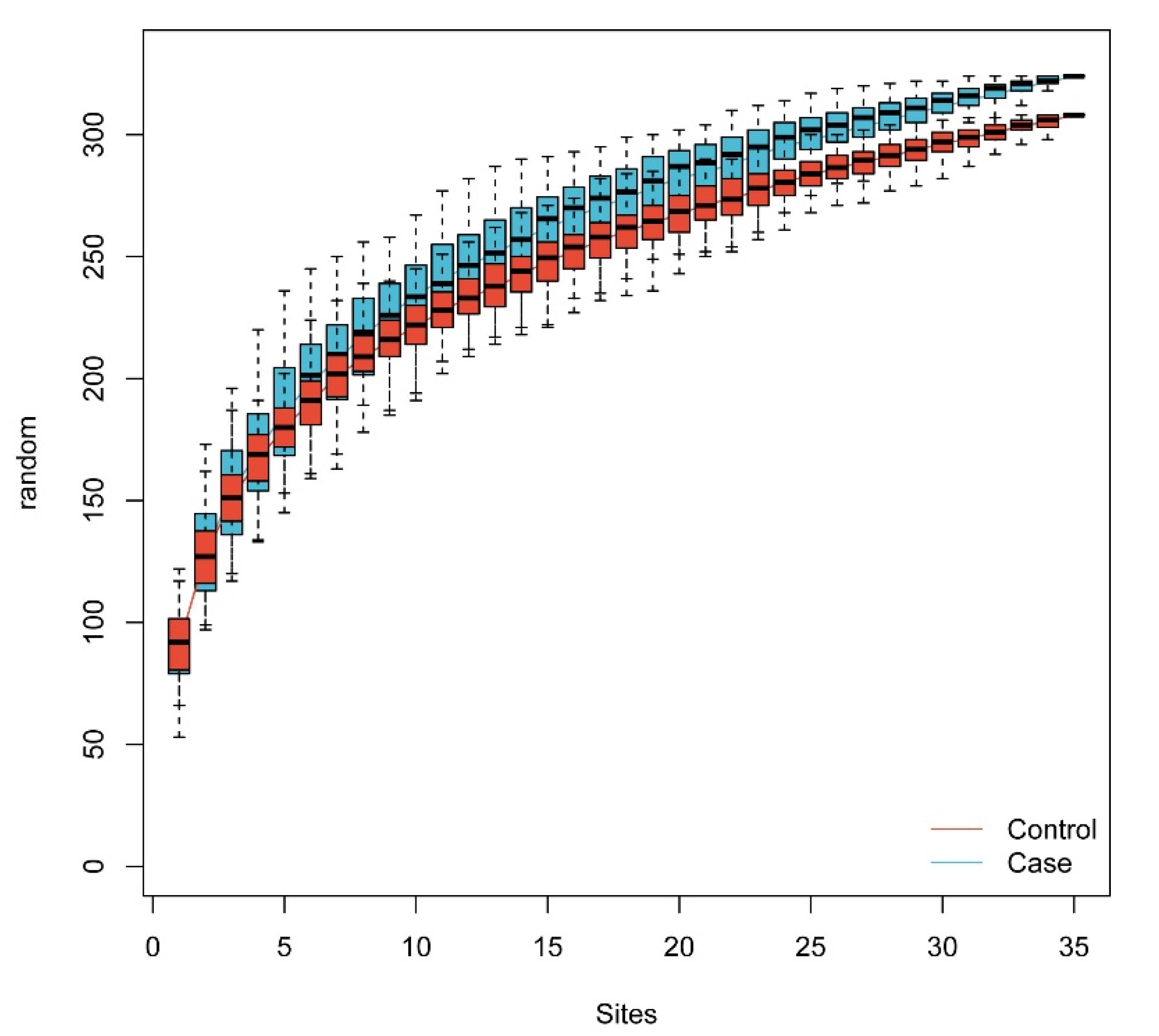
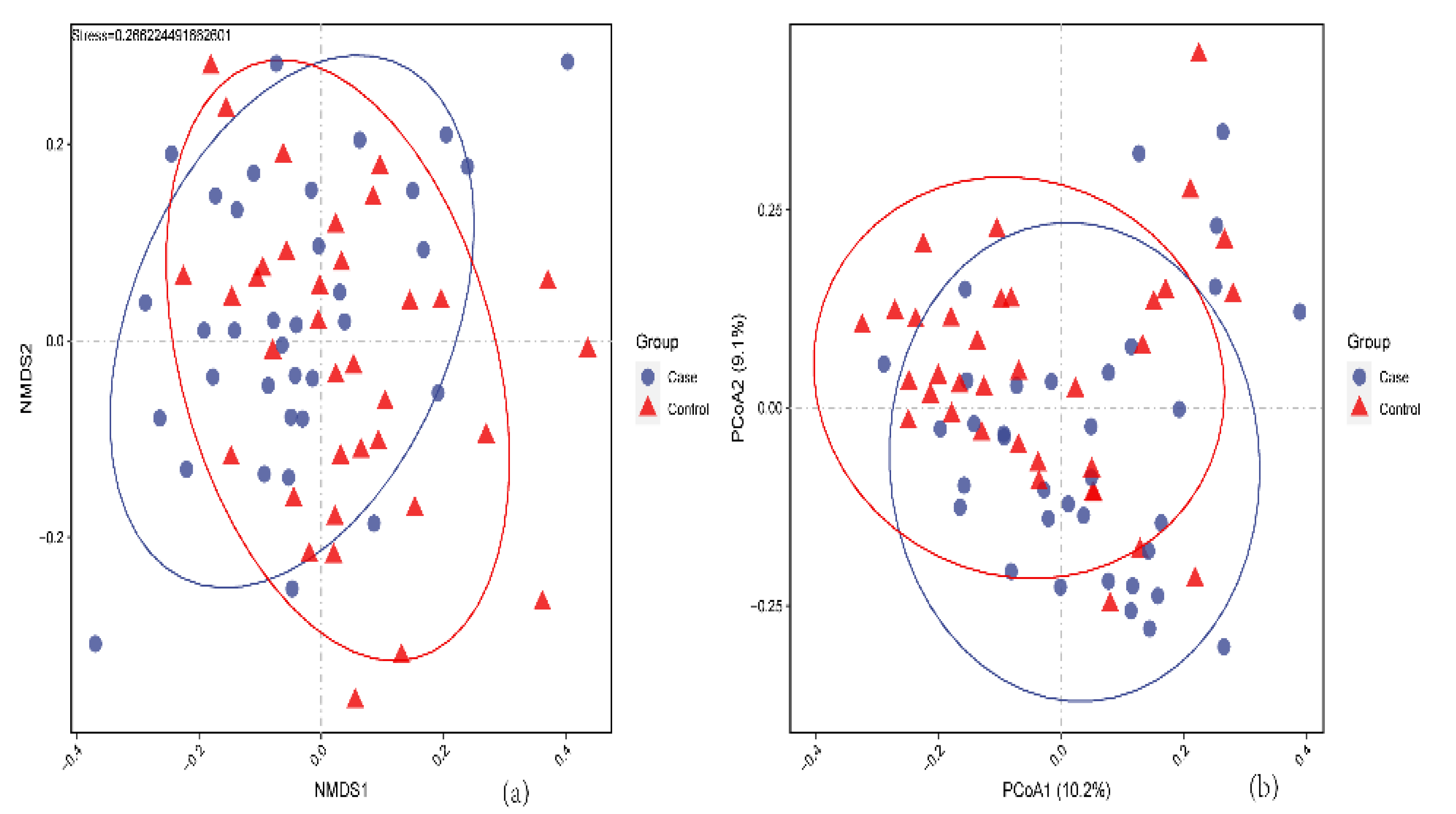
3.1.2. The Gut Microbiome Diversity Alteration

The rarefaction curve was performed according to the gut microbiome data from metagenomic sequencing. The curve was flat in our sample size, indicating that sufficient sequencing data were obtained to reflect nearly all microbial diversity in the subjects (Figure 1).
The results show no significant differences in alpha diversity between the two groups. Inversely, the beta diversity between the PIH and control groups did differ statistically according to the PCoA results and NMDS analysis (Figure 2A,B). (The result of diversity analysis between three groups is showed in Figure S1).

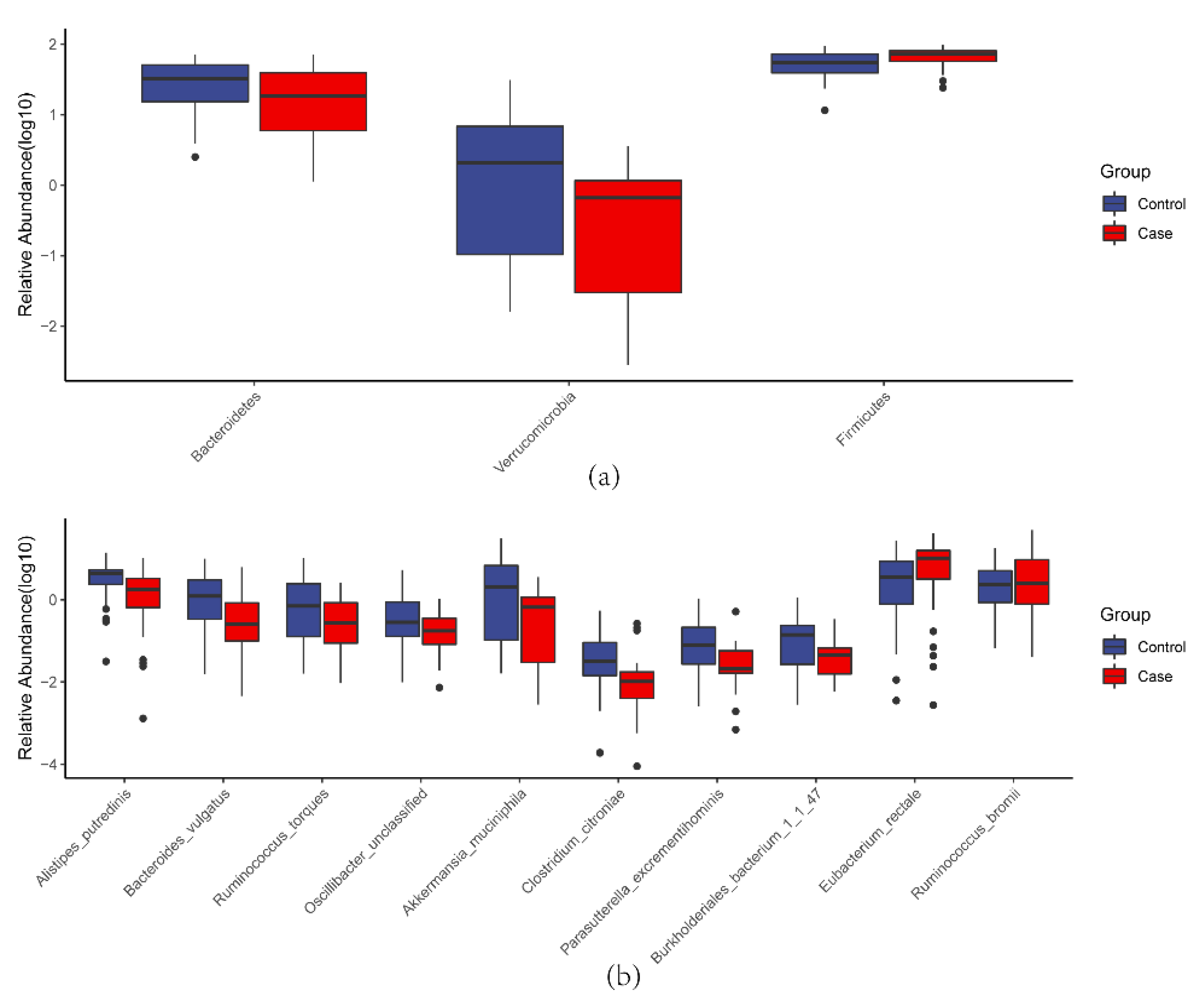
3.1.3. Microbial Taxa Alteration

Comparison of the PIH and control groups revealed three, four, four, six, seven and ten different taxa at the phylum, class, order, family, genus and species level, respectively.
At the phylum level, PIH patients had a higher abundance of Firmicutes (p = 0.025) and a lower abundance of Bacteroidetes (p = 0.020) and Verrucomicrobia (p = 0.026) (Figure 3A).
At the species level, PIH patients had significantly higher abundance of Eubacterium rectale (p = 0.009) and Ruminococcus bromii (p = 0.039) and a lower abundance of Alistipes putredinis (p = 0.003), Bacteroides vulgatus (p = 0.007), Ruminococcus torques (p = 0.011), Oscillibacter unclassified (p = 0.006), Akkermansia muciniphila (p = 0.026), Clostridium citroniae (p = 0.039), Parasutterella excrementihominis (p = 0.043), and Burkholderiales bacterium_1_1_47 (p = 0.047) (Figure 3B). (The taxa alterations between three groups are showed in Figure S2).

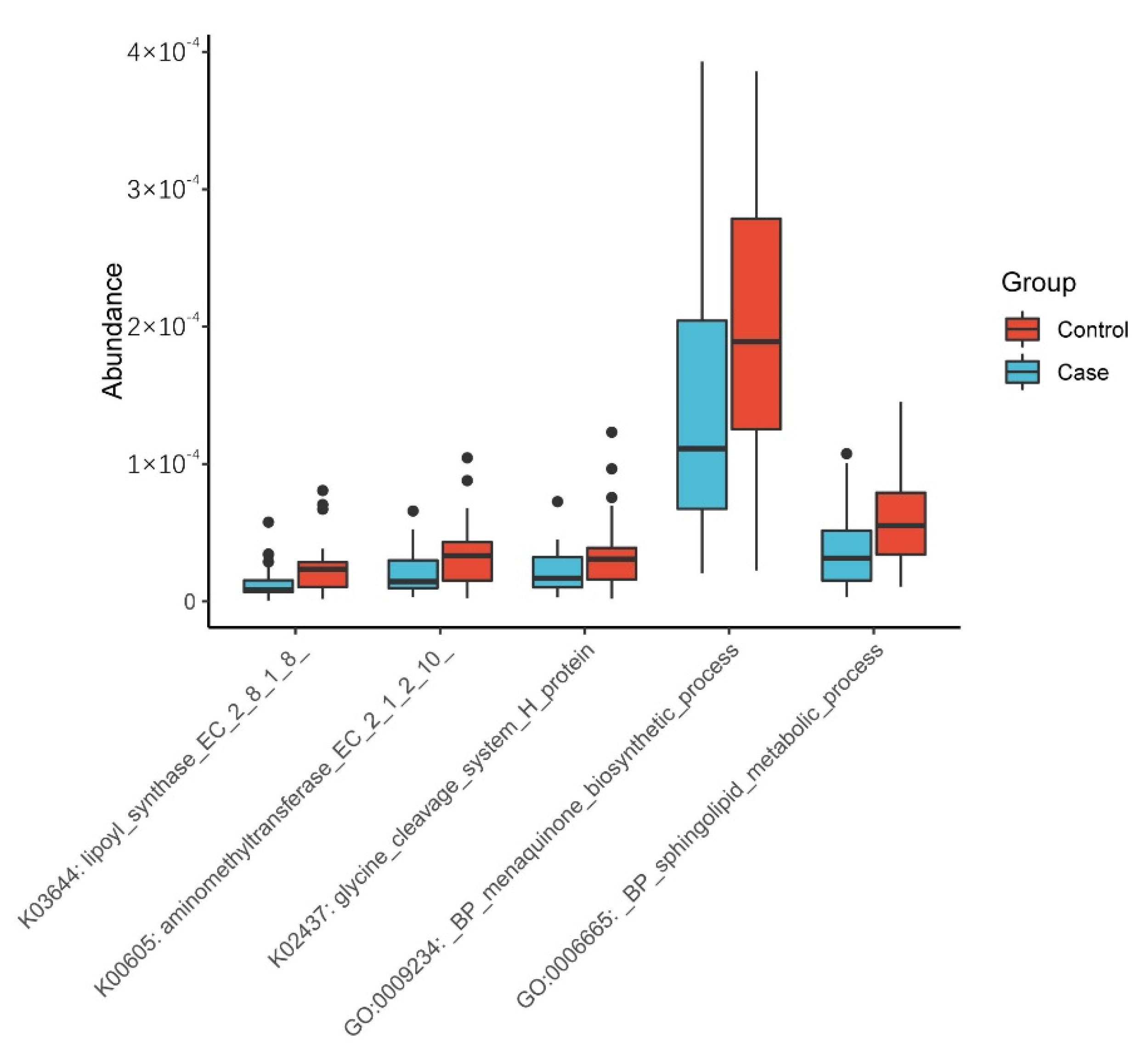
3.1.4. Functional Prediction Analysis

The transcriptome information of samples was compared with KO and GO standard libraries to explore the potential function and mechanism of the gut microbiome in the occurrence of PIH. The preliminary screening of different functions was conducted according to “p < 0.01”, then through literature review, related functions were chosen and visually presented in a heatmap. As shown in Figure 4, PIH patients had significantly lower levels in five functions. (The functional alteration between three groups is showed in Figure S3).

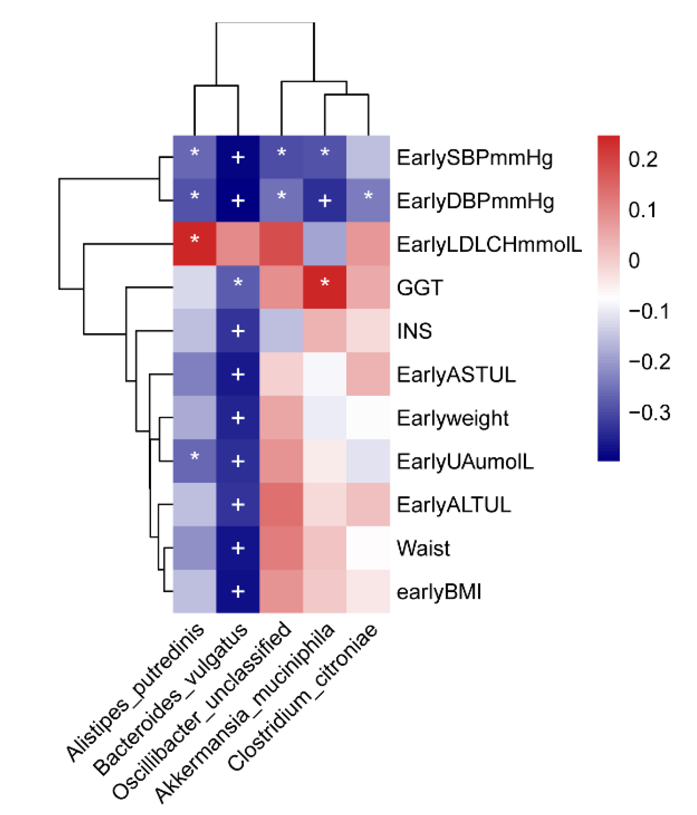
3.1.5. Correlation Analysis

Correlation between the Gut Microbiome and Clinical Factors

The correlation analysis was conducted on clinical factors and the 10 different microbial species screened in the PIH and control groups. Five bacteria were associated with 11 clinical indicators. The abundance of A. tredinis was negatively correlated with early UA, early SBP, and early DBP and positively correlated with LDLCH. The abundance of B. vulgatus was negatively correlated with early BMI, waist, early ALT, early UA, early weight, early AST, INS, GGT, as well as early SBP and early DBP. The abundance of O. unclassified and A. muciniphila was negatively correlated with early SBP and early DBP, and A. muciniphila was positively correlated with GGT. The abundance of C. citroniae was negatively correlated with early DBP (Figure 5). (The correlation between gut microbiome and clinical factors of three groups is presented in Figure S4).

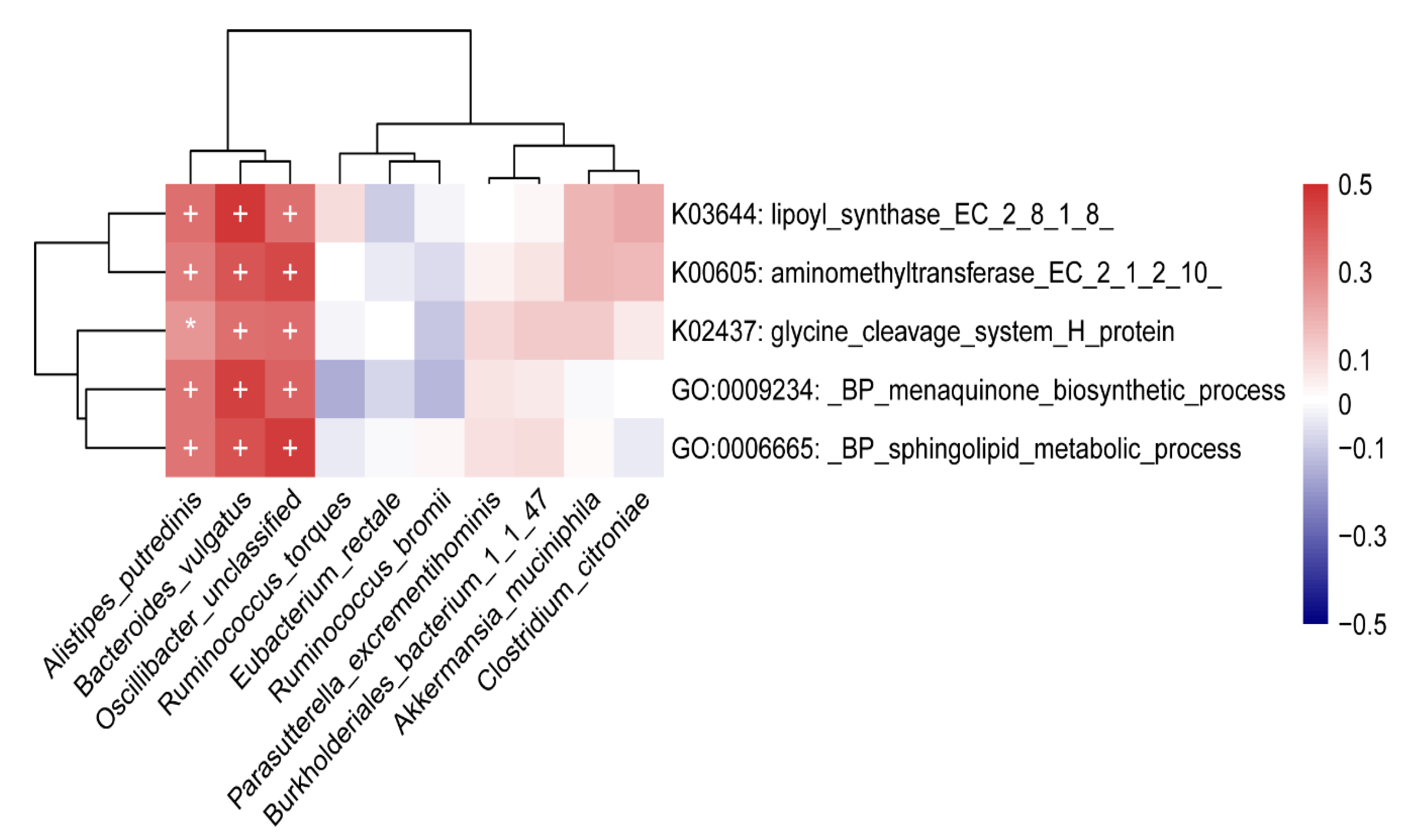
Correlation between Gut Microbiome and Functions

The results of the association analysis between different microbial species and functions indicated that A. putredinis, B. vulgatus and O. unclassified were positively associated with the five functions (Figure 6). (The correlation between gut microbiome and functions of three groups was presented in Figure S5).

4. Discussion

In this study, we explored the differences in gut microbiome between PIH patients and healthy controls in the first trimester of pregnancy. The results show that the gut microbiome is changed in early pregnancy, and the changes might be associated with the occurrence of PIH. Moreover, all the subjects did not take drugs two weeks before enrollment, and they are all permanent residents in Hunan, with no obvious difference in diet. The influence of external factors in the gut microbiome was excluded as far as possible.

4.1. Diversity Alteration

The gut microbiome is interdependent with the host in normal physiology, and this interdependency maintains dynamic balance. When microbiota diversity is reduced, a variety of diseases might occur [36,37]. Our study showed no significant differences in alpha diversity but greatly significant changes in beta diversity. Similarly, Wang et al. [38] obtained the same result in PE patients. However, another study found that alpha and beta diversity in PE patients were both significantly reduced [29]. This indicates that the bacterial proportions change in the first trimester of pregnancy, suggesting that the occurrence of PIH might be related to specific bacteria.

4.2. Microbial Taxa Alteration

Most significantly different strains were mainly focused on Firmicutes and Bacteroidetes in our study. It was been reported in 2019 that PE patients have a decreased abundance of Firmicutes and an enriched abundance of Bacteroidetes [38], which is contrary to our study’s results. On the contrary, another study found that the abundance of Firmicutes and Bacteroidetes was not significantly different in PE patients and healthy pregnant women in the second trimester, however, the opposite difference was observed in the third trimester [39]. This indicates that the gut microbiome is in flux during disease and gestation and opposing patterns may be present depending on the sampling time and study population.
E. rectale is an opportunistic pathogen in PIH-enriched species, which can easily cause a series of health problems. Significant enrichment of this species has been found in hypertensive patients [40], indicating that it could increase the host’s blood pressure.
In PIH-reduced species, C. citroniae, together with Oscillibacter, has been reported to be negatively associated with visceral obesity and metabolic syndrome, and it is significantly enriched in healthy people [41,42]. Moreover, studies found that PE patients had an enriched abundance of Clostridium [38]. Although the role of B. vulgatus and A. putredinis in PIH has not yet been reported, animal experiments have found that B. vulgatus can inhibit inflammatory processes and enhance the intestinal barrier [43]. Furthermore, reports suggest that Alistipes may reduce inflammation [44], and that it may have protective effects on colitis, immune diseases, and cardiovascular diseases, and that the abundance of Alistipes is decreased in PE patients [38,45]. The decreased abundance indicates that PIH patients might already have an obvious inflammation in the first trimester, and long-term low-grade inflammation during pregnancy has been proven to induce elevated blood pressure.
In recent years, A. muciniphila has been regarded as a potentially beneficial bacterium [46], closely associated with metabolic and gastrointestinal diseases [47,48]. It has been reported that the abundance of Akkermansia is significantly decreased in PE women [30]. The decreased abundance of A. muciniphila in PIH women could increase intestinal permeability and the probability of harmful intestinal metabolites entering the bloodstream. R. torques can damage the intestinal barrier and cause inflammation [49], which is contrary to our study’s results. The mechanism of such reverse alteration remains unclear, and this may be related to the compensatory increase in the beneficial bacteria in the early stages of PIH.

4.3. Exploration of the Potential Mechanisms of the Gut Microbiome

We found that the menaquinone biosynthetic process module was lower in PIH patients. Menaquinone is a generic term for vitamin K2, which is synthesized by the gut microbiome. Studies have found that vitamin K2 is a protective factor against cardiovascular and metabolic disease [50]. Its deficiency might cause vasculopathy and increase in blood pressure. Other studies have suggested a reduced vitamin K2 level could be associated with insulin resistance and damage to the kidney and liver [51,52,53]. On the other hand, PIH patients had a significantly higher level of UA, GGT and INS in our study, which indicated the possibility of kidney and liver damage and metabolic abnormalities, and these alterations were found to be associated with PIH [54,55,56,57]. It is consistent that the changes of these indicators and functional modules, suggesting that they might interact with each other to participate in the occurrence of PIH.
The lower sphingolipid metabolic process module in PIH patients meant a higher level of sphingolipids. Studies suggest that ceramide, one of the most common sphingolipids, is related to the angiotensin-aldosterone system and could elevate blood pressure [58]. In addition, ceramide accumulation could promote liver steatosis and cause liver damage [59]. Studies have found that a higher ceramide level in obese people enhances insulin resistance [60,61], and liver injury and insulin metabolism disorders are both related to PIH. It is suggested that lower sphingolipid metabolism in PIH patients might be involved in the occurrence of PIH through the interaction with the level of blood pressure and insulin.
The lipoyl synthase (LipA) module was lower in PIH patients, and LipA is a key enzyme in lipoic acid synthesis. The lower synthesis of LipA in PIH patients suggests lower lipoic acid levels. Lipoic acid could relieve oxidative stress and insulin resistance [62], and could reduce blood pressure in hypertension [63]. Lower lipoic acid levels, higher oxidative and insulin level in the first trimester might work together to form the basics of PIH.
The glycine cleavage system (GCS) is the main pathway of glycine metabolism. Our study found that the aminomethyltransferase and glycine cleavage system H protein, which were components of GCS, were lower in PIH women. The decreased GCS contents suggest higher glycine in PIH patients. Glycine is an amino acid with anti-inflammatory effects [64]. The increased content in PIH patients indicates that some physiological adjustments have been made in these patients in early pregnancy in response to the pathological changes before PIH occurred. This might be related to compensations mechanisms.
Taken together, these results suggest that differences in the gut microbiome might affect the metabolic functional modules, which interact with physiological function alterations, ultimately the PIH happened.
The main limitation of our study was the small sample size which may reduce the statistical power of our results. Moreover, this study did not explore the function of the inflammatory factors and beneficial microbial metabolites involved in the association between the gut microbiome and PIH. Further studies are needed to explore the mechanisms of the gut microbiome in PIH.

5. Conclusions

This is the first study to investigate the association between gut microbiome alterations in the first trimester and the occurrence of PIH. The results show that alterations in the microbiome begin in the first trimester, including alterations in the microbial diversity, taxa, and function. These differences in the gut microbiome affect the metabolic functional modules and clinical indicators, thereby promoting PIH occurrence.

Supplementary Materials

The following supporting information can be downloaded at: https://www.mdpi.com/article/10.3390/nu14214582/s1. Table S1. Basic characteristics of subjects in PE and GH group; Table S2. Clinical characteristics of subjects in PE and GH group; Figure S1. The beta diversity of subgroup and healthy controls; Figure S2. Boxplot of different microbial taxa; Figure S3. Boxplot of different function modules in three groups; Figure S4. Heatmap of Spearman correlation between different species and clinical indices between three groups; Figure S5. Heatmap of Spearman correlation between different species and function modules between three groups.

Author Contributions

Conceptualization, H.T. and S.M.; methodology, H.T., S.M. and H.L.; investigation, S.M., H.L. and R.A.; software, J.C. and H.L.; writing—original draft preparation, H.L., J.C.; writing—review and editing, H.T. and X.L.; project administration, H.T. and S.M.; funding acquisition, H.T. and S.M. All authors have read and agreed to the published version of the manuscript.

Funding

This research was funded by National Natural Science Foundation of China, grant number 81773535, and Key Research and Development Program of Hunan Province, grant number 2018SK2061.

Institutional Review Board Statement

This study involving human subject samples was conducted according to the guidelines of Declaration of Helsinki and approved by the Medical Ethics Committee of Hunan Maternal and Child Health Hospital (No. EC201624).

Informed Consent Statement

Informed consent was obtained from all subjects involved in the study, including publication of the data.

Data Availability Statement

The data set supporting the results of this article has been deposited in the EMBL European Nucleotide Archive (ENA) under BioProject accession code PRJEB55996 [http://www.ebi.ac.uk/ena/data/view/PRJEB13870 (accessed on 26 October 2022)].

Acknowledgments

We acknowledge Maternity and Child Health Hospital of Hunan Province, China for helping us in collecting data and performing biochemical test.

Conflicts of Interest

The authors declare no conflict of interest.

References

  1. Kintiraki, E.; Papakatsika, S.; Kotronis, G.; Goulis, D.G.; Kotsis, V. Pregnancy-Induced hypertension. Hormones 2015, 14, 211–223. [Google Scholar] [CrossRef] [Green Version]
  2. Savitz, D.A.; Danilack, V.A.; Engel, S.M.; Elston, B.; Lipkind, H.S. Descriptive Epidemiology of Chronic Hypertension, Gestational Hypertension, and Preeclampsia in New York State, 1995–2004. Matern. Child. Health J. 2014, 18, 829–838. [Google Scholar] [CrossRef] [PubMed] [Green Version]
  3. Umesawa, M.; Kobashi, G. Epidemiology of hypertensive disorders in pregnancy: Prevalence, risk factors, predictors and prognosis. Hypertens. Res. 2016, 40, 213–220. [Google Scholar] [CrossRef] [PubMed]
  4. American College of Obstetricians and Gynecologists. Hypertension in pregnancy. Report of the American College of Obstetricians and Gynecologists’ Task Force on Hypertension in Pregnancy. Obstet. Gynecol. 2013, 122, 1122–1131. [Google Scholar]
  5. Liu, Y.; An, H.; Li, Z.; Zhang, L.; Li, H.; Zhang, Y.; Ye, R.; Li, N.; Li, Z.; Zhang, L.; et al. Impact of gestational hypertension and preeclampsia on low birthweight and small-for-gestational-age infants in China: A large prospective cohort study. J. Clin. Hypertens 2021, 23, 835–842. [Google Scholar] [CrossRef]
  6. Abalos, E.; Cuesta, C.; Carroli, G.; Qureshi, Z.; Widmer, M.; Vogel, J.; Souza, J. Pre-eclampsia, eclampsia and adverse maternal and perinatal outcomes: A secondary analysis of the World Health Organization Multicountry Survey on Maternal and Newborn Health. BJOG Int. J. Obstet. Gynaecol. 2014, 121, 14–24. [Google Scholar] [CrossRef] [Green Version]
  7. Khumanthem, P.D.; Chanam, M.S.; Samjetshabam, R.D. Maternal Mortality and Its Causes in a Tertiary Center. J. Obstet. Gynecol. India 2012, 62, 168–171. [Google Scholar] [CrossRef] [Green Version]
  8. You, F.; Huo, K.; Wang, R.; Xu, N.; Deng, J.; Wei, Y.; Shi, F.; Liu, H.; Cheng, G.; Zhang, Z.; et al. Maternal Mortality in Henan Province, China: Changes between 1996 and 2009. PLoS ONE 2012, 7, e47153. [Google Scholar] [CrossRef] [Green Version]
  9. Männistö, T.; Mendola, P.; Vääräsmäki, M.; Jarvelin, M.-R.; Hartikainen, A.-L.; Pouta, A.; Suvanto, E. Elevated Blood Pressure in Pregnancy and Subsequent Chronic Disease Risk. Circulation 2013, 127, 681–690. [Google Scholar] [CrossRef] [Green Version]
  10. Mitka, M. Any Hypertension during Pregnancy Raises Risk for Several Chronic Diseases. JAMA 2013, 309, 971–972. [Google Scholar] [CrossRef]
  11. Kajantie, E.; Eriksson, J.G.; Osmond, C.; Thornburg, K.; Barker, D.J. Pre-Eclampsia Is Associated with Increased Risk of Stroke in the Adult Offspring the Helsinki Birth Cohort Study. Stroke 2009, 40, 1176–1180. [Google Scholar] [CrossRef] [PubMed]
  12. Miettola, S.; Hartikainen, A.-L.; Vääräsmäki, M.; Bloigu, A.; Ruokonen, A.; Järvelin, M.-R.; Pouta, A. Offspring’s blood pressure and metabolic phenotype after exposure to gestational hypertension in utero. Eur. J. Epidemiol. 2013, 28, 87–98. [Google Scholar] [CrossRef] [PubMed]
  13. Kuciene, R.; Dulskiene, V. Associations of maternal gestational hypertension with high blood pressure and overweight/obesity in their adolescent offspring: A retrospective cohort study. Sci. Rep. 2022, 12, 3800. [Google Scholar] [CrossRef] [PubMed]
  14. Tuovinen, S.; Eriksson, J.G.; Kajantie, E.; Lahti, J.; Pesonen, A.K.; Heinonen, K.; Osmond, C.; Barker, D.J.; Räikkönen, K. Maternal hypertensive disorders in pregnancy and self-reported cognitive impairment of the offspring 70 years later: The Helsinki Birth Cohort Study. Am. J. Obstet. Gynecol. 2013, 208, e1–e9. [Google Scholar] [CrossRef] [PubMed]
  15. Jansen, M.A.; Pluymen, L.P.; Dalmeijer, G.W.; Groenhof, T.K.J.; Uiterwaal, C.S.; Smit, H.A.; Van Rossem, L. Hypertensive disorders of pregnancy and cardiometabolic outcomes in childhood: A systematic review. Eur. J. Prev. Cardiol. 2019, 26, 1718–1747. [Google Scholar] [CrossRef] [Green Version]
  16. Minter, M.R.; Zhang, C.; Leone, V.; Zhang, C.; Leone, V.; Zhang, X.; Oyler-Castrillo, P.; Zhang, X.; Musch, M.W.; Shen, X.; et al. Antibiotic-induced perturbations in gut microbial diversity influences neuro-inflammation and amyloidosis in a murine model of Alzheimer’s disease. Sci. Rep. 2016, 6, 30018. [Google Scholar] [CrossRef]
  17. Hall, A.B.; Tolonen, A.C.; Xavier, R.J. Human genetic variation and the gut microbiome in disease. Nat. Rev. Genet. 2017, 18, 690–699. [Google Scholar] [CrossRef]
  18. Tang, W.H.W.; Li, D.Y.; Hazen, S.L. Dietary metabolism, the gut microbiome, and heart failure. Nat. Rev. Cardiol. 2019, 16, 137–154. [Google Scholar] [CrossRef]
  19. Patterson, E.; Ryan, P.M.; Cryan, J.F.; Dinan, T.G.; Ross, R.P.; Fitzgerald, G.F.; Stanton, C. Gut microbiota, obesity and diabetes. Postgrad. Med. J. 2016, 92, 286–300. [Google Scholar] [CrossRef] [Green Version]
  20. Verhaar, B.J.H.; Prodan, A.; Nieuwdorp, M.; Muller, M. Gut Microbiota in Hypertension and Atherosclerosis: A Review. Nutrients 2020, 12, 2982. [Google Scholar] [CrossRef]
  21. Wang, J.; Zheng, J.; Shi, W.; Du, N.; Xu, X.; Zhang, Y.; Ji, P.; Zhang, F.; Jia, Z.; Wang, Y.; et al. Dysbiosis of maternal and neonatal microbiota associated with gestational diabetes mellitus. Gut 2018, 67, 1614–1625. [Google Scholar] [CrossRef] [PubMed]
  22. Balci, S.; Tohma, Y.A.; Esin, S.; Onalan, G.; Tekindal, M.A.; Zeyneloglu, H.B. Gut dysbiosis may be associated with hyperemesis gravidarum. J. Matern. Neonatal Med. 2022, 3, 2041–2045. [Google Scholar] [CrossRef] [PubMed]
  23. Karbach, S.H.; Schonfelder, T.; Brandao, I.; Wilms, E.; Hörmann, N.; Jäckel, S.; Schüler, R.; Finger, S.; Knorr, M.; Lagrange, J.; et al. Gut Microbiota Promote Angiotensin II-Induced Arterial Hypertension and Vascular Dysfunction. J. Am. Heart Assoc. 2016, 5, e003698. [Google Scholar] [CrossRef] [PubMed] [Green Version]
  24. Yang, F.; Chen, H.; Gao, Y.; An, N.; Li, X.; Pan, X.; Yang, X.; Tian, L.; Sun, J.; Xiong, X.; et al. Gut microbiota-derived short-chain fatty acids and hypertension: Mechanism and treatment. Biomed. Pharmacother. 2020, 130, 110503. [Google Scholar] [CrossRef]
  25. Marques, F.Z.; Mackay, C.R.; Kaye, D.M. Beyond gut feelings: How the gut microbiota regulates blood pressure. Nat. Rev. Cardiol. 2018, 15, 20–32. [Google Scholar] [CrossRef]
  26. Tomasova, L.; Dobrowolski, L.; Jurkowska, H.; Wróbel, M.; Huc, T.; Ondrias, K.; Ostaszewski, R.; Ufnal, M. Intracolonic hydrogen sulfide lowers blood pressure in rats. Nitric Oxide 2016, 60, 50–58. [Google Scholar] [CrossRef]
  27. De Siena, M.; Laterza, L.; Matteo, M.V.; Mignini, I.; Schepis, T.; Rizzatti, G.; Ianiro, G.; Rinninella, E.; Cintoni, M.; Gasbarrini, A. Gut and Reproductive Tract Microbiota Adaptation during Pregnancy: New Insights for Pregnancy-Related Complications and Therapy. Microorganisms 2021, 9, 473. [Google Scholar] [CrossRef]
  28. Gomez-Arango, L.F.; Barrett, H.L.; McIntyre, H.D.; Callaway, L.K.; Morrison, M.; Nitert, M.D. Increased Systolic and Diastolic Blood Pressure Is Associated with Altered Gut Microbiota Composition and Butyrate Production in Early Pregnancy. Hypertension 2016, 68, 974–981. [Google Scholar] [CrossRef]
  29. Chen, X.; Li, P.; Liu, M.; Zheng, H.; He, Y.; Chen, M.X.; Tang, W.; Yue, X.; Huang, Y.; Zhuang, L.; et al. Faculty Opinions recommendation of Gut dysbiosis induces the development of pre-eclampsia through bacterial translocation. Gut 2020, 69, 513–522. [Google Scholar] [CrossRef]
  30. Lv, L.J.; Li, S.H.; Li, S.C.; Zhong, Z.C.; Duan, H.L.; Tian, C.; Li, H.; He, W.; Chen, M.C.; He, T.W.; et al. Early-Onset Preeclampsia Is Associated with Gut Microbial Alterations in Antepartum and Postpartum Women. Front. Cell. Infect. Microbiol. 2019, 9, 224. [Google Scholar] [CrossRef] [Green Version]
  31. Bolger, A.M.; Lohse, M.; Usadel, B. Trimmomatic: A flexible trimmer for Illumina sequence data. Bioinformatics 2014, 30, 2114–2120. [Google Scholar] [CrossRef] [PubMed]
  32. Li, R.; Yu, C.; Li, Y.; Lam, T.-W.; Yiu, S.-M.; Kristiansen, K.; Wang, J. SOAP2: An improved ultrafast tool for short read alignment. Bioinformatics 2009, 25, 1966–1967. [Google Scholar] [CrossRef] [PubMed] [Green Version]
  33. Truong, D.T.; Franzosa, E.A.; Tickle, T.L.; Scholz, M.; Weingart, G.; Pasolli, E.; Tett, A.; Huttenhower, C.; Segata, N. MetaPhlAn2 for enhanced metagenomic taxonomic profiling. Nat. Methods 2015, 12, 902–903. [Google Scholar] [CrossRef] [PubMed]
  34. Franzosa, E.A.; McIver, L.J.; Rahnavard, G.; Thompson, L.R.; Schirmer, M.; Weingart, G.; Lipson, K.S.; Knight, R.; Caporaso, J.G.; Segata, N.; et al. Species-level functional profiling of metagenomes and metatranscriptomes. Nat. Methods 2018, 15, 962–968. [Google Scholar] [CrossRef] [PubMed]
  35. Mallick, H.; Rahnavard, A.; Mciver, L.J.; Ma, S.; Zhang, Y.; Nguyen, L.H.; Tickle, T.L.; Weingart, G.; Ren, B.; Schwager, E.H.; et al. Multivariable association discovery in population-scale meta-omics studies. PLoS Comput. Biol. 2021, 17, e1009442. [Google Scholar]
  36. Hu, J.; Zhong, X.; Yan, J.; Zhou, D.; Qin, D.; Xiao, X.; Zheng, Y.; Liu, Y. High-throughput sequencing analysis of intestinal flora changes in ESRD and CKD patients. BMC Nephrol. 2020, 21, 12. [Google Scholar] [CrossRef] [Green Version]
  37. Yan, H.-X.; An, W.-C.; Chen, F.; An, B.; Pan, Y.; Jin, J.; Xia, X.-P.; Cui, Z.-J.; Jiang, L.; Zhou, S.-J.; et al. Intestinal microbiota changes in Graves’ disease: A prospective clinical study. Biosci. Rep. 2020, 40, BSR20191242. [Google Scholar] [CrossRef]
  38. Wang, J.; Gu, X.; Yang, J.; Wei, Y.; Zhao, Y. Gut Microbiota Dysbiosis and Increased Plasma LPS and TMAO Levels in Patients With Preeclampsia. Front. Cell. Infect. Microbiol. 2019, 9, 409. [Google Scholar] [CrossRef] [Green Version]
  39. Wang, J.; Shi, Z.-H.; Yang, J.; Wei, Y.; Wang, X.-Y.; Zhao, Y.-Y. Gut microbiota dysbiosis in preeclampsia patients in the second and third trimesters. Chin. Med. J. 2020, 133, 1057–1065. [Google Scholar] [CrossRef]
  40. Liu, J.; An, N.; Ma, C.; Li, X.; Zhang, J.; Zhu, W.; Zhang, Y.; Li, J. Correlation analysis of intestinal flora with hypertension. Exp. Ther. Med. 2018, 16, 2325–2330. [Google Scholar] [CrossRef] [Green Version]
  41. Qin, Q.; Yan, S.; Yang, Y.; Chen, J.; Li, T.; Gao, X.; Yan, H.; Wang, Y.; Wang, J.; Wang, S.; et al. A Metagenome-Wide Association Study of the Gut Microbiome and Metabolic Syndrome. Front. Microbiol. 2021, 12, 682721. [Google Scholar] [CrossRef] [PubMed]
  42. Nie, X.; Chen, J.; Ma, X.; Ni, Y.; Shen, Y.; Yu, H.; Panagiotou, G.; Bao, Y. A metagenome-wide association study of gut microbiome and visceral fat accumulation. Comput. Struct. Biotechnol. J. 2020, 18, 2596–2609. [Google Scholar] [CrossRef] [PubMed]
  43. Yoshida, N.; Emoto, T.; Yamashita, T.; Watanabe, H.; Hayashi, T.; Tabata, T.; Hoshi, N.; Hatano, N.; Ozawa, G.; Sasaki, N.; et al. Bacteroides vulgatus and Bacteroides dorei Reduce Gut Microbial Lipopolysaccharide Production and Inhibit Atherosclerosis. Circulation 2018, 138, 2486–2498. [Google Scholar] [CrossRef] [PubMed]
  44. Parker, B.J.; Wearsch, P.A.; Veloo, A.C.M.; Rodriguez-Palacios, A. The GenusAlistipes: Gut Bacteria with Emerging Implications to Inflammation, Cancer, and Mental Health. Front. Immunol. 2020, 11, 906. [Google Scholar] [CrossRef]
  45. Chang, Y.; Chen, Y.; Zhou, Q.; Wang, C.; Chen, L.; Di, W.; Zhang, Y. Short-chain fatty acids accompanying changes in the gut microbiome contribute to the development of hypertension in patients with preeclampsia. Clin. Sci. 2020, 134, 289–302. [Google Scholar] [CrossRef] [Green Version]
  46. Cheng, D.; Xie, M.Z. A review of a potential and promising probiotic candidate—Akkermansia muciniphila. J. Appl. Microbiol. 2021, 130, 1813–1822. [Google Scholar] [CrossRef]
  47. Macchione, I.G.; Lopetuso, L.R.; Ianiro, G.; Napoli, M.; Gibiino, G.; Rizzatti, G.; Petito, V.; Gasbarrini, A.; Scaldaferri, F. Akkermansia muciniphila: Key player in metabolic and gastrointestinal disorders. Eur. Rev. Med. Pharmacol. 2019, 23, 8075–8083. [Google Scholar]
  48. Roshanravan, N.; Bastani, S.; Tutunchi, H.; Kafil, B.; Nikpayam, O.; Mesri Alamdari, N.; Hadi, A.; Sotoudeh, S.; Ghaffari, S.; Ostadrahimi, A. A comprehensive systematic review of the effectiveness of Akkermansia muciniphila, a member of the gut microbiome, for the management of obesity and associated metabolic disorders. Arch. Physiol. Biochem. 2021, 15, 1–11. [Google Scholar] [CrossRef]
  49. Zeng, Z.; Guo, X.; Zhang, J.; Yuan, Q.; Chen, S. Lactobacillus paracasei modulates the gut microbiota and improves inflammation in type 2 diabetic rats. Food Funct. 2021, 12, 6809–6820. [Google Scholar] [CrossRef]
  50. Dam, V.; Dalmeijer, G.W.; Vermeer, C.; Drummen, N.E.; Knapen, M.H.; Van Der Schouw, Y.T.; Beulens, J.W. The association between vitamin K and the metabolic syndrome: A ten year follow-up study in adults. J. Clin. Endocrinol. Metab. 2015, 100, 2472–2479. [Google Scholar] [CrossRef] [Green Version]
  51. Stępień, A.; Koziarska-Rościszewska, M.; Rysz, J.; Stępień, M. Biological Role of Vitamin K—With Particular Emphasis on Cardiovascular and Renal Aspects. Nutrients 2022, 14, 262. [Google Scholar] [CrossRef] [PubMed]
  52. Ho, H.J.; Komai, M.; Shirakawa, H. Beneficial Effects of Vitamin K Status on Glycemic Regulation and Diabetes Mellitus: A Mini-Review. Nutrients 2020, 12, 2485. [Google Scholar] [CrossRef] [PubMed]
  53. Sakakima, Y.; Hayakawa, A.; Nagasaka, T.; Nakao, A. Prevention of hepatocarcinogenesis with phosphatidylcholine and menaquinone-4: In vitro and in vivo experiments. J. Hepatol. 2007, 47, 83–92. [Google Scholar] [CrossRef] [PubMed]
  54. Chen, Y.; Ou, W.; Lin, D.; Lin, M.; Huang, X.; Ni, S.; Chen, S.; Yong, J.; O’Gara, M.C.; Tan, X.; et al. Increased Uric Acid, Gamma-Glutamyl Transpeptidase and Alkaline Phosphatase in Early-Pregnancy Associated with the Development of Gestational Hypertension and Preeclampsia. Front. Cardiovasc. Med. 2021, 8, 756140. [Google Scholar] [CrossRef] [PubMed]
  55. Anusha, T.; Sankaranarayana, T. Study of Serum Calcium, Magnesium, Uric Acid and Liver Enzymes in Pregnancy Induced Hypertension. J. Evol. Med. Dent. Sci. 2018, 7, 1347–1352. [Google Scholar] [CrossRef]
  56. Kumar, N.; Singh, A.K. Maternal Serum Uric Acid as a Predictor of Severity of Hypertensive Disorders of Pregnancy: A Prospective Cohort Study. Curr. Hypertens Rev. 2019, 15, 154–160. [Google Scholar] [CrossRef]
  57. Cundiff, D.K.; Agutter, P.S. Cardiovascular Disease Death before Age 65 in 168 Countries Correlated Statistically with Biometrics, Socioeconomic Status, Tobacco, Gender, Exercise, Macronutrients, and Vitamin, K. Cureus 2016, 8, e748. [Google Scholar] [CrossRef] [Green Version]
  58. Spijkers, L.J.A.; van den Akker, R.F.P.; Janssen, B.J.A.; Debets, J.J.; De Mey, J.G.R.; Stroes, E.S.G.; van den Born, B.-J.H.; Wijesinghe, D.S.; Chalfant, C.E.; MacAleese, L.; et al. Hypertension Is Associated with Marked Alterations in Sphingolipid Biology: A Potential Role for Ceramide. PLoS ONE 2011, 6, e21817. [Google Scholar] [CrossRef] [Green Version]
  59. Maldonado-Hernández, J.; Saldaña-Dávila, G.E.; Piña-Aguero, M.I.; Núñez-García, B.A.; López-Alarcón, M.G. Association between Plasmatic Ceramides Profile and AST/ALT Ratio: C14:0 Ceramide as Predictor of Hepatic Steatosis in Adolescents Independently of Obesity. Can. J. Gastroenterol. Hepatol. 2017, 2017, 3689375. [Google Scholar] [CrossRef] [Green Version]
  60. Chaurasia, B.; Summers, S.A. Ceramides—Lipotoxic Inducers of Metabolic Disorders. Trends Endocrinol. Metab. 2015, 26, 538–550. [Google Scholar] [CrossRef]
  61. Zici, D.; Sezer, H. Insulin Resistance, Obesity and Lipotoxicity. Adv. Exp. Med. Biol. 2017, 960, 277–304. [Google Scholar]
  62. El Midaoui, A.; de Champlain, J. Prevention of hypertension, insulin resistance, and oxidative stress by alpha-lipoic acid. Hypertension 2002, 39, 303–307. [Google Scholar] [CrossRef] [PubMed] [Green Version]
  63. Ergür, B.U.; Çilaker Micili, S.; Yilmaz, O.; Akokay, P. The effects of α-lipoic acid on aortic injury and hypertension in the rat remnant kidney (5/6 nephrectomy) model. Anatol. J. Cardiol. 2015, 15, 443–449. [Google Scholar] [CrossRef] [PubMed] [Green Version]
  64. Simmons, R.M.; Mcknight, S.M.; Edwards, A.K.; Wu, G.; Satterfield, M.C. Obesity increases hepatic glycine dehydrogenase and aminomethyltransferase expression while dietary glycine supplementation reduces white adipose tissue in Zucker diabetic fatty rats. Amino Acids 2020, 52, 1413–1423. [Google Scholar] [CrossRef]
Figure 1. Rarefaction curve.
Nutrients 14 04582 g001
Figure 2. The beta diversity of PIH and healthy controls. (a) NMDS analysis in two groups. (b) PCoA analysis in two groups.
Nutrients 14 04582 g002
Figure 3. Boxplot of different microbial taxa. (a) Different phylum level taxa in two groups. (b) Different species level taxa in two groups.
Nutrients 14 04582 g003
Figure 4. Boxplot of different function modules in two groups.
Nutrients 14 04582 g004
Figure 5. Heatmap of Spearman correlation between different species and clinical indices between case and control groups. Positive correlations are indicated in red text and negative correlations are indicated in blue text. (* p < 0.05; + p < 0.01). SBP, systolic blood pressure; DBP, diastolic blood pressure; LDLCH, low density lipoprotein cholesterol; GGT, glutamyltransferase; INS, insulin; AST, glutamic oxalacetic transaminase; UA, uric acid; ALT, glutamic pyruvic transaminase.
Nutrients 14 04582 g005
Figure 6. Heatmap of Spearman correlation between different species and function modules between case and control groups. Positive correlations are indicated in red text and negative correlations are indicated in blue text. (* p < 0.05; + p < 0.01).
Nutrients 14 04582 g006
Table 1. Basic characteristics of subjects.
VariableControl (N = 35)PIH (N = 35)p-Value
Age (year)31.3 ± 3.931.6 ± 4.20.221
Gestational age (week)12.70 ± 0.8613.15 ± 3.260.428
Race 0.221
Han33 (94.3%)34 (97.1%)
Others2 (5.7%)1 (2.9%)
Occupation 0.954
Professionals9 (25.7%)8 (22.9%)
Company employee14 (40.0%)15 (42.9%)
Others12 (34.3%)12 (34.3%)
Education
Junior college and below18 (51.4%)14 (40.0%)
Undergraduate and above17 (48.6%)21 (60.0%)
Monthly income 0.334
10,000 and below18 (51.4%)22 (62.9%)
10,000 and above17 (48.6%)13 (37.1%)
Table 2. Clinical characteristics of subjects.
VariableControl (N = 35)PIH (N = 35)p-Value
Early waist (cm)79.47 ± 7.0182.27 ± 9.080.041
Early BMI (kg/m2)21.80 ± 2.2023.27 ± 3.560.004
Early SBP (mmHg)114.77 ± 9.54125.06 ± 7.82<0.001
Early DBP (mmHg)73.49 ± 8.4681.74 ± 7.28<0.001
HGB (g/L)122.71 ± 10.51127.61 ± 7.270.026
GLU (mmol/L)4.52 ± 0.394.72 ± 0.470.062
ALB (g/L)45.84 ± 2.2644.99 ± 2.480.150
ALT (U/L)17.57 ± 10.5422.74 ± 16.280.123
AST (U/L)18.46 ± 5.4821.34 ± 8.150.093
CREA (umol/L)43.23 ± 8.1644.09 ± 7.440.640
UA (umol/L)197.89 ± 38.93230.62 ± 54.070.006
UREA (mmol/L)2.50 ± 0.552.62 ± 0.780.497
TG (mmol/L)1.42 ± 0.401.67 ± 0.650.057
TCHOL (mmol/L)4.74 ± 0.864.60 ± 0.790.500
HDLCH (mmol/L)1.89 ± 0.391.90 ± 0.440.960
LDLCH (mmol/L)2.74 ± 0.782.55 ± 0.710.313
hsCRP (mg/L)3.86 ± 4.204.95 ± 5.010.324
INS (mU/L)19.19 ± 31.7445.28 ± 60.050.020
GGT (U/L)13.57 ± 5.7922.33 ± 16.940.003
BMI, body mass index; SBP, systolic blood pressure; DBP, diastolic blood pressure; HGB, hemoglobin; ALB, albumin; ALT, glutamic pyruvic transaminase; AST, glutamic oxalacetic transaminase; CREA, creatinine; UA, uric acid; TG, triglyceride; TCHOL, total cholesterol; HDLCH, high density lipoprotein cholesterol; LDLCH, low density lipoprotein cholesterol; hsCRP, hypersensitive C-reactive protein; INS, insulin; GGT, glutamyltransferase.
Publisher’s Note: MDPI stays neutral with regard to jurisdictional claims in published maps and institutional affiliations.

