
Increased Body Mass Index and Risk of Left Atrial Thrombus in Nonvalvular Atrial Fibrillation Patients—Data from the Left Atrial Thrombus on Transesophageal Echocardiography (LATTEE) Registry
 , , , , , , , , , , , , , add
Show full author list
, , , , , , , , , , , , , add
Show full author list
Abstract
:1. Introduction
2. Methods
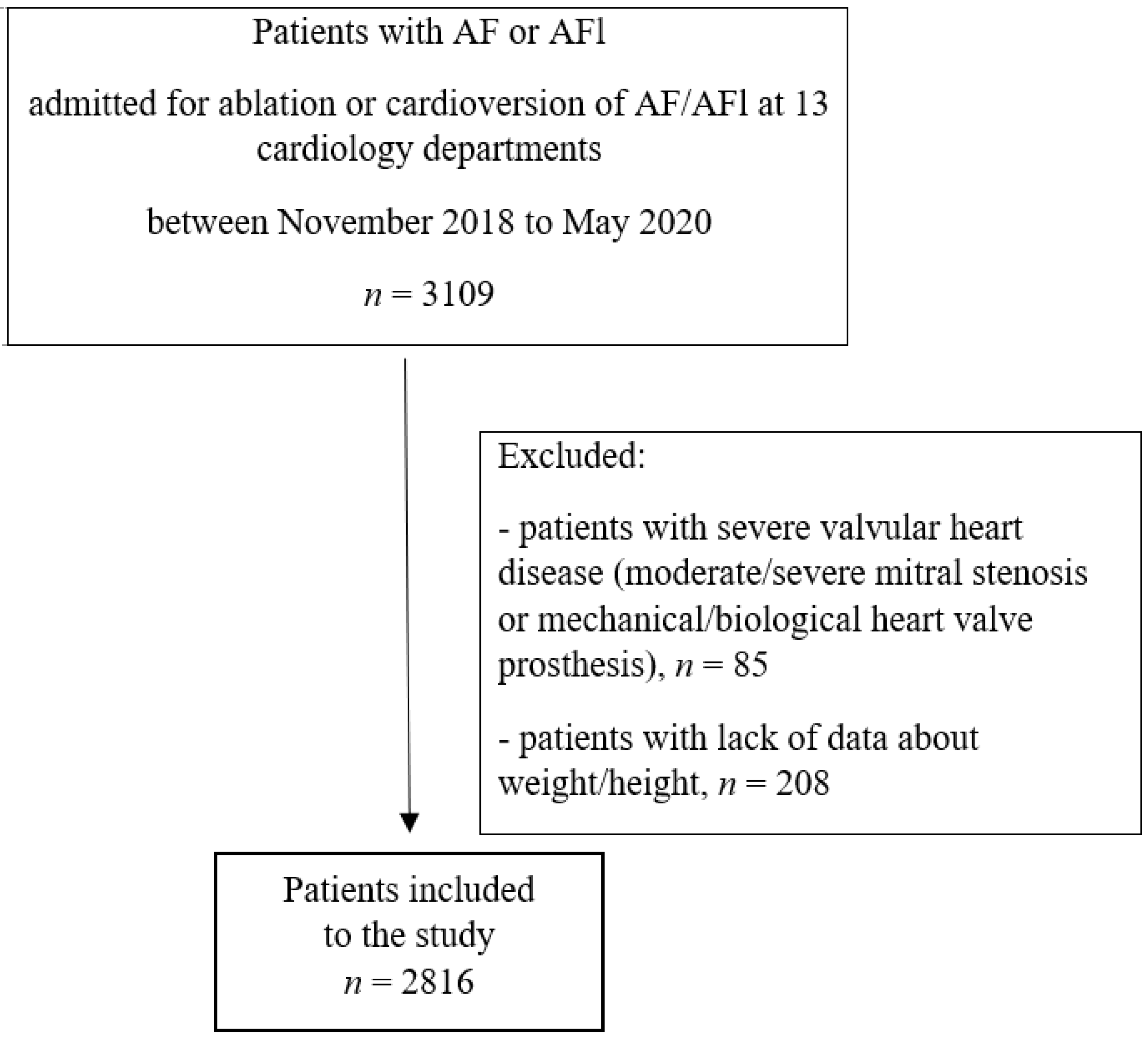
2.1. Patients
2.2. Data Collection
2.3. Statistical Analysis
3. Results
3.1. General Study Population
3.2. BMI and the Risk of Left Atrial Thrombus
4. Discussion
5. Conclusions
Limitations
Author Contributions
Funding
Institutional Review Board Statement
Informed Consent Statement
Data Availability Statement
Acknowledgments
Conflicts of Interest
References
- Schnabel, R.B.; Yin, X.; Gona, P.; Larson, M.G.; Beiser, A.S.; McManus, D.D.; Newton-Cheh, C.; Lubitz, S.A.; Magnani, J.W.; Ellinor, P.T.; et al. 50 year trends in atrial fibrillation prevalence, incidence, risk factors, and mortality in the Framingham Heart Study: A cohort study. Lancet 2015, 386, 154–162. [Google Scholar] [CrossRef]
- Asghar, O.; Alam, U.; Hayat, S.A.; Aghamohammadzadeh, R.; Heagerty, A.M.; Malik, R.A. Obesity, diabetes and atrial fibrillation; epidemiology, mechanisms and interventions. Curr. Cardiol. Rev. 2012, 8, 253–264. [Google Scholar] [CrossRef] [PubMed]
- Samad, F.; Ruf, W. Inflammation, obesity, and thrombosis. Blood 2013, 122, 3415–3422. [Google Scholar] [CrossRef] [PubMed]
- Hindricks, G.; Potpara, T.; Dagres, N.; Arbelo, E.; Bax, J.J.; Blomström-Lundqvist, C.; Boriani, G.; Castella, M.; Dan, G.A.; Dilaveris, P.E.; et al. 2020 ESC Guidelines of the diagnosis and management of atrial fibrillation developed in collaboration with the European Association for Cardio-Thoracic Surgery (EACTS): The Task Force for the diagnosis and management of atrial fibrillation of the European Society of Cardiology (ESC) Developed with the special contribution of the European Heart Rhythm Association (EHRA) of the ESC. Eur. Heart. J. 2021, 42, 373–498. [Google Scholar] [PubMed]
- Overvad, T.F.; Rasmussen, L.H.; Skjøth, F.; Overvad, K.; Lip, G.J.H.; Larsen, T.B. Body mass index and adverse events in patients with incident atrial fibrillation. Am. J. Med. 2013, 126, 640.e9–640.e17. [Google Scholar] [CrossRef] [PubMed]
- Decker, J.J.; Norby, F.L.; Rooney, M.R.; Soliman, E.Z.; Lutsey, P.L.; Pankow, J.S.; Alonso, A.; Chen, L.Y. Metabolic Syndrome and Risk of Ischemic Stroke in Atrial Fibrillation: ARIC Study. Stroke 2019, 50, 3045–3050. [Google Scholar] [CrossRef] [PubMed]
- Steffel, J.; Collins, R.; Antz, M.; Cornu, P.; Desteghe, L.; Haeusler, K.G.; Oldgren, J.; Reinecke, H.; Roldan-Schiling, V.; Rowell, N.; et al. 2021 European Heart Rhythm Association Practical Guide on the Use of Non-Vitamin K Antagonist Oral Anticoagulants in Patients with Atrial Fibrillation. Europace 2021, 23, 1612–1676. [Google Scholar] [CrossRef]
- Tang, R.B.; Liu, X.H.; Kalifa, J.; Li, Z.A.; Dong, J.Z.; Yang, Y.; Liu, X.P.; Long, D.Y.; Yu, R.H.; Ma, C.S. Body mass index and risk of left atrial thrombus in patients with atrial fibrillation. Am. J. Cardiol. 2009, 104, 1699–1703. [Google Scholar] [CrossRef]
- Cohoon, K.P.; McBane, R.D.; Ammash, N.; Slusser, J.P.; Grill, D.E.; Wysokiński, W.E. Relationship between body mass index and left atrial appendage thrombus in nonvalvular atrial fibrillation. J. Thromb. Thrombolysis 2016, 41, 613–618. [Google Scholar] [CrossRef]
- Kapłon-Cieślicka, A.; Gawałko, M.; Budnik, M.; Uziębło-Życzkowska, B.; Krzesiński, P.; Starzyk, K.; Gorczyca-Głowacka, I.; Daniłowicz-Szymanowicz, L.; Kaufmann, D.; Wójcik, M.; et al. Left Atrial Thrombus in Atrial Fibrillation/Flutter Patients in Relation to Anticoagulation Strategy: LATTEE Registry. J. Clin. Med. 2022, 11, 2705. [Google Scholar] [CrossRef]
- Kapłon-Cieślicka, A.; Budnik, M.; Gawałko, M.; Wójcik, M.; Błaszczyk, R.; Uziębło-Życzkowska, B.; Krzesiński, P.; Starzyk, K.; Gorczyca, I.; Szymańska, A.; et al. The rationale and design of the LATTEE registry—the first multicenter project on the Scientific Platform of the “Club 30” of the Polish Cardiac Society. Kardiol. Pol. 2019, 77, 1078–1080. [Google Scholar] [CrossRef] [PubMed]
- Ponikowski, P.; Voors, A.A.; Anker, S.D.; Bueno, H.; Cleland, J.G.F.; Coats, A.J.S.; Falk, V.; González-Juanatey, J.R.; Harjola, V.P.; Jankowska, E.A.; et al. 2016 ESC Guidelines for the diagnosis and treatment of acute and chronic heart failure: The Task Force for the diagnosis and treatment of acute and chronic heart failure of the European Society of Cardiology (ESC) Developed with the special contribution of the Heart Failure Association (HFA) of the ESC. Eur. Heart J. 2016, 37, 2129–2200. [Google Scholar]
- Manning, W.J.; Weintraub, R.M.; Waksmonski, C.A.; Hearing, J.M.; Rooney, P.S.; Maslow, A.D.; Johnson, R.G.; Douglas, P.S. Accuracy of transesophageal echocardiography for identifying left atrial thrombi. A prospective, intraoperative study. Ann. Intern. Med. 1995, 123, 817–822. [Google Scholar] [CrossRef]
- Lang, R.M.; Badano, L.P.; Mor-Avi, V.; Afilalo, J.; Armstrong, A.; Ernande, L.; Flachskampf, F.A.; Foster, E.; Goldstein, S.A.; Kuznetsova, T.; et al. Recommendations for Cardiac Chamber Quantification by Echocardiography in Adults: An Update from the American Society of Echocardiography and the European Association of Cardiovascular Imaging. J. Am. Soc. Echocardiogr. 2015, 16, 233–271. [Google Scholar]
- Noubiap, J.J.; Agbaedeng, T.A.; Ndoadoumgue, A.L.; Nyaga, U.F.; Kengne, A.P. Atrial thrombus detection on transoesophageal echocardiography in patients with atrial fibrillation undergoing cardioversion or catheter ablation: A pooled analysis of rates and predictors. J. Cardiovasc. Electrophysiol. 2021, 32, 2179–2188. [Google Scholar] [CrossRef] [PubMed]
- Lurie, A.; Wang, J.; Hinnegan, K.J.; McIntyre, W.F.; Belley- Côté, E.P.; Amit, G.; Healey, J.S.; Connolly, S.J.; Wong, J.A. Prevalence of Left Atrial Thrombus in Anticoagulated Patients With Atrial Fibrillation. J. Am. Coll. Cardiol. 2021, 77, 2875–2886. [Google Scholar] [CrossRef]
- Seravalle, G.; Grassi, G. Obesity and hypertension. Pharmacol. Res. 2017, 122, 1–7. [Google Scholar] [CrossRef]
- Aiad, N.N.; Hearon, C.J.; Hieda, M.; Dias, K.; Levine, B.D.; Sarma, S. Mechanisms of Left Atrial Enlargement in Obesity. Am. J. Cardiol. 2019, 24, 442–447. [Google Scholar] [CrossRef]
- Providência, R.; Adragão, P.; de Asmundis, C.; Chun, J.; Chierchia, G.; Defaye, P.; Anselme, F.; Creta, A.; Lambiase, P.D.; Schmidt, B.; et al. Impact of Body Mass Index on the Outcomes of Catheter Ablation of Atrial Fibrillation: A European Observational Multicenter Study. J. Am. Heart. Assoc. 2019, 8, e012253. [Google Scholar] [CrossRef]
- Uziębło-Życzkowska, B.; Krzesiński, P.; Jurek, A.; Kapłon-Cieślicka, A.; Gorczyca, I.; Budnik, M.; Gielerak, G.; Kiliszek, M.; Gawałko, M.; Scisło, P.; et al. Left Ventricular Ejection Fraction Is Associated with the Risk of Thrombus in the Left Atrial Appendage in Patients with Atrial Fibrillation. Cardiovasc. Ther. 2020, 2020, 3501749. [Google Scholar] [CrossRef]
- Gawałko, M.; Budnik, M.; Uziębło-Życzkowska, B.; Gorczyca, I.; Krzesiński, P.; Scisło, P.; Kochanowski, J.; Michalska, A.; Jelonek, O.; Starzyk, K.; et al. Risk of left atrial appendage thrombus in older patients with atrial fibrillation. Arch. Med. Sci. 2020, 5. [Google Scholar] [CrossRef]
- Lip, G.Y.; Nieuwlaat, R.; Pisters, R.; Lane, D.A.; Crijns, H.J.G.M. Refining clinical risk stratification for predicting stroke and thromboembolism in atrial fibrillation using a novel risk factor-based approach: The euro heart survey on atrial fibrillation. Chest 2010, 137, 263–272. [Google Scholar] [CrossRef] [PubMed]
- Kapłon-Cieślicka, A.; Budnik, M.; Gawałko, M.; Peller, M.; Gorczyca, I.; Michalska, A.; Babiarz, A.; Bodys, A.; Uliński, R.; Żochowski, M.; et al. Atrial fibrillation type and renal dysfunction as important predictors of left atrial thrombus. Heart 2019, 105, 1310–1315. [Google Scholar] [CrossRef]
- Inao, K.; Hirai, T.; Nakagawa, K.; Numa, S.; Ohara, K.; Fukuda, N.; Kinugawa, K.; Inoue, H. Transesophageal echocardiographic thromboembolic risk is associated with smoking status in patients with atrial fibrillation. J. Arrhythm. 2017, 33, 613–618. [Google Scholar] [CrossRef]
- Albertsen, I.E.; Rasmussen, L.H.; Lane, D.A.; Overvad, T.F.; Skjøth, F.; Overvad, K.; Lip, G.J.H.; Larsen, T.B. The Impact of Smoking on Thromboembolism and Mortality in Patients With Incident Atrial Fibrillation: Insights from the Danish Diet, Cancer, and Health study. Chest 2014, 145, 559–566. [Google Scholar] [CrossRef]
- Beyer-Westendorf, J.; Kreutz, R.; Posch, F.; Ay, C. The CHA2DS2-VASc score strongly correlates with glomerular filtration rate and predicts renal function decline over time in elderly patients with atrial fibrillation and chronic kidney disease. Int. J. Cardiol. 2018, 253, 71–77. [Google Scholar] [CrossRef] [PubMed]
- Sikorska, A.; Baran, J.; Pilichowska-Paszkiet, E.; Sikora-Frąc, M.; Kryński, T.; Piotrowski, R.; Stec, S.; Zaborska, B.; Kułakowski, P. Risk of left atrial appendage thrombus in patients scheduled for ablation for atrial fibrillation: Beyond the CHA2DS2VASc score. Pol. Arch. Med. Wewn. 2015, 125, 921–928. [Google Scholar] [CrossRef] [PubMed] [Green Version]


| Variable | BMI (kg/m2) | p-Value | |||
|---|---|---|---|---|---|
| <25 N = 474; 16.8% Normal Weight | 25.0–29.9 N = 1077; 38.2% Overweight | >30 N = 1265; 44.9% Obese | |||
| The reason for admission | |||||
| Catheter ablation | 219 (47) | 517 (48.8) | 580 (46.4) | 0.51 | |
| Cardioversion | 247 (53) | 542 (51.2) | 669 (53.6) | 0.51 | |
| Type of AF/AFl | |||||
| AF/AFl non-paroxysmal | 261 (55.2) | 605 (56.4) | 805 (63.8) | <0.001 | |
| Time from AF/AFl diagnosis (years) | 2 (1–5) | 2 (1–5) | 2 (1–5) | 0.31 | |
| Demographic data | |||||
| Age (years) | 68 (60–76) | 67 (60–73) | 66 (59–72) | <0.001 | |
| Sex | Female | 202 (42.6) | 363 (33.7) | 460 (36.4) | 0.004 |
| Male | 272 (57.4) | 714 (66.3) | 805 (63.6) | ||
| Concomitant diseases | |||||
| Heart failure | 193 (41) | 447 (41.5) | 564 (44.7) | 0.2 | |
| HFrEF | 75 (15.9) | 171 (15.9) | 187 (14.8) | 0.73 | |
| HFmrEF | 56 (11.9) | 128 (11.9) | 140 (11.1) | 0.81 | |
| HFpEF | 63 (13.4) | 153 (14.2) | 241 (19.1) | 0.001 | |
| Hypertension | 278 (58.6) | 809 (75.1) | 1061 (83.9) | <0.001 | |
| Diabetes mellitus | 66 (13.9) | 224 (20.8) | 422 (33.4) | <0.001 | |
| Previous TIA | 14 (3) | 31 (2.9) | 37 (2.9) | 0.99 | |
| Previous stroke | 35 (7.4) | 85 (7.9) | 89 (7) | 0.74 | |
| Coronary artery disease | 130 (27.4) | 328 (30.5) | 379 (30) | 0.47 | |
| Chronic kidney disease | 83 (17.5) | 154 (14.3) | 220 (17.4) | 0.09 | |
| Smoking | 135 (28.7) | 346 (32.9) | 462 (37.3) | 0.002 | |
| Malignant tumor | 15 (3.2) | 37 (3.4) | 40 (3.2) | 0.93 | |
| COPD | 32 (6.8) | 50 (4.6) | 67 (5.3) | 0.23 | |
| CHA2DS2-VASc score (points) | 3 (2–4) | 3 (2–4) | 3 (2–5) | 0.002 | |
| CHADS2 score (points) | 1 (1–3) | 2 (1–3) | 2 (1–3) | <0.001 | |
| Laboratory and echocardiography data | |||||
| Hemoglobin (g/dL) | 13.7 (12.8–14.7) | 14.1 (13–15.1) | 14.3 (13.2–15.3) | <0.001 | |
| eGFR (mL/min/1.73 m2) | 59.8 (46.5–80.9) | 59.1 (48.7–78.5) | 57.1 (45.6–77.2) | 0.07 | |
| LVEF (%) | 55 (42–60) | 55 (45–60) | 55 (45–60) | 0.85 | |
| LAd (cm) | 43 (39–47) | 45 (41–48.8) | 46 (43–50) | <0.001 | |
| LA area (cm2) | 24.9 (21.2–29) | 25.4 (21.5–30) | 27 (23.4–31) | <0.001 | |
| LAA emptying velocity (cm/s) | 37 (25–53.3) | 40 (26–57) | 38 (27–50) | 0.74 | |
| Oral anticoagulation therapy | |||||
| OAC therapy | 401 (84.6) | 954 (88.7) | 1132 (89.5) | 0.02 | |
| VKA | 54 (13.3) | 161 (16.6) | 182 (15.8) | 0.3 | |
| warfarin | 21 (4.4) | 66 (6.1) | 86 (6.8) | 0.19 | |
| acenocoumarol | 33 (7) | 95 (8.8) | 96 (7.6) | 0.37 | |
| NOAC | 352 (86.7) | 807 (83.4) | 972 (84.2) | 0.3 | |
| rivaroxaban | 142 (30) | 397 (36.9) | 449 (35.5) | 0.03 | |
| dabigatran | 132 (27.8) | 289 (26.9) | 352 (27.8) | 0.86 | |
| apixaban | 78 (16.5) | 121 (11.3) | 171 (13.5) | 0.02 | |
| Variable | BMI (kg/m2) | p-Value | ||
|---|---|---|---|---|
| <25 N = 474; 16.8% Normal Weight | >24.99 N = 2342; 83.2% Overweight/Obese | |||
| The reason for admission | ||||
| Catheter ablation | 219 (47) | 1097 (47.5) | 0.83 | |
| Cardioversion | 247 (53) | 1211 (52.5) | 0.83 | |
| Type of AF/AFl | ||||
| AF/AFl non-paroxysmal | 261 (55.2) | 1410 (60.4) | 0.04 | |
| Time from AF/AFl diagnosis (years) | 2 (1–5) | 2 (1–5) | 0.57 | |
| Demographic data | ||||
| Age (years) | 68 (60–76) | 67 (59–73) | <0.001 | |
| Sex | Female | 202 (42.6) | 823 (35.1) | 0.002 |
| Male | 272 (57.4) | 1519 (64.9) | ||
| Concomitant diseases | ||||
| Heart failure | 193 (41) | 1011 (43.3) | 0.39 | |
| HFrEF | 75 (15.9) | 358 (15.3) | 0.73 | |
| HFmrEF | 56 (11.9) | 268 (11.5) | 0.81 | |
| HFpEF | 63 (13.4) | 394 (16.9) | 0.07 | |
| Hypertension | 278 (58.6) | 1870 (79.9) | <0.001 | |
| Diabetes mellitus | 66 (13.9) | 646 (27.6) | <0.001 | |
| Previous TIA | 14 (3) | 48 (2.9) | 0.88 | |
| Previous stroke | 35 (7.4) | 174 (7.4) | 1 | |
| Coronary artery disease | 130 (27.4) | 707 (30.2) | 0.25 | |
| Chronic kidney disease | 83 (17.5) | 374 (16) | 0.41 | |
| Smoking | 135 (28.7) | 808 (35.3) | 0.006 | |
| Malignant tumor | 15 (3.2) | 77 (3.3) | 1 | |
| COPD | 32 (6.8) | 117 (5) | 0.14 | |
| CHA2DS2-VASc score (points) | 3 (2–4) | 3 (2–4) | 0.1 | |
| CHADS2 score (points) | 1 (1–3) | 2 (1–3) | <0.001 | |
| Laboratory and echocardiography data | ||||
| Hemoglobin (g/dL) | 13.7 (12.8–14.7) | 14.2 (13.1–15.2) | <0.001 | |
| eGFR (mL/min/1.73 m) | 59.8 (46.5–80.9) | 58 (46.9–78.2) | 0.35 | |
| LVEF (%) | 55 (42–60) | 55 (45–60) | 0.65 | |
| LAd (cm) | 43 (39–47) | 46 (42–50) | <0.001 | |
| LA area (cm2) | 24.9 (21.2–29) | 26.5 (22.5–30.3) | <0.001 | |
| LAA emptying velocity (cm/s) | 37 (25–53.2) | 39 (26–54) | 0.79 | |
| Oral anticoagulation therapy | ||||
| OAC therapy | 401 (84.6) | 2086 (89.1) | 0.005 | |
| VKA | 54 (13.3) | 343 (16.2) | 0.16 | |
| warfarin | 21 (4.4) | 152 (6.5) | 0.09 | |
| acenocoumarol | 33 (7) | 191 (8.2) | 0.4 | |
| NOAC | 352 (86.7) | 1779 (83.8) | 0.16 | |
| rivaroxaban | 142 (30) | 846 (36.2) | 0.01 | |
| dabigatran | 132 (27.8) | 641 (27.4) | 0.87 | |
| apixaban | 78 (16.5) | 292 (12.5) | 0.06 | |
| Variable | LAT(−) | LAT(+) | p-Value |
|---|---|---|---|
| BMI (kg/m2) | |||
| <25 | 432 (91.1) | 42 (8.9) | 0.5 |
| 25.0–29.9 | 989 (91.8) | 88 (8.2) | |
| >30 | 1173 (92.7) | 92 (7.3) | |
| BMI (kg/m2) | |||
| <25 | 432 (91.1) | 42 (8.9) | 0.4 |
| >24.99 | 2162 (92.3) | 180 (7.7) | |
Publisher’s Note: MDPI stays neutral with regard to jurisdictional claims in published maps and institutional affiliations. |
© 2022 by the authors. Licensee MDPI, Basel, Switzerland. This article is an open access article distributed under the terms and conditions of the Creative Commons Attribution (CC BY) license (https://creativecommons.org/licenses/by/4.0/).
Share and Cite
Uziębło-Życzkowska, B.; Kapłon-Cieślicka, A.; Kiliszek, M.; Gawałko, M.; Budnik, M.; Starzyk, K.; Wożakowska-Kapłon, B.; Daniłowicz-Szymanowicz, L.; Kaufmann, D.; Wójcik, M.; et al. Increased Body Mass Index and Risk of Left Atrial Thrombus in Nonvalvular Atrial Fibrillation Patients—Data from the Left Atrial Thrombus on Transesophageal Echocardiography (LATTEE) Registry. Nutrients 2022, 14, 3652. https://doi.org/10.3390/nu14173652
Uziębło-Życzkowska B, Kapłon-Cieślicka A, Kiliszek M, Gawałko M, Budnik M, Starzyk K, Wożakowska-Kapłon B, Daniłowicz-Szymanowicz L, Kaufmann D, Wójcik M, et al. Increased Body Mass Index and Risk of Left Atrial Thrombus in Nonvalvular Atrial Fibrillation Patients—Data from the Left Atrial Thrombus on Transesophageal Echocardiography (LATTEE) Registry. Nutrients. 2022; 14(17):3652. https://doi.org/10.3390/nu14173652
Chicago/Turabian StyleUziębło-Życzkowska, Beata, Agnieszka Kapłon-Cieślicka, Marek Kiliszek, Monika Gawałko, Monika Budnik, Katarzyna Starzyk, Beata Wożakowska-Kapłon, Ludmiła Daniłowicz-Szymanowicz, Damian Kaufmann, Maciej Wójcik, and et al. 2022. "Increased Body Mass Index and Risk of Left Atrial Thrombus in Nonvalvular Atrial Fibrillation Patients—Data from the Left Atrial Thrombus on Transesophageal Echocardiography (LATTEE) Registry" Nutrients 14, no. 17: 3652. https://doi.org/10.3390/nu14173652