
A Review of Methods for Removal of Ceftriaxone from Wastewater


Abstract
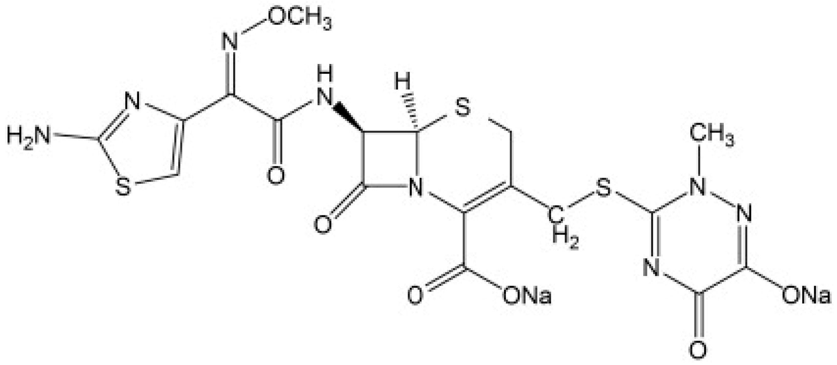
:1. Introduction
2. Methods Used to Analyze Antibiotics
2.1. Chromatographic Methods
2.2. Spectrophotometric Methods
2.3. Electrochemical Methods
2.4. Methods Studied for Analysis of Ceftriaxone in Aquatic and Biological Samples
3. Methods Used for Removal of Antibiotics from Wastewater
3.1. Constructed Wetland
3.2. Biological Treatment
3.3. Advanced Oxidation Processes (AOP)
3.4. Membrane Technology
4. Methods Studied for Removal of Ceftriaxone from Water and Wastewater
5. Conclusions
Author Contributions
Funding
Institutional Review Board Statement
Informed Consent Statement
Data Availability Statement
Conflicts of Interest
References
- Shi, X.; Karachi, A.; Hosseini, M.; Yazd, M.S.; Kamyab, H.; Ebrahimi, M.; Parsaee, Z. Ultrasound wave assisted removal of Ceftriaxone sodium in aqueous media with novel nano composite g-C3N4/MWCNT/Bi2WO6 based on CCD-RSM model. Ultrason. Sonochemistry 2019, 68, 104460. [Google Scholar] [CrossRef] [PubMed]
- Zhao, Y.; Liang, X.; Shi, H.; Wang, Y.; Ren, Y.; Liu, E.; Zhang, X.; Fan, J.; Hu, X. Photocatalytic activity enhanced by synergistic effects of nano-silver and ZnSe quantum dots co-loaded with bulk g-C3N4 for Ceftriaxone sodium degradation in aquatic environment. Chem. Eng. J. 2018, 353, 56–68. [Google Scholar] [CrossRef]
- Kordestani, B.; Yengejeh, R.J.; Takdastan, A.; Neisi, A. A new study on photocatalytic degradation of meropenem and ceftriaxone antibiotics based on sulfate radicals: Influential factors, biodegradability, mineralization approach. Microchem. J. 2019, 146, 286–292. [Google Scholar] [CrossRef]
- Amiri, S.; Sohrabi, M.R.; Motiee, F. Optimization Removal of the Ceftriaxone Drug from Aqueous Media with Novel Zero-Valent Iron Supported on Doped Strontium Hexaferrite Nanoparticles by Response Surface Methodology. ChemistrySelect 2020, 5, 5831–5840. [Google Scholar] [CrossRef]
- Kordestani, B.; Takdastan, A.; Yengejeh, R.J.; Neisi, A. Photo-Fenton oxidative of pharmaceutical wastewater containing meropenem and ceftriaxone antibiotics: Influential factors, feasibility, and biodegradability studies. Toxin Rev. 2018, 39, 292–302. [Google Scholar] [CrossRef]
- Kaur, B.; Kuntus, L.; Tikker, P.; Kattel, E.; Trapido, M.; Dulova, N. Photo-induced oxidation of ceftriaxone by persulfate in the presence of iron oxides. Sci. Total Environ. 2019, 676, 165–175. [Google Scholar] [CrossRef]
- Thalji, M.R. Nanotechnologies for Removal of Pharmaceuticals from Wastewater. Med. Pharm. Sci. 2021, 1, 25–28. [Google Scholar]
- Puddoo, H.; Nithyanandam, R.; Nguyenhuynh, T.; Taylor’s University Malaysia. Degradation of the Antibiotic Ceftriaxone by Fenton Oxidation Process and Compound Analysis. J. Phys. Sci. 2017, 28, 95–114. [Google Scholar] [CrossRef] [Green Version]
- AttariKhasraghi, N.; Zare, K.; Mehrizad, A.; Modirshahla, N.; Behnajady, M.A. Achieving the Enhanced Photocatalytic Degradation of Ceftriaxone Sodium Using CdS-g-C3N4 Nanocomposite under Visible Light Irradiation: RSM Modeling and Optimization. J. Inorg. Organomet. Polym. Mater. 2021, 31, 3164–3174. [Google Scholar] [CrossRef]
- Mahmoud, M.E.; El-Ghanam, A.M.; Mohamed, R.H.A.; Saad, S.R. Enhanced adsorption of Levofloxacin and Ceftriaxone antibiotics from water by assembled composite of nanotitanium oxide/chitosan/nano-bentonite. Mater. Sci. Eng. C 2019, 108, 110199. [Google Scholar] [CrossRef]
- Khorsandi, H.; Teymori, M.; Aghapour, A.A.; Jafari, S.J.; Taghipour, S.; Bargeshadi, R. Photodegradation of ceftriaxone in aqueous solution by using UVC and UVC/H2O2 oxidation processes. Appl. Water Sci. 2019, 9, 81. [Google Scholar] [CrossRef] [Green Version]
- Owens, H.M.; Dash, A.K. Ceftriaxone Sodium: Comprehensive Profile. Profiles Drug Subst. Excip. Relat. Methodol. 2003, 30, 21–57. [Google Scholar] [CrossRef]
- Zhang, P.O.; Zhang, L.; Ma, C. Degradation of Ceftriaxone Sodium in Pharmaceutical Wastewater by Photocatalytic Oxidation. In Proceedings of the 2020 2nd World Congress on Chemistry, Biotechnology and Medicine (WCCBM 2020), Zurich, Switzerland, 6–8 April 2020; pp. 217–221. [Google Scholar] [CrossRef]
- Da Trindade, M.T.; Salgado, H.R.N. A Critical Review of Analytical Methods for Determination of Ceftriaxone Sodium. Crit. Rev. Anal. Chem. 2018, 48, 95–101. [Google Scholar] [CrossRef] [PubMed]
- Rohimmahtunnissa, A.; Alfan, D.A.; Firdayani, A.S. The Influence Study of the Mole Ratio Reactant in Ceftriaxone Sodium Synthesis Against The Yield of The Production. Int. J. Innov. Eng. Sci. Res. 2018, 2, 6. [Google Scholar] [CrossRef]
- Scholar, E. Ceftriaxone. In xPharm: The Comprehensive Pharmacology Reference; Elsevier: Amsterdam, The Netherlands, 2007. [Google Scholar]
- Rebec, G.V. Vitamin C and Glutamate Uptake: Implications for Huntington’s Disease. In Diet and Nutrition in Dementia and Cognitive Decline; Academic Press: San Diego, CA, USA; Elsevier, Inc.: San Diego, CA, USA, 2015. [Google Scholar]
- Cetecioglu, Z.; Atasoy, M. Biodegradation and Inhibitory Effects of Antibiotics on Biological Wastewater Treatment Systems. In Toxicity and Biodegradation Testing. Methods in Pharmacology and Toxicology; Humana Press: New York, NY, USA, 2018; pp. 29–55. [Google Scholar] [CrossRef]
- Muriuki, C.W.; Home, P.G.; Raude, J.M.; Ngumba, E.K.; Munala, G.K.; Kairigo, P.K.; Gachanja, A.N.; Tuhkanen, T.A. Occurrence, distribution, and risk assessment of pharmerciuticals in wastewater and open surface drains of peri-urban areas: Case study of Juja town, Kenya. Environ. Pollut. 2020, 267, 115503. [Google Scholar] [CrossRef] [PubMed]
- Rodriguez-Mozaz, S.; Vaz-Moreira, I.; Della Giustina, S.V.; Llorca, M.; Barceló, D.; Schubert, S.; Berendonk, T.U.; Michael-Kordatou, I.; Fatta-Kassinos, D.; Martinez, J.L.; et al. Antibiotic residues in final effluents of European wastewater treatment plants and their impact on the aquatic environment. Environ. Int. 2020, 140, 105733. [Google Scholar] [CrossRef] [PubMed]
- Polianciuc, S.I.; Gurzău, A.E.; Kiss, B.; Ștefan, M.G.; Loghin, F. Antibiotics in the environment: Causes and consequences. Med. Pharm. Rep. 2020, 93, 231–240. [Google Scholar] [CrossRef] [PubMed]
- Wajahat, R.; Yasar, A.; Khan, A.M.; Tabinda, A.B.; Bhatti, S.G. Ozonation and Photo-Driven Oxidation of Ciprofloxacin in Pharmaceutical Wastewater: Degradation Kinetics and Energy Requirements. Pol. J. Environ. Stud. 2019, 28, 1933–1938. [Google Scholar] [CrossRef]
- Huang, A.; Yan, M.; Lin, J.; Xu, L.; Gong, H.; Gong, H. A Review of Processes for Removing Antibiotics from Breeding Wastewater. Int. J. Environ. Res. Public Health 2021, 18, 4909. [Google Scholar] [CrossRef]
- Ncube, S.; Nuapia, Y.B.; Chimuka, L.; Madikizela, L.M.; Etale, A. Trace Detection and Quantitation of Antibiotics in a South African Stream Receiving Wastewater Effluents and Municipal Dumpsite Leachates. Front. Environ. Sci. 2021, 9, 365. [Google Scholar] [CrossRef]
- Nantaba, F.; Wasswa, J.; Kylin, H.; Palm, W.-U.; Bouwman, H.; Kümmerer, K. Occurrence, distribution, and ecotoxicological risk assessment of selected pharmaceutical compounds in water from Lake Victoria, Uganda. Chemosphere 2019, 239, 124642. [Google Scholar] [CrossRef] [PubMed]
- Berglund, B.; Khan, G.A.; Weisner, S.E.; Ehde, P.M.; Fick, J.; Lindgren, P.-E. Efficient removal of antibiotics in surface-flow constructed wetlands, with no observed impact on antibiotic resistance genes. Sci. Total Environ. 2014, 476–477, 29–37. [Google Scholar] [CrossRef] [PubMed]
- Moreira, F.C.; Soler, J.; Alpendurada, M.; Boaventura, R.A.; Brillas, E.; Vilar, V.J. Tertiary treatment of a municipal wastewater toward pharmaceuticals removal by chemical and electrochemical advanced oxidation processes. Water Res. 2016, 105, 251–263. [Google Scholar] [CrossRef] [PubMed]
- Lien, L.T.Q.; Hoa, N.Q.; Chuc, N.T.K.; Thoa, N.T.M.; Phuc, H.D.; Diwan, V.; Dat, N.T.; Tamhankar, A.J.; Lundborg, C.S. Antibiotics in Wastewater of a Rural and an Urban Hospital before and after Wastewater Treatment, and the Relationship with Antibiotic Use—A One Year Study from Vietnam. Int. J. Environ. Res. Public Health 2016, 13, 588. [Google Scholar] [CrossRef] [PubMed] [Green Version]
- Yang, Q.; Gao, Y.; Ke, J.; Show, P.L.; Ge, Y.; Liu, Y.; Guo, R.; Chen, J. Antibiotics: An overview on the environmental occurrence, toxicity, degradation, and removal methods. Bioengineered 2021, 12, 7376–7416. [Google Scholar] [CrossRef]
- Mahdavi, H.; Bagherifar, R. Cellulose acetate/SiO2-poly(2-Acrylamido-2-methylpropane sulfonic acid) hybrid nanofiltration membrane: Application in removal of ceftriaxone sodium. J. Iran. Chem. Soc. 2018, 15, 2839–2849. [Google Scholar] [CrossRef]
- Abramović, B.F.; Uzelac, M.M.; Finčur, N.L. Photocatalytic degradation of thiotriazinone, stable hydrolysis product of antibiotic ceftriaxone. Acta Period. Technol. 2019, 50, 1–11. [Google Scholar] [CrossRef] [Green Version]
- Shipingana, L.N.N.; Shivaraju, H.P.; Yashas, S.R. Quantitative assessment of pharmaceutical drugs in a municipal wastewater and overview of associated risks. Appl. Water Sci. 2022, 12, 16. [Google Scholar] [CrossRef]
- Faleye, A.; Adegoke, A.A.; Ramluckan, K.; Bux, F.; Stenström, T.A. Antibiotic Residue in the Aquatic Environment: Status in Africa. Open Chem. 2018, 16, 890–903. [Google Scholar] [CrossRef]
- Bekele, L.K.; Gebeyehu, G.G. Application of Different Analytical Techniques and Microbiological Assays for the Analysis of Macrolide Antibiotics from Pharmaceutical Dosage Forms and Biological Matrices. ISRN Anal. Chem. 2012, 2012, 859473. [Google Scholar] [CrossRef] [Green Version]
- Davani, B. Pharmaceutical Analysis for Small Molecules; John Wiley & Sons, Inc.: Hoboken, NJ, USA, 2017. [Google Scholar]
- Dasgupta, A.; Krasowski, M.D. Application of chromatographic techniques for therapeutic drug monitoring. In Therapeutic Drug Monitoring Data; Academic Press: London, UK, 2020; pp. 53–63. [Google Scholar] [CrossRef]
- Pauter, K.; Szultka-Młyńska, M.; Buszewski, B. Determination and Identification of Antibiotic Drugs and Bacterial Strains in Biological Samples. Molecules 2020, 25, 2556. [Google Scholar] [CrossRef] [PubMed]
- Raeisi, A.; Ramezani, M.; Ravazadeh, H.; Taher, M.A. Chromatographic Behaviour of Antibiotics on Thin Layers of Zeolite. J. Pharm. Res. Int. 2019, 30, 1–8. [Google Scholar] [CrossRef]
- Hancu, G.; Simon, B.; Kelemen, H.; Rusu, A.; Mircia, E.; Gyéresi, Á. Thin Layer Chromatographic Analysis of Beta-Lactam Antibiotics. Adv. Pharm. Bull. 2013, 3, 367–371. [Google Scholar] [CrossRef] [PubMed]
- Asan, A.; Seddiq, N. A Simple Spectrophotometric Determination of Amoxicillin in Drug samples. J. Turk. Chem. Soc. 2022, 9, 423–432. [Google Scholar] [CrossRef]
- De Paula, C.E.R.; Almeida, V.G.K.; Cassella, R.J. Determinação espectrofotométrica de cefalexina em formulações farmacêuticas explorando a sua reação de transferência de carga com a quinalizarina. Quim. Nova 2010, 33, 914–919. [Google Scholar] [CrossRef] [Green Version]
- Moldoveanu, S.C.; David, V. Short Overviews of the Main Analytical Techniques Containing a Separation Step. In Selection of the HPLC Method in Chemical Analysis; Elsevier Inc.: Amsterdam, The Netherlands, 2017; pp. 55–85. [Google Scholar] [CrossRef]
- Chew, Y.-L.; Khor, M.-A.; Lim, Y.-Y. Choices of chromatographic methods as stability indicating assays for pharmaceutical products: A review. Heliyon 2021, 7, e06553. [Google Scholar] [CrossRef] [PubMed]
- Parys, W.; Dołowy, M.; Pyka-Pająk, A. Significance of Chromatographic Techniques in Pharmaceutical Analysis. Processes 2022, 10, 172. [Google Scholar] [CrossRef]
- Ahuja, S. Derivatization in gas chromatography. J. Pharm. Sci. 1976, 65, 163–182. [Google Scholar] [CrossRef] [PubMed]
- Lozano-Sánchez, J.; Borrás-Linares, I.; Sass-Kiss, A.; Segura-Carretero, A. Chromatographic Technique: High-Performance Liquid Chromatography (HPLC). In Modern Techniques for Food Authentication; Academic Press: London, UK, 2018; pp. 459–526. [Google Scholar]
- Jena, A.K.A. HPLC: Highly Accessible Instrument in Pharmaceutical Industry for Effective Method Development. Pharm. Anal. Acta 2012, 3, 1–9. [Google Scholar] [CrossRef] [Green Version]
- Locatelli, M.; Melucci, D.; Carlucci, G.; Locatelli, C. Recent hplc strategies to improve sensitivity and selectivity for the analysis of complex matrices. Instrum. Sci. Technol. 2012, 40, 112–137. [Google Scholar] [CrossRef]
- Gallagher, T.; Riedel, S.; Kapcia, J.; Caverly, L.J.; Carmody, L.; Kalikin, L.M.; Lu, J.; Phan, J.; Gargus, M.; Kagawa, M.; et al. Liquid Chromatography Mass Spectrometry Detection of Antibiotic Agents in Sputum from Persons with Cystic Fibrosis. Antimicrob. Agents Chemother. 2021, 65, 2. [Google Scholar] [CrossRef] [PubMed]
- Usman, M.R.; Prasasti, A.; Islamiah, S.; Firdaus, A.N.; Marita, A.W.; Fajriyah, S.; Yanti, E.F. Ceftriaxone Degradation by Titanium Dioxide (TiO2) Nanoparticles: Toxicity and Degradation Mechanism. J. Kim. Val. 2020, 6, 82–89. [Google Scholar] [CrossRef]
- Nováková, L.; Svoboda, P.; Pavlík, J. Ultra-high performance liquid chromatography. In Liquid Chromatography: Fundamentals and Instrumentation, 2nd ed.; Elsevier: Amsterdam, The Netherlands, 2017; Volume 1, pp. 719–769. [Google Scholar]
- Zhang, A.-H.; Wang, P.; Sun, H.; Yan, G.-L.; Han, Y.; Wang, X.-J. High-throughput ultra-performance liquid chromatography-mass spectrometry characterization of metabolites guided by a bioinformatics program. Mol. BioSyst. 2013, 9, 2259–2265. [Google Scholar] [CrossRef] [PubMed]
- Rathod, R.H.; Chaudhari, S.R.; Patil, A.S.; Shirkhedkar, A.A. Ultra-high performance liquid chromatography-MS/MS (UHPLC-MS/MS) in practice: Analysis of drugs and pharmaceutical formulations. Futur. J. Pharm. Sci. 2019, 5, 6. [Google Scholar] [CrossRef] [Green Version]
- Zhang, Y.; Li, X.Q.; Li, H.M.; Zhang, Q.H.; Gao, Y.; Li, X.J. Antibiotic residues in honey: A review on analytical methods by liquid chromatography tandem mass spectrometry. TrAC Trends Anal. Chem. 2018, 110, 344–356. [Google Scholar] [CrossRef]
- Chen, F.; Cheng, Z.; Peng, Y.; Wang, Z.; Huang, C.; Liu, D.; Wang, B.; Pan, B.; Guo, W. A liquid chromatography-tandem mass spectrometry (LC-MS/MS)-based assay for simultaneous quantification of aldosterone, renin activity, and angiotensin II in human plasma. J. Chromatogr. B 2021, 1179, 122740. [Google Scholar] [CrossRef]
- Keskar, M.R.; Jugade, R.M. Spectrophotometric Investigations of Macrolide Antibiotics: A Brief Review. Anal. Chem. Insights 2015, 10, ACI.S31857. [Google Scholar] [CrossRef] [Green Version]
- Rachidi, M.; Elharti, J.; Digua, K.; Cherrah, Y.; Bouklouze, A. New Spectrophotometric Method for Azithromycin Determination. Anal. Lett. 2006, 39, 1917–1926. [Google Scholar] [CrossRef]
- Rufino, J.L.; Fernandes, F.C.B.; Ruy, M.S.; Pezza, H.R.; Pezza, L. A simple spectrophotometric method for the determination of tetracycline and doxycycline in pharmaceutical formulations using chloramine-t. Eclet. Quim. 2018, 35, 139–145. [Google Scholar] [CrossRef]
- Keskar, M.R.; Jugade, R.M. Spectrophotometric Determination of Cefixime Trihydrate in Pharmaceutical Formulations Based on Ion-Pair Reaction with Bromophenol Blue. Anal. Chem. Insights 2015, 10, 11–16. [Google Scholar] [CrossRef]
- Omar, M.A.; Nagy, D.M.; Hammad, M.A.; Aly, A.A. Validated spectrophotometric methods for determination of certain aminoglycosides in pharmaceutical formulations. J. Appl. Pharm. Sci. 2013, 3, 151–161. [Google Scholar] [CrossRef]
- Salem, H.; Askal, H. Colourimetric and AAS determination of cephalosporins using Reineck‘s salt. J. Pharm. Biomed. Anal. 2002, 29, 347–354. [Google Scholar] [CrossRef]
- Choudhary, Y.S.; Jothi, L.; Nageswaran, G. Electrochemical Characterization. In Spectroscopic Methods for Nanomaterials Characterization; Elsevier: Amsterdam, The Netherlands, 2017; Volume 2, pp. 19–54. [Google Scholar]
- Westbroek, P.; Priniotakis, G.; Kiekens, P. Electrochemical methods. In Analytical Electrochemistry in Textiles; Woodhead Publishing Limited: Cambridge, UK, 2005; pp. 37–69. [Google Scholar]
- Svorc, L. Determination of Caffeine: A Comprehensive Review on Electrochemical Methods. Int. J. Electrochem. Sci. 2013, 8, 5755–5773. [Google Scholar]
- Avramov, M.L.; Petrovi, S.D.; Mijin, Ž. Contribution to the Recent Advances in Electrochemical Analysis of Pharmaceuticals. In Biomedical and Pharmaceutical Applications of Electrochemistry. Modern Aspects of Electrochemistry; Springer International Publishing: Cham, Switzerland, 2016. [Google Scholar]
- Aboul-Enein, H.Y.; Sibel, A. Ozkan: Electroanalytical Methods in Pharmaceutical Analysis and Their Validation. Chromatographia 2012, 75, 811. [Google Scholar] [CrossRef] [Green Version]
- Ayankojo, A.G.; Reut, J.; Ciocan, V.; Öpik, A.; Syritski, V. Molecularly imprinted polymer-based sensor for electrochemical detection of erythromycin. Talanta 2019, 209, 120502. [Google Scholar] [CrossRef]
- Da Cunha, C.E.P.; Rodrigues, E.S.B.; Fernandes Alecrim, M.; Thomaz, D.V.; Macêdo, I.Y.L.; Garcia, L.F.; de Oliveira Neto, J.R.; Moreno, E.K.G.; Ballaminut, N.; de Souza Gil, E. Voltammetric Evaluation of Diclofenac Tablets Samples through Carbon Black-Based Electrodes. Pharmaceuticals 2019, 12, 83. [Google Scholar] [CrossRef]
- Shah, J.; Jan, M.R.; Shah, S.; Naeem, M. Spectrofluorimetric Protocol for Ceftriaxone in Commercial Formulation and Human Plasma After Condensation with Formaldehyde and Ethyl Acetoacetate. J. Fluoresc. 2011, 21, 2155–2163. [Google Scholar] [CrossRef]
- Tariq, A.; Siddiqui, M.R.; Kumar, J.; Reddy, D.; Negi, P.S.; Chaudhary, M.; Srivastava, S.M.; Singh, R.K. Development and validation of high performance liquid chromatographic method for the simultaneous determination of ceftriaxone and vancomycin in pharmaceutical formulations and biological samples. Sci. Asia 2010, 36, 297–304. [Google Scholar] [CrossRef]
- Tange, M.; Yoshida, M.; Nakai, Y.; Uchida, T. The Role of an Impurity in Ceftriaxone Sodium Preparation for Injection in Determining Compatibility with Calcium-Containing Solutions. Chem. Pharm. Bull. 2016, 64, 207–214. [Google Scholar] [CrossRef] [Green Version]
- Diwan, V.; Tamhankar, A.J.; Khandal, R.K.; Sen, S.; Aggarwal, M.; Marothi, Y.; Iyer, R.V.; Sundblad-Tonderski, K.; Lundborg, C.S. Antibiotics and antibiotic-resistant bacteria in waters associated with a hospital in Ujjain, India. BMC Public Health 2010, 10, 414. [Google Scholar] [CrossRef] [Green Version]
- Kratzer, A.; Liebchen, U.; Schleibinger, M.; Kees, M.G.; Kees, F. Determination of free vancomycin, ceftriaxone, cefazolin and ertapenem in plasma by ultrafiltration: Impact of experimental conditions. J. Chromatogr. B 2014, 961, 97–102. [Google Scholar] [CrossRef] [PubMed]
- Kale, R.S.; Jain, H.K.; Ghode, P.D.; Mhaske, G.S.; Puri, M.V.; Raut, M.D.; Patil, H.S. An rphplc method for simultaneous estimation of Ceftriaxone sodium and sulbactam sodium in parenteral dosage form. Int. J. Pharm. Pharm. Sci. 2011, 3, 406–409. [Google Scholar]
- Akl, M.A.; Ahmed, M.A.; Ramadan, A. Validation of an HPLC-UV method for the determination of ceftriaxone sodium residues on stainless steel surface of pharmaceutical manufacturing equipments. J. Pharm. Biomed. Anal. 2011, 55, 247–252. [Google Scholar] [CrossRef] [PubMed]
- Shrivastava, S.M.; Singh, R.; Tariq, A.; Siddiqui, M.R.; Yadav, J.; Negi, P.S.; Chaudhary, M. A Novel High Performance Liquid Chromatographic Method for Simultaneous Determination of Ceftriaxone and Sulbactam in Sulbactomax. Int. J. Biomed. Sci. IJBS 2009, 5, 37–43. [Google Scholar] [PubMed]
- Rehm, S.; Rentsch, K.M. LC-MS/MS method for nine different antibiotics. Clin. Chim. Acta 2020, 511, 360–367. [Google Scholar] [CrossRef]
- Herrera-Hidalgo, L.; Gil-Navarro, M.; Penchala, S.D.; López-Cortes, L.; de Alarcón, A.; Luque-Márquez, R.; Gutiérrez-Valencia, A. Ceftriaxone pharmacokinetics by a sensitive and simple LC–MS/MS method: Development and application. J. Pharm. Biomed. Anal. 2020, 189, 113484. [Google Scholar] [CrossRef]
- Wongchang, T.; Winterberg, M.; Tarning, J.; Sriboonvorakul, N.; Muangnoicharoen, S.; Blessborn, D. Determination of ceftriaxone in human plasma using liquid chromatography–tandem mass spectrometry. Wellcome Open Res. 2019, 4, 47. [Google Scholar] [CrossRef] [Green Version]
- Mohamed, D.; Kamal, M. Enhanced HPLC-MS/MS method for the quantitative determination of the co-administered drugs ceftriaxone sodium and lidocaine hydrochloride in human plasma following an intramuscular injection and application to a pharmacokinetic study. Biomed. Chromatogr. 2018, 32, e4322. [Google Scholar] [CrossRef]
- Ongas, M.; Standing, J.; Ogutu, B.; Waichungo, J.; Berkley, J.A.; Kipper, K. Liquid chromatography–tandem mass spectrometry for the simultaneous quantitation of ceftriaxone, metronidazole and hydroxymetronidazole in plasma from seriously ill, severely malnourished children. Wellcome Open Res. 2017, 2, 43. [Google Scholar] [CrossRef] [Green Version]
- Shrestha, B.; Bhuyan, N.R.; Sinha, B.N. Simultaneous determination of Ceftriaxone and Tazobactam in injectables by UHPLC method. Pharm. Methods 2013, 4, 46–51. [Google Scholar] [CrossRef]
- Sun, H.; Xing, H.; Tian, X.; Zhang, X.; Yang, J.; Wang, P. UPLC-MS/MS Method for Simultaneous Determination of 14 Antimicrobials in Human Plasma and Cerebrospinal Fluid: Application to Therapeutic Drug Monitoring. J. Anal. Methods Chem. 2022, 2022, 7048605. [Google Scholar] [CrossRef] [PubMed]
- Yu, X.; Tang, X.; Zuo, J.; Zhang, M.; Chen, L.; Li, Z. Distribution and persistence of cephalosporins in cephalosporin producing wastewater using SPE and UPLC–MS/MS method. Sci. Total Environ. 2016, 569-570, 23–30. [Google Scholar] [CrossRef]
- Salman, A.T. RP-HPLC Estimation of Ceftriaxone Sodium in Pharmaceuticals. Egypt. J. Chem. 2021, 64, 4901–4906. [Google Scholar] [CrossRef]
- De Aléssio, P.V.; Kogawa, A.C.; Salgado, H.R.N. Quality of Ceftriaxone Sodium in Lyophilized Powder for Injection Evaluated by Clean, Fast, and Efficient Spectrophotometric Method. J. Anal. Methods Chem. 2017, 2017, 7530242. [Google Scholar] [CrossRef] [PubMed] [Green Version]
- Ethiraj, R.; Thiruvengadam, E.; Sampath, V.S.; Vahid, A.; Raj, J. Development and Validation of Stability Indicating Spectroscopic Method for Content Analysis of Ceftriaxone Sodium in Pharmaceuticals. Int. Sch. Res. Not. 2014, 2014, 278173. [Google Scholar] [CrossRef]
- Abu, T.M.M.; Ghithan, J.; Abu-Taha, M.I.; Darwish, S.M.; Abu-Hadid, M.M. Spectroscopic approach of the interaction study of ceftriaxone and human serum albumin. J. Biophys. Struct. Biol. 2014, 6, 1–12. [Google Scholar] [CrossRef]
- Pasha, C.; Narayana, B. A simple method for the spectrophotometric determination of cephalosporins in pharmaceuticals using variamine blue. Eclét. Quím. 2008, 33, 41–46. [Google Scholar] [CrossRef]
- Gunasekaran, S.; Charles, J. Spectral measurements and qualitative analysis of ceftriaxone and cefotaxime. Asian J. Chem. 2008, 20, 1343–1356. [Google Scholar]
- Manimekalai, P.; Dhanalakshmi, R.; Manavalan, R. Preparation and characterization of ceftriaxone sodium encapsulated chitosan nanoparticles. Int. J. Appl. Pharm. 2017, 9, 10. [Google Scholar] [CrossRef] [Green Version]
- Feng, Y.-C.; Ni, Z.; Hu, C.-Q. Variable selection in near infrared spectroscopy for quantitative models of homologous analogs of cephalosporins. J. Innov. Opt. Health Sci. 2014, 7, 1450005. [Google Scholar] [CrossRef]
- Shah, J.; Jan, M.R.; Shah, S. Inayatullah Development and validation of a spectrofluorimetric method for the quantification of ceftriaxone in pharmaceutical formulations and plasma. Luminescence 2013, 28, 516–522. [Google Scholar] [CrossRef] [PubMed]
- Dafale, N.A.; Semwal, U.P.; Agarwal, P.K.; Sharma, P.; Singh, G.N. Quantification of ceftriaxone sodium in pharmaceutical preparations by a new validated microbiological bioassay. Anal. Methods 2012, 4, 2490–2498. [Google Scholar] [CrossRef]
- Aléssio, P.V.; Salgado, H.R.N. Development and Validation of a Successful Microbiological Agar Assay for Determination of Ceftriaxone Sodium in Powder for Injectable Solution. Pharmaceutics 2012, 4, 334–342. [Google Scholar] [CrossRef] [PubMed] [Green Version]
- Solangi, A.; Memon, S.; Mallah, A.; Memon, N.; Khuhawar, M.Y.; Bhanger, M.I. Determination of ceftriaxone, ceftizoxime, paracetamol, and diclofenac sodium by capillary zone electrophoresis in pharmaceutical formulations and in human blood serum. Turk. J. Chem. 2010, 34, 921–934. [Google Scholar] [CrossRef]
- Crini, G.; Lichtfouse, E. Advantages and disadvantages of techniques used for wastewater treatment. Environ. Chem. Lett. 2019, 17, 145–155. [Google Scholar] [CrossRef]
- Vymazal, J. Constructed Wetlands for Wastewater Treatment. Water 2010, 2, 530–549. [Google Scholar] [CrossRef] [Green Version]
- Li, Y.; Zhu, G.; Ng, W.J.; Tan, S.K. A review on removing pharmaceutical contaminants from wastewater by constructed wetlands: Design, performance and mechanism. Sci. Total Environ. 2014, 468–469, 908–932. [Google Scholar] [CrossRef] [PubMed]
- Ávila, C.; Nivala, J.; Olsson, L.; Kassa, K.; Headley, T.; Mueller, R.A.; Bayona, J.M.; García, J. Emerging organic contaminants in vertical subsurface flow constructed wetlands: Influence of media size, loading frequency and use of active aeration. Sci. Total Environ. 2014, 494–495, 211–217. [Google Scholar] [CrossRef]
- Ilyas, H.; Masih, I.; van Hullebusch, E.D. Pharmaceuticals’ removal by constructed wetlands: A critical evaluation and meta-analysis on performance, risk reduction, and role of physicochemical properties on removal mechanisms. J. Water Health 2020, 18, 253–291. [Google Scholar] [CrossRef]
- Choi, Y.-J.; Kim, L.-H.; Zoh, K.-D. Removal characteristics and mechanism of antibiotics using constructed wetlands. Ecol. Eng. 2016, 91, 85–92. [Google Scholar] [CrossRef]
- Guan, Y.; Wang, B.; Gao, Y.; Liu, W.; Zhao, X.; Huang, X.; Yu, J. Occurrence and Fate of Antibiotics in the Aqueous Environment and Their Removal by Constructed Wetlands in China: A review. Pedosphere 2017, 27, 42–51. [Google Scholar] [CrossRef]
- Cetecioglu, Z.; Ince, B.; Azman, S.; Gokcek, N.; Coskun, N.; Ince, N.C.A.O. Determination of Anaerobic and Anoxic Biodegradation Capacity of Sulfamethoxasole and the Effects on Mixed Microbial Culture. In Biodegradation-Engineering and Technology; Intech: Rijeka, Croatia, 2013. [Google Scholar] [CrossRef] [Green Version]
- Chen, J.; Liu, Y.-S.; Zhang, J.-N.; Yang, Y.-Q.; Hu, L.-X.; Yang, Y.-Y.; Zhao, J.-L.; Chen, F.-R.; Ying, G.-G. Removal of antibiotics from piggery wastewater by biological aerated filter system: Treatment efficiency and biodegradation kinetics. Bioresour. Technol. 2017, 238, 70–77. [Google Scholar] [CrossRef] [PubMed]
- Wang, R.; Feng, F.; Chai, Y.; Meng, X.; Sui, Q.; Chen, M.; Wei, Y.; Qi, K. Screening and quantitation of residual antibiotics in two different swine wastewater treatment systems during warm and cold seasons. Sci. Total Environ. 2019, 660, 1542–1554. [Google Scholar] [CrossRef]
- Zheng, W.; Zhang, Z.; Liu, R.; Lei, Z. Removal of veterinary antibiotics from anaerobically digested swine wastewater using an intermittently aerated sequencing batch reactor. J. Environ. Sci. 2018, 65, 8–17. [Google Scholar] [CrossRef] [PubMed]
- Xu, Z.; Song, X.; Li, Y.; Li, G.; Luo, W. Removal of antibiotics by sequencing-batch membrane bioreactor for swine wastewater treatment. Sci. Total Environ. 2019, 684, 23–30. [Google Scholar] [CrossRef]
- Akbari, M.Z.; Xu, Y.; Lu, Z.; Peng, L. Review of antibiotics treatment by advance oxidation processes. Environ. Adv. 2021, 5, 100111. [Google Scholar] [CrossRef]
- Cuerda-correa, E.M.; Alexandre-franco, M.F.; Fern, C. Advanced Oxidation Processes for the Removal of Antibiotics from Water. An Overview. Water 2020, 12, 102. [Google Scholar] [CrossRef] [Green Version]
- Saharan, V.K.; Pinjari, D.V.; Gogate, P.R.; Pandit, A.B. Advanced Oxidation Technologies for Wastewater Treatment: An Overview. In Industrial Wastewater Treatment, Recycling and Reuse; Butterworth-Heinemann: Oxford, UK, 2014; pp. 141–191. [Google Scholar]
- Pandis, P.K.; Kalogirou, C.; Kanellou, E.; Vaitsis, C.; Savvidou, M.G.; Sourkouni, G.; Zorpas, A.A.; Argirusis, C. Key Points of Advanced Oxidation Processes (AOPs) for Wastewater, Organic Pollutants and Pharmaceutical Waste Treatment: A Mini Review. ChemEngineering 2022, 6, 8. [Google Scholar] [CrossRef]
- Miyata, M.; Ihara, I.; Yoshid, G.; Toyod, K.; Umetsu, K. Electrochemical oxidation of tetracycline antibiotics using a Ti/IrO2 anode for wastewater treatment of animal husbandry. Water Sci. Technol. 2011, 63, 456–461. [Google Scholar] [CrossRef]
- Zhou, Y.; Hu, B.; Zhuang, X.; Qiu, J.; Xu, T.; Zeng, M.; He, X.; Yu, G. Investigation on Mechanism of Tetracycline Removal from Wastewater by Sinusoidal Alternating Electro-Fenton Technique. Sustainability 2022, 14, 2328. [Google Scholar] [CrossRef]
- Mahdi, M.H.; Mohammed, T.J.; A Al-Najar, J. Advanced Oxidation Processes (AOPs) for treatment of antibiotics in wastewater: A review. IOP Conf. Ser. Earth Environ. Sci. 2021, 779, 012109. [Google Scholar] [CrossRef]
- Saleh, T.A.; Gupta, V.K. An Overview of Membrane Science and Technology. In Nanomaterial and Polymer Membranes; Elsevier: Amsterdam, The Netherlands, 2016; pp. 1–23. [Google Scholar] [CrossRef]
- Nqombolo, A.; Mpupa, A.; Moutloali, R.; Nomngongo, P. Wastewater Treatment Using Membrane Technology. In Wastewater and Water Quality; Yonar, T., Ed.; IntechOpen: London, UK, 2018; pp. 29–40. [Google Scholar]
- Sethy, N.K.; Arif, Z.; Sista, K.S.; Mishra, P.K.; Kumar, P.; Kushwaha, A.K. Advances in Remediation of Water Pollution Advances in Membrane Technology Used in the Wastewater Treatment Process. In Pollutants and Water Management: Resources, Strategies and Scarcity; Singh, P., Singh, R., Singh, V.K., Bhadouria, R., Eds.; John Wiley & Sons Ltd.: Hoboken, NJ, USA, 2021. [Google Scholar]
- Deegan, A.M.; Shaik, B.; Nolan, K.; Urell, K.; Oelgemöller, M.; Tobin, J.; Morrissey, A. Treatment options for wastewater effluents from pharmaceutical companies. Int. J. Environ. Sci. Technol. 2011, 8, 649–666. [Google Scholar] [CrossRef] [Green Version]
- Ezugbe, E.O.; Rathilal, S. Membrane Technologies in Wastewater Treatment: A Review. Membranes 2020, 10, 89. [Google Scholar] [CrossRef] [PubMed]
- Liu, P.; Zhang, H.; Feng, Y.; Yang, F.; Zhang, J. Removal of trace antibiotics from wastewater: A systematic study of nanofiltration combined with ozone-based advanced oxidation processes. Chem. Eng. J. 2014, 240, 211–220. [Google Scholar] [CrossRef]
- Lan, L.; Kong, X.; Sun, H.; Li, C.; Liu, D. High removal efficiency of antibiotic resistance genes in swine wastewater via nanofiltration and reverse osmosis processes. J. Environ. Manag. 2018, 231, 439–445. [Google Scholar] [CrossRef]
- Badi, M.Y.; Azari, A.; Pasalari, H.; Esrafili, A.; Farzadkia, M. Modification of activated carbon with magnetic Fe3O4 nanoparticle composite for removal of ceftriaxone from aquatic solutions. J. Mol. Liq. 2018, 261, 146–154. [Google Scholar] [CrossRef]
- Reynoso, E.; Spesia, M.B.; García, N.A.; Biasutti, M.A.; Criado, S. Riboflavin-sensitized photooxidation of Ceftriaxone and Cefotaxime. Kinetic study and effect on Staphylococcus aureus. J. Photochem. Photobiol. B: Biol. 2015, 142, 35–42. [Google Scholar] [CrossRef] [Green Version]
- Hashemi, S.Y.; Badi, M.Y.; Pasalari, H.; Azari, A.; Arfaeinia, H.; Kiani, A. Degradation of Ceftriaxone from aquatic solution using a heterogeneous and reusable O3/UV/Fe3O4@TiO2 systems: Operational factors, kinetics and mineralisation. Int. J. Environ. Anal. Chem. 2020, 1–17. [Google Scholar] [CrossRef]
- Shokri, M.; Isapour, G.; Shamsvand, S.; Kavousi, B. Photocatalytic degradation of ceftriaxone in aqueous solutions by immobilized TiO2 and ZnO nanoparticles: Investigating operational parameters. J. Mater. Environ. Sci. 2016, 7, 2843–2851. [Google Scholar]
- Tutunaru, B.; Samide, A.; Iordache, S.; Tigae, C.; Simionescu, A.; Popescu, A. Ceftriaxone Degradation in the Presence of Sodium Halides Investigated by Electrochemical Methods Assisted by UV-Vis Spectrophotometry. Appl. Sci. 2021, 11, 1376. [Google Scholar] [CrossRef]
- Takdastan, A.; Sadeghi, H.; Dobaradaran, S.; Ma, L.; Sorooshian, A.; Ravanbakhsh, M.; Niari, M.H. Synthesis and characterization of γ-Fe2O3 encapsulated NaY zeolites as solid adsorbent for degradation of ceftriaxone through heterogeneous catalytic advanced oxidation processes. J. Iran. Chem. Soc. 2019, 17, 725–734. [Google Scholar] [CrossRef]


| Characteristics | Value |
|---|---|
| Physical properties | Crystalline white powder |
| Solubility | Soluble in water (app. 40 g/100 mL at 25 °C) |
| Ionization constants (pKa) | 4.1 (enolic OH), 3.2 (NH3+) and 3 (COOH) |
| Route of elimination | By glomerular filtration, ceftriaxone is eliminated unaltered in the urine. Bile excretes around 35–45% of a given dosage of ceftriaxone. |
| Type of Technique | Sample Used | Limit of Detection (µgL−1) | Range of Linearity (µgL−1) | % Recovery |
|---|---|---|---|---|
| HPLC-UV | Hospital wastewater | 2.0 | 5.0–600 | 152.38 |
| HPLC-MS/MS | Human plasma | 3.0–300 | 87.35 | |
| HPLC | Sterile powder for injection | 20–150 | 99.42 | |
| HPLC | Human urine | 0.05 | 0.24–250 | 97.73–100.7 |
| RP-HPLC | Pharmaceutical formulation | 0.51–1.54 | 2.5–25 | ˃98.1 |
| Method | Results | Reference |
|---|---|---|
| Chemical oxidation | Degradation occurs through Type I and Type II mechanisms. | [124] |
| UVC/H2O2 and UVC | At a solution pH of 5 and an H2O2 concentration of 10 mg/L, the most ceftriaxone degradation was observed. Pseudo-first- and second-order kinetics models with reaction rate constants of 0.0165 and 0.0012 min−1, respectively, better represent UVC/H2O2 and UVC processes. | [11] |
| O3/UV/Fe3O4@TiO2 | Maximum ceftriaxone removal 92.40% Organic carbon reduction 72.5% Optimal conditions, time: 30 min, photocatalyst dosage: 2 g/L, pH: 9, initial ceftriaxone concentration: 10 mg/L, and ozone dosage: 0.2 g/h) | [125] |
| Immobilized TiO2 and ZnO | Results revealed that photodegradation using UV/TiO2 process was more effective than photodegradation using the UV/ZnO process. Ceftriaxone photodegradation followed pseudo-first-order kinetics in both systems. | [126] |
| Electrochemical in aqueous solutions containing sodium halides | Ceftriaxone gradually decomposes, but not fully, in the presence of fluoride ions in about 60 min without yielding a reaction product. The electro (degradation/transformation) of ceftriaxone is practically complete in 10 and 5 min with completion of the electro-transformation reaction, which take 60 and 30 min, respectively. Ceftriaxone and the iodide ions formed instantaneous interactions. | [127] |
| Heterogeneous catalytic AOP γ-Fe2O3 encapsulated NaY zeolites solid adsorbent | initial concentration of 20 mg/L, catalyst 1.17 g/L, H2O2 30 mM, and UV light, ceftriaxone may be effectively removed within 90 min at pH 4.0. The adsorption mechanism was investigated using the kinetic and isotherm model, and the results demonstrate that the model and data are in good agreement. | [128] |
Publisher’s Note: MDPI stays neutral with regard to jurisdictional claims in published maps and institutional affiliations. |
© 2022 by the authors. Licensee MDPI, Basel, Switzerland. This article is an open access article distributed under the terms and conditions of the Creative Commons Attribution (CC BY) license (https://creativecommons.org/licenses/by/4.0/).
Share and Cite
Karungamye, P.; Rugaika, A.; Mtei, K.; Machunda, R. A Review of Methods for Removal of Ceftriaxone from Wastewater. J. Xenobiot. 2022, 12, 223-235. https://doi.org/10.3390/jox12030017
Karungamye P, Rugaika A, Mtei K, Machunda R. A Review of Methods for Removal of Ceftriaxone from Wastewater. Journal of Xenobiotics. 2022; 12(3):223-235. https://doi.org/10.3390/jox12030017
Chicago/Turabian StyleKarungamye, Petro, Anita Rugaika, Kelvin Mtei, and Revocatus Machunda. 2022. "A Review of Methods for Removal of Ceftriaxone from Wastewater" Journal of Xenobiotics 12, no. 3: 223-235. https://doi.org/10.3390/jox12030017