Technical Note

A Detailed Exploration of the Ex Utero Intrapartum Treatment Procedure with Center-Specific Advancements

by
Marta Domínguez-Moreno
1,
Ángel Chimenea
1,2,
María Remedios Viegas-González
3,
Clara Morales-Muñoz
3,
Lutgardo García-Díaz
1,4,* and
Guillermo Antiñolo
1,2,4,5,*
1
Department of Materno-Fetal Medicine, Genetics and Reproduction, Institute of Biomedicine of Seville (IBIS), Hospital Universitario Virgen del Rocio/CSIC/University of Seville, 41013 Seville, Spain
2
Fetal, IVF and Reproduction Simulation Training Center (FIRST), 41010 Seville, Spain
3
Department of Anesthesiology, Hospital Universitario Virgen del Rocio, 41013 Seville, Spain
4
Department of Surgery, University of Seville, 41002 Seville, Spain
5
Center for Biomedical Network Research on Rare Diseases (CIBERER), 41013 Seville, Spain
*
Authors to whom correspondence should be addressed.
Surg. Tech. Dev. 2024, 13(1), 76-86; https://doi.org/10.3390/std13010005
Submission received: 18 November 2023 / Revised: 24 January 2024 / Accepted: 20 February 2024 / Published: 23 February 2024

Abstract

:
The Ex Utero Intrapartum Treatment (EXIT) procedure has long been an invaluable tool in managing complex fetal conditions requiring airway interventions during the transition from intrauterine to extrauterine life. This technical note offers an in-depth examination of the EXIT procedure, emphasizing the refinements and innovations introduced at our center. The technique focuses on meticulous preoperative assessment and uses distinctive techniques and anesthetic methodologies. A multidisciplinary team assembles to plan the EXIT procedure, emphasizing patient communication and risk discussion. Our technique involves atraumatic access to the uterine cavity, achieved through the application of a uterine progressive distractor developed for this purpose. Following the use of this distractor, vascular clamps and a stapling device (Premium Poly Cs-57 Autosuture®, Medtronic) are employed. Our anesthetic approach employs general anesthesia with epidural catheter placement. Maternal operation involves low transverse laparotomy and intraoperative ultrasonography-guided hysterotomy. Fetal exposure includes gentle extraction or external version, ensuring airway access. After securing fetal airway access, umbilical cord clamping and maternal abdominal closure conclude the procedure. By revisiting the core principles of EXIT and incorporating center-specific advancements, we enhance our understanding and technical expertise. To our knowledge, this is the first time a detailed description of the technique has been published.

1. Introduction

Ex utero intrapartum treatment (EXIT) is a specialized procedure developed to ensure a patent airway during the birth of fetuses that may face challenges in successfully transitioning to postnatal life, all while preserving the feto–placental circulation [1,2,3]. This scenario typically involves fetuses with airway obstructions resulting from intrinsic factors, such as conditions like Congenital High Upper Airway Obstruction Syndrome (CHAOS) or laryngeal atresia, or more frequently, due to extrinsic compressions from cervical, pharyngeal, or thoracic masses [1,4,5].
Advancements in antenatal diagnostics have significantly increased the detection of fetal airway obstructive lesions [4,6,7,8,9], resulting in a broadened scope of indications and modifications to the EXIT procedure [10,11,12,13]. Presently, the EXIT procedure should be considered in any prenatal diagnosis where difficulties or compromises in neonatal adaptation to postnatal life are anticipated [4,6,14,15].
Originating in the United States, the EXIT strategy made its early appearances as far back as 1990 [10]. Initially designed to secure the airway of fetuses who had undergone in-utero tracheal occlusion for severe congenital diaphragmatic hernia [6,12,16], it has since gained widespread recognition and expertise, finding application in an increasing number of fetal medicine and fetal therapy centers worldwide [4,17,18].
The primary objective of EXIT is to provide critical time during delivery to achieve neonatal cardiopulmonary stability [3]. This is achieved through various airway interventions, including laryngoscopy, bronchoscopy, vascular access establishment, cannulation for extracorporeal membrane oxygenation (ECMO), and, if necessary, resection of neck or lung masses, all while sustaining feto–placental circulation [4,6,19]. In doing so, the ultimate goal is to transform an emergency neonatal airway situation into a controlled, elective procedure that ensures the well-being of both the mother and the newborn, ultimately facilitating a life-saving transition for the neonate [4,10,12,15,20]. Thanks to the expansion of EXIT indications, the procedure has been increasingly utilized for the correction of non-cardiorespiratory fetal pathologies, allowing a complete repositioning of the prolapsed bowel during the fetal stage and preventing bowel expansion after neonatal breathing [21,22,23].

1.1. Diagnostic Suspicion and Preoperative Assessment

The successful outcome of the EXIT procedure hinges significantly on the precision and thoroughness of prenatal diagnostic assessments [24]. Thus, when suspicions of fetal airway obstruction or other fetal conditions associated with a potential cardiopulmonary instability at birth emerge during routine prenatal ultrasonography screening [4], it becomes imperative to initiate a well-coordinated and timely process. This entails promptly referring the patient to a tertiary-level medical center equipped with specialized EXIT services, ensuring seamless collaboration among the various specialists essential to the procedure’s success.
The preoperative assessment for EXIT encompasses a thorough evaluation of the fetal condition, maternal health, and logistical considerations. This includes an in-depth analysis of the nature and extent of the fetal problem, potential comorbidities, and fetal well-being, as well as the assessment of maternal–fetal compatibility for the procedure. The entire process is aimed at minimizing risks, optimizing patient safety, and ensuring that the EXIT procedure can be executed with the highest probability of success.
Moreover, the referral to a tertiary-level center ensures access to state-of-the-art equipment, advanced medical interventions, and a highly skilled team with expertise in EXIT procedures. It enables a streamlined approach to preoperative planning, preparation, and communication among the multidisciplinary team members, all of which are fundamental to achieving a favorable outcome for both the mother and the fetus.

1.2. Multidisciplinary Team

The EXIT procedure is widely recognized as a complex and meticulously planned intervention [5,10]. Central to its success is the assembly of a multidisciplinary team with diverse expertise in the realm of fetal intervention [3,25,26]. This collective effort brings together professionals from various specialties, including anesthesiologists, pediatric surgeons, neonatologists, maternal–fetal medicine specialists, and operating room nurses [27]. Their combined skills and knowledge are indispensable for navigating the complexities of the procedure.
Upon the patient’s referral to our center, we initiate a comprehensive evaluation that leverages advanced imaging modalities such as fetal echocardiography, including 3D or 4D scans to obtain the most precise assessment of the fetal anomaly. This advanced imaging plays a pivotal role in confirming the indication for the EXIT procedure. Subsequently, the surgical technique is presented to the patient in detail, including a discussion of potential risks inherent to the proposed procedure for both the mother and the fetus [3,4,28]. Maternal and fetal risks are summarized in Figure 1.
The subsequent step involves a thorough pre-anesthetic assessment to eliminate any potential contraindications to the procedure. In most cases, fetal magnetic resonance imaging (MRI) is conducted to validate and improve ultrasound findings and to provide precise delineation of the lesion. Notably, it serves as an invaluable tool for surgical planning.
Whenever feasible, we aim to carry EXIT delivery at term. Depending on the fetal condition, other procedures such as amniodrainage may be necessitated.
Of particular note, this study represents a pioneering effort as the first to focus on providing an in-depth, comprehensive exposition of the surgical technique employed in EXIT procedures. While there are existing studies that briefly describe the intervention, they often lack the intricate level of detail we offer in this exploration. Our work will not only elucidate our center’s approach, but also facilitate a nuanced comparison with other studies, thereby enhancing the collective knowledge in the field of fetal medicine and surgical techniques.

2. Materials and Methods

This technical note outlines the methodology employed in the implementation of the EXIT procedure at the Virgen del Rocío University Hospital in Seville, Spain. As a tertiary care facility specializing in fetal therapy, the hospital conducts approximately 6000 deliveries annually.
As a descriptive study, the primary objective is to detail each step of the EXIT procedure. Description of the surgical technique involves a step-by-step analysis of both anesthetic and technical aspects. This includes the maternal and fetal time, highlighting specific procedures and interventions.
A narrative review of the existing literature was undertaken to establish a contextual framework for the EXIT procedure. This involved an in-depth exploration of previously published studies and protocols, allowing for a critical assessment and comparison of our center’s approach with established practices in the field.
In accordance with ethical standards and institutional protocols, this technical note does not involve the collection, analysis, or interpretation of individual patient data or outcomes. Consequently, the need for approval from the Institutional Review Board was deemed unnecessary.

3. Results

In the EXIT procedure, the surgical approach may appear to closely parallel a maternal and fetal outcome, with anesthetic management being the first concern [29,30].

3.1. Anesthesia

Thus, at the core of our methodology lies our distinctive anesthetic approach. Specifically, we employ a profound general anesthesia that guarantees complete relaxation of the uterus. This entails the induction of general anesthesia, incorporating agents such as remifentanil, propofol, and rocuronium. Subsequently, endotracheal intubation and controlled ventilation follow swiftly. To maintain uterine relaxation and ensure the preservation of uteroplacental circulation and fetal gas exchange, we employ deep inhalational anesthesia with sevoflurane. Additionally, we strategically place an epidural catheter to facilitate postoperative pain management for the mother.
The steps of anesthetic management are summarized in Table 1.

3.2. Surgical Procedure

The crucial steps of the overall procedure are summarized in Figure 2.

3.2.1. Maternal Aspect of the Operation

The procedure should be performed in an operating theatre that has enough space for the different specialists involved in EXIT, taking care of the mother and the fetus at the same time. Detailed organization of the equipment and an extreme level of coordination between all the members of the multidisciplinary team are essential. Our operating room layout is shown in Figure 3.
The maternal aspect of the operation commences with the positioning of the mother in dorsal decubitus, with a slight leftward tilt to prevent hypotension and avoid inferior vena cava and aorta compression. To gain access to the uterine cavity and the amniotic sac, we initiate a low transverse laparotomy, traversing subcutaneous cellular tissue to unveil the uterus. Once the uterus is exposed, the use of intraoperative sterile ultrasonography enables us to accurately map the placental position and fetal presentation.
This technique guides the placement of the hysterotomy, ensuring a margin of at least 5 cm from the lower placental edge. To access the amniotic sac, we employ a uterine progressive distractor, Satinsky vascular clamps, and a stapling device (Premium Poly Cs-57 Autosuture®, Medtronic. Dublin, Ireland) (Figure 4 and Figure 5a–d). This approach minimizes trauma and uterine bleeding, upholding the utmost care and precision.

3.2.2. Fetal Aspect of the Operation
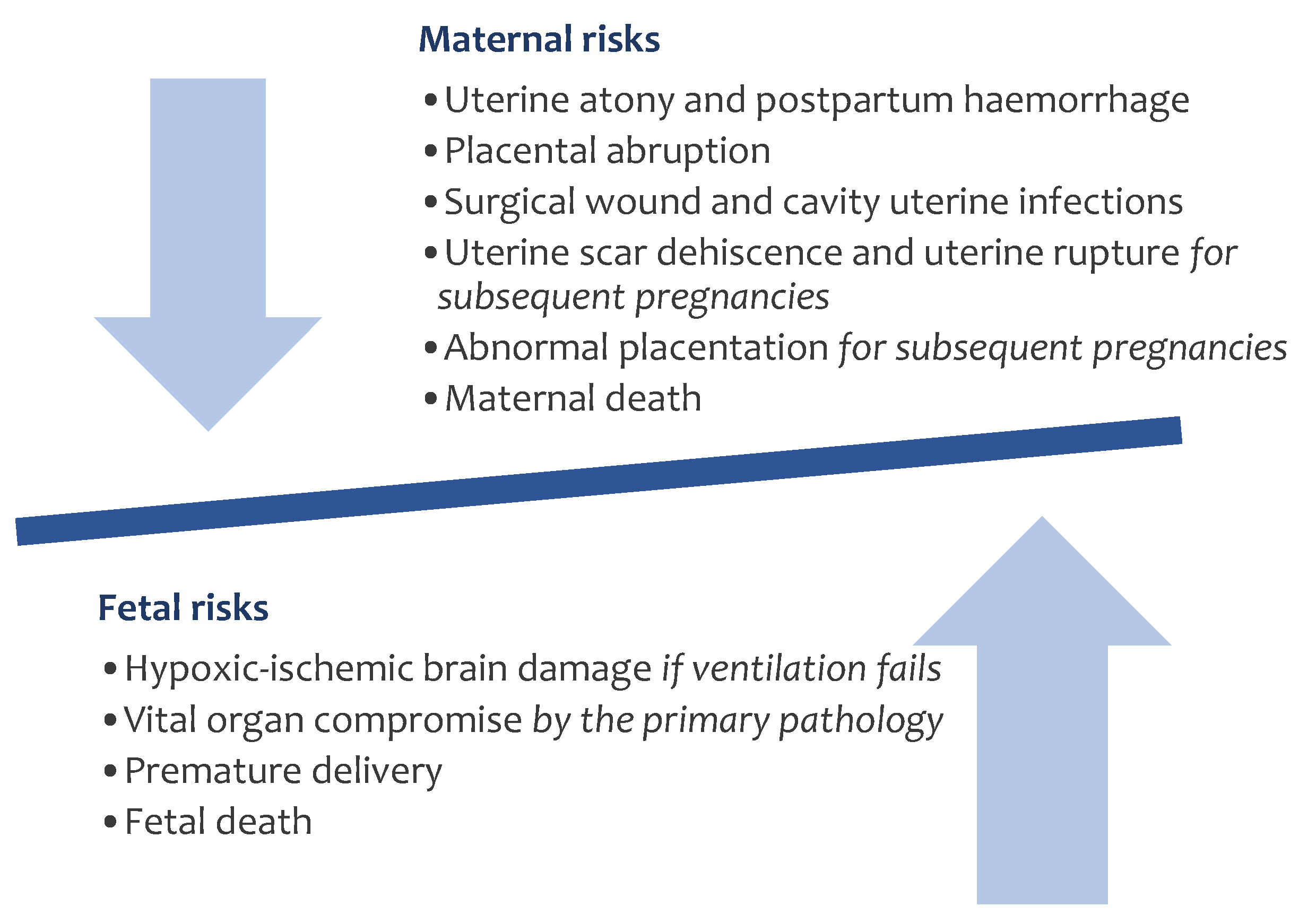
Key steps of the EXIT procedure are summarized in Figure 5. The subsequent pivotal phase pertains to fetal exposure. In cases where the fetus is in cephalic presentation, gentle extraction is carried out with the aid of a single-use suction vacuum (Kiwi Omnicup®, Laborie Obstetrics) (Figure 5d), being crucial in the presence of oropharyngeal and cervical tumors. If a non-cephalic presentation is observed during the intraoperative ultrasound performed for placental mapping, we perform an external version before accessing the amniotic sac. In most of the procedures, we expose only the fetal head and shoulders to avoid complete uterine contraction. Exceptionally, it may be necessary to expose part of the fetal abdomen depending on the fetal pathological condition and its complexity, even when completely delivered and positioned approximately at the placental level, as described elsewhere [12].
Following fetal exposure (Figure 5f), anesthesia is administered to the fetus via an intramuscular injection of a three-agent combination (fentanyl 1–20 mcg/kg, rocuronim bromide 0.8–1 mg/kg, and atropine 20 mcgr/kg). Subsequently, the fetal head is carefully rotated into an occipitoposterior position, facilitating direct laryngoscopy or bronchoscopy for airway access. In cases involving large compressive masses, repositioning them away from the airway may be necessary to facilitate intubation.
Throughout this intricate process, we maintain uterine volume through amnioinfusion using Ringer’s solution warmed to body temperature, well-tolerated by the fetus. Simultaneously, fetal well-being is continuously monitored via echocardiography. In specific cases, pulse oximetry is added.
Finally, with fetal airway access secured, we proceed to clamp and divide the umbilical cord. The placenta is delivered, and immediate restoration of uterine tone is achieved through intravenous administration of carbetocin (100 mcg), oxytocin (10 UI), and methylergonovine (0.2 mg intramuscular via or 0.1–0.2 intravenous line), a critical step to prevent uterine atony. In addition, the minimum alveolar concentration (CAM) of the volatile anesthetic should be decreased to 0.5–1, or even disconnected and replaced by propofol infusion to maintain maternal anesthesia while removing the tocolytic effect. Finally, the conclusion of the procedure mirrors that of a conventional cesarean section, involving the closure of the uterus and maternal abdominal wall.

4. Discussion

Few fetal anomalies wield as profound an impact on perinatal management as airway obstructions [6]. They represent a life-threatening scenario associated with elevated morbidity and mortality [8,31]. The transition from fetal to neonatal life constitutes an immense challenge, requiring complex physiologic changes that must occur in a relatively short period of time [2,32,33,34]. A delay in establishing a secure airway at the moment of birth can lead to hypoxia and acidosis [6,35]. If this delay exceeds five minutes, it may result in brain damage due to anoxia [6,24].
Given the limited number of centers performing the EXIT procedure, available data on maternal and fetal outcomes remain relatively scarce [4]. The reported maternal risks include a higher incidence of uterine atonia after placental delivery, related to the uterine relaxation required to maintain uteroplacental circulation during the procedure [1,3,24].
In our experience [15], spanning from 2007 to 2022, we have undertaken a total of 34 EXIT procedures. Notably, our surgical approach has revealed no significant differences in terms of maternal complications when compared to those observed in standard cesarean sections. This observation underscores the similarity in the average postoperative hemoglobin reduction between the two approaches [36].
The primary fetal risk lies in the potential compromise of effective placental gas exchange, which may be secondary to excessive uterine tone, placental abruption, or cord compression. To mitigate these risks, it is essential to maintain adequate uterine relaxation. Managing uterine tone must be executed with great care, considering the increased risk of uterine atony for the mother as previously mentioned [4]. As such, close communication between the surgical and anesthetic teams is paramount to promptly restore uterine tone after umbilical cord clamping.
A critical point to note is the limited number of studies available that comprehensively examine and describe the EXIT technique. Kenneth W. Liechty [6] details the technique developed at the Children’s Hospital of Philadelphia, which involves a rapid sequence anesthetic induction using different anesthetic agents (thiopental, succinylcholine, and fentanyl) and the use of desflurane to maintain anesthesia instead of sevoflurane. They employ oxytocin bolus and continuous infusion to prevent uterine atony. Furthermore, they perform the hysterotomy using a uterine stapling device, though the specific device is not described.
Recognizing the critical importance of managing maternal bleeding during fetal interventions, Jain et al. [37] highlighted their utilization of reefing sutures, given the unavailability of a uterine stapler in their center. This divergence in techniques is further exemplified by the approach of Porter et al. [38], who omit an intermediary step involving stapling or suturing the individual hysterotomy edges to reduce blood loss, deeming it non-essential to their practice.
In contrast, Florencia Varela et al. [20] advocate for the use of a hemostatic uterine stapling device as the ideal choice during hysterotomy. Notably, a distinctive element in our technique involves atraumatic access to the uterine cavity, achieved through the application of a uterine progressive distractor developed for this purpose. Following the use of this distractor, vascular clamps and a stapling device (Premium Poly Cs-57 Autosuture®, Medtronic) are employed in succession.
The uterine progressive distractor comprises cylindric and semi-cylindric pieces of increasing diameters and decreasing lengths that telescopically fit into each other. The sequential extraction of inner pieces facilitates a gradual distraction of muscular fibers without inducing traction, thereby minimizing the risk of significant hemorrhages. Additionally, this approach allows for a controlled outflow of amniotic fluid from the uterine cavity.
Vascular clamps, integral to this technique, serve as specialized hemostatic instruments designed to occlude major vascular structures or delicate tissues. These clamps feature two serrated jaws that provide a stable grip, minimizing avoidable bleeding. The elongated slim shanks of the clamps further enhance their utility by facilitating access to deep spaces during the procedure.
The single-use liner stapler places two rows of lactomer absorbable copolymer staples while a knife blade cuts between them, creating a 57 mm opening. The instrument is activated by squeezing the handle, creating a temporary opening. The automatic staple and cut allows a rapid controlled hemostatic hysterectomy, reducing blood loss. It could extend the duration of the procedure without increasing maternal risks, giving pediatric surgeons more time to perform life-saving airway interventions. In addition, prolonged hysterotomy incision during fetal airway manipulation could be avoided.
Moreover, the staples are totally absorbable, so no foreign materials are left in the body. This approach could enhance maternal and obstetric outcomes, making it a promising technique for broader implementation in various medical institutions.
In this context, Kern et al. [3] describe their experience with the EXIT procedure, detailing modified EXIT procedures based on three key modifications, which include the use of spinal anesthesia, complete delivery of the child, and specific uterine relaxation (without specific details provided).
Regarding the anesthetic technique, spinal anesthesia, although occasionally used, may not consistently achieve complete uterine relaxation. Most recently described EXIT procedures have utilized maternal general anesthesia [3,29,38,39,40]. This anesthetic technique with extensive uterine relaxation is related by these authors to a greater risk of maternal hemorrhage, maternal hypotension, ventricular systolic dysfunction, and fetal bradycardia [39].
In our center, we employ a combination of spinal anesthesia and deep general anesthesia to ensure uterine relaxation during the procedure while also providing effective postoperative pain management. Using this anesthetic approach, in our center we have not identified any significant hemorrhagic complications during or after the surgical procedure.
In delineation from the conventional full fetal delivery, our advocacy for exposing only the upper portion of the fetal thorax when technically feasible aligns closely with the established fetal management practices of specialized EXIT centers [14,39]. This deliberate strategy aims to address key concerns, notably minimizing amniotic fluid loss and averting the rapid temperature decline of the fetus, as well as mitigating risks associated with complete uterine contraction and placental abruption.
The rationale behind this nuanced approach is deeply rooted in the well-documented escalation of intrauterine pressure following childbirth, reaching levels significantly higher than systolic blood pressure [41]. The consequence of this heightened pressure is an abrupt decrease in maternal placental perfusion, despite the persistence of umbilical pulsations for a brief duration post-delivery [1].
Acknowledging the need for adaptability in the procedure, especially in cases where pathologies impact lung volume, is paramount. Notably, Kern et al. [3] reported cases where complete fetal delivery was employed for ease of access to the thorax and abdomen, positioning the fetuses approximately at the placental level. However, the absence of specificity regarding any accompanying surgical steps raises critical questions, particularly concerning the prevention of rapid uterine decompression, which could lead to amniotic fluid loss and expose the patient to the inherent risk of placental abruption.
This underscores the importance of a tailored approach, emphasizing the need for careful consideration of individual case complexities and potential pathologies. While the partial exposure strategy remains a focus in our methodology, there exists a recognition that certain circumstances may necessitate deviations, underscoring the ongoing refinement and adaptability within the field of fetal interventions. It is imperative to strike a delicate balance between procedural efficiency and patient safety, incorporating evolving insights from clinical experiences and research endeavors to optimize outcomes.
In considering alternatives to in utero fetal intervention, the EXIT procedure may offer a post-delivery surgical approach for cases like gastroschisis. The procedure involves the primary closure of the prolapsed bowel before the newborn initiates breathing, facilitating bowel expansion. However, this indication remains controversial and has not been universally adopted across all fetal surgery centers. In specific cases of gastroschisis performed in our center (three cases, year 2011) it was also necessary to expose the entire fetal thorax and abdomen, leaving only the pelvis and legs in utero, but instilling warmed Ringer’s solution into the uterine cavity to maintain uterine volume.
We believe that optimal uterine volume management is one of the key aspects of a successful procedure and could partly explain the maternal and neonatal outcomes achieved in our center. A careful manipulation of the fetal body and the maintenance of intrauterine tension could prevent placental abruption given that a loss of uterine volume is a strong stimulus for uterine contraction [13].
This issue is especially important when a prolonged duration of the procedure is foreseen, such as in cases of twin pregnancies. As we described in the longest series published about EXIT in twin pregnancies [12], the complexity and risks of the procedure are multiplied [42]. Priority should always be given to the normal twin in case of risk of complication. It does not necessarily mean to deliver the normal twin first [12]. In our experience, adequate uterine relaxation, maintenance of uterine volume, and careful monitoring of both fetuses and the mother allow the treatment of the affected twin before delivery of the unaffected one without risk [12].
Additionally, it is crucial to ensure adequate fetal anesthesia to optimize conditions for airway interventions [43]. While halogenated agents can diffuse from the mother to the fetus, we also administer anesthesia directly to the fetus to guarantee a sufficient level of fetal anesthesia. This approach is consistent with those reported in other studies [37,44].

5. Conclusions

Today, the EXIT procedure should be increasingly considered in many cases of fetal anomalies, not only those where potential compromise of the airway at birth is suspected.
By ensuring an optimal approach to accessing the uterine cavity and employing refined anesthetic techniques, we aim to maximize fetal benefits while mitigating maternal risks.
As our experience continues to grow, we anticipate further refinements and enhancements in these recommendations. The evolution of this technique will undoubtedly pave the way for improved outcomes and a deeper understanding of the delicate balance between fetal and maternal well-being in these complex cases.

Author Contributions

M.D.-M., Á.C., L.G.-D. and G.A. contributed to the conception and design. M.D.-M. was responsible for the acquisition of data. All authors contributed to drafting the article or revising it critically for important intellectual content. All authors have read and agreed to the published version of the manuscript.

Funding

This research received no external funding.

Institutional Review Board Statement

Not applicable.

Informed Consent Statement

Not applicable.

Data Availability Statement

The datasets used during the current study are available from the corresponding author on reasonable request.

Conflicts of Interest

The authors declare no conflicts of interest.

References

  1. Zimmermann, R. Ex utero intrapartum treatment. Swiss Med. Wkly. 2007, 137, 271. [Google Scholar] [PubMed]
  2. Chimenea, A.; Dominguez-Moreno, M.; Barrera-Talavera, M.; García-Díaz, L.; Antiñolo, G. Maternal and Neonatal Outcomes after Ex-Utero Intrapartum Treatment for Congenital Diaphragmatic Hernia: A Case Series. Eur. J. Pediatr. Surg. 2023; Ahead of print. [Google Scholar]
  3. Christian, K.; Morales, M.A.; Peiry, B.; Pfiser, R.E. Ex utero intrapartum treatment (EXIT), a resuscitation option for intra-thoracic foetal pathologies. Swiss Med. Wkly. 2007, 137, 279–285. [Google Scholar]
  4. Butler, C.R.; Maughan, E.F.; Pandya, P.; Hewitt, R. Ex utero intrapartum treatment (EXIT) for upper airway obstruction. Curr. Opin. Otolaryngol. Head Neck Surg. 2017, 25, 119–126. [Google Scholar] [CrossRef] [PubMed]
  5. Spiers, A.; Legendre, G.; Biquard, F.; Descamps, P.; Corroenne, R. Ex utero intrapartum technique (EXIT): Indications, procedure methods and materno-fetal complications—A literature review. J. Gynecol. Obstet. Hum. Reprod. 2022, 51, 102252. [Google Scholar] [CrossRef] [PubMed]
  6. Liechty, K.W. Ex-utero intrapartum therapy. Semin. Fetal. Neonatal. Med. 2010, 15, 34–39. [Google Scholar] [CrossRef]
  7. MacKenzie, T.C.; Crombleholme, T.M.; Flake, A.W. The ex-utero intrapartum treatment. Curr. Opin. Pediatr. 2002, 14, 453–458. [Google Scholar] [CrossRef]
  8. Garcia de Paredes, J.; Gosnell, J.; Strug, M.; Giuliani, E.; Thakur, M.; Romero, V.C.; Cordoba, M. Antenatal Three-Dimensional Printing for Ex Utero Intrapartum Treatment Procedures. Obs. Gynecol. 2022, 139, 313–316. [Google Scholar] [CrossRef]
  9. Mufti, N.; Ebner, M.; Patel, P.; Aertsen, M.; Gaunt, T.; Humphries, P.D.; Bredaki, F.E.; Hewitt, R.; Butler, C.; Sokolska, M.; et al. Super-resolution Reconstruction MRI Application in Fetal Neck Masses and Congenital High Airway Obstruction Syndrome. OTO Open 2021, 5, 43–45. [Google Scholar] [CrossRef]
  10. Bence, C.M.; Wagner, A.J. Ex utero intrapartum treatment (EXIT) procedures. Semin. Pediatr. Surg. 2019, 28, 150820. [Google Scholar] [CrossRef]
  11. Novoa, R.H.; Quintana, W.; Castillo-Urquiaga, W.; Ventura, W. EXIT (ex utero intrapartum treatment) surgery for the management of fetal airway obstruction: A systematic review of the literature. J. Pediatr. Surg. 2020, 55, 1188–1195. [Google Scholar] [CrossRef] [PubMed]
  12. García-Díaz, L.; de Agustín, J.C.; Ontanilla, A.; Marenco, M.L.; Pavón, A.; Losada, A.; Antiñolo, G. EXIT procedure in twin pregnancy: A series of three cases from a single center. BMC Pregnancy Childbirth 2014, 14, 1–4. [Google Scholar] [CrossRef] [PubMed]
  13. Hoagland, M.A.; Chatterjee, D. Anesthesia for fetal surgery. Paediatr. Anaesth. 2017, 27, 346–357. [Google Scholar] [CrossRef]
  14. Zhao, Y.; Wang, Y.; Liu, C.; Jiang, Y.; Wei, Y.; Meng, H.; Jian, S.; Zhu, X.; Pei, L.; Bai, X.; et al. Ex utero intrapartum therapy in infants with congenital diaphragmatic hernia: A propensity score matching analysis. World J. Ped. Surg. 2022, 5, 1–8. [Google Scholar] [CrossRef] [PubMed]
  15. García-Díaz, L.; Chimenea, A.; De Agustín, J.C.; Pavón, A.; Antiñolo, G. Ex-Utero Intrapartum Treatment (EXIT): Indications and outcome in fetal cervical and oropharyngeal masses. BMC Pregnancy Childbirth 2020, 20, 1–6. [Google Scholar] [CrossRef]
  16. Jiang, S.; Yang, C.; Bent, J.; Yang, C.J.; Gangar, M.; Nassar, M.; Suskin, B.; Dar, P. Ex utero intrapartum treatment (EXIT) for fetal neck masses: A tertiary center experience and literature review. Int. J. Pediatr. Otorhinolaryngol. 2019, 127, 109642. [Google Scholar] [CrossRef]
  17. Zamora, I.J.; Ethun, C.G.; Evans, L.M.; Olutoye, O.O.; Ivey, R.T.; Haeri, S.; Belfort, M.A.; Lee, T.C.; Cass, D.L. Maternal morbidity and reproductive outcomes related to fetal surgery. J. Pediatr. Surg. 2013, 48, 951–955. [Google Scholar] [CrossRef] [PubMed]
  18. Weber, S.U.; Kranke, P. Anesthesia for predelivery procedures: Ex-utero intrapartum treatment/intrauterine transfusion/surgery of the fetus. Curr. Opin. Anaesthesiol. 2019, 32, 291–297. [Google Scholar] [CrossRef]
  19. Morales, C.Z.; Barrette, L.X.; Vu, G.H.; Kalmar, C.L.; Oliver, E.; Gebb, J.; Feygin, T.; Howell, L.J.; Javia, L.; Hedrick, H.L.; et al. Postnatal outcomes and risk factor analysis for patients with prenatally diagnosed oropharyngeal masses. Int. J. Pediatr. Otorhinolaryngol. 2022, 152, 110982. [Google Scholar] [CrossRef]
  20. Varela, M.F.; Pinzon-Guzman, C.; Riddle, S.; Parikh, R.; McKinney, D.; Rutter, M.; Lim, F.-Y.; Peiro, J.L. EXIT-to-airway: Fundamentals, prenatal work-up, and technical aspects. Semin. Pediatr. Surg. 2021, 30, 151066. [Google Scholar] [CrossRef]
  21. de Oliveira, G.H.; Svetliza, J.; Vaz-Oliani, D.C.M.; Liedtke, H., Jr.; Oliani, A.H.; Pedreira, D.A.L. Novel multidisciplinary approach to monitor and treat fetuses with gastroschisis using the Svetliza Reducibility Index and the EXIT-like procedure. Einstein 2017, 15, 395–402. [Google Scholar] [CrossRef]
  22. Velhote, M.C.P. Monitoring and treating fetuses with gastroschisis using the Svetliza Reducibility Index (SRI) and the EXIT-like procedure—A novel approach. Einstein 2017, 15, 10–11. [Google Scholar] [CrossRef] [PubMed]
  23. Durmaz, L.O.; Brunner, S.E.; Meinzer, A.; Krebs, T.F.; Bergholz, R. Fetal Surgery for Gastroschisis—A Review with Emphasis on Minimally Invasive Procedures. Children 2022, 9, 416. [Google Scholar] [CrossRef] [PubMed]
  24. Mohammad, S.; Olutoye, O.A. Airway management for neonates requiring ex utero intrapartum treatment (EXIT). Paediatr. Anaesth. 2020, 30, 248–256. [Google Scholar] [CrossRef] [PubMed]
  25. Barrette, L.X.; Morales, C.Z.; Oliver, E.R.; Gebb, J.S.; Feygin, T.; Lioy, J.; Howell, L.J.; Hedrick, H.L.; Jackson, O.A.; Adzick, N.S.; et al. Risk factor analysis and outcomes of airway management in antenatally diagnosed cervical masses. Int. J. Pediatr. Otorhinolaryngol. 2021, 149, 110851. [Google Scholar] [CrossRef]
  26. Shamshirsaz, A.A.; Aalipour, S.; Stewart, K.A.; Nassr, A.A.; Furtun, B.Y.; Erfani, H.; Sundgren, N.C.; Cortes, M.S.; Donepudi, R.V.; Lee, T.C.; et al. Perinatal characteristics and early childhood follow up after ex-utero intrapartum treatment for head and neck teratomas by prenatal diagnosis. Prenat. Diagn. 2021, 41, 497–504. [Google Scholar] [CrossRef] [PubMed]
  27. Reeve, N.H.; Kahane, J.B.; Spinner, A.G.; O-Lee, T.J. Ex utero intrapartum treatment to extracorporeal membrane oxygenation: Lifesaving management of a giant cervical teratoma. J. Laryngol. Otol. 2020, 134, 650–653. [Google Scholar] [CrossRef]
  28. Scully Noah, M.M.; Norton, M.E.; Sandberg, P.; Esakoff, T.; Farrell, J.; Albanese, C.T. Short-term maternal outcomes that are associated with the EXIT procedure, as compared with cesarean delivery. Am. J. Obs. Gynecol. 2002, 186, 773–777. [Google Scholar] [CrossRef]
  29. Chung, W.; Lim, C. Intraoperative management for ex-utero intrapartum treatment: Focusing on the fetus. Anesth. Pain. Med. 2021, 16, 329–337. [Google Scholar] [CrossRef]
  30. Marwan, A.; Crombleholme, T.M.; Taghavi, K.; Beasley, S.; Nnamani, N. The ex utero intrapartum treatment (EXIT) procedure: Application of a new therapeutic paradigm. J. Anesth. Clin. Res. 2015, 49, 107–115. [Google Scholar]
  31. Puricelli, M.D.; Rahbar, R.; Allen, G.C.; Balakrishnan, K.; Brigger, M.T.; Daniel, S.J.; Fayoux, P.; Goudy, S.; Hewitt, R.; Hsu, W.-C.; et al. International Pediatric Otolaryngology Group (IPOG): Consensus recommendations on the prenatal and perinatal management of anticipated airway obstruction. Int. J. Pediatr. Otorhinolaryngol. 2020, 138, 110281. [Google Scholar] [CrossRef]
  32. Askin, D.F. Fetal-to-neonatal transition—What is Normal and What is Not ? Neonatal. Netw. 2009, 28, 33–40. [Google Scholar] [CrossRef]
  33. Vento, M. Improving fetal to neonatal transition of the very preterm infant: Novel approaches. Chin. Med. J. 2010, 123, 2924–2928. [Google Scholar]
  34. Noah, M.M.S.; Norton, M.E.; Sandberg, P.; Esakoff, T.; Farrell, J.; Albanese, C.T. Does the ex utero intrapartum treatment to extracorporeal membrane oxygenation procedure change morbidity outcomes for high-risk congenital diaphragmatic hernia survivors? J. Pediatr. Surg. 2017, 52, 22–25. [Google Scholar]
  35. Askin, D.F. Complications in the transition from fetal to neonatal life. J. Obstet. Gynecol. Neonatal. Nurs. 2002, 31, 318–327. [Google Scholar] [CrossRef]
  36. Domínguez-Moreno, M.; Chimenea, A.; García-Díaz, L.; Antiñolo, G. Maternal and obstetric outcomes after Ex-Utero Intrapartum Treatment (EXIT): A single center experience. BMC Pregnancy Childbirth, 2023; Ahead of print. [Google Scholar]
  37. Jain, P.; Prasad, A.; Rahul, K.M.; Ankur, K. Difficult airway of fetus: Making a safe Ex Utero intrapartum treatment. J. Indian Assoc. Pediatr. Surg. 2021, 26, 448–450. [Google Scholar] [CrossRef]
  38. Porter, H.; Trivedi, A.; Marquez, M.; Gibson, P.; Melov, S.J.; Mishra, U.; Jani, P.; Cheng, A.T.; Nayyar, R.; Alahakoon, T.I. Changing indications and antenatal prognostic factors for ex-utero intrapartum treatment procedures. Prenat. Diagn. 2022, 42, 1420–1428. [Google Scholar] [CrossRef] [PubMed]
  39. Wang, W.; Pei, L.; Zhang, Y.; Chen, W.; Liu, J.; Jiang, Y.; Lv, Y.; Li, Z.; Jian, S.; Ma, L.; et al. Neuraxial anesthesia in ex utero intrapartum therapy for parturients with fetal congenital diaphragmatic hernia: A prospective observational study. Int. J. Obstet. Anesth. 2022, 52, 103599. [Google Scholar] [CrossRef] [PubMed]
  40. Noguchi, S.; Tanaka, M.; Terui, K. The first national survey of anesthesia techniques for fetal therapies in Japan. J. Anesth. 2019, 33, 665–669. [Google Scholar] [CrossRef] [PubMed]
  41. King, A.; Keswani, S.G.; Belfort, M.A.; Nassr, A.A.; Shamshirsaz, A.A.; Espinoza, J.; Bedwell, J.R.; Mehta, D.K.; Doughty, C.B.; Leong-Kee, S.M.; et al. EXIT (ex utero Intrapartum Treatment) to Airway Procedure for Twin Fetuses with Oropharyngeal Teratomas: Lessons Learned. Front. Surg. 2020, 7, 1–4. [Google Scholar] [CrossRef] [PubMed]
  42. Harison, M.R.; Evans, M.I.; Adzick, N.S.; Holzgreve, W. The Fetus with Airway Obstruction; Saunders: Philadelphia, PA, USA, 2001. [Google Scholar]
  43. Al-Hindi, M.; Al Sayari, T.A.; Al Solami, R.; Al Baiti, A.K.; Alnemri, J.A.; Mirza, I.M.; Alattas, A.; A Faden, Y. Association of Antenatal Risk Score with Maternal and Neonatal Mortality and Morbidity. Cureus 2020, 12, 1–8. [Google Scholar] [CrossRef] [PubMed]
  44. Yanko, F.; Nathani, H.; Alden, T.; Valika, T.; Rastatter, J.; Alhajjat, A.; Ballard, H.A. An operation on placental support in a fetus with a nasopharyngeal teratoma. Anaesth. Rep. 2023, 11, e12219. [Google Scholar] [CrossRef] [PubMed]
Figure 1. Maternal and fetal risks associated with EXIT procedure.
Std 13 00005 g001
Figure 2. Steps of EXIT procedure.
Std 13 00005 g002
Figure 3. Scheme of the distribution of fetal–maternal team in the operating room.
Std 13 00005 g003
Figure 4. Surgical instruments by order of use in EXIT procedure: arrow guide (1), uterine progressive distractor (2), Satinsky vascular clamps (3), stapling device (4).
Std 13 00005 g004
Figure 5. Key steps of EXIT procedure: atraumatic uterine opening by uterine progressive distractor (a,b), placement of Satinsky vascular clamps (c), sealing of hysterotomy edges by stapling device Premium Poly Cs-57 Autosuture®, Medtronic (d), gentle fetal extraction with the aid of a single-use suction vacuum Kiwi© (Laborie, Mississauga, ON, Canada) (e), exposure of the fetal head and upper thorax (f).
Std 13 00005 g005
Table 1. Steps of anesthetic technique.
Steps of Anesthetic TechniqueDrugs and Dose
Pre-inductionBlood reserve availability check
Placement of epidural catheter
Maternal hemodynamic monitoring
Broncho-aspiration prophylaxis: Omeprazole 40 mg or Metoclopramide 10 mg
Antibiotic prophylaxis: Cefazolin 2 gr
Slight left lateral decubitus
Anesthetic inductionAtropine (0.01 mg/kg)
Remifentanile (continuous infusion, 3 ng/mL)
Maternal preoxygenation (for, at least, five minutes)
Dexametasone (4–8 mg, single dose)
General Anesthesia with rapid sequence inductionPropofol (2.5 mg/kg)
Rocuronium bromide (1–2 mg/kg)
Sellick maneuver
Endotracheal intubation (with videolaryngoscope support)
Controlled ventilation
Anesthesia maintenancePhenylephrine (1 mg diluted in 100 mL SSF, 15 mL/h, to maintain maternal mean blood pressure > 65 mmHg)
Tranexamic acid (1 g diluted in 100 mL SSF, administered after 15 min, as intraoperative bleeding prophylaxis)
Mechanical ventilation (exhaled CO2 28–34 mmHg to maintain normocapnia)
Remifentanil (dosing according to hemodynamic response)
Rocuronim bromide (0.2 mg/kg or bolus of 0.15 mg/kg, when neuromuscular block reaches 25% of its recovery)
Immediately after clamping umbilical cord:
Carbetocin 100 mcg (slow infusion)
+/− Oxitocin 10 UI (slow injection)
+/− Methylergonovine 0.2 mg via intramuscular or 0.1–0.2 intravenous slow infusion
After hysterotomy closure:
Sevoflurane (Minimum alveolar concentration: 0.5)
Local anesthetic via epidural
+/− Fentanyl 50 mcg
Additional fetal anesthesiaFetal cocktail via intramuscular
Atropine 20 mcg/kg
Fentanyl 10–20 mcg/kg
Rocuronim bromide (0.8–1 mg/kg)
Disclaimer/Publisher’s Note: The statements, opinions and data contained in all publications are solely those of the individual author(s) and contributor(s) and not of MDPI and/or the editor(s). MDPI and/or the editor(s) disclaim responsibility for any injury to people or property resulting from any ideas, methods, instructions or products referred to in the content.

