


Surg. Tech. Dev. 2024, 13(2), 122-161; https://doi.org/10.3390/std13020009 - 22 Apr 2024
Abstract
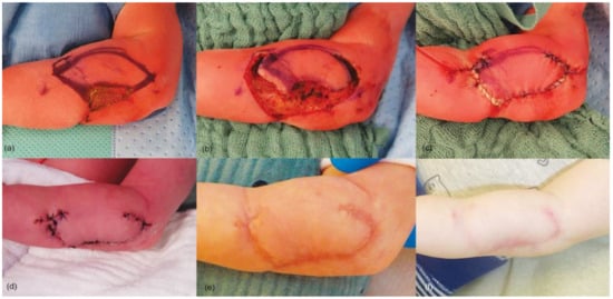
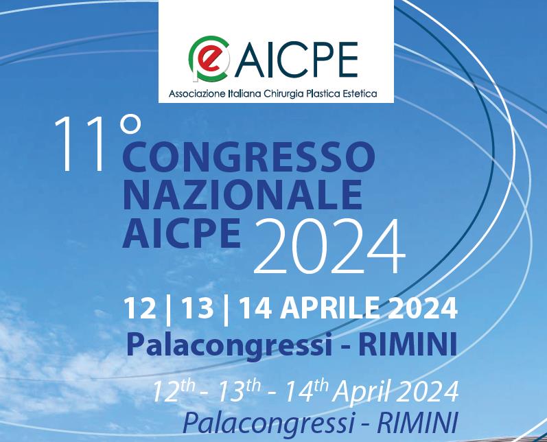
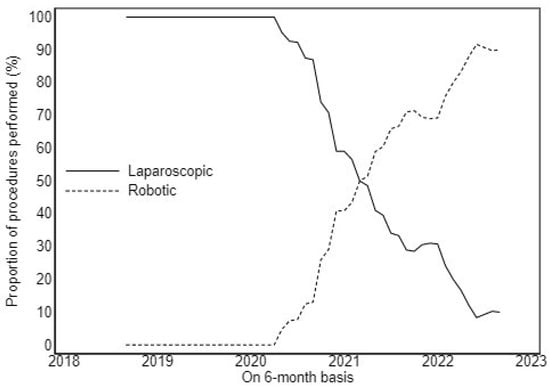
The annual congress of the Italian Association of Plastic Aesthetic Surgery (AICPE) is one of the most relevant conference meetings in Europe concerning aesthetic plastic surgery as there are a number of participants and a parterre of invited speakers chosen for their renowned
[...] Read more.
The annual congress of the Italian Association of Plastic Aesthetic Surgery (AICPE) is one of the most relevant conference meetings in Europe concerning aesthetic plastic surgery as there are a number of participants and a parterre of invited speakers chosen for their renowned scientific value. [...]
Full article


